HAL Id: hal-01988182
https://hal.archives-ouvertes.fr/hal-01988182
Submitted on 29 Jan 2019
HAL is a multi-disciplinary open access
archive for the deposit and dissemination of
sci-entific research documents, whether they are
pub-lished or not. The documents may come from
teaching and research institutions in France or
abroad, or from public or private research centers.
L’archive ouverte pluridisciplinaire HAL, est
destinée au dépôt et à la diffusion de documents
scientifiques de niveau recherche, publiés ou non,
émanant des établissements d’enseignement et de
recherche français ou étrangers, des laboratoires
publics ou privés.
Interactions of an Essential Bacillus subtilis GTPase,
YsxC, with Ribosomes
Catherine Wicker-Planquart, Anne-Emmanuelle Foucher, Mathilde Louwagie,
Robert Britton, Jean-Michel Jault
To cite this version:
Catherine Wicker-Planquart, Anne-Emmanuelle Foucher, Mathilde Louwagie, Robert Britton,
Jean-Michel Jault. Interactions of an Essential Bacillus subtilis GTPase, YsxC, with Ribosomes. Journal
of Bacteriology, American Society for Microbiology, 2008, 190 (2), pp.681-690. �10.1128/jb.01193-07�.
�hal-01988182�
J
OURNAL OFB
ACTERIOLOGY, Jan. 2008, p. 681–690
Vol. 190, No. 2
0021-9193/08/$08.00
⫹0 doi:10.1128/JB.01193-07
Copyright © 2008, American Society for Microbiology. All Rights Reserved.
Interactions of an Essential Bacillus subtilis GTPase, YsxC,
with Ribosomes
䌤
†
Catherine Wicker-Planquart,
1* Anne-Emmanuelle Foucher,
1Mathilde Louwagie,
2Robert A. Britton,
3and Jean-Michel Jault
1Institut de Biologie Structurale, UMR 5075 Universite´ Joseph Fourier/CEA/CNRS, 41 rue Jules Horowitz 38027 Grenoble Cedex 1,
France
1; CEA, DSV, iRTSV, Laboratoire d’Etude de la Dynamique des Prote´omes, INSERM, U880,
Universite´ Joseph Fourier, Grenoble, F-38054, France
2; and Department of Microbiology and
Molecular Genetics, Michigan State University, East Lansing, Michigan 48824
3Received 26 July 2007/Accepted 24 October 2007
YsxC is a small GTPase of Bacillus subtilis with essential but still unknown function, although recent works
have suggested that it might be involved in ribosome biogenesis. Here, purified YsxC overexpressed in
Escherichia coli was found to be partly associated with high-molecular-weight material, most likely rRNA, and
thus eluted from gel filtration as a large complex. In addition, purification of ribosomes from an E. coli strain
overexpressing YsxC allowed the copurification of the YsxC protein. Purified YsxC was shown to bind
pref-erentially to the 50S subunit of B. subtilis ribosomes; this interaction was modulated by nucleotides and was
stronger in the presence of a nonhydrolyzable GTP analogue than with GTP. Far-Western blotting analysis
performed with His
6-YsxC and ribosomal proteins separated by sodium dodecyl sulfate-polyacrylamide gel
electrophoresis showed that YsxC interacted with at least four ribosomal proteins from the 50S subunit. Two
of these putative protein partners were identified by mass spectrometry as L1 and L3, while the third reactive
band in the one-dimensional gel contained L6 and L10. The fourth band that reacted with YsxC contained a
mixture of three proteins, L7/L12, L23, and L27, suggesting that at least one of them binds to YsxC.
Coimmobilization assays confirmed that L1, L6, and L7/L12 interact with YsxC. Together, these results suggest
that YsxC plays a role in ribosome assembly.
GTPases are found in all three kingdoms of life (9, 13, 50,
63) and can be broadly classified into four large subfamilies
according to their cellular roles and molecular weights: small
GTP-binding proteins involved in cell proliferation,
transla-tional GTPases,
␣-subunits of heterotrimeric G proteins
in-volved in cell signaling, and large GTP-binding proteins (50).
In eukaryotes, all these families are present, and the last two
decades have witnessed a tremendous increase in our
under-standing of the structures and functions of many of their
mem-bers (9, 63, 67, 70, 71). In contrast, GTPases are rather scarce
in prokaryotes, as some families, such as the
␣-subunits of
heterotrimeric G proteins or large GTP-binding proteins,
ap-pear to be missing, and small GTPases are largely
underrep-resented (13, 50). Besides, apart from the well-characterized
translation factors, including EF-G, EF-Tu, and IF2, there is
still a paucity of information concerning most of the
remain-ing bacterial GTPases. At first glance, this might seem
para-doxical, as many putative GTPases inferred from the
genome-sequencing programs have been shown to be essential for
bacterial growth, but their discovery is still in an early stage
(11, 13, 45). This emphasizes the need to urgently address the
fundamental question regarding the cellular roles of these
un-characterized enzymes (56).
Many reports have suggested that the functions of most of
the unknown conserved putative bacterial GTPases are
some-how linked with the ribosomes and nucleic acid binding (11, 12,
15). Over the past few years, biochemical evidence has
accu-mulated to support this view for several GTPases, including
YlqF/RbgA (40, 66), YloQ (14, 16), Era (32, 41, 60), Obg/CgtA
(59, 72, 75), and EngA/Der (7, 28, 61). Bacillus subtilis YsxC
(termed YihA in Escherichia coli) is part of another family of
GTPases broadly conserved in bacteria that has been recently
selected as a priority target for functional characterization
(22). Genetic studies have shown that this protein is essential
to the growth of B. subtilis and E. coli (2, 53, 69). Depletion of
YsxC resulted in cell elongation, abnormal cell curvature, and
nucleoid condensation in B. subtilis (45). Depletion of YihA in
E. coli results in impaired cell division (17). The ysxC gene was
shown to be transcribed together with the lon gene (55), and
both gene products are considered heat shock proteins, since
they could be induced by heat and other stresses (3, 52, 55).
Both YihA and YsxC have been purified (35, 57), and
deter-mination of the three-dimensional structure of YsxC (57)
con-firmed that the protein belongs to the large superfamily of
translation factor-related (TRAFAC) GTPases, as defined by
Leipe and coworkers (36). Regarding the function of YsxC, a
recent study suggested that it is required for
large-ribosomal-subunit biogenesis in B. subtilis, since YsxC-depleted cells
ac-cumulate immature ribosomal-subunit intermediates (61).
These immature subunits lack three ribosomal proteins, L16,
L27, and L36 (61).
In the current work, we show that during purification of
YsxC overexpressed in E. coli, a high-molecular-weight RNA
* Corresponding author. Mailing address: Institut de Biologie
Struc-turale, UMR 5075 Universite
´ Joseph Fourier/CEA/CNRS, 41 rue Jules
Horowitz, 38027 Grenoble Cedex 1, France. Phone: 33 4 38 78 31 19.
Fax: 33 4 38 78 54 94. E-mail: catherine.wicker-planquart@ibs.fr.
† Supplemental material for this article may be found at http://jb
.asm.org/.
䌤
Published ahead of print on 2 November 2007.
species, possibly rRNA, copurified with the protein. We have
further investigated the association between YsxC and B.
sub-tilis ribosomes using pelleting assays and velocity gradient
cen-trifugation techniques. YsxC was shown to preferentially bind
to the 50S ribosomal subunit, and this binding was
strength-ened in the presence of a nonhydrolyzable GTP analogue. A
far-Western approach was then used to pinpoint the putative
ribosomal-protein partners of YsxC, revealing four reactive
bands, all from the large ribosomal subunit. Two of these
bands were assigned to L1 and L3 by mass spectrum analysis,
while the two additional bands contained a mixture of two (L6
and L10) or three (L7/L12, L23, and L27) subunits, suggesting
that at least one of them in each band was an interacting
partner. Of these proteins, six could be overexpressed under a
soluble form: L1, L6, L10, L7/L12, L23, and L27. Pull-down
experiments were then performed with these proteins and
con-firmed that YsxC could interact with at least L1, L6, and
L7/L12. Overall, our results strongly support a direct role of
YsxC during the biogenesis of the large ribosomal subunit.
MATERIALS AND METHODS
Plasmids, bacterial strains, and culture conditions.To clone the ysxC gene from B. subtilis, the genomic DNA of B. subtilis strain 168 was prepared accord-ing to the method of te Riele et al. (64). The ysxC gene was amplified from B.
subtilis genomic DNA by PCR using Pfu Turbo DNA polymerase from
Strata-gene and oligonucleotides a (CGGGATCCATGAAAGTCACAAAGTCAGAA ATCGTGATC; a BamHI site is underlined) and b (GGGCTCGAGCGACCG GTTTATCATTTTTTTGATCG; an XhoI site is underlined), cut with BamHI and XhoI, and cloned into the BamHI-XhoI sites of pGEX-4T1 (Amersham Biosciences). The His6-ysxC fusion plasmid has already been described (61).
Oligonucleotides b and c (GGCATATGAAAGTCACAAAGTCAGAAATC; an NdeI site is underlined) were used to perform another PCR, and the PCR product was digested with NdeI and XhoI and ligated into the NdeI-XhoI sites of pET-21b (Novagen) (to express YsxC without any tag). After transformation of the ligation mixtures into E. coli DH5␣ cells, positive clones were confirmed by DNA sequencing (Genome Express).
B. subtilis ribosomal-protein genes coding for L1, L3, L6, L7, L10, L23, and
L27 were amplified from B. subtilis genomic DNA by PCR using Pfu Turbo DNA polymerase and the oligonucleotides listed in Table S1 in the supplemental material. The resulting PCR products were digested either by NdeI and BamHI and ligated into the NdeI-BamHI sites of pET-15b, by BamHI and XhoI and ligated into the BamHI-XhoI sites of pET-21b, or by BamHI and XhoI and ligated into the BamHI-XhoI sites of pGEX-4T-1 (see Table S2 in the supple-mental material). After transformation of the ligation mixtures into E. coli DH5␣ cells, positive clones were confirmed by DNA sequencing (Genome Express).
E. coli cells, strain BL21(DE3), were transformed with the YsxC plasmids or
the plasmids encoding the ribosomal proteins. All E. coli strains that express the
ysxC gene or the ribosomal genes were first grown overnight at 37°C with
vigorous shaking in LB medium (Sigma) supplemented with 100g of ampicillin per ml. The overnight cultures were inoculated into LB medium containing ampicillin and then induced with 1 mM IPTG (isopropyl--D -thiogalactopyrano-side) (Euromedex) at an optical density at 600 nm (OD600) of 0.6 for 3 h at 37°C
for YsxC proteins or overnight at 15°C for ribosomal proteins when necessary (see Table S2 in the supplemental material). Cells were harvested by centrifu-gation at 4,000⫻ g for 10 min. The pellets were frozen at ⫺80°C until they were used.
Purification of YsxC proteins.The bacterial pellets were thawed on ice and resuspended in 25 ml of cold lysis buffer (50 mM NaPO4, 1 mM MgCl2, 1 mM
phenylmethylsulfonyl fluoride) (Fluka), 10⌴ leupeptin (Euromedex), and 6 M pepstatin (Euromedex) at pH 8 for YsxC and 8.5 for glutathione S-trans-ferase (GST)-YsxC and His6-YsxC. The cells were disrupted by ultrasonication
three times for 20 s each time and spun at 39,000⫻ g for 20 min at 4°C. The supernatant fraction was saved.
The GST-YsxC protein was purified by glutathione-Sepharose affinity column chromatography by loading the supernatant onto a 4-ml glutathione-Sepharose column (Sigma). The column was washed with 100 ml of buffer A (50 mM NaPO4, 1 mM MgCl2, 0.1 M NaCl, pH 8). At this point, either the GST-tagged
protein was eluted with about 15 ml of freshly prepared 20 mM glutathione in
buffer A or the GST tag was cleaved by proteolysis while bound to glutathione-Sepharose. In the latter case, the resin was suspended in the same volume of buffer A, and thrombin (Calbiochem; 25 U/ml of resin) was added. The digestion proceeded for 2 h at 4°C under gentle rotation to keep the beads in suspension. The flowthrough was then recovered, and the column was rinsed with 1 resin volume of buffer A. Phenylmethylsulfonyl fluoride (1 mM) was added to inacti-vate the thrombin.
His6-YsxC was purified as described previously (61).
The native (nontagged) YsxC protein was purified in two steps. The superna-tant fraction was loaded onto a 10-ml DEAE-cellulose (Sigma) column equili-brated with buffer C (50 mM NaPO4, 1 mM MgCl2, pH 8). The YsxC protein was
recovered in the flowthrough, and the column was rinsed with 1 resin volume of buffer C. The protein solution was then loaded onto a 5-ml SP-Sepharose column (Sigma) equilibrated with buffer C, and the column was washed with 100 ml buffer C containing 0.3 M NaCl and eluted with 10 ml 0.6 M NaCl in buffer C. GST was purified from E. coli cells, strain BL21(DE3), transformed with pGEX-4T1 vector (Amersham) by using a glutathione-Sepharose affinity column as described above for the GST-YsxC protein.
The eluted fractions containing nontagged or tagged YsxC were subjected to sodium dodecyl sulfate-polyacrylamide gel electrophoresis (SDS-PAGE) (34) and stained with Coomassie blue (protein molecular weight marker from Fer-mentas). They were then concentrated in centrifugal filter devices (Centricon from Amicon) up to a final OD280of 1 to 2. Glycerol was added to the YsxC
preparations at a final concentration of 15% (vol/vol), and after a freezing step in liquid nitrogen, aliquots were stored at⫺80°C. The protein concentrations were determined using a Lowry Protein Assay Reagent (Pierce), with bovine serum albumin as a standard.
In a few cases, protein-concentrated aliquots were gel filtered on a Superdex 200 10/300GL (Amersham) equilibrated in buffer A (50 mM NaPO4, pH 8, 0.1
M NaCl). The resulting fractions (0.5 ml each) were subjected to SDS-PAGE and stained with Coomassie blue or electroblotted onto Immobilon-P transfer membranes (Millipore) for Western blotting analysis. The blots were blocked with 25 mg albumin bovine fraction V (Sigma) per ml Tris-buffered saline (10 mM Tris, 0.137 M NaCl, pH 7.6)–Tween 0.05% for 1 h. Primary antibodies against GST (anti-GST–horseradish peroxidase [HPR] conjugate; Euromedex) or His tag (HisProbe-HPR; Pierce) were incubated with membranes in the presence of albumin in Tris-buffered saline–Tween for 1 h at room temperature at dilutions of 1:5,000. The antibodies were detected by fluorography with an enhanced chemiluminescent substrate (Super-Signal Pico chemiluminescent sub-strate; Pierce) as recommended by the manufacturer.
Purification of recombinant ribosomal proteins.His6-ribosomal proteins were
purified following the same protocol as described for His6-YsxC (61). The
GST-ribosomal proteins were purified by glutathione-Sepharose affinity column chro-matography as described above for the GST-YsxC protein.
Analysis of the high-molecular-weight material associated with YsxC.To analyze the material associated with YsxC, GST-YsxC was treated with either 1 mg/ml RNase A (Sigma) or 0.2 mg/ml DNase I (Euromedex) for 15 min at room temperature and loaded onto a Superdex 200 10/300GL (equilibrated with buffer A), as described above, that had been calibrated with the following protein standards (Amersham): RNase (13.7 kDa), ovalbumin (43 kDa), bovine serum albumin (67 kDa), and Blue Dextran (2,000 kDa). Each peak resolved was subjected to SDS-PAGE or Western blotting analysis as described above.
To check for the presence of RNA in the high-molecular-weight material associated with YsxC, purified GST-YsxC was extracted with an equal volume of phenol-chloroform-isoamyl alcohol, and RNA was precipitated with ethanol (58). The pellet was resuspended in water and analyzed with 1.5% agarose gels containing formaldehyde (58), with E. coli rRNAs as molecular weight markers.
E. coli ribosome preparation from strain BL21(DE3) is described below, and
rRNA extraction was performed as described above (phenol-chloroform-isoamyl alcohol extraction).
Ribosome purification and preparation of 30S and 50S subunits. Highly purified ribosomes were prepared from B. subtilis (strain 168) and from E. coli cells [strain BL21(DE3), wild type or transformed with YsxC or GST plasmids and induced with IPTG for 3 h, thus expressing YsxC proteins or GST, respec-tively], essentially as described by Daigle and Brown (16). For all strains, 2 liters of LB was inoculated with 20 ml of an overnight culture of B. subtilis or E. coli. The cells were grown in LB medium at 37°C and collected at an OD600of 0.8.
Chloramphenicol was added to a final concentration of 100g/ml 3 min prior to harvesting (37). The ribosomal pellet was resuspended in a small volume of buffer (0.1 mM magnesium acetate, 0.005 mM EDTA, 10 mM Tris-HCl, pH 7.5, 30 mM KCl, 6 mM mercaptoethanol, 1.25 mM dithiothreitol) and stored at ⫺80°C until they were used.
Separation of the 70S ribosome into 30S and 50S subunits.Chloramphenicol (100g/ml) was added to the ribosome preparation, and 5 OD260units of the
suspension was layered onto 5 to 20% (wt/vol) sucrose RNase- and DNase-free-gradients made up in 10 mM Tris-HCl, pH 7.5, 0.1 mM magnesium acetate, 30 mM KCl, followed by centrifugation at 200,000⫻ g at 4°C for 190 min in a SW41Ti rotor (Beckman). Fractions (0.5 ml) were collected, the pump flow rate was set to 0.75 ml/min, and the UV absorbance (OD260) of the sample was
monitored. Each fraction was precipitated by the addition of 10% trichloroacetic acid (TCA) and 0.02% deoxycholate, resuspended in SDS loading buffer, ana-lyzed by SDS-12% PAGE, and immunoblotted with anti-His antibodies.
When 50S and 30S subunits were needed, fractions containing the 50S and 30S subunits were collected, combined, and precipitated in the presence of 20 mM magnesium acetate by mixing the solution with an equal volume of ice-cold ethanol. After incubation at⫺20°C for 1 h, subunits were pelleted by centrifu-gation at 10,000⫻ g for 30 min at 4°C and resuspended in a small volume of buffer (0.1 mM magnesium acetate, 5M EDTA, 10 mM Tris-HCl, pH 7.5, 30 mM KCl, 6 mM-mercaptoethanol, 1.25 mM dithiothreitol). They were frac-tionated again on a 5 to 20% sucrose gradient, collected, and stored at⫺80°C. Quantitation of subunits was done by absorbance at 260 nm (1 A260unit is
equivalent to 69 or 34.5 pmol of 30S or 50S ribosomes, respectively) (16).
Binding of YsxC to 70S ribosomes. (i) Pelleting assays.Pelleting assays were performed with a mixture of B. subtilis ribosomes (total A260⫽ 10, i.e., 114
picomoles) and 1g His6-YsxC (180 picomoles), as described previously (61). (ii) Effects of excess guanine nucleotides on YsxC association with ribosome 30S and 50S subunits.A mixture of B. subtilis ribosomes (OD260⫽ 10)
prein-cubated with 1g His6-YsxC in the presence of 1 mM GDP, GTP, GMPPNP, or
no added nucleotides was sedimented through a 5 to 20% (wt/vol) sucrose gradient in standard buffer (10 mM Tris-HCl, pH 7.5, 0.1 mM magnesium acetate, 30 mM KCl), with an additional 100M GDP, GTP, GMPPNP (gua-nylyl-imidodiphosphate), or nothing, respectively, at 200,000⫻ g at 4°C for 190 min in a SW41Ti rotor (Beckman), and the resulting fractions were analyzed as described above (absorbance at 260 nm, precipitation by 10% TCA and 0.02% deoxycholate, and immunoblotting with anti-His antibodies).
(iii) Far-Western blot analysis. Ribosomal proteins from B. subtilis were separated by SDS-PAGE, using the electrophoretic system Protean II xi Cell device from Bio-Rad and a 14 to 18% or 14 to 20% acrylamide gradient. A prestained protein molecular weight marker (Fermentas), Seeblue Plus 2-stained standard (Invitrogen), or Mark 12 MW Standard (Invitrogen), was used. Proteins from the gel were then transferred to Immobilon-P transfer membranes (Milli-pore), washed, and incubated with histidine-tagged YsxC following precisely the protocol described by Bouvet et al. (10). Histidine-tagged YsxC was revealed by immunoblotting with anti-His antibodies, as described above.
Association of YsxC and recombinant ribosomal proteins. (i) Pull-down assay.
About 50g GST-tagged bait protein (GST-YsxC, GST-L10, GST-L27, or GST protein) was bound to about 50l of an immobilized glutathione support (glu-tathione-Sepharose) for 35 min in a minicolumn in buffer A (50 mM NaPO4, 0.3
M NaCl, pH 8). The column was then washed with 3 ml buffer A; loaded with L1, L6, L7, or L23 (when the bait protein was GST-YsxC or GST protein) or His6-YsxC (when the bait was GST-L10, GST-L27, or GST); incubated for 45
min; and then washed again with 3 ml buffer A, and the protein complex was eluted from the affinity support with 100l elution buffer (buffer A plus 100 mM glutathione). Protein complexes contained in eluted samples were then visual-ized by SDS-PAGE, and when necessary, histidine-tagged prey proteins were revealed by immunoblotting with anti-His antibodies, as described before.
(ii) Protein digestion and mass spectrometry analysis.Protein bands were manually excised from Coomassie-blue-stained one-dimensional gels and washed several times with destaining solutions (25 mM NH4HCO3for 15 min and then
50% [vol/vol] acetonitrile containing 25 mM NH4HCO3for 15 min). Gel pieces
were then dehydrated with 100% acetonitrile and dried under vacuum on a centrifugal evaporator. Samples were incubated with 7% H2O2for 15 min for
cysteine bond reduction before being washed again several times with the destaining solutions described above. After dehydration with 100% acetonitrile, the gel pieces were dried under vacuum. Depending on the protein amount, 0.2 g to 0.5 g of modified trypsin (Promega; sequencing grade) in 25 mM NH4HCO3was added to the dehydrated gel bands. After 30 min of incubation at
room temperature, 10l to 20 l of 25 mM NH4HCO3was added to submerge
the gel pieces before incubation overnight at 37°C (29).
(iii) Nano-LC-MS/MS analysis of samples.For nano-liquid chromatography-tandem mass spectrometry (nano-LC-MS/MS) analysis, peptides were extracted from gel pieces by incubation for 15 min first in 50% acetonitrile, followed by 5% formic acid and finally 100% acetonitrile. The pooled supernatants were then transferred into microcentrifuge tubes and dried under vacuum. The dried ex-tracted peptides were solubilized in water containing 2% acetonitrile and 7%
trifluoroacetic acid before being transferred in vials compatible with nano-LC-MS/MS analysis (CapLC [Waters] and Q-TOF Ultima [Micromass], United Kingdom). The method consisted of a 60-min run at a flow rate of 200 nl/min using a gradient from two solvents, A (5% acetonitrile and 0.1% formic acid in water) and B (80% acetonitrile and 0.08% formic acid in water). The system included a 300-m by 5-mm PepMap C18precolumn to preconcentrate peptides
and a 75-m by 150-mm C18column (LC Parkings Dionex) used for peptide
elution. For automatic LC-MS/MS analysis, the QTOF Ultima instrument was run in data-dependent mode. In MS/MS mode, spectra were calibrated thanks to the fragmentation of the glufibrino-peptide. MS and MS/MS data were acquired and processed automatically using MassLynx 4.0 software (Waters). Each sample was submitted to searches against the Swiss Prot Trembl database (4,967 se-quences for the B. subtilis taxonomy) using an in-house Mascot server (version 2.1; Matrix Sciences, London, United Kingdom). The Mascot search parameters used were as follows: enzyme, trypsin/P; one missed cleavage allowed; peptide tolerance, 0.4 Da; MS/MS tolerance, 0.4 Da; monoisotopic peptide charges, 2⫹ and 3⫹; and variable modifications, acetyl (N-ter)/oxidized methionine under sulfone and sulfoxide form/FMA⫹ 1/FMA ⫺ 1/cysteic acid. Proteins that were identified with at least two peptides, both showing Mascot scores higher than 40, were validated without any manual validation. For proteins identified by only one peptide having a score higher than 40, the peptide sequence was checked man-ually. Peptides, with scores higher than 20 and lower than 40 were systematically checked and/or interpreted manually to confirm or cancel the Mascot suggestion.
RESULTS
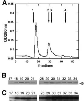
Purification of GST-YsxC overexpressed in E. coli pulls
down high-molecular-weight compounds.
After overexpression
of the GST-YsxC fusion protein in E. coli, its purification by a
standard procedure using glutathione affinity chromatography
gave essentially a single “pure” band when analyzed by
SDS-PAGE, with an apparent molecular mass of
⬃45 kDa (data not
shown). Unexpectedly, however, analysis of the “purified”
GST-YsxC fusion protein by size exclusion chromatography
revealed the presence of two main peaks, eluting in fractions
18 to 20 and 29 to 31 (Fig. 1A). The second peak (fractions 29
to 31) eluted with an apparent molecular mass of
⬃45 kDa
and, as expected, contained the GST-YsxC protein, as shown
in Fig. 1B. This was confirmed by Western blot analysis using
anti-GST antibodies (Fig. 1C). The first peak (fractions 18 to
20) eluted in the void volume of the column and therefore had
an estimated molecular mass of over 400 kDa. SDS-PAGE
showed that these fractions contained only protein(s) with an
apparent molecular mass of
⬃45 kDa (Fig. 1B), suggesting that
GST-YsxC also eluted in these fractions. No additional protein
could be visualized on the stained gel. The presence of the
GST moiety was confirmed in these fractions using anti-GST
antibodies (Fig. 1C), thus demonstrating that GST-YsxC was
indeed the protein present in this peak. The amount of
GST-YsxC in the first peak was estimated to be
⬃50% of that in the
second peak (Fig. 1B and C), whereas the absorbance at 280
nm of the first peak reached a value almost twice as high as that
in the second peak (Fig. 1A). A further analysis of this peak of
high molecular mass revealed that it absorbed strongly at 260
nm (OD
260/OD
280⬎ 1.6) suggesting that it likely contained
nucleic acid associated with the GST-YsxC protein.
The B. subtilis YsxC protein strongly interacts with
ribo-somal material from E. coli.
In order to analyze the nature of
the material associated with the GST-YsxC fusion protein,
samples were first treated with RNase or DNase prior to being
subjected to size exclusion chromatography, as previously
re-ported for the Era protein (41). When the GST-YsxC
prepa-ration was first treated with 1 mg/ml of RNase A (previously
boiled for 15 min at 100°C to inactivate the possibly
nating DNases), the peak of high molecular mass totally
dis-appeared from the elution profile (Fig. 2A). Concomitantly,
the peak with an apparent molecular mass of about 45 kDa,
corresponding to the GST-YsxC protein alone, increased
slightly but significantly. Two new peaks also appeared: one,
containing fractions 39 to 40, corresponded to RNase
(appar-ent molecular mass,
⬃14 kDa), and the last peak (around
fraction 45) corresponded to very low-molecular-mass material
(probably nucleotides and salts). These results strongly
sug-gested that the GST-YsxC fusion protein formed a complex
with some E. coli RNA. By contrast, prior treatment of
GST-YsxC with 0.2 mg/ml of DNase I (RNase free) did not modify
the elution profile of GST-YsxC (Fig. 2B).
To assess which moiety of the GST-YsxC fusion interacted
with E. coli RNA, several controls were made. First,
purifica-tion of the His
6-YsxC tagged protein led to a column profile
similar to that seen with the GST-YsxC fusion protein when
analyzed by size exclusion chromatography (data not shown).
Conversely, purification of the GST tag alone run on size
exclusion chromatography did not allow the detection of
high-molecular-mass species (data not shown). Also, a thrombin
digestion was performed to remove the GST moiety from the
GST-YsxC fusion protein, thereby allowing the purification of
the YsxC moiety alone (apparent molecular mass,
⬃22 kDa).
This sample was then submitted to size exclusion
chromatog-raphy as before, and again, a peak of high molecular mass was
obtained, which contained the YsxC protein alone (fractions
18 to 20), while free YsxC eluted from the column with a
higher retention time (fractions 35 to 37), due, as expected, to
its lower molecular mass (Fig. 2C). Collectively, these results
show that high-molecular-mass material, most likely the
ribo-some, is associated with the YsxC moiety and not with the GST
moiety.
To further analyze the nature of the high-molecular-mass
compounds copurified with the GST-YsxC fusion, the mobility
of phenol-chloroform-extracted material from a GST-YsxC
preparation was checked using denaturing agarose gel
electro-phoresis and staining with ethidium bromide. Two bands with
apparent mobilities of
⬃1.5 and ⬃3 kb were visualized for
GST-YsxC-associated material, and two bands with similar
apparent mobilities were also observed for the E. coli RNA
extracted from the ribosomes (Fig. 3, lanes 1 and 2). Together,
these experiments show that rRNA is part of the
high-molec-ular-mass material copurified with GST-YsxC.
YsxC overexpressed in E. coli can be copurified with the
ribosomal fraction.
To further show that overexpressed YsxC
was bound to the ribosome, we purified ribosomes from E. coli
bacteria following overexpression of GST alone, GST-YsxC, or
His
6-YsxC, as checked by an analytical SDS-PAGE (data not
shown). The ribosomal fractions were then loaded onto a 12%
SDS-PAGE gel, and the overexpressed proteins putatively
co-purified with the ribosomes were visualized by Western
blot-ting. The presence of the GST-YsxC fusion protein was
de-tected, with an expected molecular mass of
⬃45 kDa, and a
faint band with an apparent molecular mass of
⬃25 kDa was
also present (Fig. 4A, lane 1). The latter band most likely
corresponded to the GST moiety, probably resulting from
some proteolytic degradation of the fusion protein. Ribosomes
prepared from bacteria overexpressing GST alone also gave a
similar faint signal (Fig. 4A, lane 2). This reflected some
non-specific binding of the GST protein alone to the ribosomes.
When a similar experiment was carried out with ribosomes
prepared from E. coli overexpressing His
6-YsxC, the presence
of this tagged-protein was detected as well, using a
HisProbe-HPR. It must be noted that control experiments performed
from ribosomes obtained from “native,” nontransformed E.
coli cells showed no detectable bands at the corresponding
molecular weights when probed with either anti-GST or
HisProbe-HPR (data not shown).
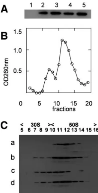
In order to pinpoint which ribosomal subunit YsxC was
bound to, ribosomes prepared from His
6-YsxC-overexpressing
E. coli were dissociated under a low Mg
2⫹concentration and
high-salt conditions in the presence of chloramphenicol. The
mixture of 30S and 50S ribosomal subunits was layered onto a
5 to 20% sucrose gradient and separated by
ultracentrifuga-tion. Each fraction of the gradient was analyzed by OD
260, and
the result is shown in Fig. 4B. To detect the presence of YsxC,
fractions of the gradient were TCA precipitated and loaded
onto an SDS-PAGE gel, and the presence of His
6-YsxC was
revealed by Western blot analysis using a HisProbe-HPR.
Clearly, His
6-YsxC was found associated with the 50S
ribo-somal subunit only (fractions 13 to 17) (Fig. 4B, inset). These
observations corroborate the conclusion that YsxC can be
found in vivo associated with ribosomes.
FIG. 1. Analysis of GST-YsxC by gel filtration chromatography.
(A) Purified GST-YsxC was subjected to gel filtration chromatography
through a Superdex-200 column, as described in Materials and
Meth-ods, and 0.5-ml fractions were collected and analyzed. The absorbance
at 280 nm (OD
280) is indicated. The elution positions of molecular
mass markers (Amersham) are indicated by arrows 1 to 4, respectively,
as follows: Blue Dextran 2000 (mass, 2,000 kDa), albumin (mass, 66
kDa), ovalbumin (mass, 45 kDa), and RNase A (mass, 13.7 kDa).
(B) Fractions were analyzed by 12% acrylamide SDS-PAGE and
stained with Coomassie blue. The position of the 45-kDa molecular
mass marker is indicated. (C) Analysis by Western blotting of the
fractions eluted from gel filtration. The fractions were transferred onto
an Immobilon-P transfer membrane and probed with an anti-GST
antibody, as described in Materials and Methods.
Purified YsxC interacts mainly with the 50S ribosomal
sub-unit of B. subtilis.
Because the previous experiments reflected
“nonnative” interaction between E. coli ribosomes and
over-expressed YsxC, we also investigated the interaction of YsxC
with its cognate ribosomes purified from B. subtilis. In a
pre-vious report, we had shown that YsxC was able to interact with
the 70S B. subtilis ribosomes, and this interaction seemed to be
increased by the presence of either GDP or GTP (61). Figure
5A confirms and extends these results, since both nucleotides
favored the binding of YsxC to ribosomes. In addition, it is
shown here that the nonhydrolyzable GTP analogue GMPPNP
strongly stimulated the binding of YsxC to ribosomes. To
iden-tify the ribosome subunit to which YsxC was bound, 30S and
50S subunits were separated by sucrose gradient, and the
frac-tion containing His
6-YsxC was revealed as shown in Fig. 4. It is
important to note here that the addition of nucleotides did not
change the overall profiles of ribosomes dissociated into 30S
and 50S subunits (only the absorbance profile obtained in the
absence of nucleotide is shown in Fig. 5B). In the absence of
any nucleotide, YsxC was found to be associated with the 50S
FIG. 2. Gel filtration chromatography profiles of GST-YsxC
prep-arations from a Superdex-200 column. The experiments were
con-ducted exactly as described in the legend to Fig. 1. The insets show
SDS-PAGE gels, stained with Coomassie blue, of the different frac
tions eluted from the gel filtration column. (A) Elution profile of
RNase-treated GST-YsxC. Elution of 45-kDa material occurs at
frac-tions 29 and 30. (B) Elution profile of DNase-treated GST-YsxC. (C)
Elution profile of GST-YsxC, where the GST moiety was digested by
thrombin while bound to glutathione-Sepharose beads prior to the gel
filtration.
FIG. 3. Analysis of the material associated with YsxC. Analysis was
performed on a 1.5% agarose gel containing formaldehyde that was
stained with ethidium bromide. Lane 1, phenol-chloroform-isoamyl
alcohol-extracted material from a GST-YsxC preparation; lane 2,
phe-nol-chloroform-isoamyl alcohol-extracted RNA from E. coli
ribo-somes. The arrows indicate the migrations of 1.5-kb and 3-kb DNA
fragments.
subunit only (Fig. 5C, line a). Addition of the nucleotide GDP,
GTP, or GMPPNP (Fig. 5C, lines b, c, and d, respectively) still
allowed binding to the 50S subunit, but marginal binding to the
30S subunit was also detected for each nucleotide. GDP or
GMPPNP was preferred over GTP for binding to the 50S
subunit, suggesting either that during GTP hydrolysis YsxC
goes through a cycle of interaction/dissociation with the 50S
subunit or that in its GTP-bound state, the affinity of YsxC for
this subunit is somehow reduced. The latter explanation,
how-ever, seems unlikely since in the presence of GMPPNP, which
mimics a GTP-bound state, YsxC appeared quite able to
in-teract with the 50S subunit.
In order to address the question of which ribosomal proteins
YsxC was able to interact with, a far-Western blotting
ap-proach was used. In the first step, 30S or 50S ribosomal
pro-teins from B. subtilis were separated by SDS-PAGE,
trans-ferred to Immobilon-P membranes, renatured on the membrane
as previously described (10), and incubated with His
6-YsxC.
After extensive washing of the membrane, the presence of the
His
6-YsxC protein bound to ribosomal proteins was detected
using the HisProbe-HPR. Under these conditions, no
ribo-somal proteins that belonged to the 30S subunit were found to
interact with YsxC (not shown). In contrast, four reactive
bands from the 50S subunit were revealed as putative partners
of His
6-YsxC (Fig. 6B). When far-Western blotting was
per-formed using total ribosomal proteins instead of 50S ribosomal
proteins, similar bands were found to react with the His probe
(data not shown). A control experiment was performed in
which the His probe was directly incubated with ribosomal
FIG. 4. Analysis of ribosomes from E. coli cells overexpressing
YsxC. (A) Western blot of purified ribosomes from E. coli cells, where
GST-YsxC (lane 1), GST (lane 2), or His
6-YsxC (lane 3) was
overex-pressed. Ribosomes (about 50 pmol) were loaded onto a 14%
SDS-PAGE gel, and proteins were transferred onto an Immobilon-P
trans-fer membrane and probed with an anti-GST antibody (lanes 1 and 2)
or with an anti-His antibody (lane 3), as described in Materials and
Methods. The positions of the 25-kDa (REase Bsp981) and 45-kDa
(ovalbumin) molecular mass markers are indicated. (B) YsxC
cofrac-tionates with the 50S ribosomal subunit by sucrose density
centrifuga-tion. E. coli ribosomes were purified from His
6-YsxC-induced cells.
Shown is the UV profile at 260 nm of the different fractions of the 5 to
20% sucrose gradient, with the top of the gradient on the left.
Frac-tions (0.5 ml) were precipitated with TCA and separated by SDS-12%
PAGE, and after transfer onto Immobilon-P, the presence of His
6-YsxC was revealed with anti-His antibodies (shown as an inset).
FIG. 5. Binding of YsxC to 70S ribosomes and ribosomal subunits.
(A) His
6-YsxC was tested for the ability to interact with 70S ribosomes
following a 15-min incubation at room temperature in the presence or
absence of GDP, GTP, or GMPPNP (1 mM each). Pelleting assays
were conducted as described in Materials and Methods. The pellets
were analyzed by SDS-PAGE and Western blotting, using anti-His
antibody, as described in Materials and Methods. Lane 1, His
6-YsxC;
lanes 2 to 5, His
6-YsxC incubated with 70S ribosomes in the absence
(lane 2) or in the presence of GDP (lane 3), GTP (lane 4), or GMPPNP
(lane 5). (B) Typical absorbance profile of B. subtilis ribosomes
sedi-mented through 5 to 20% sucrose gradients, with the top of the
gra-dient on the left. (C) Effects of nucleotides on YsxC sedimentation.
Shown is Western blot analysis of fractions obtained by velocity
cen-trifugation of B. subtilis ribosomes through 5 to 20% sucrose gradients
containing no nucleotide (line a), 100
M GDP (line b), 100 M GTP
(line c), or 100
M GMPPNP (line d) after a prior incubation with
His
6-YsxC with no added nucleotide (line a), 1 mM GDP (line b), 1
mM GTP (line c), or 1 mM GMPPNP (line d).
proteins previously separated by SDS-PAGE and transferred
to an Immobilon-P membrane. Under these conditions, where
no YsxC was added, no signal was detected, showing that no
nonspecific recognition of ribosomal proteins occurred using
the His probe (data not shown). To assign the ribosomal
pro-teins to which the His
6-YsxC protein was bound,
superimpo-sition of the detected bands with those on the membrane
previously stained with Ponceau red was carried out and
com-pared to an SDS-PAGE of the 50S ribosomal proteins
migrat-ing under similar conditions. The correspondmigrat-ing bands, shown
in Fig. 6A, were excised, and the protein content of each band
was analyzed by nano-LC-MS/MS. Bands 1 and 2 were in some
cases well separated on the gradient gel, and both were
reac-tive toward His
6-YsxC. The identified proteins are listed in
Table 1. The proteins present in the doublet of the two upper
bands in Fig. 6B were L1 and L3 in both samples (Table 1, no.
1 and 2) but in different ratios; the upper band contained
essentially L1 (11 peptides compared to 1), whereas the lower
band contained a little bit more L3 (11 peptides out of 20). In
the third band, the most abundant protein was L10 (with nine
peptides and a coverage of the sequence of 38.2%), and one
peptide corresponded to L6. In the fourth band, the most
abundant protein was L27 (7 peptides out of 11), but L7/L12
was also present (3 peptides), as well as L23 (1 peptide).
To validate our results, we used a pull-down assay, in which
one of the proteins (YsxC or a ribosomal protein) was cloned
with a GST tag at one of its extremities and bound to a
gluta-thione-agarose column. If a putative interacting protein
(lack-ing a GST tag) bound to the immobilized target protein and
coeluted from the column when the target was eluted with
reduced glutathione, then one could infer that the two proteins
interacted. All of the putative interacting ribosomal proteins
(L1, L3, L6, L7/L12, L10, L23, and L27) were thus cloned with
a His
6tag to facilitate their purification and, when soluble (see
Table S2 in the supplemental material for production and
solubility), were used as “prey” proteins, the immobilized
tar-get protein (the “bait” protein) being GST-YsxC (or GST
protein as a control). In these cases, L7/L12 (mass, 14.9 kDa),
L1 (mass, 29 kDa), and L6 (mass, 22.7 kDa) were eluted from
the glutathione-agarose column, together with GST-YsxC
(mass, 45 kDa) (but not with GST protein alone, as a control),
whereas L23 (mass, 12.6 kDa) did not coelute with GST-YsxC
(Fig. 7A). During the pull-down assay, there was some
prote-olysis of the GST-YsxC fusion protein, resulting in the
pres-ence of two protein bands on the gel: GST-YsxC and GST
(YsxC was washed away during the washing step). In the case
of L6 (mass, 22.7 kDa), its migration was almost identical to
that of the GST protein (25.2 kDa); therefore, the presence of
His
6-L6 was checked and confirmed by immunoblotting, using
anti-His antibody (Fig. 7B). His
6-L10 and His
6-L27 were not
soluble. GST-L10 and GST-L27 were therefore produced but
were poorly soluble (see Table S2 in the supplemental
mate-rial). Coimmobilization assays were then performed with
GST-L10 and GST-L27 as the prey proteins, using His
6-YsxC as
bait, but no interaction could be detected between these two
ribosomal proteins and YsxC. However, one has to keep in
FIG. 6. Interaction of YsxC with a subset of ribosomal proteins.
Proteins from B. subtilis 50S ribosomal subunits were analyzed by
SDS-PAGE. After transfer onto an Immobilon-P membrane, the
pro-teins were stained with Ponceau red dye. Ribosomal propro-teins bound to
the membrane were renatured and incubated with purified His
6-YsxC.
After extensive washing, YsxC was detected by immunoblotting using
an anti-His antibody and revealed on a film using enhanced
chemilu-minescence. (A) Separation of 50S ribosomal proteins through a 14 to
18% SDS-PAGE gel. The 20-, 27-, and 36-kDa molecular mass
mark-ers were the Prestained Protein Molecular Weight Markmark-ers from
Fer-mentas, and the 6- and 16-kDa molecular mass markers were the
Seeblue Plus 2-stained standard from Invitrogen. (B) Detection of
bound YsxC by immunoblotting. The film was superimposed on the
corresponding Immobilon-P membrane stained with Ponceau red dye
to localize the protein bands (indicated by arrows).
TABLE 1. Protein identification by MS/MS
aBand no. Protein description Accession no. Scoreb
Mass (Da) Coverage (%)c
No. of peptidesd
1
L1 (RL1_BACSU)
Q06797
762.4
24,776
47.2
11
L3 (RL3_BACSU)
P42920
92.4
22,669
7.3
1
2
L3 (RL3_BACSU)
P42920
722.9
22,669
50.7
11
L1 (RL1_BACSU)
Q06797
542.3
24,776
36.4
9
3
L10 (RL10_BACSU)
P42923
401.4
17,887
38.2
9
L6 (RL6_BACSU)
P46898
69.6
19,366
6.8
1
4
L27 (RL27_BACSU)
P05657
320.5
10,366
47.9
7
L7/L12 (RL7_BACSU)
P02394
170.8
12,612
23.8
3
L23 (RL23_BACSU)
P42924
35.2
10,922
13.1
1
aProteins were identified by nano-LC-ESI (electrospray ionization) MS/MS, followed by protein database mining. bScore attributed to the identified protein by Mascot software (Matrix Science).
cCoverage (%), percentage of the full-length sequence covered by the matching peptides. dNo of peptides, number of peptides assigned to the protein.
mind that an additional interaction(s) between YsxC and some
other putative ribosomal partner(s) might have occurred but
that it was too loose to be retained during the washing steps. In
conclusion, we were able to show that the L1, L6, and L7/L12
ribosomal proteins interact in vitro with YsxC, with the
possi-bility that an additional interaction(s) between YsxC and some
other soluble putative ribosomal partner(s) might have occurred
but was too loose to be retained during the washing steps.
DISCUSSION
In the past few years, numerous nonribosomal proteins,
in-cluding many GTPases, have been found to play key roles in
ribosome assembly in eukaryotic cells (19, 31), and a similar
trend is now emerging for the prokaryotic ribosome (15). YsxC
has recently been suggested to be involved in ribosome
bio-genesis (61), and we report here that YsxC interacts with
several ribosomal proteins of the 50S subunit.
We observed above that heterologous expression of
recom-binant YsxC from B. subtilis in E. coli cells resulted in the
association of YsxC with ribosomes, which involved, at least in
part, a direct interaction with rRNA. A possible interaction
between YsxC and RNA was initially proposed based on the
three-dimensional structure of YsxC due to the presence on its
surface of a patch of conserved basic residues that might act as
a hook to anchor the protein to RNA (57). Moreover, among
the newly discovered bacterial GTPases that interact with
ri-bosome, many have been shown to have the ability to bind to
rRNA (5, 25, 27, 40, 42, 43), and this is probably a conserved
trait originating from an ancient GTPase traceable to the last
universal common ancestor (13, 36).
We were able to show that YsxC preferentially binds the 50S
subunit and that guanine nucleotide occupancy of YsxC affects
its association with the ribosome. This result is consistent with
the different conformations adopted by YsxC in the
GTP-bound, GDP-GTP-bound, and apo-enzyme states (57). Thus, it is
likely that the ribosome-associated GTP-bound YsxC is
re-leased from the ribosome as a consequence of the hydrolysis of
GTP.
To identify the possible ribosomal-protein partners of YsxC,
a far-Western approach was used, leading to a positive
inter-action with L1, L3, at least one protein of a mixture of L6 and
L10, and one protein of a mixture of L7/L12 (L7 and L12 are
the same protein, but L7 is acetylated), L23, and L27. It is
important to stress here the importance of protein L27 for both
the assembly and function of the ribosome, since L27 has been
suggested to be lacking in the ribosomes of YsxC-depleted
cells (61) and deletion of the gene results in a severe growth
defect (38, 73). All of the putative protein partners that we
identified belong to the 50S subunit, while the 30S subunit
failed to reveal any positive band, a result consistent with the
interaction of YsxC primarily with the 50S subunit.
In order to validate our far-Western results, we performed
pull-down assays with YsxC and recombinant ribosomal
pro-teins and confirmed a direct interaction between L1, L6, or
L7/L12 and YsxC. We were unable to detect any interaction
between L10 or L27 and YsxC by this approach. However, for
L10, its N-terminal part is free in the ribosome, while its
C-terminal part interacts with two L7/L12 dimers in E. coli (20,
23, 24, 51). In contrast, in the recombinant protein, its
N-terminal part is linked to the GST moiety, and this could have
prevented its interaction with YsxC. Thus, we cannot rule out
the possibility that L10 is a physiological partner of YsxC.
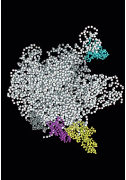
It is remarkable that most of the proteins that potentially
interact with YsxC, except L1, are in the same location on the
FIG. 8. Components of the 50S subunit of Thermus thermophilus
ribosomes. 23S and 5S rRNAs are shown in white. Only proteins L1
(cyan), L3 (gray), L6 (magenta), and L7/L12 (yellow) are represented.
The figure was generated using PYMOL software and the structure of
the T. thermophilus 50S ribosome (Protein Data Bank accession
num-ber 1GIY) (74). In this structure, L10 and L27 were missing, and only
one of the two L7/L12 dimers was represented.
FIG. 7. Pull-down assay of YsxC and His-tagged recombinant
ri-bosomal proteins. (A) SDS-PAGE of coeluted GST-YsxC and
somal prey proteins from a glutathione-agarose column. The
ribo-somal proteins used as prey proteins were L7 (lane 1), L23 (lane 2), L1
(lane 3), and L6 (lane 4). GST-YsxC alone was loaded as a control
(lane 5) and gave two bands (GST-YsxC and GST, due to partial
cleavage of the YsxC moiety). The values of the molecular mass
mark-ers (Fermentas) are indicated on the right of the gel in kDa. (B)
West-ern blot analysis using an anti-His antibody of coeluted ribosomal
proteins with GST-YsxC. Lane 1, L7; lane 2, L23; lane 3, L1; lane
4, L6.
ribosome (Fig. 8 shows the protein localization). The L7/L12
proteins are the only ribosomal proteins that are present in
multiple copies in prokaryotic ribosomes. They are organized
as two L7/L12 dimers associated with the C-terminal region of
protein L10, and at least one of the dimers comprises the stalk
protuberance (20, 23, 24, 51). The detailed structure of the
pentameric complex has not yet been resolved by X-ray
crys-tallography of ribosomes (4, 6, 26, 33, 62, 74). On the basis of
immune electron microscopy, the two L7/L12 dimers have
been proposed to bind to different locations and also to adopt
different conformations (49): one nonstalk dimer of protein
L7/L12 is in a folded conformation on the subunit body, while
the second dimer is in an extended conformation in the subunit
stalk. Other studies (cross-linking data and cryoelectron
mi-croscopic localization) also suggest multiple locations for L7/
L12 in the large subunit (18, 44, 65). The ribosomal stalk is
highly flexible, and various conformations of the stalk are
thought to occur in response to elongation factor binding and
GTP hydrolysis (8). As for L1, it has been proposed to be
located on the opposite side of the ribosome from the stalk
position (39, 68). However, intrinsic flexibility of L1 has been
suggested, depending on RNA binding (46–48); it adopts a
closed conformation in the absence of RNA (47) but opens
upon RNA binding (46, 48). This probably explains the
diffi-culty in precisely determining the structure of the L1
protu-berance within the ribosome, due to the high mobility of the
region (1, 48). The flexibility of these regions might lead to a
possible location of L1 closer to L7/L12 in the
three-dimen-sional structure. Alternatively, one might propose that YsxC is
able to interact with L1 and L7/L12 at different locations on
the ribosome. The different patterns of interaction of YsxC
with the 50S ribosomal proteins, depending on the nature of
the nucleotide added (Fig. 5), might support such a hypothesis.
YsxC is required for 50S ribosomal-subunit assembly in vivo,
since a 44.5S ribosomal intermediate accumulates in cells
de-pleted of YsxC (61). This intermediate lacks ribosomal
pro-teins L16, L27, and L36 (61), and we also have evidence that
YsxC interacts with the 44.5S complex (data not shown).
Al-together, these results strongly suggest that YsxC participates
in the assembly and/or the processing steps of the preribosomal
particle. We propose that YsxC binds to the 44.5S
preriboso-mal complex through interaction with the partner ribosopreriboso-mal
proteins identified here and/or possibly through association
with rRNA. This association would somehow locally modify
the conformation of the presubunit, thereby allowing the
in-corporation of the other missing proteins.
ACKNOWLEDGMENTS
We are grateful to Je
´ro
ˆme Garin for preparing Table 1 and for
critical reading of the manuscript.
This work was supported by a CNRS “Young Investigator” ATIP
Program (J.-M.J.).
REFERENCES
1. Agrawal, R. K., R. K. Lata, and J. Frank. 1999. Conformational variability in
Escherichia coli 70S ribosome as revealed by 3D cryo-electron microscopy.
Int. J. Biochem. Cell Biol. 31:243–254.
2. Arigoni, F., F. Talabot, M. Peitsch, M. D. Edgerton, E. Meldrum, E. Allet, R.
Fish, T. Jamotte, M. L. Curchod, and H. Loferer.1998. A genome-based approach for the identification of essential bacterial genes. Nat. Biotechnol.
16:851–856.
3. Arnosti, D. N., V. L. Singer, and M. J. Chamberlin. 1986. Characterization of heat shock in Bacillus subtilis. J. Bacteriol. 168:1243–1249.
4. Ban, N., P. Nissen, J. Hansen, P. B. Moore, and T. A. Steitz. 2000. The complete atomic structure of the large ribosomal subunit at 2.4 Å resolution. Science 289:905–920.
5. Bassler, J., M. Kallas, and E. Hurt. 2006. The NUG1 GTPase reveals and N-terminal RNA-binding domain that is essential for association with 60 S pre-ribosomal particles. J. Biol. Chem. 281:24737–24744.
6. Berk, V., W. Zhang, R. D. Pai, and J. H. Cate. 2006. Structural basis for mRNA and tRNA positioning on the ribosome. Proc. Natl. Acad. Sci. USA
103:15830–15834.
7. Bharat, A., M. Jiang, S. M. Sullivan, J. R. Maddock, and E. D. Brown. 2006. Cooperative and critical roles for both G domains in the GTPase activity and cellular function of ribosome-associated Escherichia coli EngA. J. Bacteriol.
188:7992–7996.
8. Bocharov, E. V., A. G. Sobol, K. V. Pavlov, D. M. Korzhnev, V. A. Jaravine,
A. T. Gudkov, and A. S. Arseniev.2004. From structure and dynamics of protein L7/L12 to molecular switching in ribosome. J. Biol. Chem. 279: 17697–17706.
9. Bourne, H. R., D. A. Sanders, and F. McCormick. 1990. The GTPase super-family: a conserved switch for diverse cell functions. Nature 348:125–132. 10. Bouvet, P., J. J. Diaz, K. Kindbeiter, J. J. Madjar, and F. Amalric. 1998.
Nucleolin interacts with several ribosomal proteins through its RGG do-main. J. Biol. Chem. 273:19025–19029.
11. Brown, E. D. 2005. Conserved P-loop GTPases of unknown function in bacteria: an emerging and vital ensemble in bacterial physiology. Biochem. Cell Biol. 83:738–746.
12. Caldon, C. E., and P. E. March. 2003. Function of the universally conserved bacterial GTPases. Curr. Opin. Microbiol. 6:135–139.
13. Caldon, C. E., P. Yoong, and P. E. March. 2001. Evolution of a molecular switch: universal bacterial GTPases regulate ribosome function. Mol. Micro-biol. 41:289–297.
14. Campbell, T. L., D. M. Daigle, and E. D. Brown. 2005. Characterization of the Bacillus subtilis GTPase YloQ and its role in ribosome function. Bio-chem. J. 389:843–852.
15. Comartin, D. J., and E. D. Brown. 2006. Non-ribosomal factors in ribosome subunit assembly are emerging targets for new antibacterial drugs. Curr. Opin. Pharmacol. 6:453–458.
16. Daigle, D. M., and E. D. Brown. 2004. Studies of the interaction of
Esche-richia coli YjeQ with the ribosome in vitro. J. Bacteriol. 186:1381–1387.
17. Dassain, M., A. Leroy, L. Colosetti, S. Carole, and J. P. Bouche. 1999. A new essential gene of the ‘minimal genome’ affecting cell division. Biochimie
81:889–895.
18. Dey, D., D. E. Bochkariov, G. G. Jokhadze, and R. R. Traut. 1998. Cross-linking of selected residues in the N- and C-terminal domains of Escherichia
coli protein L7/L12 to other ribosomal proteins and the effect of elongation
factor Tu. J. Biol. Chem. 273:1670–1676.
19. Dez, C., and D. Tollervey. 2004. Ribosome synthesis meets the cell cycle. Curr. Opin. Microbiol. 7:631–637.
20. Diaconu, M., U. Kothe, F. Schlunzen, N. Fischer, J. M. Harms, A. G.
Tonevitsky, H. Stark, M. V. Rodnina, and M. C. Wahl.2005. Structural basis for the function of the ribosomal L7/12 stalk in factor binding and GTPase activation. Cell 121:991–1004.
21. Falson, P., A. Di Pietro, and D. C. Gautheron. 1986. Chemical modification of thiol groups of mitochondrial F1-ATPase from the yeast
Schizosaccharo-myces pombe. Involvement of alpha- and gamma-subunits in the enzyme
activity. J. Biol. Chem. 261:7151–7159.
22. Galperin, M. Y., and E. V. Koonin. 2004. ‘Conserved hypothetical’ proteins: prioritization of targets for experimental study. Nucleic Acids Res. 32:5452– 5463.
23. Griaznova, O., and R. R. Traut. 2000. Deletion of C-terminal residues of
Escherichia coli ribosomal protein L10 causes the loss of binding of one
L7/L12 dimer: ribosomes with one L7/L12 dimer are active. Biochemistry
39:4075–4081.
24. Gudkov, A. T., L. G. Tumanova, S. Y. Venyaminov, and N. N.
Khechinash-villi.1978. Stoichiometry and properties of the complex between ribosomal proteins L7 and L10 in solution. FEBS Lett. 93:215–218.
25. Hang, J. Q., T. I. Meier, and G. Zhao. 2001. Analysis of the interaction of 16S rRNA and cytoplasmic membrane with the C-terminal part of the
Strepto-coccus pneumoniae Era GTPase. Eur. J. Biochem. 268:5570–5577.
26. Harms, J., F. Schluenzen, R. Zarivach, A. Bashan, S. Gat, I. Agmon, H.
Bartels, F. Franceschi, and A. Yonath.2001. High resolution structure of the large ribosomal subunit from a mesophilic eubacterium. Cell 107:679–688. 27. He, W. J., S. Tang, and W. Y. Liu. 2002. In vitro interaction of eukaryotic
elongation factor 2 with synthetic oligoribonucleotide that mimics GTPase domain of rat 28S ribosomal RNA. Int. J. Biochem. Cell Biol. 34:263–268. 28. Hwang, J., and M. Inouye. 2006. The tandem GTPase, Der, is essential for the biogenesis of 50S ribosomal subunits in Escherichia coli. Mol. Microbiol.
61:1660–1672.
29. Jaquinod, M., F. Villiers, S. Kieffer-Jaquinod, V. Hugouvieux, C. Bruley, J.
Garin, and J. Bourguignon.2007. A proteomics dissection of Arabidopsis
thaliana vacuoles isolated from cell culture. Mol. Cell Proteomics 6:394–412.
30. Jeffares, D. C., A. M. Poole, and D. Penny. 1998. Relics from the RNA world. J. Mol. Evol 46:18–36.
31. Johnson, A. W., E. Lund, and J. Dahlberg. 2002. Nuclear export of ribosomal subunits. Trends Biochem. Sci. 27:580–585.
32. Johnstone, B. H., A. A. Handler, D. K. Chao, V. Nguyen, M. Smith, S. Y. Ryu,
E. L. Simons, P. E. Anderson, and R. W. Simons.1999. The widely conserved Era G-protein contains an RNA-binding domain required for Era function
in vivo. Mol. Microbiol. 33:1118–1131.
33. Korostelev, A., S. Trakhanov, M. Laurberg, and H. F. Noller. 2006. Crystal structure of a 70S ribosome-tRNA complex reveals functional interactions and rearrangements. Cell 126:1065–1077.
34. Laemmli, U. K. 1970. Cleavage of structural proteins during the assembly of the head of bacteriophage T4. Nature 227:680–685.
35. Lehoux, I. E., M. J. Mazzulla, A. Baker, and C. M. Petit. 2003. Purification and characterization of YihA, an essential GTP-binding protein from
Esch-erichia coli. Protein Expr. Purif. 30:203–209.
36. Leipe, D. D., Y. I. Wolf, E. V. Koonin, and L. Aravind. 2002. Classification and evolution of P-loop GTPases and related ATPases. J. Mol. Biol. 317: 41–72.
37. Lin, B., D. A. Thayer, and J. R. Maddock. 2004. The Caulobacter crescentus CgtAC protein cosediments with the free 50S ribosomal subunit. J. Bacteriol.
186:481–489.
38. Maguire, B. A., A. D. Beniaminov, H. Ramu, A. S. Mankin, and R. A.
Zimmermann.2005. A protein component at the heart of an RNA machine: the importance of protein L27 for the function of the bacterial ribosome. Mol. Cell 20:427–435.
39. Malhotra, A., P. Penczek, R. K. Agrawal, I. S. Gabashvili, R. A. Grassucci,
R. Junemann, N. Burkhardt, K. H. Nierhaus, and J. Frank.1998.
Esche-richia coli 70 S ribosome at 15 Å resolution by cryo-electron microscopy:
localization of fMet-tRNAfMet and fitting of L1 protein. J. Mol. Biol. 280: 103–116.
40. Matsuo, Y., T. Morimoto, M. Kuwano, P. C. Loh, T. Oshima, and N.
Oga-sawara.2006. The GTP-binding protein YlqF participates in the late step of 50 S ribosomal subunit assembly in Bacillus subtilis. J. Biol. Chem. 281:8110– 8117.
41. Meier, T. I., R. B. Peery, S. R. Jaskunas, and G. Zhao. 1999. 16S rRNA is bound to Era of Streptococcus pneumoniae. J. Bacteriol. 181:5242–5249. 42. Meier, T. I., R. B. Peery, K. A. McAllister, and G. Zhao. 2000. Era GTPase
of Escherichia coli: binding to 16S rRNA and modulation of GTPase activity by RNA and carbohydrates. Microbiology 146:1071–1083.
43. Moazed, D., J. M. Robertson, and H. F. Noller. 1988. Interaction of elon-gation factors EF-G and EF-Tu with a conserved loop in 23S RNA. Nature
334:362–364.
44. Montesano-Roditis, L., D. G. Glitz, R. R. Traut, and P. L. Stewart. 2001. Cryo-electron microscopic localization of protein L7/L12 within the
Esche-richia coli 70 S ribosome by difference mapping and Nanogold labeling.
J. Biol. Chem. 276:14117–14123.
45. Morimoto, T., P. C. Loh, T. Hirai, K. Asai, K. Kobayashi, S. Moriya, and N.
Ogasawara.2002. Six GTP-binding proteins of the Era/Obg family are es-sential for cell growth in Bacillus subtilis. Microbiology 148:3539–3552. 46. Nevskaya, N., S. Tischenko, R. Fedorov, S. Al-Karadaghi, A. Liljas, A. Kraft,
W. Piendl, M. Garber, and S. Nikonov.2000. Archaeal ribosomal protein L1: the structure provides new insights into RNA binding of the L1 protein family. Structure 8:363–371.
47. Nikonov, S., N. Nevskaya, I. Eliseikina, N. Fomenkova, A. Nikulin, N.
Ossina, M. Garber, B. H. Jonsson, C. Briand, S. Al-Karadaghi, A. Svensson, A. Aevarsson, and A. Liljas.1996. Crystal structure of the RNA binding ribosomal protein L1 from Thermus thermophilus. EMBO J. 15:1350–1359. 48. Nikulin, A., I. Eliseikina, S. Tishchenko, N. Nevskaya, N. Davydova, O.
Platonova, W. Piendl, M. Selmer, A. Liljas, D. Drygin, R. Zimmermann, M. Garber, and S. Nikonov.2003. Structure of the L1 protuberance in the ribosome. Nat. Struct. Biol 10:104–108.
49. Olson, H. M., A. Sommer, D. S. Tewari, R. R. Traut, and D. G. Glitz. 1986. Localization of two epitopes of protein L7/L12 to both the body and stalk of the large ribosomal subunit. Immune electron microscopy using monoclonal antibodies. J. Biol. Chem. 261:6924–6932.
50. Pandit, S. B., and N. Srinivasan. 2003. Survey for g-proteins in the prokary-otic genomes: prediction of functional roles based on classification. Proteins
52:585–597.
51. Pettersson, I., S. J. Hardy, and A. Liljas. 1976. The ribosomal protein L8 is a complex L7/L12 and L10. FEBS Lett. 64:135–138.
52. Phillips, T. A., R. A. VanBogelen, and F. C. Neidhardt. 1984. lon gene product of Escherichia coli is a heat-shock protein. J. Bacteriol. 159:283–287. 53. Pragai, Z., and C. R. Harwood. 2000. YsxC, a putative GTP-binding protein
essential for growth of Bacillus subtilis 168. J. Bacteriol. 182:6819–6823. 54. Pullman, M. E., H. S. Penefsky, A. Datta, and E. Racker. 1960. Partial
resolution of the enzymes catalyzing oxidative phosphorylation. I. Purifica-tion and properties of soluble dinitrophenol-stimulated adenosine triphos-phatase. J. Biol. Chem. 235:3322–3329.
55. Riethdorf, S., U. Volker, U. Gerth, A. Winkler, S. Engelmann, and M.
Hecker.1994. Cloning, nucleotide sequence, and expression of the Bacillus
subtilis lon gene. J. Bacteriol. 176:6518–6527.
56. Roberts, R. J. 2004. Identifying protein function—a call for community action. PLoS Biol. 2:E42.
57. Ruzheinikov, S. N., S. K. Das, S. E. Sedelnikova, P. J. Baker, P. J. Artymiuk,
J. Garcia-Lara, S. J. Foster, and D. W. Rice.2004. Analysis of the open and closed conformations of the GTP-binding protein YsxC from Bacillus subtilis. J. Mol. Biol. 339:265–278.
58. Sambrook, J., and D. W. Russell. 2001. Molecular cloning: a laboratory manual, 3rd ed. Cold Spring Harbor Laboratory Press, Cold Spring Har-bor, NY.
59. Sato, A., G. Kobayashi, H. Hayashi, H. Yoshida, A. Wada, M. Maeda, S.
Hiraga, K. Takeyasu, and C. Wada.2005. The GTP binding protein Obg homolog ObgE is involved in ribosome maturation. Genes Cells 10:393–408. 60. Sayed, A., S. Matsuyama, and M. Inouye. 1999. Era, an essential Escherichia
coli small G-protein, binds to the 30S ribosomal subunit. Biochem. Biophys.
Res. Commun. 264:51–54.
61. Schaefer, L., W. C. Uicker, C. Wicker-Planquart, A. E. Foucher, J. M. Jault,
and R. A. Britton.2006. Multiple GTPases participate in the assembly of the large ribosomal subunit in Bacillus subtilis. J. Bacteriol. 188:8252–8258. 62. Selmer, M., C. M. Dunham, F. V. T. Murphy, A. Weixlbaumer, S. Petry, A. C.
Kelley, J. R. Weir, and V. Ramakrishnan.2006. Structure of the 70S ribo-some complexed with mRNA and tRNA. Science 313:1935–1942. 63. Sprang, S. R. 1997. G protein mechanisms: insights from structural analysis.
Annu. Rev. Biochem. 66:639–678.
64. te Riele, H., B. Michel, and S. D. Ehrlich. 1986. Single-stranded plasmid DNA in Bacillus subtilis and Staphylococcus aureus. Proc. Natl. Acad. Sci. USA 83:2541–2545.
65. Traut, R. R., D. Dey, D. E. Bochkariov, A. V. Oleinikov, G. G. Jokhadze, B.
Hamman, and D. Jameson.1995. Location and domain structure of
Esche-richia coli ribosomal protein L7/L12: site specific cysteine crosslinking and
attachment of fluorescent probes. Biochem. Cell Biol. 73:949–958. 66. Uicker, W. C., L. Schaefer, and R. A. Britton. 2006. The essential GTPase
RbgA (YlqF) is required for 50S ribosome assembly in Bacillus subtilis. Mol. Microbiol. 59:528–540.
67. Vetter, I. R., and A. Wittinghofer. 2001. The guanine nucleotide-binding switch in three dimensions. Science 294:1299–1304.
68. Walleczek, J., D. Schuler, M. Stoffler-Meilicke, R. Brimacombe, and G.
Stoffler.1988. A model for the spatial arrangement of the proteins in the large subunit of the Escherichia coli ribosome. EMBO J. 7:3571–3576. 69. Wang, B., and H. K. Kuramitsu. 2003. Assessment of the utilization of the
antisense RNA strategy to identify essential genes in heterologous bacteria. FEMS Microbiol. Lett. 220:171–176.
70. Wittinghofer, A., and E. F. Pai. 1991. The structure of Ras protein: a model for a universal molecular switch. Trends Biochem. Sci. 16:382–387. 71. Wittinghofer, A., K. Scheffzek, and M. R. Ahmadian. 1997. The interaction
of Ras with GTPase-activating proteins. FEBS Lett. 410:63–67.
72. Wout, P., K. Pu, S. M. Sullivan, V. Reese, S. Zhou, B. Lin, and J. R.
Maddock.2004. The Escherichia coli GTPase CgtAE cofractionates with the 50S ribosomal subunit and interacts with SpoT, a ppGpp synthetase/hydro-lase. J. Bacteriol. 186:5249–5257.
73. Wower, I. K., J. Wower, and R. A. Zimmermann. 1998. Ribosomal protein L27 participates in both 50 S subunit assembly and the peptidyl transferase reaction. J. Biol. Chem. 273:19847–19852.
74. Yusupov, M. M., G. Z. Yusupova, A. Baucom, K. Lieberman, T. N. Earnest,
J. H. Cate, and H. F. Noller.2001. Crystal structure of the ribosome at 5.5 Å resolution. Science 292:883–896.
75. Zhang, S., and W. G. Haldenwang. 2004. Guanine nucleotides stabilize the binding of B. subtilis Obg to ribosomes. Biochem. Biophys. Res. Commun.