HAL Id: hal-01348945
https://hal.archives-ouvertes.fr/hal-01348945
Submitted on 26 Jul 2016
HAL is a multi-disciplinary open access
archive for the deposit and dissemination of
sci-entific research documents, whether they are
pub-lished or not. The documents may come from
teaching and research institutions in France or
abroad, or from public or private research centers.
L’archive ouverte pluridisciplinaire HAL, est
destinée au dépôt et à la diffusion de documents
scientifiques de niveau recherche, publiés ou non,
émanant des établissements d’enseignement et de
recherche français ou étrangers, des laboratoires
publics ou privés.
Intracellular I-II Loop
Arnaud Monteil, Patrick Chausson, Katia Boutourlinsky, Alexandre
Mezghrani, Iulia Blesneac, Isabelle Bidaud, Sylvaine Huc-Brandt, Céline
Lemmers, Nathalie Leresche, Régis C Lambert, et al.
To cite this version:
Arnaud Monteil, Patrick Chausson, Katia Boutourlinsky, Alexandre Mezghrani, Iulia Blesneac, et al..
Inhibition of Cav3.2 T-type Calcium Channels by Its Intracellular I-II Loop. Journal of Biological
Chemistry, American Society for Biochemistry and Molecular Biology, 2015, 290 (26), pp.16168-16176.
�10.1074/jbc.M114.634261�. �hal-01348945�
Inhibition of Cav3.2 T-type Calcium Channels by Its
Intracellular I-II Loop
*
Received for publication, December 19, 2014, and in revised form, April 30, 2015Published, JBC Papers in Press, April 30, 2015, DOI 10.1074/jbc.M114.634261
Arnaud Monteil‡§¶储, Patrick Chausson**‡‡§§, Katia Boutourlinsky**‡‡§§, Alexandre Mezghrani‡§储,
Sylvaine Huc-Brandt‡§储, Iulia Blesneac‡§储, Isabelle Bidaud‡§储, Céline Lemmers‡§¶储, Nathalie Leresche**‡‡§§,
Régis C. Lambert**‡‡§§, and Philippe Lory‡§储1
From the‡Université de Montpellier, CNRS UMR 5203, Département de Physiologie, Institut de Génomique Fonctionnelle, Montpellier, F-34094 France,§INSERM, U1191, Montpellier, F-34094 France,¶Plateforme de Vectorologie, Biocampus Montpellier CNRS UMS 3426, INSERM US009, Montpellier, F-34094 France,储LabEx “Ion Channel Science and Therapeutics,” Montpellier, F-34094 France, **Sorbonne Universités, Université Pierre et Marie Curie Université Paris 06, UM 119, Neuroscience Paris Seine (NPS), Paris F-75005, France,‡‡CNRS UMR 8246, NPS, Paris F-75005, France, and§§INSERM, U1130, NPS, Paris F-75005, France
Background:Novel strategies are needed to characterize the properties of T-type (Cav3) calcium channel isoforms.
Results:The I-II loop of the Cav3.2 protein potently inhibits both recombinant and neuronal Cav3.1 and Cav3.2 channels.
Conclusion:This I-II loop region can be used to selectively silence Cav3.1/Cav3.2 channels.
Significance:This study reveals a new approach to differentiate among the activity of native Cav3 channels.
Voltage-dependent calcium channels (Cav) of the T-type fam-ily (Cav3.1, Cav3.2, and Cav3.3) are activated by low threshold membrane depolarization and contribute greatly to neuronal network excitability. Enhanced T-type channel activity, espe-cially Cav3.2, contributes to disease states, including absence epilepsy. Interestingly, the intracellular loop connecting do-mains I and II (I-II loop) of Cav3.2 channels is implicated in the control of both surface expression and channel gating, indicat-ing that this I-II loop plays an important regulatory role in T-type current. Here we describe that co-expression of this I-II loop or its proximal region (⌬1-Cav3.2; Ser423–Pro542) together with recombinant full-length Cav3.2 channel inhibited T-type current without affecting channel expression and membrane incorporation. Similar T-type current inhibition was obtained in NG 108-15 neuroblastoma cells that constitutively express Cav3.2 channels. Of interest,⌬1-Cav3.2 inhibited both Cav3.2 and Cav3.1 but not Cav3.3 currents. Efficacy of⌬1-Cav3.2 to inhibit native T-type channels was assessed in thalamic neurons using viral transduction. We describe that T-type current was significantly inhibited in the ventrobasal neurons that express Cav3.1, whereas in nucleus reticularis thalami neurons that express Cav3.2 and Cav3.3 channels, only the fast inactivating T-type current (Cav3.2 component) was significantly inhibited. Altogether, these data describe a new strategy to differentially inhibit Cav3 isoforms of the T-type calcium channels.
T-type, low voltage-activated calcium channels are present in many excitable cells, especially in neurons, where they are critical to operate multiple physiological processes, including neuronal firing, neurotransmitter release, and slow wave sleep
(1). In addition, substantial evidence supports a role of T-type calcium channels in diseases, especially in epilepsy and pain (2, 3). Indeed, enhanced activity of neuronal T-type channels would play a pathogenic role in the initiation of seizures and pain signal transmission (4).
Importantly, T-type calcium channels exhibit diversity in function and structure. They are encoded by three Cav32 sub-units (Cav3.1, Cav3.2, and Cav3.3) (for a review, see Ref. 5). Functional expression studies have revealed that these Cav3 subunits all display low threshold activity. However, they mark-edly differ in some other electrophysiological properties. Strik-ingly, the Cav3.1 and Cav3.2 subunits are typical fast inactivat-ing T-type channels, whereas the Cav3.3 subunit exhibits much slower activation and inactivation kinetics (6, 7).
Within the central nervous system, the Cav3.1, Cav3.2, and Cav3.3 subunits are differentially expressed in the various neu-ronal populations (8). However, the precise contributions of each of the three Cav3 subunits to neuron function remain elusive. This is well exemplified in the thalamus where the GABAergic neurons from the nucleus reticularis thalami (NRT) express both Cav3.2 and Cav3.3 subunits, whereas the glutamatergic thalamocortical relay neurons only express the Cav3.1 channels (8, 9). The development of knock-out/trans-genic mice has been useful to identify the main roles of the Cav3 subunits (9 –11). Unfortunately, no pharmacological tools are available to date to discriminate more precisely among the spe-cific roles of the Cav3 subunits.
Toward this goal, we have hypothesized that selective mod-ulation of the Cav3 subunits could be possibly achieved using alternative molecular strategies. Recent studies have identified that the intracellular loop connecting domains I and II (I-II loop) of the Cav3.2 subunit plays an important role in control-*This work was supported by Agence Nationale de la Recherche Grants
ANR-10-BLAN-1601 (to P. L.) and ANR-09-MNPS-035 grants (to P. L., R. C. L., and N. L.). The authors declare that they have no conflicts of interest with the contents of this article.
1To whom correspondence should be addressed: Inst. de Génomique
Fonc-tionnelle, 141 rue de la Cardonille, 34094 Montpellier Cedex 5, France. Tel.: 33-434-359-251; Fax: 33-467-542-432; E-mail: philippe.lory@igf.cnrs.fr.
2The abbreviations used are: Cav, voltage-dependent calcium channel; I-II
loop, intracellular loop connecting domains I and II; VB, ventrobasal com-plex; NRT, nucleus reticularis thalami; TEA, tetraethylammonium; I, current;
V, voltage; Nter, N-terminal region; D, domain; L, loop; Ctrl, control;⍀,
ohms; LTS, low threshold calcium spike.
THE JOURNAL OF BIOLOGICAL CHEMISTRY VOL. 290, NO. 26, pp. 16168 –16176, June 26, 2015 © 2015 by The American Society for Biochemistry and Molecular Biology, Inc. Published in the U.S.A.
at BIOCHIMIE CHU ST ANTOINE on July 26, 2016
http://www.jbc.org/
ling both expression at the plasma membrane and gating of the channel. The proximal part of this I-II loop (subsequent to domain I) was described as a “gating brake” (12). Interestingly, this I-II loop plays distinct roles in plasma membrane expres-sion among the Cav3 channel family (13–15) with a major inhibitory role for Cav3.2, modest inhibitory role in Cav3.1, and forward trafficking role in Cav3.3 channels. Building on these early findings, we show in the present study using two heterol-ogous expression systems, cell lines and thalamic slices, that the proximal I-II loop region of the Cav3.2 subunit can be designed as a molecular tool to specifically inhibit Cav3.1 and Cav3.2 currents without affecting Cav3.3 channel activity.
Experimental Procedures
Constructs—The cDNAs encoding the Cav3.1, Cav3.2, and Cav3.3 subunits were described earlier (7) as well as epitope-tagged Cav3 subunits (15, 16). The various truncated forms of Cav3.2 as well as the I-II loop fragments were generated using PCR techniques, cloned into pEGFP-C1 expression vector (BD Biosciences), and verified using automated DNA sequencing.
Cell Lines—Human embryonic kidney (HEK)-293 and rat neuroblastoma NG 108-15 cells were cultured as described pre-viously (7, 15). Cells were plated at 50 –70% confluence for opti-mal transfection using the JetPEI transfection reagent accord-ing to the manufacturer’s protocol (QBiogene, Irvine, CA). The Cav1.2 construct was cotransfected with␣2/␦1 and 1b sub-units (1:2:2 molar ratio) as described earlier (17). In all condi-tions, corresponding empty plasmids were used to adjust the quantity of the transfected material.
Subcloning and Virus Production—⌬1-Cav3.2 and
Nter-Cav3.2 were subcloned into the Sindbis viral vector SINrep-(nsP2S726) kindly provided by H. Marie (Institut de Pharmacolo-gie Moléculaire et Cellulaire-CNRS UMR 7275). Recombinant SINrep(nsP2S726) constructs and helper plasmid DH-BB(tRNA/ TE12) (18) were transcribed in vitro into capped RNA using the Megascript SP6 kit (Ambion). Baby hamster kidney-21 (ATCC CCL-10) cells were electroporated with ⌬1-Cav3.2 or Nter-Cav3.2 RNA and the helper RNA (20⫻ 106cells/ml, 950 micro-farads, 230 V) and grown for 24 h at 37 °C at 5% CO2in DMEM containing 5% fetal calf serum before collecting the cell super-natant containing viruses. The virus titer (3⫻ 109infectious particles/ml) was determined after counting fluorescent baby hamster kidney-21 cells infected using a serial dilution of the virus stock.
Preparation and Infection of Brain Slices—All procedures involving experimental animals were carried out in accordance with the European Union Council Directive 86-609 and local ethics committee guidelines. All efforts were made to minimize animal suffering and the number of animals used. Brains were excised from 8 –12-day-old Wistar rats. A block of tissue con-taining the thalamus was removed, placed in a cold (⬍4 °C) oxygenated (95% O2and 5% CO2) solution of artificial cerebro-spinal fluid (125 mMNaCl, 2.5 mMKCl, 0.4 mMCaCl2, 1 mM MgCl2, 1.25 mMNaH2PO4, 26 mMNaHCO3, 25 mMglucose, 5
mMsodium pyruvate, and 1 mMkynurenic acid (pH 7.3,
osmo-larity 310 mOsM)). The block of tissue was glued, ventral surface
uppermost, to the stage of a Vibroslice (Leica VT1000S), and 280 –300-m-thick horizontal slices containing the
ventro-basal complex (VB) and the NRT were prepared using the inter-nal capsule and the medial lemniscus as landmarks.
Slices (three to four per hemisphere) were stored for 30 min in an oxygenated incubation chamber containing artificial cere-brospinal fluid of the above composition and then transferred onto a Millicell CM membrane (Millipore) pre-equilibrated with culture medium (50% minimum Eagle’s medium, 50% Hanks’ balanced salt solution, 5.5 g/liter glucose, and 1 ml/liter penicillin-streptomycin; Invitrogen).
A 2-l drop of the viral solution (titrated at 3 ⫻ 109infectious particles/ml) was added in the thalamic area containing the VB and NRT. The slices were incubated overnight at 35 °C at 5% CO2. Slices were then transferred in an incubating chamber and equilibrated in continuously oxygenated (95% O2and 5% CO2) artificial cerebrospinal fluid of the above composition but without kynurenic acid and with 2 mMCaCl2for at least 1 h at room temperature before being transferred to the recording chamber.
Patch Clamp Recordings on Cultured Cells—In HEK-293 and NG 108-15 cells, T-type whole-cell patch clamp recordings were performed 2–3 days after transfection as described earlier (7). T-type currents were recorded using 2 mMCa2⫹at room
temperature using a test pulse to⫺30 mV from a holding potential at⫺100 mV or a ramp stimulus protocol ranging from ⫺100 to ⫹100 mV over 200 ms. The current density was calcu-lated according to the capacitance of the cell and expressed in pA/picofarad. Extracellular solution contained 2 mM CaCl2, 160 mM TEACl, and 10 mM HEPES (pH 7.4 with TEAOH).
Pipettes (2–3 M⍀) were filled with a solution containing 110 mMCsCl, 10 mMEGTA, 10 mMHEPES, 3 mMMg-ATP, and 0.6
mMGTP (pH 7.2 with CsOH). Detailed acquisition and analysis
procedures can be found elsewhere (7).
Patch Clamp Recordings on Slices—Slices were perfused (2.5 ml/min) continuously with oxygenated artificial cerebrospinal fluid recording solution containing 2 mMCa2⫹at room tem-perature. Using the patch clamp technique (Axopatch 200B amplifier, Clampex 10, Molecular Devices), whole-cell record-ings in current or voltage clamp mode were performed in VB and NRT neurons visualized with an Olympus BX51WI (⫻60 lens). Infected neurons were identified on the basis of their GFP fluorescence. Recordings were filtered by a four-pole Bessel fil-ter set at a corner frequency of 2 kHz, digitalized at 10 kHz, and later analyzed using Clampfit10 (Molecular Devices) and Igor 6 (Wavemetrics). For current clamp recordings, the electrodes (borosilicate glass capillaries; World Precision Instruments) were filled with the following solution: 140 mMK-MeSO4, 0.1
mMCaCl2, 5 mMMgCl2, 1 mMEGTA, 10 mMHEPES, 4 mM Na-ATP, 15 mM phosphocreatine, and 50 units/ml creatine
phosphokinase (pH 7.3, osmolarity 290 mOsM). For voltage
clamp recording electrodes were filled with 110 mMCsCl, 10
mMHEPES, 10 mMEGTA, 5 mMMgCl2, 4 mMNa-ATP, 0.6 mM Na-GTP, 15 mMphosphocreatine, and 150 units/ml creatine
phosphokinase (pH 7.3 with CsOH, osmolarity 280 mOsM) (tip
resistances, 1.5–2.2 M⍀; access resistance, ⬍5 M⍀). At least 80% of the cell capacitance and series resistance were compen-sated. To isolate Ca2⫹currents, the following were added to the extracellular solution: 10 mMTEACl (Sigma), 1 mM
4-amin-opyridine (Sigma), 0.5 Mtetrodotoxin (Tocris), and 2 mM
at BIOCHIMIE CHU ST ANTOINE on July 26, 2016
http://www.jbc.org/
CsCl (Sigma). Glutamatergic (50M DL
-2-amino-5-phosphon-ovaleric acid and 10M6-cyano-7-nitroquinoxaline-2,3-dione)
and GABAergic (3MSR-95531) receptor antagonists (Tocris) were added to the perfusion medium to block the spontaneous synaptic activities.
Leak currents were subtracted off line using templates con-structed from the current induced by a 10-mV hyperpolarizing step applied from⫺100 mV at the end of each recording. Acti-vation current-voltage (I-V) curves were constructed from the currents obtained by successive depolarization steps ranging from⫺80 to ⫺40 mV with 2.5-mV increments preceded by a 1-s-long hyperpolarizing period to⫺100 mV. Each curve was fitted with a modified Boltzmann equation: Imax ⫽ G(V ⫹ 30)/[1⫹ exp((V1/2⫺ V)/k)] where G is maximal conductance, V is membrane potential, V1/2is potential of half-activation, and k is the slope. Steady-state I-V curves were constructed from the maximal current amplitude evoked at⫺50 mV following a 1-s-long hyperpolarization ranging from ⫺110 to ⫺60 mV with 5-mV increments. Curves were fitted with a modified Boltz-mann equation: I/Imax⫽ 1/[1 ⫹ exp((V ⫺ V1/2)/k)].
Biochemistry—Surface expression measurement was per-formed using luminometry-ELISA analysis as described previ-ously (15, 19). Briefly, 40 h following transfection, cells were fixed for 5 min in 4% paraformaldehyde followed by two washes in PBS. The surface expression was measured in non-permea-bilized cells, and total expression was measured after Triton X-100 (0.1%) permeabilization. The expression of HA-tagged channels was measured using rat anti-HA (3F10, Roche Applied Science; 1:1000) and secondary goat anti-rat antibody coupled to horseradish peroxidase (1:5000). After extensive washes in PBS, SuperSignal substrate (Femto, Pierce) was added, and luminescence was measured (Victor 2 luminome-ter). The percentage of surface expression corresponds to the ratio of surface/total relative light unit values.
Western blotting and immunoprecipitation experiments were performed as described previously (19). Briefly, 48 h after transfection, HEK-293 cells were lysed with Nonidet P-40 buffer and a mixture of protease inhibitors (Roche Applied Sci-ence), and lysates were spun at 10,000⫻ g for 30 min at 4 °C. Protein content of the supernatant was determined using the BCA Protein Assay kit (Pierce), and 60 g of proteins was loaded for SDS-PAGE. Proteins were then transferred onto nitrocellulose membranes and blocked with 5% powdered non-fat milk. Primary antibodies used to detect HA-tagged Cav3.2 protein (the rat 3F10 monoclonal antibody or anti-Cav3.2 (sc-16263, Santa Cruz Biotechnology)) or the GFP constructs (rab-bit anti-enhanced GFP, Cliniscience) were incubated for 1 h at room temperature in PBS and 0.05% Tween buffer. After two washes, secondary HRP-coupled antibodies were incubated for 1 h in PBS and 0.05% Tween buffer. The signal was detected using the SuperSignal West Pico Chemiluminescent system (Pierce). For immunoprecipitation, cells were lysed in Nonidet P-40 buffer and a mixture of protease inhibitors supplemented with 10 mMN-ethylmaleimide for 30 min on ice. Clarified cell
lysateswerethenincubatedwithanti-HA-oranti-GFP-conjugat-ed magnetic beads (Sigma) or anti-enhanclysateswerethenincubatedwithanti-HA-oranti-GFP-conjugat-ed GFP antibodies for 4 h at 4 °C. The beads were washed four times with lysis buffer and resuspended in 4⫻ loading buffer, and the
immuno-precipitates were then analyzed by SDS-PAGE and Western blotting.
Statistical Analysis—Data are presented as mean⫾ S.E. The
nvalues indicated in the histogram bars indicate the number of recorded cells. Statistical significance was determined using either Student’s unpaired t test or one-way analysis of variance with Dunnett’s correction for multiple comparisons with * indi-cating p⬍ 0.05, ** indicating p ⬍ 0.01, and *** indicating p ⬍ 0.001.
Results
Whole cell patch clamp recordings using heterologous expression of the full-length wild-type Cav3.2 channel in HEK-293 cells was used to identify regions of the Cav3.2 protein able to modulate Cav3.2 channel activity. In a previous study (19), we had described that co-expression of a truncated form of the Cav3.2 channel protein comprising domains I and II produced a dominant-negative effect on the wild-type Cav3.2 channels, leading to degradation of the full-length Cav3.2 protein and silencing of the corresponding T-type current. Fig. 1A shows that silencing of the Cav3.2 current was obtained with trun-cated Cav3.2 proteins that contain domain I (D1), domain II (D2), or the I-II loop (L12). The latter is responsible for a⬃ 5-fold decrease in T-type current density (n⫽ 30, p ⬍ 0.001; Fig. 1A). On the contrary, no change in T-type current density was observed when the full-length Cav3.2 channels were coex-pressed with the domain III-IV distal part of the protein (D34) or with the N-terminal region (Nter), the loop between domains II and III (L23), the loop between domains III and IV (L34), and the C-terminal region (p⬎ 0.05; Fig. 1A). Significant inhibition of the Cav3.2 T-type current was obtained with var-ious L12 constructs (375-amino acid, GFP fusion, and internal ribosome entry site GFP constructs) as well as with shorter fragments of the I-II loop (Fig. 1B) designed as illustrated in Fig. 2A. Indeed, significant T-type current inhibition was obtained with a construct containing the first 119 amino acids, named⌬1 (Ser423–Pro542), enabling us to identify a given domain involved in this L12 inhibition of Cav3.2 T-type current. Importantly, inhibition of the current amplitude was not accompanied with a change in kinetics (Fig. 2B) or in the current-voltage relation-ship recorded using a ramp protocol (Fig. 2C) of the remaining Cav3.2 current.
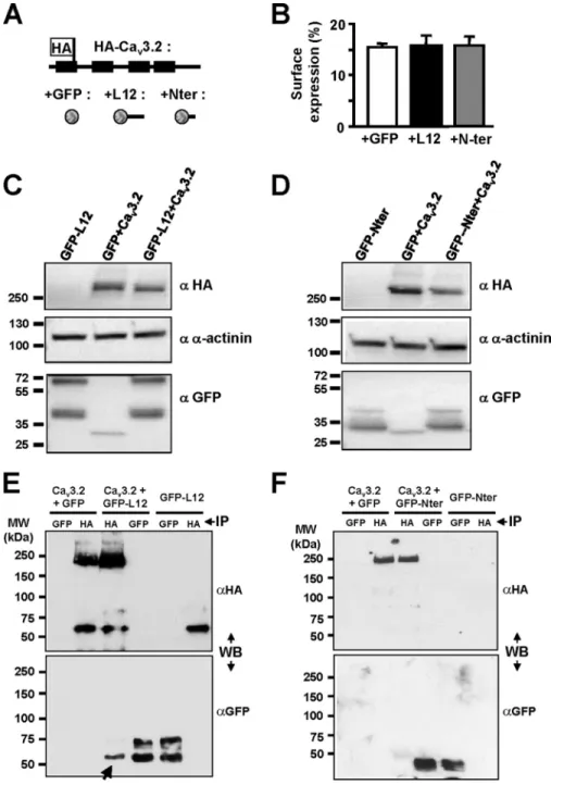
Using a full-length Cav3.2 channel HA-tagged construct (Fig. 3A and Refs. 15 and 16), we performed luminometry experi-ments to evaluate the level of Cav3.2 protein at the plasma membrane when coexpressed with constructs inhibiting (L12) or not (Nter) Cav3.2 current. As reported previously,⬃15% of the total HA-tagged Cav3.2 protein is present at the cell surface (see Ref. 15), and no change in surface expression of the HA-tagged Cav3.2 protein could be observed when coexpressed with either L12 or Nter GFP-fused construct (Fig. 3, A and B). To identify any change in the total amount of Cav3.2 protein, Western blotting was performed (Fig. 3, C and D). These exper-iments revealed that a similar amount of Cav3.2 protein was detected in the presence of GFP-L12 construct compared with GFP alone (Fig. 3C) and similarly when the experiments were conducted with the GFP-Nter construct (Fig. 3D).
Cav3.2 T-type Channel Silencing by Its Intracellular I-II Loop
at BIOCHIMIE CHU ST ANTOINE on July 26, 2016
http://www.jbc.org/
Whether the I-II loop could interact with the full-length Cav3.2 protein was further investigated using co-immuno-precipitation experiments (Fig. 3, E and F). When the HA-tagged Cav3.2 protein was coexpressed with the GFP-L12 construct, the presence of the latter was detected following immunoprecipitation of the HA-tagged Cav3.2 protein (Fig. 3E, arrow), indicating that the I-II loop can form a protein complex with the full-length Cav3.2 protein. On the con-trary, no such protein interaction could be detected when
the HA-Cav3.2 protein was coexpressed with the GFP-Nter construct (Fig. 3F).
Considering that the I-II loop sequence of the Cav3.2 protein shares some similarities with other T-type/Cav3 channels, we also investigated its ability to inhibit Cav3.1 or Cav3.3 currents. When recombinant Cav3.1 channels were coexpressed to-gether with the ⌬1-Cav3.2 construct in HEK-293 cells, the T-type current density was reduced by⬃3.2-fold (n ⫽ 11, p ⬍ 0.01; Fig. 4A). On the contrary, when this⌬1-Cav3.2 construct was coexpressed with Cav3.3 channels, no change in the cur-rent density could be observed (n⫽ 14, p ⬎ 0.05; Fig. 4B). These data indicate that the ⌬1 construct made of Cav3.2 protein could target both Cav3.2 and Cav3.1 channels but not Cav3.3 channels. In addition, coexpression of the⌬1-Cav3.2 construct with the L-type Cav1.2 channels (in the presence of auxiliary ␣2␦1 and 3 subunits), resulted in no change in L-type current density (n⫽ 10, p ⬎ 0.05; Fig. 4C). Similarly, no alteration of the P/Q-type current density (Cav2.1 in the presence of␣2␦1 and 3) was observed in the presence of the ⌬1-Cav3.2 construct (not shown).
We next explored the ability of the I-II loop constructs (L12 and⌬1) to inhibit native Cav3.2 T-type current in the neuro-blastoma cell line NG 108-15 (Fig. 5). Representative T-type currents traces obtained in the control condition (⫹GFP) and in the presence of L12 or⌬1 construct are shown in Fig. 5A. Significant inhibition (⬎10-fold) of the native T-type current density was obtained with the L12 and⌬1 constructs (n ⫽ 15 and n⫽ 10, respectively; Fig. 5, A and B), demonstrating the efficacy of these I-II loop constructs to silence native Cav3.2 channels. Taking advantage of the presence of native Cav3.2 channels in NG 108-15 cells to further validate the specificity of I-II loop inhibition, we next investigated the inhibitory effect of the Cav3.1, Cav3.2, and Cav3.3 I-II loop constructs on NG 108-15 T-type currents (Fig. 5C). Importantly, the Cav3.1 I-II loop could silence native Cav3.2 T-type current in NG 108-15 cells (⬎20-fold, n ⫽ 9), but the Cav3.3 I-II loop could not (n ⫽ 8). These data demonstrate that I-II loop constructs (L12 and ⌬1) made either of Cav3.1 or Cav3.2 proteins selectively target and silence Cav3.1 and Cav3.2 T-type channels. To further val-idate these data obtained using transient transfection proce-dures in NG 108-15, we developed a secondary NG 108-15 cell line expressing the Cav3.2 L12 in which the permanent expres-sion of this L12 construct completely abolished the presence of the T-type current (not shown). Altogether, these data demon-strate that expression of the L12 or⌬1 construct of the Cav3.2 protein efficiently eliminates the presence of T-type currents in cells expressing Cav3.1 or Cav3.2 channels.
Next, we assessed the ability of the⌬1 fragment to suppress native T-type currents in thalamic neurons. The thalamus is one of the brain areas showing the strongest expression of Cav3 channels with thalamocortical neurons expressing the Cav3.1 T-type channel isoform, whereas the GABAergic neurons of the NRT express both Cav3.2 and Cav3.3 isoforms (8, 20). In both neuronal types, an accurate biophysical characterization of the T-type current can only be performed in young pups (P8 –P12) due to the development profile of the T-type cur-rents and space-clamp limitations. To meet this temporal
FIGURE 1. T-type current inhibition obtained with various truncated forms of the Cav3.2 protein coexpressed with Cav3.2 channel. A, normal-ized T-type current density recorded in HEK-293 cells expressing Cav3.2 chan-nel in the presence of GFP alone or GFP-fused truncated forms of the Cav3.2 channel protein. The schematic structure of the Cav3.2 constructs is pre-sented in the left part of the figure. B, normalized T-type current density in cells expressing Cav3.2 channel in the presence of various truncated forms of the I-II loop of Cav3.2 channel protein (⌬1–⌬7 constructs; see Fig. 2A for their amino acid sequences). For these constructs, the normalized residual Cav3.2 current densities (ICa,T/ICa,T-Ctrl) were as follows:⌬1, 0.12 ⫾ 0.05, n ⫽ 20, p ⬍
0.001;⌬2, 0.32 ⫾ 0.07, n ⫽ 21, p ⬍ 0.01; ⌬3, 0.47 ⫾ 0.14, n ⫽ 16, p ⬍ 0.01; ⌬4, 0.72⫾ 0.14, n ⫽ 17, p ⬎ 0.05; ⌬5, 0.39 ⫾ 0.17, n ⫽ 22, p ⬍ 0.001; ⌬6, 0.91 ⫾ 0.13, n⫽ 20, p ⬎ 0.05; ⌬7, 1.04 ⫾ 0.15, n ⫽ 20, p ⬎ 0.05. Note that coexpres-sion of the I-II loop produces similar Cav3.2 current inhibition using either fusion (ICa,T/ICa,T-Ctrl⫽ 0.18 ⫾ 0.04, n ⫽ 21, p ⬍ 0.001) or internal ribosome
entry site (IRES) (ICa,T/ICa,T-Ctrl⫽ 0.25 ⫾ 0.08, n ⫽ 8, p ⬍ 0.001) GFP constructs.
Statistical significance was determined using one-way analysis of variance with Dunnett’s correction for multiple comparisons (**, p⬍ 0.01; ***, p ⬍ 0.001). Error bars represent S.E. Cter, C-terminal region.
at BIOCHIMIE CHU ST ANTOINE on July 26, 2016
http://www.jbc.org/
constraint, we selected a fast expressing system using Sind-bis viral constructs.
Freshly prepared thalamic slices were infected with either a GFP-⌬1-expressing virus (⌬1) or a control GFP-Nter-express-ing virus (Ctrl), and whole-cell recordGFP-Nter-express-ings of neurons located in the VB and NRT were performed 12–24 h after infection (see “Experimental Procedures”). Current clamp recordings showed that thalamocortical⌬1-infected neurons, GFP-Nter-express-ing neurons (Ctrl) and non-fluorescent (i.e. non-transfected) neurons displayed similar input resistances (Ctrl, 314.3⫾ 59.8 M⍀, n ⫽ 12; ⌬1, 388.8 ⫾ 47.5 M⍀, n ⫽ 12; non-transfected, 353.6⫾ 63.6 M⍀, n ⫽ 7; p ⬎ 0.5) and resting membrane poten-tials (Ctrl:,⫺73.7 ⫾ 1.7 mV, n ⫽ 12; ⌬1, ⫺68.5 ⫾ 2.1 mV, n ⫽ 12; non-transfected,⫺71.9 ⫾ 1.8 mV, n ⫽ 7; p ⬎ 0.5). However, the maximal firing rates evoked during depolarizing current injections were identical in ⌬1 and control neurons (Ctrl, 19.5⫾ 1.3 Hz, n ⫽ 10; ⌬1, 22 ⫾ 1.8 Hz, n ⫽ 10; p ⫽ 0.27) but significantly higher in non-transfected neurons (28.9⫾ 1.3 Hz,
n⫽ 7; p ⬍ 0.02) indicating that the virus per se may change
some active properties of the neurons.
Considering the voltage-gated calcium currents, VB ⌬1-in-fected neurons showed a drastic decrease in the total T-type
current evoked at⫺50 mV after a 1-s hyperpolarizing prepulse to⫺100 mV (Ctrl, 1425 ⫾ 112 pA, n ⫽ 18; ⌬1, 377 ⫾ 37 pA, n ⫽ 26; p⬍ 0.001; Fig. 6, A and B), whereas the amplitude of the currents recorded in control and in non-transfected neurons (1518⫾ 120 pA, n ⫽ 15) were identical (p ⫽ 0.6). Inactivation decay was best fitted with a single time constant typical of Cav3.1 current of VB neurons (21), and no difference was observed between the⌬1 (, 13.7 ⫾ 0.3 ms, n ⫽ 18) and control conditions (, 12.8 ⫾ 1.6 ms, n ⫽ 18; p ⫽ 0.065; Fig. 6C). Acti-vation and inactiActi-vation voltage dependences were similar in both conditions (activation V1/2:⌬1, ⫺60.0 ⫾ 0.5 mV, n ⫽ 8 versusCtrl,⫺61.4 ⫾ 0.8 mV, n ⫽ 8; p ⫽ 0.15 and inactivation
V1/2:⌬1, ⫺86.3 ⫾ 1.5 mV, n ⫽ 8 versus Ctrl, ⫺86.2 ⫾ 1.1 mV,
n⫽ 10; p ⫽ 0.95). Importantly, the amplitude of the high
volt-age-activated current evoked by a step depolarization from⫺60 to⫹10 mV was unchanged for ⌬1-infected and control neu-rons (2700.5⫾ 284 pA (n ⫽ 12) and 2864 ⫾ 259 pA (n ⫽ 11, respectively; p⫽ 0.67).
In NRT neurons infected by the control GFP-Nter-express-ing virus, the inactivation decay of T-type current was described by a double exponential function with the fast (1, 16.4⫾ 1.0 ms) and slow (2, 82 ⫾ 3.0 ms; n ⫽ 10) components
FIGURE 2. Design of short (⌬) I-II loop constructs and representative electrophysiological properties of the ⌬1 construct. A, the amino acid sequences of the⌬1–⌬7 constructs were identified according to the human Cav3.2 protein entry in the UniProt database (O95180): ⌬1 (Ser423–Pro542),⌬2 (Ser423–His521),
⌬3 (Ser423–Pro501),⌬4 (Ser423–Ala481),⌬5 (Ser452–Pro544),⌬6 (Val472–Pro544), and⌬7 (Asp493–Pro544). These constructs are compared in this multiple alignment
with the Cav3.2 deletion mutants (D1–D4) used in Vitko et al. (15). B, superimposed representative T-type current traces recorded using a test pulse at⫺30 mV from a holding potential of⫺100 mV (100-ms duration) on HEK-293 cells transfected with Cav3.2 and Nter constructs (Ctrl; gray trace) and Cav3.2 and ⌬1 constructs (⫹⌬1; black trace). The traces were normalized in amplitude (inset), which revealed no difference in the activation and inactivation kinetics of the residual Cav3.2 current in cells expressing the⌬1 construct. C, representative T-type currents recorded using a ramp test (⫺100/⫹100 mV; holding potential, ⫺100 mV; 200-ms duration) on HEK-293 cells transfected with Cav3.2 and Nter constructs (Ctrl; gray trace) and Cav3.2 and ⌬1 constructs (⫹⌬1; black trace). Traces were normalized in amplitude (inset), revealing no shift in the current-voltage relationship of the residual Cav3.2 current in cells expressing the⌬1 construct.
Cav3.2 T-type Channel Silencing by Its Intracellular I-II Loop
at BIOCHIMIE CHU ST ANTOINE on July 26, 2016
http://www.jbc.org/
reflecting the presence of the Cav3.2 and Cav3.3 channels, respectively. Similarly to VB neurons, all NRT⌬1-infected neu-rons showed a significant decrease in the total T-type current (Ctrl; 1048⫾ 122 pA; n ⫽ 10; ⌬1; 537 ⫾ 57 pA, n ⫽ 9; p ⬍ 0.001; Fig. 6, D and E). In three of the nine⌬1-infected NRT neurons, the inactivation decay was fitted by a single exponential func-tion with a time constant of 61.7⫾ 8.5 ms, which is similar to the slow time constant (2) of the control T-type current (Fig. 6F). In the six remaining⌬1-infected neurons, the decay of the T-type current was still described by a double exponential func-tion with time constants similar to those of the control current.
Of importance, the weight of the fast time constant was mark-edly reduced (⌬1, 267 ⫾ 27 pA; Ctrl, 685 ⫾ 217 pA; p ⬍ 0.01), whereas the weight of the slow time constant was unchanged (⌬1, 304 ⫾ 19 pA; Ctrl, 300 ⫾ 36 pA; p ⫽ 0.94). Hence, in all ⌬1-infected NRT neurons, the fast component of the T-type current presumably due to the Cav3.2 channels was almost fully blocked. Altogether, these data confirm the results obtained in the recombinant system showing that only the fast inactivating Cav3.1 and Cav3.2 currents are inhibited in the presence of the ⌬1 construct, whereas Cav3.3 and high voltage-activated cur-rents are unchanged.
FIGURE 3. Biochemical properties of the Cav3.2 channel when coexpressed with I-II loop construct. A and B, luminometry detection of the surface expression of the HA-Cav3.2 construct (schematic structure presented in A) in the presence of free GFP (B, white bar), GFP-tagged I-II loop (B, black bar), and GFP-tagged Nter constructs (B, gray bar). C, Western blot analysis of the total HA-Cav3.2 protein amount in cells co-transfected with I-II loop (right lane) compared with the total amount of Cav3.2 protein in cells co-transfected with GFP (middle lane). D, Western blotting (WB) experiments similar to those in C performed in the presence of the Nter construct. E and F, co-immunoprecipitation (IP) experiments for the transfection conditions indicated at the top of the figure showing that the I-II loop protein could be detected (E, arrow) when the HA-Cav3.2 protein was immunoprecipitated but not when the HA-Cav3.2 protein was coexpressed with the Nter (F). Error bars represent S.E.
at BIOCHIMIE CHU ST ANTOINE on July 26, 2016
http://www.jbc.org/
Finally, to assess the effects of the⌬1-induced decrease in T-type current amplitude on neuronal excitability, we evoked a rebound low threshold calcium spike (LTS) in thalamocortical neurons. Following 1-s-long hyperpolarization to ⫺100 mV that allows de-inactivation of the whole T-type channel
popu-lation, rebound LTS crowned by at least one action potential was evoked in the 12 control neurons recorded (Fig. 6G, left
panel) but failed to evoked an action potential in two of the 12 neurons expressing the⌬1 construct (Fig. 6G, middle panel). Considering that the⌬1 construct decreased by 74% the ampli-tude of the T-type current, this moderate failure rate in ⌬1 neurons is in agreement with our previous data showing that more than 70% of the T-type channel population needs to be blocked to hamper the LTS-associated firing in thalamic neu-rons (22). Accordingly, when the hyperpolarizing potential was reduced to⬃⫺85 mV, a value corresponding to the half-inac-tivation of the T-type current, LTS crowned by an action poten-tial was still recorded in six of the 12 control neurons but only in three of the 12⌬1-infected neurons (Fig. 6G, right panel).
Discussion
Here we document that overexpression of the I-II loop of the Cav3.2 subunit of T-type calcium channels induces silencing of the Cav3.1/Cav3.2 T-type currents without affecting Cav3 pro-tein expression at the plasma membrane. This inhibitory effect on Cav3.1/Cav3.2 currents is produced by the proximal part of the I-II loop, and Cav3.2 currents were unchanged upon expression of the other intracellular regions of the Cav3.2 chan-nel. This study reveals a novel aspect of T-type Cav3 channel regulation that involves its I-II loop and can be used to block selectively the activity of native Cav3.1/Cav3.2 channels, such as in neurons.
Importantly, we found that the amount of Cav3.2 protein as well as its expression at the plasma membrane was unchanged in the presence of the I-II loop. This finding indicates that the silencing effect produced by the I-II loop does not involve a retention/degradation mechanism of the wild-type Cav3.2 channel, which was observed with the truncated forms of Cav3.2 that include domain I and/or domain II (19). To char-acterize further the mechanism involved in the I-II loop silenc-ing effect, we searched for the domain involved by ussilenc-ing shorter fragments, but we were unsuccessful in identifying the specific protein domain(s) as truncated forms of the I-II loop shorter than the proximal⬃100-amino acid-long peptide were less effi-cient in producing the inhibitory effect. The precise molecular mechanism supporting this silencing effect remains to be iden-tified. However, several observations should be considered. (i) It likely involves molecular interactions as co-immunoprecipi-tation experiments have revealed association of the I-II loop with the full-length Cav3.2 protein. (ii) Stability and plasma membrane expression of Cav3.2 protein are not altered, indi-cating that Cav3.2 channels are correctly processed to the cell surface when the I-II loop is overexpressed. (iii) The remaining T-type calcium currents in cells overexpressing the I-II loop are unaffected in their kinetics and steady-state properties, sug-gesting an “all or none” silencing effect for channels associated with the I-II loop. To account for this silencing mechanism, we hypothesize that the Cav3.1/Cav3.2 channels are locked in a non-conducting closed state or inactivated state in the presence of a I-II loop overload. Our data are also well supportive of the previous description of a gating brake role of the I-II loop in Cav3.2 and to a lesser extent in Cav3.1 but not Cav3.3 channels (12, 13, 15). Altogether, this “dominant-negative’” strategy
rep-FIGURE 4. No Cav3.3 current and Cav1.2 current inhibition in the pres-ence of the I-II loop construct. The effect of the shorter I-II loop construct⌬1 on other Cav channels is shown. There was an inhibitory effect of the⌬1 construct on Cav3.1 current (A) but not on Cav3.3 current (B) and Cav1.2 current (C). Representative current traces are presented in the top part of the panels (Ctrl and⫹⌬1), and the average current densities are shown in the
bottom part of the panels. Statistical significance was determined using
Student’s unpaired t test (**, p⬍ 0.01; ns, non-significant). Error bars represent S.E. pF, picofarad.
FIGURE 5. Silencing effect of the I-II loop and⌬1 constructs on native T-type current (Cav3.2) channels in NG 108-15 cells. A, examples of T-type current traces recorded in NG 108-15 cells (holding potential,⫺100 mV; test pulse,⫺30 mV) transfected with GFP (left part), with the I-II loop construct (L12; middle part), with the⌬1 construct (right part). B, average inhibitory effects of the Cav3.2 I-II loop and⌬1 constructs on native T-type current in NG 108-15 cells. C, efficacy of the I-II loop constructs generated from the Cav3.1, Cav3.2, and Cav3.3 channels on native T-type current in NG 108-15 cells. Note the absence of inhibitory effect of the I-II loop construct generated from the Cav3.3 channel. Statistical significance was determined using Student’s unpaired t test (***, p⬍ 0.001; ns, non-significant). Error bars represent S.E.
Norm., normalized.
Cav3.2 T-type Channel Silencing by Its Intracellular I-II Loop
at BIOCHIMIE CHU ST ANTOINE on July 26, 2016
http://www.jbc.org/
resents an original alternative to eliminate Cav3.1/Cav3.2 chan-nels at the functional level without altering their protein expression.
Indeed, an important finding of our study is that delivery of the I-II loop into neurons could efficiently silence native Cav3.2 and Cav3.1 channels. Clear evidence was first obtained in the
neuroblastoma cell line NG 108-15, which expresses T-type calcium channels made of Cav3.2 subunit (7). Both transient and stable expression of I-II loop constructs (L12 and ⌬1) resulted in a complete disappearance of the T-type current in NG 108-15 cells. Importantly, similar findings were obtained in VB and NRT thalamic neurons following viral delivery of the⌬1
FIGURE 6. Silencing effect of the⌬1 construct on thalamic T-type currents. A, mean amplitude of T-type currents recorded in VB neurons infected with either the⌬1-expressing virus or the control GFP-Nter-expressing virus (Ctrl). T-type currents were evoked by step depolarization (200 ms long) to ⫺50 mV after a 1-s hyperpolarizing prepulse to⫺100 mV. B, examples of T-type currents recorded in both conditions. C, mean values of the inactivation time constant. D, mean amplitude of T-type currents recorded in NRT neurons infected with either the⌬1-expressing virus or the control GFP-Nter-expressing virus (Ctrl). The protocol used was the same protocol as in A. E, examples of T-type currents recorded in both conditions. The top traces show T-type currents recorded in ⌬1-infected neurons displaying an inactivation kinetic described with either one or two time constants. F, mean values of the inactivation time constants. Note that when the decay of the T-type current recorded in⌬1-infected neurons is described by a single time constant its value is identical to those of the slow time constant (2) of the T-type currents recorded in ⌬1-infected and control neurons. Statistical significance was determined using Student’s unpaired t test (***,
p⬍ 0.001). G, typical rebound LTS evoked by 1-s-long hyperpolarizing steps in control (left traces) and ⌬1-infected (middle and right traces) thalamocortical
neurons. Note that rebound LTS-associated firing was evoked following⫺100-mV hyperpolarizing steps in the 12 recorded control neurons (left, gray trace) and was still present after a⫺85-mV hyperpolarization in six neurons (left, black trace). When considering the ⌬1-infected neurons, if a rebound firing was still observed following⫺100-mV hyperpolarizing steps in 10 of 12 neurons (right versus middle, gray traces), then reducing the hyperpolarizing potential to ⫺85 mV suppressed LTS firing in nine of the 12 neurons (middle and right, black traces). Error bars represent S.E.
at BIOCHIMIE CHU ST ANTOINE on July 26, 2016
http://www.jbc.org/
construct. In VB neurons that express the Cav3.1 isoform, the T-type current density was significantly reduced with no change in current properties, such as inactivation kinetics and activation and inactivation voltage dependences. In NRT neu-rons that express both the Cav3.2 and the Cav3.3 isoforms, the T-type current density was also significantly reduced. Inactiva-tion kinetics analysis revealed that only the Cav3.2 fast inacti-vating component was eliminated, whereas the slow inactivat-ing component attributable to Cav3.3 T-type current was unaffected. Altogether, these findings demonstrate that silenc-ing of native Cav3.1/Cav3.2 channels can be achieved ussilenc-ing delivery of I-II loop constructs. This strategy greatly expands the current means to investigate the role of the neuronal Cav3 channels as it allows manipulating the Cav3.1 and Cav3.2 sub-unit activities in a time- and region-selective manner even in chronically monitored animals without affecting the amount of Cav3 protein as observed in knock-out animals or using RNA interference strategies. In addition, its lack of effect on the Cav3.3 isoform will allow deciphering the respective role of fast and slow inactivating T-type currents in brain areas or neurons where both channels are expressed.
References
1. Lambert, R. C., Bessaïh, T., Crunelli, V., and Leresche, N. (2014) The many faces of T-type calcium channels. Pflugers Arch. 466, 415– 423
2. François, A., Laffray, S., Pizzoccaro, A., Eschalier, A., and Bourinet, E. (2014) T-type calcium channels in chronic pain: mouse models and spe-cific blockers. Pflugers Arch. 466, 707–717
3. Zamponi, G. W., Lory, P., and Perez-Reyes, E. (2010) Role of voltage-gated calcium channels in epilepsy. Pflugers Arch. 460, 395– 403
4. Cheong, E., and Shin, H. S. (2013) T-type Ca2⫹channels in normal and abnormal brain functions. Physiol. Rev. 93, 961–992
5. Perez-Reyes, E. (2003) Molecular physiology of low-voltage-activated T-type calcium channels. Physiol. Rev. 83, 117–161
6. Cain, S. M., and Snutch, T. P. (2010) Contributions of T-type calcium channel isoforms to neuronal firing. Channels 4, 475– 482
7. Chemin, J., Monteil, A., Perez-Reyes, E., Bourinet, E., Nargeot, J., and Lory, P. (2002) Specific contribution of human T-type calcium channel isotypes (␣1G,␣1Hand␣1I) to neuronal excitability. J. Physiol. 540, 3–14
8. Talley, E. M., Cribbs, L. L., Lee, J. H., Daud, A., Perez-Reyes, E., and Bayliss, D. A. (1999) Differential distribution of three members of a gene family encoding low voltage-activated (T-type) calcium channels. J. Neurosci. 19, 1895–1911
9. Kim, D., Song, I., Keum, S., Lee, T., Jeong, M. J., Kim, S. S., McEnery, M. W., and Shin, H. S. (2001) Lack of the burst firing of thalamocortical relay neurons and resistance to absence seizures in mice lacking␣1G
T-type Ca2⫹channels. Neuron 31, 35– 45
10. Astori, S., and Lüthi, A. (2013) Synaptic plasticity at intrathalamic connec-tions via CaV3.3 T-type Ca2⫹channels and GluN2B-containing NMDA receptors. J. Neurosci. 33, 624 – 630
11. Ernst, W. L., Zhang, Y., Yoo, J. W., Ernst, S. J., and Noebels, J. L. (2009) Genetic enhancement of thalamocortical network activity by elevating alpha 1g-mediated low-voltage-activated calcium current induces pure absence epilepsy. J. Neurosci. 29, 1615–1625
12. Arias-Olguín, I. I., Vitko, I., Fortuna, M., Baumgart, J. P., Sokolova, S., Shumilin, I. A., Van Deusen, A., Soriano-García, M., Gomora, J. C., and Perez-Reyes, E. (2008) Characterization of the gating brake in the I-II loop of Cav3.2 T-type Ca2⫹channels. J. Biol. Chem. 283, 8136 – 8144
13. Baumgart, J. P., Vitko, I., Bidaud, I., Kondratskyi, A., Lory, P., and Perez-Reyes, E. (2008) I-II loop structural determinants in the gating and surface expression of low voltage-activated calcium channels. PLoS One 3, e2976 14. Shcheglovitov, A., Vitko, I., Bidaud, I., Baumgart, J. P., Navarro-Gonzalez, M. F., Grayson, T. H., Lory, P., Hill, C. E., and Perez-Reyes, E. (2008) Alternative splicing within the I-II loop controls surface expression of T-type Cav3.1 calcium channels. FEBS Lett. 582, 3765–3770
15. Vitko, I., Bidaud, I., Arias, J. M., Mezghrani, A., Lory, P., and Perez-Reyes, E. (2007) The I-II loop controls plasma membrane expression and gating of Cav3.2 T-type Ca2⫹channels: a paradigm for childhood absence
epi-lepsy mutations. J. Neurosci. 27, 322–330
16. Dubel, S. J., Altier, C., Chaumont, S., Lory, P., Bourinet, E., and Nargeot, J. (2004) Plasma membrane expression of T-type calcium channel␣1
sub-units is modulated by high voltage-activated auxiliary subsub-units. J. Biol.
Chem. 279,29263–29269
17. Bidaud, I., and Lory, P. (2011) Hallmarks of the channelopathies associated with L-type calcium channels: a focus on the Timothy mutations in Cav1.2
channels. Biochimie 93, 2080 –2086
18. Kim, J., Dittgen, T., Nimmerjahn, A., Waters, J., Pawlak, V., Helmchen, F., Schlesinger, S., Seeburg, P. H., and Osten, P. (2004) Sindbis vector SINrep(nsP2S726): a tool for rapid heterologous expression with attenu-ated cytotoxicity in neurons. J. Neurosci. Methods 133, 81–90
19. Mezghrani, A., Monteil, A., Watschinger, K., Sinnegger-Brauns, M. J., Barrère, C., Bourinet, E., Nargeot, J., Striessnig, J., and Lory, P. (2008) A destructive interaction mechanism accounts for dominant-negative ef-fects of misfolded mutants of voltage-gated calcium channels. J. Neurosci. 28,4501– 4511
20. Huguenard, J. R., and Prince, D. A. (1992) A novel T-type current under-lies prolonged Ca2⫹-dependent burst firing in GABAergic neurons of rat thalamic reticular nucleus. J. Neurosci. 12, 3804 –3817
21. Leresche, N., Parri, H. R., Erdemli, G., Guyon, A., Turner, J. P., Williams, S. R., Asprodini, E., and Crunelli, V. (1998) On the action of the anti-absence drug ethosuximide in the rat and cat thalamus. J. Neurosci. 18, 4842– 4853
22. Dreyfus, F. M., Tscherter, A., Errington, A. C., Renger, J. J., Shin, H. S., Uebele, V. N., Crunelli, V., Lambert, R. C., and Leresche, N. (2010) Selec-tive T-type calcium channel block in thalamic neurons reveals channel redundancy and physiological impact of ITwindow. J. Neurosci. 30, 99 –109
Cav3.2 T-type Channel Silencing by Its Intracellular I-II Loop
at BIOCHIMIE CHU ST ANTOINE on July 26, 2016
http://www.jbc.org/
Leresche, Régis C. Lambert and Philippe Lory
Sylvaine Huc-Brandt, Iulia Blesneac, Isabelle Bidaud, Céline Lemmers, Nathalie
Arnaud Monteil, Patrick Chausson, Katia Boutourlinsky, Alexandre Mezghrani,
doi: 10.1074/jbc.M114.634261 originally published online April 30, 2015 2015, 290:16168-16176.
J. Biol. Chem.
10.1074/jbc.M114.634261
Access the most updated version of this article at doi: Alerts:
When a correction for this article is posted
•
When this article is cited
•
to choose from all of JBC's e-mail alerts
Click here
http://www.jbc.org/content/290/26/16168.full.html#ref-list-1
This article cites 22 references, 12 of which can be accessed free at
at BIOCHIMIE CHU ST ANTOINE on July 26, 2016
http://www.jbc.org/