HAL Id: tel-02517664
https://tel.archives-ouvertes.fr/tel-02517664v2
Submitted on 25 Mar 2020
HAL is a multi-disciplinary open access archive for the deposit and dissemination of sci-entific research documents, whether they are pub-lished or not. The documents may come from teaching and research institutions in France or abroad, or from public or private research centers.
L’archive ouverte pluridisciplinaire HAL, est destinée au dépôt et à la diffusion de documents scientifiques de niveau recherche, publiés ou non, émanant des établissements d’enseignement et de recherche français ou étrangers, des laboratoires publics ou privés.
influence on stress regulation, sleep, and cognitive
executive performance
Sylvain Laborde
To cite this version:
Sylvain Laborde. Slow-paced breathing and cardiac vagal activity : influence on stress regulation, sleep, and cognitive executive performance. Psychology. Normandie Université, 2019. English. �NNT : 2019NORMC044�. �tel-02517664v2�
Pour obtenir le diplôme de doctorat
Spécialité PSYCHOLOGIE
Préparée au sein de l'Université de Caen Normandie
Slοw-paced breathing and cardiac vagal activity: Ιnfluence οn
stress regulatiοn, sleep, and cοgnitive executive perfοrmance
Présentée et soutenue par
Sylvain LABORDE
Thèse soutenue publiquement le 07/12/2019 devant le jury composé de
M. ULRICH ETTINGER Professeur, Université de Bonn - Allemagne Rapporteur du jury Mme ILSE VAN DIEST Professeur, Katholieke Universiteit Leuven- Belgique Rapporteur du jury M. DAMIEN DAVENNE Professeur des universités, Université Caen Normandie Président du jury
Thèse dirigée par FABRICE DOSSEVILLE, Mobilités : vieillissement, pathologie, santé - COMETE
“Yes, they are elves," Legolas said. "and they say that you breathe so loud they could shoot you in the dark."
Sam hastily covered his mouth.
Summary
1 INDEXES ... 5
1.1 FIGURES INDEX ... 5
1.2 TABLES INDEX ... 6
1.3 ABBREVIATIONS INDEX ... 7
1.4 PUBLICATIONS INCLUDED IN THE PHD THESIS ... 8
1.5 SCIENTIFIC COMMUNICATIONS RELATED TO THIS PHD THESIS ... 8
1.5.1 Symposia ... 8
1.5.2 Oral talks ... 8
1.5.3 Posters ... 9
1.5.4 Applied workshops... 9
1.6 AWARDS RELATED TO THIS PHD THESIS ...10
2 PREFACE ...11
3 INTRODUCTION ...13
4 THEORETICAL BACKGROUND ...15
4.1 BREATHING ...15
4.1.1 Main role: Gas exchange between the body and the environment ...15
4.1.2 Respiratory passageways: The crucial role of the nose...15
4.1.3 Mechanics of pulmonary ventilation ...16
4.1.4 Pulmonary volumes ...18
4.1.5 Alveolar ventilation...19
4.1.6 Transport of oxygen and carbon dioxide in blood and tissue fluids ...21
4.1.7 The central regulation of respiration ...22
4.2 AT THE CROSSROADS BETWEEN THE RESPIRATORY AND THE CARDIOVASCULAR SYSTEMS ...24
4.2.1 Coupling between heart beat and respiration: Respiratory sinus arrhythmia...24
4.2.2 Slow-paced breathing ...26
4.3 HOW SLOW-PACED BREATHING MAY INFLUENCE SELF-REGULATION: THE ROLE OF THE VAGUS NERVE...30
4.3.1 The role of the vagus nerve in self-regulation ...30
4.3.2 Cardiac vagal activity: the output of the central autonomic network ...33
4.3.3 The neurovisceral integration model ...34
4.3.4 Non-invasive assessment of cardiac vagal activity with heart rate variability ...35
4.4 THE 3RS OF VAGUS NERVE FUNCTIONING ...36
4.5 SELF-REGULATION PHENOMENA INVESTIGATED WITH SLOW-PACED BREATHING AND CARDIAC VAGAL ACTIVITY WITHIN THIS PHD THESIS ...37
4.5.1 Stress ...37
4.5.1.1 Psychological stress ... 37
4.5.1.2 Physical stress ... 38
4.5.1.3 Stress and slow-paced breathing ... 38
4.5.1.4 Stress and intellectual disability ... 39
4.6 SLEEP ...39
4.6.1 Sleep and cardiac vagal activity ...40
4.6.2 Sleep and slow-paced breathing...41
4.7 EXECUTIVE FUNCTIONING ...42
4.7.1 Inhibition and cardiac vagal activity ...42
4.7.2 Working memory and cardiac vagal activity ...43
4.7.3 Cognitive flexibility and cardiac vagal activity ...44
4.7.4 Executive functioning and slow-paced breathing ...45
5 RESEARCH QUESTIONS & HYPOTHESES...47
6 GENERAL METHOD ...49
6.1 CARDIAC VAGAL ACTIVITY MEASUREMENT ...49
6.2 SLOW-PACED BREATHING EXERCISE ...50
7 STUDY 1 (PUBLISHED) - THE EFFECT OF SLOW-PACED BREATHING ON STRESS MANAGEMENT IN ADOLESCENTS WITH INTELLECTUAL DISABILITY ...54
8 STUDY 2 (PUBLISHED) - INFLUENCE OF A 30-DAY SLOW PACED BREATHING INTERVENTION
COMPARED TO SOCIAL MEDIA USE ON SUBJECTIVE SLEEP QUALITY AND CARDIAC VAGAL ACTIVITY ...62
9 STUDY 3 (SUBMITTED) – THE INFLUENCE OF SLOW-PACED BREATHING ON EXECUTIVE FUNCTION 74 9.1 INTRODUCTION ...74
9.2 METHOD ...75
9.2.1 Participants ...75
9.2.2 Material and Measures ...75
9.2.2.1 Cardiac vagal activity ... 75
9.2.2.2 Slow-paced breathing technique ... 75
9.2.2.3 TV neutral documentary (control condition) ... 76
9.2.3 Executive Function Tasks...76
9.2.3.1 Inhibition task ... 76 9.2.3.2 Working memory ... 77 9.2.3.3 Cognitive flexibility ... 77 9.2.4 Procedure ...78 9.2.5 Data Analysis ...81 9.3 RESULTS...83
9.3.1 Slow-paced Breathing and Physiological Measures...83
9.3.2 Slow-paced Breathing and Behavioral Measures ...84
9.3.3 Mediation Analysis ...85
9.4 DISCUSSION ...86
9.5 CONCLUSION ...89
10 STUDY 4 (PUBLISHED) - INFLUENCE OF SLOW-PACED BREATHING ON INHIBITION AFTER PHYSICAL EXERTION 90 11 GENERAL DISCUSSION ... 105 11.1 OVERVIEW... 105 11.2 LIMITATIONS ... 108 11.3 PERSPECTIVES... 109 12 CONCLUSION ... 112 13 REFERENCES ... 113 14 ACADEMIC CV ... 132
15 A TOKEN OF THANKS - WITH OR WITHOUT YOU ... 138
16 SYNTHESE DE LA THESE EN FRANÇAIS ... 140
16.1 CADRE THEORIQUE ... 140
16.1.1 Introduction... 140
16.1.2 À la croisée des chemins entre les systèmes respiratoire et cardiovasculaire ... 141
16.1.2.1 Couplage entre le rythme cardiaque et la respiration : L’arythmie sinusale respiratoire ... 141
16.1.2.2 La respiration lente contrôlée ... 143
16.1.3 Comment la respiration lente contrôlée peut-elle influencer l'autorégulation : le rôle du nerf vague 146 16.1.3.1 Le rôle du nerf vague dans l'autorégulation ... 146
16.1.3.2 Activité cardiaque vagale : le produit du réseau central autonome ... 146
16.1.3.3 Le modèle d'intégration neuroviscérale ... 147
16.1.3.4 Évaluation non invasive de l'activité vagale cardiaque avec la variabilité de la fréquence cardiaque 147 16.1.4 Les 3Rs du fonctionnement du nerf vague ... 148
16.1.5 Phénomènes d'autorégulation étudiés avec la respiration lente contrôlée et l'activité vagale cardiaque dans cette thèse de doctorat ... 149
16.1.5.1 Stress ... 149
16.1.5.1.1 Stress psychologique ... 149
16.1.5.1.2 Stress physique ... 149
16.1.5.1.3 Stress et respiration lente contrôlée ... 150
16.1.5.1.4 Stress et handicap mental ... 150
16.1.5.2 Sommeil ... 151
16.1.5.2.1 Sommeil et activité vagale cardiaque ... 151
16.1.5.2.2 Sommeil et respiration lente ... 152
16.1.5.3 Les fonctions exécutives ... 153
16.1.5.3.3 Flexibilité cognitive et activité vagale cardiaque ... 154
16.1.5.3.4 Fonctions exécutives et respiration lente contrôlée... 155
16.2 QUESTIONS DE RECHERCHE ET HYPOTHESES ... 156
16.3 METHODE GENERALE ... 157
16.3.1 Mesure de l’activité vagale cardiaque ... 157
16.3.2 La respiration lente contrôlée ... 158
16.4 ÉTUDES ... 159 16.4.1 Étude 1 - résumé ... 159 16.4.2 Étude 2 - résumé ... 160 16.4.3 Étude 3 - résumé ... 161 16.4.4 Étude 4 - résumé ... 161 16.5 DISCUSSION GENERALE ... 162
16.5.1 Synthèse des résultats ... 162
16.5.2 Limites ... 164
16.5.3 Perspectives ... 164
16.6 CONCLUSION... 165
17 FRENCH SHORT ABSTRACT – LA RESPIRATION LENTE CONTROLEE ET L’ACTIVITE VAGALE CARDIAQUE : INFLUENCE SUR LA GESTION DU STRESS, LE SOMMEIL, ET LES PERFORMANCES COGNITIVES EXECUTIVES ... 167
18 GERMAN SHORT ABSTRACT - LANGSAM KONTROLLIERTE ATMUNG UND KARDIOVAGALE AKTIVITÄT: EINFLUSS AUF STRESSREGULATION, SCHLAF UND KOGNITIVE EXEKUTIVFUNKTIONEN ... 168
Indexes
1 Indexes
1.1 Figures Index
Figure 1: Schematic representation of the respiratory system displaying the upper and lower respiratory
tract regions (from Tu, Inthavong, & Ahmadi, 2013, p. 20) ...16
Figure 2: Diaphragm movements during inhalation and exhalation (from Colbert et al., 2016, p. 293) ...17
Figure 3: Illustration of the mechanics of pulmonary ventilation (from Hall, 2011a, p. 466) ...17
Figure 4: Respiratory patterns during normal breathing and during maximal inspiration and maximal expiration (from Hall, 2011a, p. 469) ...19
Figure 5: Coupling between alveoli and pulmonary capillary (from Hall, 2011a, p. 472) ...20
Figure 6: Gas exchange at the level of the alveoli, illustration of the diffusion process (from Colbert et al., 2016, p. 293) ...21
Figure 7: Gas exchange at the level of the tissues (from Colbert et al., 2016, p. 293) ...21
Figure 8: The Medulla Oblongata (from Waldman, 2009, p. 208) ...23
Figure 9: Organization of the respiratory center (from Hall, 2011d, p. 506) ...23
Figure 10: Illustration of the suggested conceptual effect of the respiratory sinus arrhythmia on the relationship between alveolar gas volume and capillary blood flow during inspiration and expiration (from Yasuma & Hayano, 2004, p. 684) ...25
Figure 11: Illustration of the functioning of the baroreflex (from Shaffer, McCraty, & Zerr, 2014, p. 9) ...29
Figure 12: Anatomy of the vagus nerve including branches (from Câmara & Griessenauer, 2015, p. 386) 32 Figure 13: A composite schematic diagram showing the pathways by which the prefrontal cortex might influence the control of heart rate (from Thayer et al., 2009, p. 143)...34
Figure 14: Electrocardiogram - Illustration of heart rate variability calculation ...36
Figure 15: The 3Rs of cardiac vagal activity functioning (from Laborde, Mosley, et al., 2017, p. 6) ...37
Figure 16: Video based on the software EZ-air (used in Study 1, 3, 4) ...51
Figure 17: Smartphone App Breath Pacer (used in Study 2) ...51
1.2 Tables Index
Table 1: Study 3 - Descriptive statistics for physiological variables...83
Table 2: Study 3 - Descriptive statistics for executive functions ...85
Table 3: Study 3 - Correlation between main study variables (Control condition) ...85
1.3 Abbreviations Index
AOSPAN: Automated Operation Span cpm: cycles per minute
CVA: Cardiac Vagal Activity ECG: Electrocardiogram HRV: Heart Rate Variability
HF-HRV: High-Frequency Heart Rate Variability MCST: Modified Card Sorting Test
min: minute mL: milliliters ms: millisecond PS: Perceived Stress
RMSSD: Root Mean Square of Successive Differences RSA: Respiratory Sinus Arrhythmia
1.4 Publications included in the PhD thesis
Laborde, S., Allen, M.S., Gohring, N., and Dosseville, F. (2017). The effect of slow-paced breathing on stress management in adolescents with intellectual disability. Journal of Intellectual Disability Research 61(6), 560-567. doi: 10.1111/jir.12350. (IF = 1.941)
Laborde, S., Hosang, T., Mosley, E., and Dosseville, F. (2019). Influence of a 30-day slow paced breathing intervention compared to social media use on subjective sleep quality and cardiac vagal activity. Journal of Clinical Medicine 8. doi: 10.3390/jcm8020193. (IF = 5.688)
Laborde, S., Allen, M., Borges, U., Hosang, T., Furley, P., Mosley, E., et al. (submitted). The influence of slow-paced breathing on executive function.
Laborde, S., Lentes, T., Hosang, T. J., Borges, U., Mosley, E., & Dosseville, F. (2019). Influence of slow-paced breathing on inhibition after physical exertion. Frontiers in Psychology, 10. doi:10.3389/fpsyg.2019.01923 (IF = 2.129)
1.5 Scientific communications related to this PhD thesis
1.5.1 Symposia
Laborde, S., Dosseville, F. (2019). Heart rate variability as a self-regulation marker. Symposium organized at the 15th European Congress for Sport Psychology (FEPSAC), 15-20th July 2019, Münster, Germany.
Laborde, S., Dosseville, F. (2018). Neuroenhancement in sport psychology. Symposium organized at the 6ème Congrès International de la Société Française de Psychologie du Sport, 13-15th June 2018, Lausanne, Switzerland.
Laborde, S. (2018). Self-regulation and cardiac vagal activity in sport psychology. Symposium organized at the 50. Jahrestagung der Arbeitsgemeinschaft für Sportpsychologie, 10-12th May 2018, Cologne, Germany.
Laborde, S., Dosseville, F. (2017, July) Personality-trait-like individual differences and psychophysiology; Symposium presented at the 14th International Congress of Sport Psychology (ISSP), Sevilla (Spain)
1.5.2 Oral talks
Laborde, S., Dosseville, F. (2018). Slow paced breathing in sport psychology. Paper presented at the 6ème Congrès International de la Société Française de Psychologie du Sport, 13-15th June 2018, Lausanne, Switzerland.
Laborde, S. (2017, July) Vagal tank theory: a functional approach to self-regulation resources, bridging the gap between neurophysiology, cognitive psychology, and social
psychology. Paper presented at the 14th International Congress of Sport Psychology (ISSP), Sevilla (Spain)
Laborde, S., Hoffmann, S., Englert, C., & Raab, M. (2017). Self-control revisited: The case for a motivational neurovisceral perspective on self-control. Paper presented at the 49. Jahrestagung der Arbeitsgemeinschaft für Sportpsychologie, 25-27th May 2017, Bern, Switzerland.
1.5.3 Posters
Laborde, S., Hosang, T., Mosley, E., & Dosseville, F. (2019, September) Influence of a 30-day slow-paced breathing intervention compared to social media use on subjective sleep quality and cardiac vagal activity. Poster presented at the 59th congress of the Society for Psychophysiological Research (SPR), Washington DC (USA)
1.5.4 Applied workshops
Laborde, S., Mosley, E. (2019). Heart rate variability in sport psychology: applications of the vagal tank theory. Workshop organized at the 15th European Congress for Sport Psychology (FEPSAC), 15-20th July 2019, Münster, Germany.
Laborde, S. (2019, May). Herzratenvariabilität im Sport und Gesundheitsmanagement. Workshop (Weiterbildung) organized at the German Sport University Cologne (Germany), 11th-12th May 2019
Laborde, S. (2019, May). Herzratenvariabilität im Sport und Gesundheitsmanagement. Workshop organized at the University of Heidelberg (Germany), 2nd-3rd May 2019 Laborde, S., Ackermann, S. (2018, November). Herzratenvariabilität im Sport und
Gesundheitsmanagement. Workshop (Weiterbildung) organized at the German Sport University Cologne (Germany), 24th-25th November 2018
Laborde, S., Ackermann, S. (2018, June). Herzratenvariabilität im Sport und Gesundheitsmanagement. Workshop (Weiterbildung) organized at the German Sport University Cologne (Germany), 2nd-3rd June 2018
Laborde, S. (2018). Heart rate variability in applied sport psychology. Workshop organized at the 50. Jahrestagung der Arbeitsgemeinschaft für Sportpsychologie, 10-12th May 2018, Cologne, Germany.
Laborde, S. (2016) Evaluación de la variabilidad de la frecuencia cardiaca, un indicador psicofisiológico de la autorregulación, XXIV Congreso Internacional FOD “Educación Física, Deporte y Ciencias Aplicadas”, 9-11 de Noviembre, Monterrey, Nuevo Léon, México
1.6 Awards related to this PhD thesis
• Science Slam « Ma thèse en 180s », Region Normandy 1st Prize Jury (March 2019,
Le Havre); then participation to the semi-final in Paris and to the national final in
Grenoble (https://www.youtube.com/watch?v=iCHv4kTHTWI)
• Science Slam DAAD (German Academic Exchange Service): 1st Prize Public
(January 2019, German Embassy Paris) (
https://www.youtube.com/watch?v=-AS6-YFWtds)
• Teaching Prize (2nd Prize Seminar) German Sport University Cologne (teaching concept based on the 30 days slow-paced breathing intervention of Study 2)
https://www.dshs-koeln.de/hochschule/studium-und-lehre/lehrpreise-foerderprogramme/lehrpreise/preistraegerinnen-2019/
Preface
2 Preface
The act of breathing is so natural to us, that we usually don’t think about it. But breathing can also be voluntarily controlled and optimized. As I discovered the scientific foundations underlying the voluntary control of breathing, and how much it could help people to enhance a large range of self-regulation phenomena in their lives (e.g., stress management, sleep, cognitive functioning), my curiosity got stimulated to understand more about it. Slow-paced breathing is the journey I am inviting you to embark on now within this PhD thesis.
Choosing this topic made me realize once more how the brain and the body are intrinsically connected, and how much we can learn from embracing a psychophysiological approach. Along my readings and the discoveries I made during this journey, I wished I had access to many more methods to investigate my phenomenon of interest, and it was sometimes hard to accept that I had to delay those endeavors for the future. Anyway, do see this work as a plea to systematically investigate the “interrelationships between the physiological and psychological aspects of brain and behavior”1 that represents the core of psychophysiology, given this is definitely to me the most exciting (and most appropriate?) road to discover the scientific truth about human behavior (quite a bold statement to start with I admit, but oh well, this is a preface, I use the chance to plead for my scientific champion’s discipline).
Very often in this PhD journey, I got to read about mind-blowing scientific discoveries that keep reminding me how much we as researchers “stand on the shoulder of giants”. If I had to name only one of those, that would be the work of Claude Bernard in the 19th century, considered as the father of neurosciences, laying the ground for the neurovisceral integration
model and the understanding of the role of the vagus nerve in the heart-brain connection (Thayer & Lane, 2009).
As a final foreword, I would like to clarify that if I do use the “I” pronoun in this manuscript, this is only to reflect the fact that I initiated this work conducted to constitute my PhD thesis, however it is by no mean an attempt to disregard the precious and invaluable help of my colleagues sharing co-authorship with me on the related studies. Any direct or indirect help related to this PhD thesis is further acknowledged at the end of this manuscript (p.138).
Pippin: “It’s so quiet” Gandalf: "It’s the deep breath before the plunge”
Introduction
3 Introduction
Since thousands of years, breathing techniques are an essential aspect of most meditative and relaxation practices (Russo, Santarelli, & O'Rourke, 2017; Zaccaro et al., 2018). Chinese (Xiuling, 2003) and Hindu (van Lysbeth & Cooper, 2007) traditions recognize breathing as strongly influencing the functioning of the brain and of the body. In Hindu philosophy for example, the Sanskrit word for breath “prana” means both breath and energy at the same time, while “prana-yama” - the stop/control but also the rising/expansion of breath – is a set of breathing techniques aiming at consciously regulating one or several parameters of respiration, such as breathing frequency, deepness, or the inspiration/expiration ratio (Zaccaro et al., 2018). Modern psychophysiologists have been building upon these ancient techniques to address some of the main challenges of our time, such as managing stress and optimizing cognitive performance. Specific voluntary respiratory patterns have been identified to optimize the adaptation to these challenges (Lehrer & Gevirtz, 2014; Van Diest et al., 2014; Vlemincx, Van Diest, & Van den Bergh, 2016; Zaccaro et al., 2018), and I will focus in this PhD thesis on those referred to as slow-paced breathing (SPB) techniques. SPB (based on Lehrer & Gevirtz, 2014; Lehrer, Vaschillo, & Vaschillo, 2000) refers to the voluntary control of the duration of the inhalation and exhalation phases (“paced”). Breathing is performed at a slower pace (around 6 cycles per minute) than spontaneous breathing, which is usually between 12 and 20 cycles per minute in adults (L. Sherwood, 2006).
So far, a large stream of research has focused on the effects of SPB combined with biofeedback, where the person practicing the breathing technique is equipped with sensors connected to devices displaying the physiological effects of SPB on the body (Gevirtz, 2013; Goessl, Curtiss, & Hofmann, 2017; Kennedy & Parker, 2018; Wheat & Larkin, 2010; Yu, Funk,
Hu, Wang, & Feijs, 2018; Zaccaro et al., 2018). However, even if the effectiveness of such interventions received extensive support, the fact that their implementation requires the use of additional technology makes them not directly accessible to everyone. My goal in this PhD thesis was to investigate further the effects of SPB without the use of biofeedback, given preliminary research argues that the psychophysiological effects would be mostly similar (Wells, Outhred, Heathers, Quintana, & Kemp, 2012), and given its implementation would be accessible to a broader population. Based on the neurovisceral integration model (Smith, Thayer, Khalsa, & Lane, 2017; Thayer, Hansen, Saus-Rose, & Johnsen, 2009) and on the resonance model (Lehrer & Gevirtz, 2014), I aim in this PhD thesis to investigate the effects of SPB without biofeedback on phenomena related to several aspects playing an important role in human adaptation, namely stress, sleep, and executive functioning.
To this end, I will first detail the main physiological mechanisms underlying breathing, then the coupling between the respiratory and cardiorespiratory functions, the way SPB is thought to improve this coupling and in turn influence positively self-regulation processes, and the role the vagus nerve is suggested to play in this optimization. After stating the main research question driving this PhD thesis and its associated hypotheses, four experimental studies will be presented. Finally, a general discussion will integrate the findings of the four experimental studies, and delineate the challenges and avenues for future research.
Theoretical Background
4 Theoretical background
4.1 Breathing
4.1.1 Main role: Gas exchange between the body and the environment
Air in the atmosphere has an approximate composition of 21% oxygen, 78% nitrogen, and less than 1% of other gases (Colbert, Ankney, & Lee, 2016). In a nutshell, breathing can be characterized as the process of moving air into and out the lungs (ventilation) to enable gas exchange with the internal environment (respiration), mostly by bringing oxygen in to provide it to the body tissues, and by removing carbon dioxide (Colbert et al., 2016; Hall, 2011a). The four major functions of breathing are: 1) Pulmonary ventilation, which represents the inflow and outflow of air between the atmosphere and the lung alveoli, 2) diffusion of oxygen and carbon dioxide between the alveoli and the blood, 3) transport of oxygen and carbon dioxide in the blood and body fluids to and from the body’s tissue cells, and 4) regulation of ventilation and other facets of respiration (Hall, 2011a).
4.1.2 Respiratory passageways: The crucial role of the nose
The respiratory passageways (Figure 1) include the nose with the nostrils and nasal passages, paranasal sinuses, the pharynx, the larynx, trachea, bronchi and bronchioles (Colbert et al., 2016; Hall, 2011a). It is usually recommended to breath in via the nose and not via the mouth, given the nasal cavities fulfil three distinct crucial respiratory functions when the air goes through (Colbert et al., 2016; Hall, 2011a; Lorig, 2011): 1) the air is warmed by the extensive surfaces of the conchae and septum, 2) the air is humidified, 3) the air is partially filtered. Altogether, those three functions are called the air conditioning functions of the upper respiratory passageways.
Figure 1: Schematic representation of the respiratory system displaying the upper and
lower respiratory tract regions (from Tu, Inthavong, & Ahmadi, 2013, p. 20)
4.1.3 Mechanics of pulmonary ventilation
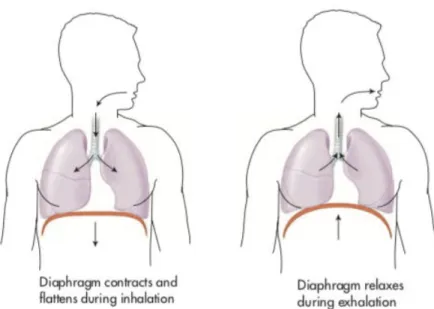
The lungs can be expanded and contracted in two ways (Colbert et al., 2016; Hall, 2011a): 1) by downward and upward movement of the diaphragm to lengthen or shorten the chest cavity (see Figure 2), and by 2) elevation and depression of the ribs to increase and decrease the anteroposterior diameter of the chest cavity (see Figure 3).
Figure 2: Diaphragm movements during inhalation and exhalation (from Colbert et al.,
2016, p. 293)
Figure 3: Illustration of the mechanics of pulmonary ventilation (from Hall, 2011a, p. 466) Note: Figure 3 shows the contraction and expansion of the thoracic cage during expiration and inspiration, demonstrating diaphragmatic contraction, function of the intercostal muscles, and elevation and depression of the rib cage. A-P: anteroposterior
Normal spontaneous breathing is achieved almost solely with the first method, that is to say by the movement of the diaphragm (Colbert et al., 2016; Hall, 2011a). During inspiration,
contraction of the diaphragm pulls the lower surfaces of the lungs downward. Then during expiration, the diaphragm simply relaxes, and the elastic recoil (also called rebound) of the lung, chest wall, and abdominal structures compresses the lungs and expels the air.
This is the breathing pattern also to be adopted with SPB – which is why it is often called diaphragmatic breathing –, to the exception that during SPB expiration would not be passive but rather active (Lehrer et al., 2000). The forced exhalation can involve abdominal muscles which, via their contraction, help to push the diaphragm back up to the thorax, thereby compressing the lungs, consequently pushing out additional air (Lorig, 2011; West, 2015; West & Luks, 2016). In SPB we would avoid to expand the lungs with the second method, that is raising the rib cage (Lehrer et al., 2000), given this would mean recruiting additional muscles, mainly the external intercostals to raise the rib cage and the abdominal recti and internal intercostals to pull the rib cage downward (Hall, 2011a; West & Luks, 2016). Recruiting additional muscles would require additional energy, which would then be counterproductive to the decrease in activation provoked by SPB (Lehrer & Gevirtz, 2014; Mather & Thayer, 2018).
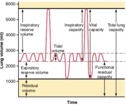
4.1.4 Pulmonary volumes
In order to understand the various events of pulmonary ventilation, the air in the lungs is usually subdivided in volumes and capacities, represented in Figure 4.
Figure 4: Respiratory patterns during normal breathing and during maximal inspiration
and maximal expiration (from Hall, 2011a, p. 469)
In order to better understand what happens during SPB, we focus here on the different kinds of pulmonary volumes (Hall, 2011a): The tidal volume is the volume of air inspired or expired with each normal breath (about 500mL in the adult male). The inspiratory reserve volume is the extra volume of air that can be inspired over and above the normal tidal volume when the person inspires with full force (about 3000mL). The expiratory reserve volume is the volume of air that can be expired by forceful expiration after the end of a normal tidal expiration (about 1100mL). The residual volume is the volume of air remaining in the lungs after the most forceful expiration (about 1200mL). SPB may require to breathe beyond the usual tidal volume and to take a deeper inhalation and exhalation (Lehrer & Gevirtz, 2014), which is why SPB is sometimes referred to as “deep breathing” .
4.1.5 Alveolar ventilation
The most important function of pulmonary ventilation is to enable to continually renew the air in the gas exchange areas of the lungs, where air is in proximity to the pulmonary blood, the alveoli (Colbert et al., 2016; Hall, 2011a; West & Luks, 2016). Other areas playing a role in
ventilation reflects the rate at which new air reaches these areas. The process of gas exchange between the alveoli and the pulmonary capillary can be seen in Figure 5. This process is suggested to be optimized by SPB, according to the resonance model (Lehrer & Gevirtz, 2014).
Figure 5: Coupling between alveoli and pulmonary capillary (from Hall, 2011a, p. 472)
More specifically, the process of gas exchange occurs via diffusion (Hall, 2011b), diffusion of oxygen from the alveoli into the pulmonary blood, and diffusion of carbon dioxide in the opposite direction, out of the blood (see Figure 6). The process of diffusion reflects the random motion of molecules in all directions through the respiratory membrane and adjacent fluid. Of importance will be the rate of diffusion.
Figure 6: Gas exchange at the level of the alveoli, illustration of the diffusion process (from
Colbert et al., 2016, p. 293)
4.1.6 Transport of oxygen and carbon dioxide in blood and tissue fluids
Once oxygen has diffused from the alveoli into the pulmonary blood, it is transported to the peripheral tissue capillaries almost entirely in combination with hemoglobin (Hall, 2011c), the protein in red blood cells in charge of carrying oxygen. Hemoglobin also returns carbon dioxide from the peripheral tissues back to the lungs.
4.1.7 The central regulation of respiration
The rate of alveolar ventilation is adjusted almost exactly according to the demands of the body by the nervous system. Consequently, the oxygen pressure and carbon dioxide pressure remains mostly constant in the arterial blood, even during different types of respiratory stress, such as heavy exercise (Hall, 2011d).
The respiratory center is composed of several groups of neurons located bilaterally in the medulla oblongata and pons of the brain stem (see Figure 8 and Figure 9). The medulla oblongata is home to all ascending and descending tracts that carry communications between the brain and the spinal cord (Waldman, 2009). In addition to the respiratory center, which fine tunes and modulates different information to regulate respiratory rate, the medulla oblongata also includes the cardiovascular center, which regulates heart rate in adjusting the strength of myocardial contractility and the dilatation and constriction of the peripheral vasculature (Waldman, 2009). Noteworthily, the sensory and motor nuclei of the vagus nerve are also found in the medulla oblongata. The vagus nerve is involved in many self-regulation processes (Thayer et al., 2009), as detailed later in section 4.3.1 (p.30), and will be a core focus of this PhD thesis.
The respiratory center contains three major groups of neurons: 1) the dorsal respiratory group, which is mainly responsible for inspiration, 2) the ventral respiratory group, which mainly causes expiration, and 3) the pneumotaxic center, which is mainly responsible for controlling rate and depth of breathing.
Figure 8: The Medulla Oblongata (from Waldman, 2009, p. 208)
Figure 9: Organization of the respiratory center (from Hall, 2011d, p. 506)
The dorsal respiratory group of neurons is thought to play the most fundamental role in the control of respiration. Most of its neurons are located within the nucleus of the tractus solitarius (Zoccal, Furuya, Bassi, Colombari, & Colombari, 2014). The nucleus of the tractus solitarius is the sensory termination of two nerves: the vagus nerve and the glossopharyngeal nerves. Both nerves transmit sensory signals into the respiratory center from 1) peripheral chemoreceptors, 2) baroreceptors, and 3) other kind of receptors in the lungs such as pulmonary stretch receptors (Hall, 2011d; Zoccal et al., 2014). The vagus nerve is already particularly important to notice here, given its role in self-regulation processes (see section 4.3.1; p.30). Finally, the activity of
the respiratory center will also be modulated by chemoreceptors based on the concentrations of oxygen, carbon dioxide, and hydrogen ions in the tissues (Hall, 2011d).
The mechanisms we just described were related to the involuntary control of respiration, but interestingly for psychophysiologists, breathing can also be modified voluntarily. Some voluntary patterns will prove very useful for self-regulation (Lehrer & Gevirtz, 2014; Lorig, 2011), such as SPB, which constitutes the main focus of this PhD thesis.
4.2 At the crossroads between the respiratory and the cardiovascular systems
4.2.1 Coupling between heart beat and respiration: Respiratory sinus arrhythmia
What makes breathing an interesting intervening variable, is that breathing is coupled to the cardiac output, in the sense that heart rate changes as a function of the respiratory cycle (Lorig, 2011). This phenomenon is called respiratory sinus arrythmia (RSA), heart rate accelerating during inhalation, and slowing down during exhalation (Angelone & Coulter, 1964; Eckberg & Eckberg, 1982). RSA is suggested to play an important role in respiration, given previous research has suggested that the efficiency of pulmonary gas exchange is improved by RSA (Giardino, Glenny, Borson, & Chan, 2003; Hayano, Yasuma, Okada, Mukai, & Fujinami, 1996; Mortola, Marghescu, & Siegrist-Johnstone, 2016, 2018; Yasuma & Hayano, 2004). The distribution of heartbeats within each respiratory cycle was suggested to improve the efficacy of respiratory gas exchange. More specifically, the matched timing of alveolar ventilation and its perfusion with RSA within each respiratory cycle was hypothesized to save energy expenditure by suppressing unnecessary heartbeats during expiration and ineffective ventilation during the ebb of perfusion (see Figure 10). In other words, heart rate tends to be higher when the air in the lungs is the richest in oxygen, and expiration occurs when carbon dioxide is at its highest level. However, this hypothesis have been questioned by further research, showing no relationship between RSA and improvement of gas exchanges (Buchheit, 2010; Sin et al., 2010; Tzeng, Sin, & Galletly, 2009). Another hypothesis, that still requires
empirical examination, is that RSA helps the heart do less work while maintaining healthy levels of blood gases (Ben-Tal, Shamailov, & Paton, 2012, 2014).
Gas exchange at the alveoli level is suggested to work best when heart rate starts to increase at the beginning of inspiration, and starts to decrease at the beginning of expiration, with would reflect a 0° phase relationship (Hayano et al., 1996), and which is coined cardioventilatory coupling (Elstad, O'Callaghan, Smith, Ben-Tal, & Ramchandra, 2018). However, in normal situations the relationship between heart rate and breathing is not completely in phase, which could be explained by allowing a greater degree of flexibility of the organism, so that greater efficiency can be achieved during phases of greater metabolic need, and less during decreased need (Lehrer & Gevirtz, 2014). SPB at 6 cycles per minute (cpm) is suggested to trigger a heart rate oscillating with breathing at a 0° phase relationship, with consequently the most efficient gas exchange (Lehrer & Gevirtz, 2014), however some posterior work strongly questioned the validity of this hypothesis (Sin, Webber, Galletly, & Tzeng, 2012). To sum up, the mechanisms by which RSA is an adaptive process still need to be understood.
Figure 10: Illustration of the suggested conceptual effect of the respiratory sinus
arrhythmia on the relationship between alveolar gas volume and capillary blood flow during inspiration and expiration (from Yasuma & Hayano, 2004, p. 684)
Note: Figure 10 shows the conceptual effects of respiratory sinus arrhythmia, the curved horizontal arrow and vertical arrows indicate the volume of blood flow circulating in the pulmonary capillaries and the direction of alveolar gas interfacing with the pulmonary capillaries.
vagal tone processes, which have different origins, dynamics, and functional consequences (Daly, 1985; Grossman, Karemaker, & Wieling, 1991; Grossman et al., 1990; Richter & Spyer, 1990). In particular, the vagus nerve is suggested to contribute to the efficiency of pulmonary gas exchange (Ito et al., 2006), which is to be linked to its role in self-regulation processes (see section 4.3.1, p.30). RSA is usually assessed via the peak-valley method (Grossman et al., 1990; Stange, Hamilton, Fresco, & Alloy, 2017), where the maximum heart rate during the expiration window of respiration is subtracted from the minimum heart rate during the inspiration window of respiration.
According to the resonance model (Lehrer & Gevirtz, 2014), RSA constitutes one of the core mechanisms targeted by the SPB technique to improve self-regulation processes.
4.2.2 Slow-paced breathing
As mentioned earlier, SPB is a breathing technique where the inhalation and exhalation durations are controlled (“paced”), and where breathing is performed at a slower pace (around 6 cycles per minute) than spontaneous breathing, which is usually comprised between 12 and 20 cycles per minute in adults (L. Sherwood, 2006). In this PhD thesis I use the term SPB instead of the other ones found in the literature, such as “deep breathing” (e.g., Tharion, Samuel, Rajalakshmi, Gnanasenthil, & Subramanian, 2012), “abdominal breathing” (e.g., Wang et al., 2010) or “diaphragmatic” breathing (e.g., Russell, Scott, Boggero, & Carlson, 2017), given it reflects the fact that a slow, paced breathing is the necessary trigger to influence positively self-regulation processes, as we now detail. The characteristics “deep”, “abdominal”, and “diaphragmatic” are not completely inaccurate and may reflect some aspects of the breathing technique, however they are not considered as the main triggers of the effects of SPB: 1) “deep” is supposed to reflect the fact that participants are required to slow down their breathing frequency, which can result – but does not have to – in increased tidal volume; however if participants get dizzy, they are immediately recommended to stop/adopt a more shallow
breathing (Lehrer et al., 2000); 2) “abdominal” or “diaphragmatic” reflects the characteristics of the most effective breathing pattern in resting conditions (for further details, see section 4.1.3, p.16).
The resonance model (Lehrer & Gevirtz, 2014) assumes that four processes are at stake to explain how SPB positively influences self-regulation mechanisms: 1) the phase relationship between heart rate oscillations and breathing at 6 cpm; 2) the phase relationship between heart rate and blood pressure oscillations at 6 cpm; 3) the activity of the baroreflex; and 4) the resonance characteristics of the cardiovascular system. These processes strengthen homeostasis in the baroreceptor (Lehrer et al., 2006; Vaschillo, Lehrer, Rishe, & Konstantinov, 2002; Vaschillo, Vaschillo, & Lehrer, 2006), which is suggested to result in improved gas exchanges at the level of the alveoli and in increased vagal afferences (Lehrer & Gevirtz, 2014).
However, empirical evidence challenged some of the suggested underlying mechanisms. Regarding the first one related to the phase relationship between heart rate oscillations and breathing at 6cpm (cardioventilatory coupling, Elstad et al., 2018), it is based on the hypothesized role of RSA in optimizing gas exchanges (Yasuma & Hayano, 2004). However, if indeed heart beat tends to redistribute towards inspiration at 6cpm (Lopes, Beda, Granja, Jandre, & Giannella-Neto, 2011), the original hypothesis regarding the physiological role of RSA to match heartbeats with pulmonary blood flow (Yasuma & Hayano, 2004) has been severely questioned (Buchheit, 2010; Sin et al., 2010; Tzeng et al., 2009), and so far no clear answer appeared to explain the adaptive physiological role of RSA. Still, the point that pulmonary gas exchange efficiency may be improved by slow breathing has received some support via reduction in ventilatory equivalents for carbon dioxide and oxygen when breathing at 6cpm (Sin et al., 2010), even if this method is not considered as the gold standard to measure gas exchange efficiency (Tharion & Subramani, 2011; Tzeng, 2011).
located in the walls of these arteries detect stretching of the arteries as blood pressure increases, and when this happens, the baroreflex causes immediate decreases in heart rate. On the contrary when blood pressure falls, the baroreflex causes immediate increases in heart rate (Eckberg & Sleight, 1992; Lehrer & Gevirtz, 2014). The mechanisms underlying the functioning of the baroreceptors is displayed in Figure 11. Similar to the phase relationship mentioned for heart rate and breathing, a similar phase relationship exists for heart rate and blood pressure, which can be depicted like this: when an external stimulation such as breathing at a specific frequency causes heart rate to rise, it also causes blood pressure to fall, consequently triggering an additional stimulus for heart rate to rise further. When the external stimulation (e.g., breathing) causes heart rate to fall, it also causes blood pressure to rise, thus causing an additional stimulus for heart rate to further decrease. Given the 0° phase relationship between heart rate and breathing occurs at approximately the same frequency that external stimulation causes maximal stimulation to the baroreflex, breathing appears a natural candidate as an external stimulator (Lehrer & Gevirtz, 2014). In line with this suggestion, large increases in baroreflex gain (number of beats per minute change in heart rate per 1 mm Hg change in blood pressure) have been observed during SPB with biofeedback, illustrating the fact that the baroreflex was strengthened (Lehrer et al., 2003).
Figure 11: Illustration of the functioning of the baroreflex (from Shaffer, McCraty, & Zerr,
2014, p. 9)
Finally, the last mechanism suggested by the resonance model (Lehrer & Gevirtz, 2014) regarding how SPB influences positively self-regulation processes is via the resonance characteristics of the cardiovascular system. Resonance is a physical principle stating that all oscillating feedback systems with a constant delay have the characteristic of resonance (Lehrer & Gevirtz, 2014). Here an example to better picture what resonance means: imagine a person being pushed on a swing (Allen & Friedman, 2012): The push must be in rhythm with the swinger’s momentum. Resonance between the natural swinging motion and the applied force makes the swing go higher than before. The same happens in the cardiovascular system when breathing at a frequency close to 6cpm, where the pattern of RSA overlaps with inherent oscillations in heart rate related to blood pressure modulation (Vaschillo et al., 2006). The point at which those two signals overlap was coined the resonant frequency, given the two signals
summate and produce large variations in heart rate (Allen & Friedman, 2012; Lehrer & Gevirtz, 2014; Vaschillo et al., 2006).
To sum up, the resonance model (Lehrer & Gevirtz, 2014) hypothesized that SPB, via several processes described above, influences positively self-regulation. The most supported mechanism so far is the strengthened homeostasis in the baroreceptor (Lehrer et al., 2003; Vaschillo et al., 2002; Vaschillo et al., 2006). The second mechanism, the optimization of pulmonary gas exchange based on RSA characteristics at 6cpm could not be fully supported so far (Buchheit, 2010; Sin et al., 2010; Tzeng et al., 2009). The third mechanism suggested was an increase in vagal afferents, however this path was so far largely based on the speculation that positive outcomes achieved with SPB interventions with biofeedback were related to brain areas related to vagal afferents (Lehrer & Gevirtz, 2014). In this PhD thesis, I focus on the possibility that this suggested action of SPB on vagal afferents would be reflected in vagal efferents and hence influence positively self-regulation mechanisms, given the links described in the next section.
4.3 How slow-paced breathing may influence self-regulation: the role of the vagus nerve
4.3.1 The role of the vagus nerve in self-regulation
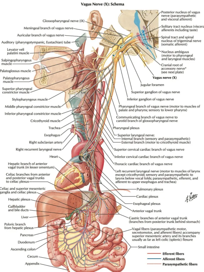
The vagus nerve is the tenth cranial nerve (see Figure 12), and it is the most important nerve of the parasympathetic nervous system (Brodal, 2010). It is composed of 80% afferent sensory fibers (sending signals from the body to the brain) and 20% efferent motor fibers (carrying information from the brain to the body). All branches of the vagus nerve with visceral efferent fibers also contain afferent sensory fibers, which makes it a highly sensitive nerve (Howland, 2014). As its name implies (the Latin translation of vagus means wandering), the vagus nerve branches to widespread regions of the body (Brodal, 2010), its fibers innervating most organs in the body including the gastrointestinal and cardiovascular systems (Bonaz,
Bazin, & Pellissier, 2018; Brodal, 2010; H. Y. Chang, Mashimo, & Goyal, 2003). Vagal fibers release acetylcholine as neurotransmitter (Brodal, 2010). In a nutshell, thanks to its extensive network the vagus nerve allows for widespread fast acting communications within the body.
Figure 12: Anatomy of the vagus nerve including branches (from Câmara & Griessenauer,
4.3.2 Cardiac vagal activity: the output of the central autonomic network
Regarding vagal efferent fibers, those that stimulate motor action, we are particularly interested here in those innervating the heart and modulating its intrinsic activity through the sinus node, which determines heart rate. Importantly, from the two branches of the autonomic nervous system, the sympathetic and the parasympathetic, the sympathetic influence on the heart is too slow to produce beat-to-beat changes (Jose & Collison, 1970), and the heart will be mainly under parasympathetic inhibitory influence through vagal efferent fibers (Jose & Collison, 1970; Saul, 1990). This cardiac autonomic balance is a way for the organism to favor energy conservation.
Regarding vagal afferent fibers, those that are linked to sensory actions, they are largely scattered through key organs in the human body (Brodal, 2010). This gives the vagal afferent system an important adaptation role as a detector of immune-related events in the human body. This peripheral sense allows for an internal signal that can generate the appropriate autonomic, endocrine, and behavioral responses via central reflex pathways going through the nucleus of the solitary tract (Berthoud & Neuhuber, 2000). These internal inputs are then integrated to external inputs, which helps to shape the appropriate response. Importantly, SPB may play a role in triggering internal inputs, given it is suggested to stimulate vagal afferents (Lehrer & Gevirtz, 2014).
A functional network based on brain structures is suggested to facilitate the organization and regulation of vagal afferent and efferent activity (Berthoud & Neuhuber, 2000). The central autonomic network (Benarroch, 1993) represents a functional unit in the central nervous system supporting goal-directed behavior, adaptability, and overall self-regulation processes. Cardiac vagal activity (CVA), the activity of the vagus nerve regulating cardiac functioning, is suggested to be the output of this central autonomic network, and is the core of the neurovisceral integration model (Smith et al., 2017; Thayer et al., 2009).
4.3.3 The neurovisceral integration model
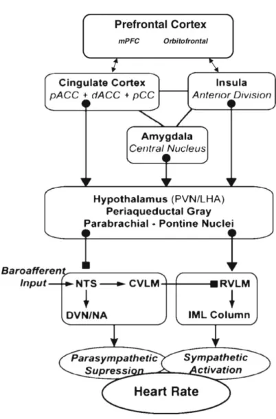
The neurovisceral integration model (Smith et al., 2017; Thayer et al., 2009) assumes a connection between the prefrontal cortex and the heart through the central autonomic network and the vagus nerve.
Figure 13: A composite schematic diagram showing the pathways by which the prefrontal
cortex might influence the control of heart rate (from Thayer et al., 2009, p. 143)
Note. mPFC: medial prefrontal cortex; pAAC: pregenual anterior cingulate cortex; dACC: dorsal anterior cingulate cortex; pCC: posterior cingulate cortex; PVN: paraventricular nucleus of hypothalamus; LHA: lateral hypothalamus; NTS: nucleus of the solitary tract; CVLM: caudal ventrolateral medulla; DVN: dorsal vagal motor nucleus; NA; RVLM: rostral ventrolateral medulla; IML: intermediolateral nucleus
The neurovisceral integration model postulates that CVA serves as an indicator of the effectiveness of this network, and consequently serves as a basis to index phenomena underpinning self-regulation, adaptation, and health (Smith et al., 2017; Thayer et al., 2009). At rest, the medial prefrontal cortex exerts inhibitory control over the amygdala, indirectly enhancing cardiac control via the vagus nerve, which is reflected in an increase of CVA (Thayer, Ahs, Fredrikson, Sollers, & Wager, 2012). This constant inhibitory control over the amygdala would support the connection of CVA to emotion regulation. The relationship between CVA and executive functioning originates from the common structures and networks involved in cardiac and cognitive regulation (Thayer et al., 2012). The effectiveness of executive functioning in the prefrontal cortex is supported via the optimal activation of neural networks, underlined with a flow of activity along neural pathways enabling to establish adequate mappings between input, internal states, and outputs needed to perform a given task (Miller & Cohen, 2001), leading to flexible responses to changing environments (Thayer et al., 2009).
4.3.4 Non-invasive assessment of cardiac vagal activity with heart rate variability
The neurovisceral integration model (Thayer et al., 2009) postulates that it is possible to index non-invasively the output of the central autonomic network, CVA, via heart rate variability (HRV, see Figure 14). HRV represents the time variation between each R peaks in the QRS complexes (Berntson et al., 1997; Laborde, Mosley, & Thayer, 2017; Malik, 1996). Two main HRV parameters reflecting CVA are the root mean square of the successive differences (RMSSD) and high-frequency HRV (Berntson et al., 1997; Laborde, Mosley, et al., 2017; Malik, 1996). SPB was found in previous research to increase vagal efferents as measured by short-term HRV (Kromenacker, Sanova, Marcus, Allen, & Lane, 2018; Lewis et al., 2015; Szulczewski & Rynkiewicz, 2018; Wells et al., 2012).
Figure 14: Electrocardiogram - Illustration of heart rate variability calculation
Note. Illustration of heart rate variability calculation, based on the time variation between each R peaks (marked here with a red cross); ECG: electrocardiogram; mV: millivolt
4.4 The 3Rs of vagus nerve functioning
Both tonic and phasic aspects of CVA functioning are important to consider (Thayer et al., 2012). Tonic CVA has also been referred to as resting CVA, and is when CVA is considered at one time point. Phasic CVA shows how the system reacts, and has been coined reactivity when depicting the change occurring between baseline and an event, and recovery, representing the change between the event to the return to resting conditions (Laborde, Mosley, & Mertgen, 2018b; Laborde, Mosley, et al., 2017). These three aspects are very important to consider from an adaptation point of view (see Figure 15). Regarding resting CVA, the neurovisceral integration model (Thayer et al., 2009) would assume in general “the higher the better”, meaning that a higher resting level of CVA is associated positively to self-regulation effectiveness. Regarding reactivity, two situations have to be differentiated (Beauchaine, 2001; Laborde, Mosley, & Mertgen, 2018b; Porges, 2007b; Thayer et al., 2012; Thayer et al., 2009): if the challenge is mainly cognitive, then a lower withdrawal or even an increase of CVA would be seen as adaptive; however if metabolic demands increase, a larger decrease of CVA would be adaptive in order to provide the necessary energy to the body. Finally, regarding recovery, a faster come back to baseline is expected to reflect a better adaptation (Stanley, Peake, & Buchheit, 2013). Taking into account the 3Rs of CVA functioning will be particularly important to derive the respective hypotheses.
EG G ( m V )
Figure 15: The 3Rs of cardiac vagal activity functioning (from Laborde, Mosley, et al., 2017, p. 6)
In summary, in the above sections we provided an overview of how SPB may be connected to self-regulation mechanisms. According to the resonance model (Lehrer & Gevirtz, 2014), if the most supported mechanism so far is the strengthening of baroreceptor homeostasis, another one being suggested is the increase in vagal afferences. This increase in vagal afferences may input into the central autonomic network (Benarroch, 1993), and according to the neurovisceral integration model (Thayer et al., 2009) consequently influence positively self-regulation phenomena. Within this PhD thesis I am going to investigate the relationship between SPB and a range of self-regulation phenomena, encompassing stress, sleep, and executive functioning.
4.5 Self-regulation phenomena investigated with slow-paced breathing and cardiac vagal activity within this PhD thesis
4.5.1 Stress
4.5.1.1 Psychological stress
Psychological stress occurs when an individual perceives that personal or environmental demands tax or exceed his or her adaptive abilities (Lazarus & Folkman, 1984). Even if psychological stress can be considered as an idiosyncratic phenomena, given it will differ across individuals and situations, a common method of inducing stress is using tasks taxing executive functions. In addition, putting an emphasis on performing well helps to represent a situation
which differs markedly from resting states in terms of psychological demands placed on the individual. This definition of stress may be linked to CVA, in that an appraisal of threat is central to a decrease of CVA, as opposed to an appraisal of safety (Thayer et al., 2009). The influence of mental stress on CVA will be very individualized according to the appraisal process (Lazarus & Folkman, 1984), and may depend for example on the degree of threat appraisal (Thayer et al., 2009). Another kind of stress may be triggered via physical stressors. 4.5.1.2 Physical stress
In terms of CVA reactivity, physical stressors require a vagal withdrawal in order for the organism to meet the physical demands of the task (Nakamura, Yamamoto, & Muraoka, 1993; Stanley et al., 2013). This reflects the evolutionary role of CVA as a “call to arms” mechanism to nurture the fight or flight response (Porges, 2007a, 2007b; Thayer, 2009). The fight or flight response is associated with near complete CVA withdrawal (Beauchaine, Gatzke-Kopp, & Mead, 2007), in order to facilitate large increases in cardiac output by the sympathetic nervous system, which is no longer facing the opposition of vagal inhibition. The level of CVA withdrawal during the physical stressor will depend mainly on the intensity of the physical stressor rather than on its duration (Stanley et al., 2013). Finally, the initial fitness levels of the person will influence both the amplitude and kinetics of vagal recovery, individuals having a greater aerobic fitness recovering faster (Stanley et al., 2013).
4.5.1.3 Stress and slow-paced breathing
The current evidence speaks strongly for a stress reducing effect of SPB. As shown by a recent meta-analysis (Goessl et al., 2017), SPB coupled with biofeedback was found to largely reduce self-reported stress and anxiety, while no moderator (e.g., number of sessions, presence of an anxiety disorder) was found to significantly influence the results. This confirms the findings of systematic and narrative reviews on the effects of SPB coupled with biofeedback on psychophysiological stress markers (Gevirtz, 2013; Kennedy & Parker, 2018; Wheat &
Larkin, 2010; Yu et al., 2018; Zaccaro et al., 2018). Interestingly, we can notice that even if SPB has been used in many contexts to reduce stress, it is still to be investigated in the field of intellectual disabilities.
4.5.1.4 Stress and intellectual disability
Having an intellectual disability is associated with high levels of stress (e.g., Forte, Jahoda, & Dagnan, 2011). This association can be explained in part by additional difficulties in adaptation and finding everyday situations over demanding, as well as being socially marginalized (de Bildt, Sytema, Kraijer, Sparrow, & Minderaa, 2005). Moreover, individual with intellectual disabilities often use maladaptive coping strategies to cope with stress (Hartley & Maclean, 2008). The stress experienced by individuals with intellectual disabilities is often transferred to family members and caregivers (e.g., Hassall & Rose, 2005). In the long term, if chronic stress in persons with intellectual disabilities is not properly addressed, it can lead to serious complications including depression (Hartley & Maclean, 2009), impaired cognitive functions (Heyman & Hauser-Cram, 2015), physical health problems (Lunsky, 2008), and maladaptive coping strategies such as substance abuse (Didden, Embregts, van der Toorn, & Laarhoven, 2009). It can also lead to family members and caregivers experiencing depression (Mutkins, Brown, & Thorsteinsson, 2011) and burnout (Innstrand, Espnes, & Mykletun, 2002). Given the prevalence of stress in people with intellectual disabilities, and the absence of stress management programs implementing SPB in people having intellectual disabilities so far, the first study will investigate whether SPB can help people with individual disabilities to cope with stress (p.54).
The second self-regulation phenomena of interest in this PhD thesis will be sleep.
4.6 Sleep
Issues with sleep are a pressing concern for individuals, given they directly impact life quality, and represent a risk factor at several levels (Davenne, 2009; Ferrie, Kumari, Salo,
Singh-Manoux, & Kivimaki, 2011). One of the main hypothesis regarding the cause of sleep disturbances is that they may be associated with a state of hyperarousal (Bonnet, Burton, & Arand, 2014; Riemann et al., 2010). Methods aiming to decrease a state of hyperarousal usually target an activation of the parasympathetic nervous system, and more specifically of its main nerve, the vagus nerve (Friedman, 2007; Prendiville, 2016). One way to do so is to use SPB (Gerritsen & Band, 2018; Lehrer, 2018; Lehrer & Gevirtz, 2014; Zaccaro et al., 2018).
4.6.1 Sleep and cardiac vagal activity
The question whether measuring CVA during sleep (CVAnight) represents an indicator of
sleep quality is still debated. Some evidence points toward an association between lower
CVAnight and sleep disorders (Stein & Pu, 2012), such as with chronic fatigue (Burton, Rahman,
Kadota, Lloyd, & Vollmer-Conna, 2010) and insomnia (Yang et al., 2011). Higher CVAnight
has also been related to higher subjective sleep quality (Brosschot, Van Dijk, & Thayer, 2007;
Patel et al., 2013; Yang et al., 2011). However, some authors argue that measuring CVAnight
across sleep stages does not provide useful information, given the variations observed in CVA during different sleep stages (Werner et al., 2015), namely CVA withdrawal during Rapid Eye Movement (REM) sleep, and CVA increase during non-REM sleep (Tobaldini et al., 2013). Further, Werner and colleagues (2015) argue that assessing CVA while sleeping is suboptimal, given CVA is supposed to reflect adaptations to environmental changes, and these do (almost) not occur during the night, so they rather recommend assessing CVA during periods where
individuals are awake. In summary, even if CVAnight measurement cannot be considered as an
index of sleep quality, it may still provide an indication of the restorative status of the body during sleep, given it indexes the activity of the parasympathetic nervous system (Laborde, Mosley, et al., 2017; Malik, 1996; Sakakibara, Hayano, Oikawa, Katsamanis, & Lehrer, 2013; L. Sherwood, 2006).
In order to address the criticisms made to CVAnight measurements, authors have suggested to measure CVA during wake periods (Werner et al., 2015). Particularly, a quiet awakening
morning period (CVAmorning) has been suggested as a good compromise, given the individual
has usually not experienced heavy environmental changes beforehand (Buchheit, Simon, Piquard, Ehrhart, & Brandenberger, 2004). CVAmorning has already been related to subjective indices of well-being and to physical training adaptations (see for example Buchheit et al., 2006; Buchheit et al., 2004), and also more recently to subjective sleep quality measurements (Flatt, Esco, & Nakamura, 2018). Investigating CVAmorning together with CVAnight measurements seems therefore an appropriate combination to further understand the effects of SPB on CVA.
4.6.2 Sleep and slow-paced breathing
To the best of our knowledge, only two previous studies investigated the effects of SPB on sleep while measuring HRV (Sakakibara et al., 2013; Tsai, Kuo, Lee, & Yang, 2015). In the first study (Sakakibara et al., 2013), SPB was delivered with the help of a biofeedback device during 20 minutes for two nights. In comparison to a control group performing autogenic training, CVA as measured with high-frequency (HF)-HRV was higher during the two nights measured, supporting the idea that it improved cardiorespiratory function. The second study (Tsai et al., 2015), focusing on self-reported insomniacs, aimed to investigate whether a 20min SPB session (6 cpm), compared to a control condition with paced breathing set at 12 cpm, would enhance objective sleep quality as assessed via polysomnography and CVA. In the SPB condition, the inspiration and expiration phases were set to 3s and 7s, while no indications were mentioned regarding the inspiration and expiration phases for the 12 cpm breathing condition. In regards to polysomnography, results showed that after a single 6 cpm session before going to sleep, sleep onset latency, number of awakenings, and awakening time during sleep were decreased, while sleep efficiency was increased, in comparison to the 12 cpm breathing
condition and to baseline. Regarding CVA, unfortunately the HRV variables mentioned in the paper (total power and R-R intervals) actually do not reflect it (Berntson et al., 1997; Laborde, Mosley, et al., 2017; Malik, 1996; Shaffer & Ginsberg, 2017; Shaffer et al., 2014), therefore it is not possible to draw any conclusions related to CVA. Moreover, HRV was not assessed during sleep, but during daytime rest. Consequently, further studies are therefore warranted to better understand the effects of SPB on subjective sleep quality and CVA, and not only on a short-term single session basis, but also on a long-term intervention basis.
The second study of this PhD thesis will target a long-term intervention (30 days) aimed to improve CVAnight and CVAmorning (p.62).
4.7 Executive functioning
Another area of self-regulation that can be indexed by CVA put forward by the neurovisceral integration model is the regulation of executive functioning, based on the common networks responsible for cardiac and executive control (Thayer et al., 2012; Thayer et al., 2009). Executive functions underpin goal-directed behavior and are essential for self-control (Diamond, 2013; Kotabe & Hofmann, 2015; Miyake et al., 2000). The three core executive functions are inhibition, working memory, and cognitive flexibility (Diamond, 2013; Miyake & Friedman, 2012; Miyake et al., 2000).
4.7.1 Inhibition and cardiac vagal activity
Inhibition reflects being able to control attention, behavior, thoughts, and/or emotions to override a strong impulse, and to do instead what is more appropriate according to the context (Diamond, 2013). A classical test for inhibition is the color word Stroop test (Stroop, 1935), where participants are requested to read out the color in which a word is printed while ignoring the meaning of the word. In the congruent condition the color matches the meaning of the word (e.g., the word “blue” expressed in the color blue), while in the incongruent condition the color differs from the meaning of the word (e.g., the word “blue” expressed in the color red). The
incongruent condition requires participants to inhibit the prepotent response of reading a word. Both the speed and accuracy of the responses can be measured. However, inhibition is primarily reflected as accuracy (error rate) (McDowd, Oseas-Kreger, & Filion, 1995), as it captures the ability to temporarily maintain the task goal in a retrievable state (Kane & Engle, 2003).
Previous research has found that Stroop task performance relates to CVA. A negative relationship has been observed between resting CVA and reaction times on incongruent and threat words (Johnsen et al., 2003), whereas a positive relationship has been observed between resting CVA and Stroop accuracy (i.e., Stroop interference score, Albinet, Abou-Dest, Andre, & Audiffren, 2016). These two findings are in line with the neurovisceral integration model (Thayer et al., 2009). One study observed mixed-findings between resting CVA (assessed with high-frequency HRV) and Stroop accuracy (i.e., Stroop interference score) (Subramanya & Telles, 2015). However, the experimental manipulation (meditation) occurring before the resting measurement might have introduced some confounding effects regarding the interpretation of high-frequency HRV, given that it is supposed to reflect CVA only when respiratory frequency is comprised between 9 and 24 cycles per minute (Berntson et al., 1997; Malik, 1996). As respiratory frequency was not assessed in the study, it is not possible to draw firm conclusions about CVA. The current experiment will investigate both accuracy and reaction time for the Stroop task controlling for respiratory frequency.
4.7.2 Working memory and cardiac vagal activity
Working memory involves working with information no longer perceptually present (Baddeley & Hitch, 1994). Stated differently, working memory involves holding information in mind and mentally working with it (Diamond, 2013). A classical test to assess working memory capacity is the automated operation span task (AOSPAN, Unsworth, Heitz, Schrock, & Engle, 2005). The AOSPAN task requires participants to solve mathematics problems while holding a number of unrelated letters in memory.