HAL Id: tel-02917926
https://tel.archives-ouvertes.fr/tel-02917926
Submitted on 20 Aug 2020HAL is a multi-disciplinary open access archive for the deposit and dissemination of sci-entific research documents, whether they are pub-lished or not. The documents may come from teaching and research institutions in France or abroad, or from public or private research centers.
L’archive ouverte pluridisciplinaire HAL, est destinée au dépôt et à la diffusion de documents scientifiques de niveau recherche, publiés ou non, émanant des établissements d’enseignement et de recherche français ou étrangers, des laboratoires publics ou privés.
Biochemical, biophysical and structural study of histone
deacetylase HDAC8 action mechanism and selective
inhibition
Tajith Baba Shaik
To cite this version:
Tajith Baba Shaik. Biochemical, biophysical and structural study of histone deacetylase HDAC8 action mechanism and selective inhibition. Biomolecules [q-bio.BM]. Université de Strasbourg, 2017. English. �NNT : 2017STRAJ057�. �tel-02917926�
UNIVERSITÉ DE STRASBOURG
ÉCOLE DOCTORALE DES SCIENCES DE LA VIE ET DE LA SANTÉ
Centre de Biologie Intégrative, IGBMC, UMR7104, Illkirch
THÈSE
présentée par :Tajith Baba SHAIK
soutenue le : 22 septembre 2017
pour obtenir le grade de : Docteur de l’université de Strasbourg Discipline/ Spécialité : Biophysique et biologie structurale
(Biophysics and Structural biology)
ETUDE BIOCHIMIQUE, BIOPHYSIQUE
ET STRUCTURALE DU MECANISME
D’ACTION ET DE L’INHIBITION
SELECTIVE DE L’HISTONE
DESACETYLASE HDAC8
THÈSE dirigée par :
Dr. ROMIER Christophe Directeur de Recherche CNRS, IGBMC, Illkirch
RAPPORTEURS :
Prof. EINSLE Oliver Professeur, Albert-Ludwigs-Universität Freiburg, Freiburg, Allemagne
Dr. FRIBOURG Sébastien Directeur de Recherche INSERM, IECB, Bordeaux AUTRES MEMBRES DU JURY :
3
Acknowledgements
First, I would like to express my sincere gratitude to my supervisor Dr. Christophe Romier for giving me this opportunity to pursue PhD in his lab. Thank you, Christophe, for giving me this fascinating subject and for being supportive from the day one. I specially thank you for being so patient with me for the past four years which have been a great experience for a great learning experience.
I take this opportunity to thank all the jury members, Dr. Catherine Schuster, Prof. Dr. Oliver Einsle, and Dr. Sébastien Fribourg who enthusiastically agreed to evaluate my work.
I would like to thank all our A-ParaDDisE collaborators, specially Dr. Raymond Pierce, Prof. Dr. Wolfgang Sippl and Prof. Dr. Manfred Jung for providing great scientific platform.
I would like to thank all the former and present members of the lab. A special thanks to Dr. Martin Marek for being a wonderful co-worker and for active discussions. I would like to thank Dr. Regis Back for such a wonderful company. I would like to thank Dr. Pierre Antony for giving me the valuable suggestions during my thesis writing.
A special thanks to Nata and Pernelle for the wonderful company in the lab. A single sentence is not enough to thank you guys, we can discuss about this in the lab. Really thank you for the nice working environment in the lab. Ah ha! A special thanks to Elizabeth for introducing adventurous Peru style of work.
A very special gratitude goes out to all the platform services, Structural biology department and administrative staff of IGBMC for helping me with lot of administrative stuff. With a special mention to Alastair, who has assisted me in collecting X-ray data at synchrotron. I am also grateful to all my friends at IGBMC, Kareem, Anna, Pau, Christophe, Moamen, and all the colleagues at CBI for providing a nice working environment.
And a special mention to Naima and Rana. Thank you for your support and for the nice food. I am very glad about all the Indians at the institute for the good company specially, Nisha, Vasugi, AK, ashique, and all others. Thank you, guys.
This list never ends without mentioning all the cricketing community of Illkirch. Especially Waseem, it’s been wonderful to play cricket with you guys.
4
Last but by no means least, I would like to thank my family members. My dad, mom, brother for their love and encouragement. Thanks mom for living my passion as your dream.
5
Introduction
L'ADN eucaryote est compacté sous forme d’une structure dynamique connue sous le nom de chromatine et dont la sous-unité minimale est le nucléosome (147 paires de bases d'ADN enroulées par les protéines histone). La structure de la chromatine module l'accessibilité à l'information génétique et régule ainsi les autres processus nucléaires. Fait important, la structure de la chromatine peut être modifiée par les mécanismes épigénétiques, permettant la régulation fine des processus nucléaires. En conséquence, les mécanismes épigénétiques ont un impact majeur sur l'homéostasie cellulaire, le type cellulaire et le développement. La dérégulation des mécanismes épigénétiques est de ce fait impliquée dans de nombreuses maladies telles que le cancer, mais aussi les maladies neurologiques et inflammatoires. Il est donc essentiel de comprendre les bases fondamentales des mécanismes épigénétiques. Mais les effecteurs épigénétiques représentent également des cibles importantes pour les interventions thérapeutiques dans de nombreuses maladies.
Parmi les mécanismes épigénétiques, « l'écriture » et « l'effacement », par des enzymes épigénétiques dédiées, des marques épigénétiques covalentes sur les histones et sur d'autres effecteurs nucléaires ont un effet majeur sur la structure de la chromatine et sa modification en réponse à divers stimuli. Plus précisément, l'addition covalente réversible de petits groupes chimiques (par exemple acétyle, méthyle, phosphate) ou macromolécules (par exemple ubiquitine, SUMO) à des résidus spécifiques des histones peut faciliter ou empêcher l'accessibilité de l'ADN par les mécanismes nucléaires.
L'acétylation est l'une des principales marques épigénétiques et est souvent associée à une structure de chromatine permissive aux processus nucléaires. L'acétylation des histones et d'autres effecteurs nucléaires est contrôlée par l'action opposée des HAT (transférases d’acétyle d’histone – histone acetyl transferases) et HDAC (desacétylases d’histone – histone deacetylases). Fait intéressant, les enzymes HDAC représentent les principales cibles des médicaments épigénétiques (épimédicaments) actuellement approuvés. Ces épimédicaments sont cependant restreints au traitement de quelques cancers car ils inhibent presque tous les membres de la famille HDAC qui ont des fonctions très différentes chez l’homme. Par conséquent, un axe de recherche majeur dans la découverte d'épimédicaments est de développer de nouvelles molécules qui montrent une sélectivité élevée pour un membre précis de la famille HDAC.
Il est important de noter que, malgré des années d'études des HDAC, la façon dont les différents membres de cette famille reconnaissent spécifiquement leurs cibles et comment ils
agissent sur ces cibles reste très obscurs. En outre, les HDAC font souvent partie de complexes macromoléculaires, et d'autres sous-unités de ces complexes peuvent affecter leur activité, la reconnaissance de leurs cibles, mais aussi l'inhibition par les épimédicaments. Il est donc essentiel de combiner à la fois des recherches fondamentales et applicatives sur les HDACs pour développer des épimédicaments plus puissants et plus sélectifs qui puissent être utilisés plus largement pour traiter un plus grand panel de maladies.
L'importance des mécanismes épigénétiques n'est pas limitée à l’homme, mais aussi à tous les organismes eucaryotes. Cela ouvre la porte à la lutte contre les parasites eucaryotes qui provoquent des millions de décès chaque année dans le monde entier. De tels parasites (par exemple le plasmodium, les trypanosomes, les schistosomes) ont souvent des cycles de vie très complexes où les mécanismes épigénétiques sont censés jouer des rôles essentiels. D’ailleurs, le traitement de parasites avec des épimédicaments approuvés ciblant les HDAC a révélé une forte sensibilité des parasites à ces épimédicaments.
Cela ouvre clairement la voie au développement de nouveaux médicaments antiparasitaires pour lutter contre des maladies pour lesquelles il n'existe que très peu de médicaments contre lesquels les phénomènes de résistance augmentent. Étant donné que le processus de découverte de médicaments nécessite beaucoup de temps et coûte cher, une stratégie de «portage» consistant à modifier les médicaments actuellement approuvés pour les rendre plus puissants contre les parasites devrait accélérer la recherche de nouveaux médicaments antiparasitaires. Encore une fois, un important goulet d'étranglement est de modifier les médicaments initiaux afin qu'ils puissent être sélectifs pour les enzymes parasitaires, mais plus pour les enzymes humaines.
Au cours du projet SEtTReND (Schistosoma Epigenetics: Targets, Regulation, New Drugs - 2010-2012) financé par l'Union Européenne, mon laboratoire a fourni la preuve de concept de cette stratégie de portage épigénétique en ciblant notamment HDAC8 du parasite
Schistosoma mansoni (smHDAC8). En résolvant la structure de smHDAC8 sous forme apo et
en complexe avec des inhibiteurs non sélectifs de HDACs, y compris le médicament déjà approuvé Vorinostat, l'équipe a fourni des informations inestimables et inattendues qui ont été utilisées pour trouver des premiers inhibiteurs présentant une bonne sélectivité mais une puissance moyenne. Les structures de smHDAC8 avec ces molécules ont par la suite été utilisées pour développer une nouvelle série d'inhibiteurs à forte puissance (dans la gamme du nM) et de sélectivité forte à excellente (Fig i).
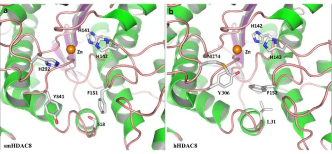
Fig i:Comparaison entre les poches du site actif smHDAC8 et hHDAC8 :
Représentation des structures cristallographiques de smHDAC8 (a) et hHDAC8 (b). Les acides aminés importants de la poche catalytique sont représentés sous forme des bâtons et sont numérotés respectivement. Le zinc catalytique est représenté par une sphère orange. Deux différences structurales sont notées au niveau de la poche catalytique, la présence de H292 contre M274 et la conformation de la phénylalanine dans les structures flipped‐in vs flipped‐out dans smHDAC8 et hHDAC8 respectivement. Ces différences de poche catalytique sont importantes dans la conception des inhibiteurs sélectifs de SmHDAC8.
Au cours de ma thèse et dans le cadre du plus grand projet financé par l'Union Européenne, A-ParaDDisE (Anti-Parasitic Drug Discovery in Epigenetics - 2014-2017), j'ai travaillé sur la caractérisation structurale des complexes entre smHDAC8 et cette nouvelle série d’inhibiteurs pour mieux comprendre les bases moléculaires de leur plus grande puissance et de leur plus grande sélectivité. J'ai complété ce travail par l'analyse biochimique, biophysique et structurale des complexes entre smHDAC8, mais aussi la HDAC8 humaine (hHDAC8), avec des inhibiteurs hautement sélectifs de hHDAC8. Ce travail m'a amené à étudier par analyse mutationnelle l'importance de l'architecture du site actif de HDAC8 pour l’activité et l’inhibition de cette enzyme. Enfin, dans une dernière partie de ma thèse, j'ai étudié des aspects plus fondamentaux de la biologie de HDAC8, en considérant notamment son interaction avec l'une de ses principales cibles, le complexe Cohésine.
Résultats
Au cours de ma thèse, j'ai résolu des dizaines de structures de smHDAC8 complexée avec divers inhibiteurs de nos collaborateurs. La structure de smHDAC8 avec la molécule la plus simple de cette série, TH31, a révélé un nouveau mécanisme de fixation de ces inhibiteurs sur smHDAC8 (Fig ii). Plus précisément, en plus de la coordination connue du
zinc catalytique de HDAC8 par un groupement hydroxamate, la liaison de TH31 dans la poche du site actif est stabilisée par l'interaction de son amide interne avec deux résidus de smHDAC8, K20 et H292. La liaison est encore plus stabilisée par des contacts hydrophobes établis entre la coiffe de TH31 et la boucle L6 de smHDAC8.
Cette liaison permet d'obtenir une sélectivité plus grande pour smHDAC8 par rapport aux autres HDAC humaines. Seule hHDAC8 est encore fortement inhibé par TH31. Par conséquent, nous avons sélectionné plusieurs autres inhibiteurs de cette série qui ont montré une puissance accrue pour smHDAC8, mais aussi une sélectivité accrue pour le smHDAC8 par rapport à hHDAC8. Ces inhibiteurs possèdent des groupes substituants supplémentaires ainsi que différentes coiffes par rapport à TH31. La structure de smHDAC8 avec ces différents inhibiteurs a fourni une foule d'informations sur la façon dont la puissance et la sélectivité peuvent être obtenues en améliorant et en étendant les interactions observées initialement entre TH31 et smHDAC8.
Fig ii Structure cristallographique de smHDAC8‐TH31 :
a) Représentation en ruban de la structure cristallographique de smHADC8‐TH31. TH31 (représenté par des bâtons de couleur cyan) est en coordination avec le zinc catalytique (sphère jaune) et d’autres résidus (représentés sous forme des bâtons et numérotés respectivement). K20 et H292 contiennent l'inhibiteur TH31 dans la poche du site actif de smHDAC8. b) Représentation structurale cristallographique de smHDAC8‐TH31 montrant la poche sélective HDAC8.
La HDAC8 humaine est l'une des HDACs pour lesquelles des inhibiteurs hautement sélectifs (par exemple PCI-34051) ont été trouvés. Mais les bases moléculaires de cette
sélectivité restent inconnues. J'ai étudié la base moléculaire de la sélectivité de HDAC8 par ces inhibiteurs par détermination de la structure de hHDAC8 et de smHDAC8 avec plusieurs de ces inhibiteurs. Ces structures ont montré que ces inhibiteurs se lient de manière similaire aux inhibiteurs sélectifs de smHDAC8 dans une poche spécifique de HDAC8 que nous avons appelée poche sélective de HDAC8 (Fig ii). Une fois de plus, toutes nos données structurales fournissent des informations très précises sur la base moléculaire de l'inhibition sélective de HDAC8, ouvrant la voie à la conception d'inhibiteurs plus puissants et plus sélectifs.
Ce travail sur l'inhibition sélective de HDAC8 a révélé l'importance de la taille et de la conformation de certaines boucles impliquées dans la formation du site actif de HDAC8. J'ai donc complété mon analyse d'inhibition par une analyse mutationnelle des boucles du site actif de HDAC8. Mes résultats montrent que la conformation de ces boucles est fortement contrainte, ce qui explique comment l'inhibition sélective peut être obtenue. Ces résultats ouvrent également la question de l'importance de ces boucles dans l’activité de HDAC8 et de sa reconnaissance de ses cibles, notamment en raison de la faible spécificité communément supposée des HDACs pour leurs cibles.
La cible la plus connue de HDAC8 est la Cohésine, un complexe qui joue un rôle important dans la cohésion des chromatides soeurs, la régulation transcriptionnelle et la réparation de l'ADN par recombinaison homologue. HDAC8 et la Cohésine sont impliquées dans de nombreux cancers, et des mutations dans HDAC8 et différentes sous-unités de la Cohésine conduisent à la même maladie, le syndrome de Cornelia de Lange, caractérisé par un nanisme et un handicap intellectuel.
Dans la dernière partie de ma thèse, j'ai commencé à étudier l'interaction entre la Cohésine et HDAC8 qui a été montrée comme désacétylant spécifiquement la sous-unité Smc3 de la Cohésine. La Cohésine est un complexe large et très flexible qu’il est difficile à caractériser structuralement. La plupart des structures actuellement publiées sont celles de petits domaines, généralement d'eucaryotes inférieurs. En utilisant la technique de co-expression développée dans l'équipe, j'ai pu reconstituer différents sous-complexes de la Cohésine humaine et, pour certains d'entre eux, j'ai déjà pu obtenir des cristaux. Ce travail ouvre la voie à la caractérisation structurale de la Cohésine humaine, mais aussi à son interaction avec HDAC8, pour mieux comprendre comment ces deux effecteurs nucléaires interagissent, avec des implications pour la maladie et l'inhibition sélective.
Conclusions
Une grande quantité de travail a été réalisée et continue d’être entreprise sur les HDACs qui représentent des cibles thérapeutiques importantes pour le développement
d’épimédicaments. En dépit de tout ce travail, des questions majeures subsistent sur les HDACs qui concernent des aspects fondamentaux tels que la reconnaissance spécifique de leurs cibles et leur mode d'action au sein de complexes, mais aussi des aspects plus applicatifs comme le développement d'inhibiteurs sélectifs pour étendre l'utilisation d’épimédicaments ciblant les HDACs.
Au cours de mon doctorat, j'ai abordé la plupart de ces problèmes en utilisant la HDAC8 humaine et du parasite Schistosoma mansoni. Plus précisément, mon travail sur l'inhibition sélective de HDAC8 a fourni des informations détaillées sur les bases moléculaires de cette inhibition sélective, ouvrant ainsi la voie au développement d'inhibiteurs plus puissants et plus sélectifs. Il est important de souligner que, à un moment où la recherche de nouveaux médicaments par des stratégies de screening à haut débit de chimiothèques et des méthodes de screening in silico sont favorisées par de nombreux chimistes médicinaux, mon travail montre comment les données structurales peuvent apporter des informations hautement complémentaires et essentielles dans le processus de découverte de nouveaux médicaments. De plus, ce travail m'a amené à étudier plus précisément l'importance de l'architecture globale du site actif des HDACs. Ce travail remet en question le dogme actuel qui considère que les enzymes HDACs sont peu sélectives en termes de cibles. Pour adresser ce problème, j'ai commencé à caractériser l'interaction entre HDAC8 et sa cible principale, le complexe Cohésine. Ce travail a déjà apporté des résultats essentiels qui permettront d'étudier finement cette interaction. Ainsi, le travail effectué lors de mon doctorat combine des recherches fondamentales et applicatives avec des implications pour des interventions thérapeutiques vers le cancer, et les maladies neurologiques et parasitaires.
6
7
The eukaryotic genomic DNA is packaged into a compact dynamic structure known as chromatin whose minimal subunit is the nucleosome (147 base pairs of DNA wrapped by histone proteins). Chromatin structure modulates the accessibility to the genomic information and thereby regulates the other nuclear processes. Importantly, chromatin structure can be modified by epigenetic mechanisms, enabling the fine tuning of nuclear processes. Accordingly, epigenetic mechanisms have a major impact on cell homeostasis, cell type and development, and deregulation of epigenetic mechanisms has been shown to be implicated in many diseases such as cancer, but also neurological and inflammatory diseases. It is therefore essential to understand the fundamental basis of epigenetic mechanisms. But epigenetic effectors also represent important targets for therapeutic interventions in many diseases.
Among the epigenetic mechanisms, the “writing” and “erasing” by dedicated epigenetic enzymes of covalent epigenetic marks on histones and other nuclear effectors has a major effect on chromatin structure and its modification in response to various stimuli. Specifically, the reversible covalent addition of either small chemical groups (e.g. acetyl, methyl, phosphate) or macromolecules (e.g. ubiquitin, SUMO) to specific residues of histone can facilitate or prevent DNA accessibility by the nuclear machineries.
Acetylation is one of the major epigenetic mark and is often associated with a chromatin structure that is permissive to nuclear processes. Acetylation of histones and other nuclear effectors is controlled by the opposing action of HATs (Histone acetyl transferases) and HDACs (histone deacetylases). Interestingly, HDAC enzymes represent the major targets of currently approved epigenetic drugs (epidrugs). These epidrugs are however restricted to the treatment of the few cancers since they inhibit almost all members of the HDAC family that have very different functions. Therefore, a major research axis in epidrug discovery is to develop new molecules that will show high selectivity for given members of the HDAC family.
Importantly, despite years of study of HDACs, it remains very obscure how the different members recognize specifically their targets and how they act on these targets. In addition, HDACs are quite often part of macromolecular complexes and other subunits of these complexes can affect activity, substrate recognition, but also inhibition by epidrugs. It is therefore essential to combine both basic and applicative research on HDACs to develop future more potent and more selective epidrugs that can be used more extensively to treat a large panel of diseases.
8
Interestingly, the importance of epigenetic mechanisms is not restricted to human but also to all eukaryotic organisms. This opens the door to the fight against eukaryotic parasites that causes yearly millions of deaths worldwide. Such parasites (e.g. plasmodium, trypanosomes, schistosomes) often have very intricate life cycles where epigenetic mechanisms are expected to play essential roles. Accordingly, treatment of parasites with approved epidrugs targeting HDACs have revealed a strong sensitivity of the parasites to these epidrugs.
This clearly opens the way for the development of new anti-parasitic drugs to fight diseases for which there are no to very few drugs available, with increasing report of resistance. Owing to the fact that the drug discovery process is extremely time-consuming and expensive, a “piggyback” strategy consisting in modifying currently approved drugs to make them highly potent against parasites is expected to speed up the search for new anti-parasitic drugs. Here again, a major bottleneck is to modify the drugs so that they can be selective for the parasitic enzymes but not anymore for the human ones.
During the course of the European-funded project SEtTReND (Schistosoma Epigenetics: Targets, Regulation, New Drugs – 2010-2012), my laboratory has provided the proof of concept of this epigenetic piggyback strategy by targeting notably HDAC8 from the parasitic flatworm
Schistosoma mansoni (smHDAC8). By solving the structure of smHDAC8 in apo form and in
complex with pan-HDAC inhibitors, including the approved Vorinostat drug, the team provided invaluable specific and unexpected information that has been used for finding initial inhibitors showing good selectivity but medium potency. Structures of smHDAC8 with these molecules were further used to develop a new series of inhibitors with high potency (nM range) and good to excellent selectivity.
During my PhD thesis and within the frame of the larger European-funded project A-ParaDDisE (Anti-Parasitic Drug Discovery in Epigenetics – 2014-2017), I have worked on the structural characterization of the complexes between smHDAC8 and the new series on inhibitors to better understand the molecular basis of high potency and selectivity. I have complemented this work by the biochemical, biophysical and structural analysis of the complex between smHDAC8, and also human HDAC8 (hHDAC8), with known highly selective hHDAC8 inhibitors. This work led me to look by mutational analysis at the importance of HDAC8 active site architecture for activity and inhibition. Finally, in a last part of my PhD thesis, I have investigated more
9
fundamental aspects of HDAC8 biology, by notably looking at its interaction with one of its main target, the Cohesin complex.
During my PhD thesis I solved tens of structures of smHDAC8 complexed with various inhibitors made by our collaborators based on our initial structures. The crystal structure of smHDAC8 with the simplest molecule of this series, TH31, revealed a novel mechanism of inhibition of these inhibitors for smHDAC8. Specifically, in addition to the common coordination of the HDAC catalytic zinc by a hydroxamate moiety, binding of TH31 in smHDAC8 active site pocket is stabilized by the interaction of its internal amide with two smHDAC8 residues, K20 and H292. Inhibitor binding is further stabilized by hydrophobic contacts made between its capping group and the L6 loop of smHDAC8.
This binding enables to gain selectivity for smHDAC8 over other human HDACs by several folds. Only hHDAC8 was still strongly inhibited by TH31. Therefore, we selected several other inhibitors of this series that showed increased potency for smHDAC8 but also increased selectivity for smHDAC8 over hHDAC8. These inhibitors had additional substitutions and different capping groups compared to TH31. The structure of smHDAC8 with these different inhibitors has provided a wealth of information how potency and selectivity can be gained by improving and extending the interactions observed initially between TH31 and smHDAC8.
Interestingly, human HDAC8 is one of the HDACs for which highly selective inhibitors (e.g. PCI-34051) have been found but the structural basis for this selectivity has remained unknown. I have investigated the molecular basis of HDAC8 selectivity with these inhibitors by determination of the structure of hHDAC8 and smHDAC8 with several of these inhibitors. Importantly, these structures showed that these inhibitors bind similarly to the smselective inhibitors to these enzymes in a specific pocket that we have termed HDAC8-selective pocket. Again, all our structural data provide precise information on the structural basis for selective inhibition, paving the way for the design of more potent and more selective inhibitors.
This work on the selective inhibition of HDAC8 has revealed the importance of the size and conformation of some of the loops building HDAC8 active site. I have therefore complemented our inhibition analysis by a mutational analysis of the active site loops of HDAC8. Our results show that the conformation of these loops is highly restricted, which further explains how selective inhibition can be achieved. This also opens the question of the importance of these
10
loops for activity and substrate recognition, notably arguing against the commonly accepted poor substrate specificity of HDACs.
HDAC8 best characterized target is Cohesin, a complex that plays an important role in sister chromatid cohesion, transcriptional regulation and DNA repair via homologous recombination. Specifically, HDAC8 and Cohesin are involved in a wide range of cancers, and mutations in both HDAC8 and different subunits of Cohesin lead to the same disease, the Cornelia de Lange syndrome, characterized by dwarfism and intellectual disability.
In the last part of my thesis, I have started to study the interaction between Cohesin and HDAC8 that has been shown to deacetylate specifically the Smc3 subunit of Cohesin. Cohesin is a large, highly flexible complex that is difficult to characterize structurally. Most currently published structures are of small domains, generally from lower eukaryotes. By using the technique of co-expression developed in the team, I have been able to reconstitute different sub-complexes of the human Cohesin and, for some of them, I have already obtained crystals. This work opens the way to the structural characterization of human Cohesin but also of its interaction with HDAC8 to better understand how these two nuclear effectors interact, with implication in disease and selective inhibition.
A large amount of work has been performed and continues to be carried out on HDACs that represent important therapeutic targets in epidrug discovery. Despite all this work, major issues remain that concern fundamental aspects such as the specific recognition of their targets by HDACs and their mode of action within complexes, but also the development of selective inhibitors against the different HDACs to extend the use of epidrugs targeting these enzymes.
During my PhD, I have addressed most of these issues using HDAC8 from human and the from parasite Schistosoma mansoni. Specifically, my work on the selective inhibition of HDAC8 has provided detailed information on the molecular basis of this selective inhibition and is paving the way for the development of more potent and more selective inhibitors. Importantly, at a time where high-throughput screening and in silico screening methods appear favoured by many medicinal chemists, my work is showing how structural data can bring highly complementary and essential data in the drug discovery process.
Importantly, this work has brought me to investigate more specifically the importance of the overall architecture of the active site of HDACs. This work questions the actual dogma that
11
implies that HDAC enzymes are poorly selective in term of substrate. To address this issue, I have started the characterization of the interaction between HDAC8 and its main target, the Cohesin complex. This work has already brought essential results that will enable from now on to investigate finely this interaction. Thus, the work done during my PhD is combining fundamental and applicative research with implication for therapeutic interventions towards cancer, neurological and parasitic diseases.
12
Table of contents
Figures ... 17
Tables List ... 19
1. Introduction ... 24
1.1. Chromatin and epigenetic mechanisms: impact on nuclear processes in health and diseases . 24 The chromatin ... 26
1.1.1.1. The nucleosome: basic unit of the chromatin ... 26
1.1.1.2. Higher order chromatin organization ... 29
Epigenetic mechanisms ... 31
1.1.2.1. Chromatin structure modulation through epigenetic mechanisms ... 31
1.1.2.2. The histone epigenetic marks ... 32
1.1.2.2.1. Post-translational modifications ... 32
1.1.2.2.2. Modification of non-histone proteins ... 36
1.1.2.2.3. Writers, readers and erasers of the epigenetic marks ... 36
1.1.2.2.4. Histone acetyltransferases and histone deacetylases ... 37
1.1.2.2.5. Histone methyltransferases and histone demethylases... 37
1.1.2.2.6. Kinases and phosphatases ... 39
1.1.2.2.7. Readers domains of the epigenetic marks ... 40
1.1.2.3. ATP-dependent chromatin remodellers ... 40
1.1.2.4. Histone variants and histone chaperones ... 43
1.1.2.5. Long non-coding RNAs ... 45
Epigenetic mechanisms in diseases and therapy ... 46
1.1.3.1. Epigenetic mechanisms in autoimmune diseases ... 47
1.1.3.2. Epigenetic mechanisms in neurological diseases ... 48
1.1.3.3. Epigenetic mechanisms in cancer ... 49
1.1.3.3.1. DNA Methylation in cancer ... 49
1.1.3.3.2. Post translational modifications of histones in cancer ... 50
1.1.3.3.3. Other epigenetic effectors in cancer ... 51
1.1.3.4. Targeting epigenetic players ... 51
1.1.3.4.1. Inhibitors targeting DNA methyltransferases ... 51
1.1.3.4.2. Inhibitors targeting bromodomain acetylation readers ... 52
13
1.1.3.4.4. Inhibitors targeting protein methyltransferases ... 52
A structural view of epigenetic targets ... 53
1.2. The acetylation mark ... 69
Role of the acetyl epigenetic mark ... 69
1.2.1.1. Acetylation and mechanisms of acetylation ... 69
1.2.1.2. A brief history of acetylation... 70
1.2.1.3. A handshake of acetylation with other PTMs ... 72
1.2.1.4. Histone acetyl transferases ... 73
1.2.1.4.1. HAT enzymes ... 73
1.2.1.4.2. HAT inhibitors and activators ... 76
1.3. Histone deacetylases: sirtuins and HDACs ... 78
Histone deacetylases classification ... 78
Sub cellular localization of histone deacetylases ... 82
Structure of histone deacetylases ... 83
1.3.3.1. HDACs ... 83
1.3.3.2. Sirtuins ... 87
Complexes of histone deacetylases ... 90
1.3.4.1. Complexes of class I HDACs ... 91
1.3.4.2. Complexes of Class II HDAC: ... 94
1.3.4.3. Sirtuin complexes: ... 95
Substrates of histone deacetylases ... 96
Mechanism of action ... 98
1.3.6.1. Mechanism of class I HDACs ... 98
1.3.6.2. Mechanism of Class II HDACs. ... 101
1.3.6.3. Mechanism of action of sirtuins ... 102
Histone deacetylases in diseases and therapy ... 103
1.3.7.1. Inhibition of HDACs in human diseases ... 103
1.3.7.2. An introduction to HDAC inhibitors ... 106
1.3.7.3. Selective inhibition of HDACs ... 110
HDAC8 ... 112
1.3.8.1. Introduction to HDAC8 ... 112
1.3.8.2. Role of HDAC8 on the Cohesin complex ... 114
14
On the interest of studying HATs and HDACs ... 119
1.4. Human neglected diseases ... 120
Neglected Tropical Diseases ... 120
Actual treatment of NTDs ... 122
Importance of epigenetics and HDACs in NTDs ... 122
Proof of concept: smHDAC8 a valid drug target to fight schistosomiasis by a piggyback strategy ... 124
1.4.4.1. Schistosomiasis ... 124
1.4.4.2. smHDAC8 ... 126
1.5. PhD thesis rationale: selective inhibition of parasitic enzymes and beyond ... 129
2. Material and methods ... 130
2.1. Cloning strategies ... 131
Expression vectors ... 131
Restriction endonuclease dependent cloning ... 132
Sequence and Ligase Independent Cloning (SLIC) ... 133
Gibson cloning ... 134
Site directed mutagenesis ... 135
Cloning of SMC-HD with linker ... 137
2.2. Expression methods ... 137
Principle of expression in case of the PET system of expression ... 138
Expression protocols ... 139
2.2.2.1. Mini expression test ... 140
2.2.2.2. Large scale production ... 140
2.3. Purification techniques ... 140
Affinity chromatography ... 141
Ion exchange chromatography ... 142
Gel filtration chromatography ... 143
Purification protocols ... 145
smHDAC8 purification ... 146
2.3.5.1. Large-Scale Overproduction and Purification of Recombinant Histone Deacetylase 8 (HDAC8) from the Human-Pathogenic Flatworm Schistosoma mansoni ... 146
2.3.5.2. Modified protocol for smHDC8 ... 147
2.4. Protein characterization ... 148
15
Dynamic Light Scattering ... 150 Differential Scanning Fluorimetry ... 151
In vitro HDAC assay ... 151
Crystallization vapor diffusion Technique ... 153 2.5. X-ray crystallography ... 154 2.5.1.1. Crystal systems ... 154 2.5.1.2. Bragg’s law ... 155 2.5.1.3. Ewald’s sphere ... 156 2.5.1.4. Theory of diffraction ... 157 2.5.2.1. Molecular replacement ... 160 2.5.2.2. Refinement ... 162 3. Results ... 164 3.1. Purification of acetyl modifying proteins from eukaryotic parasites ... 165 Histone Acetyltransferase 1 from Trypanosoma cruzi (TcHAT1) ... 165 3.1.1.1. Initial expression tests of TcHAT1 ... 165 3.1.1.2. TcHAT1-N2C2 Purification ... 167 3.1.1.3. Optimization of production ... 167 3.1.1.4. Purification of dl-TcHAT1-N2C2 ... 168 3.1.1.5. Purification of lh-TcHAT1-N2C2 ... 169 Deacetylase 3 from Leishmania braziliensis (LbDAC3) ... 170 3.1.2.1. Purification of LbDAC3: ... 171 3.1.2.2. Optimization of expression and purification: ... 173 Conclusion: ... 175 3.2. Elucidating smHDAC8 selective inhibition mechanism ... 175 Production of HDACs ... 175 3.2.1.1. Purification of smHDAC8 and hHDAC8 ... 176 3.2.1.2. Crystallization and soaking with inhibitors of smHDAC8 ... 178 Publication 1 ... 181 Publication 2 ... 182 Crystal structure of smHDAC8 with J1075 derived TB compounds ... 183 Crystal structures of smHDAC8 with miscellaneous inhibitors ... 185 3.2.5.1. Triazole derivatives ... 185 3.2.5.1. Crystal structures of smHDAC8 with Uracil based compounds ... 189
16
3.3. Cohesin complex purification ... 191 Expression of cohesin complex subunits ... 192 Purification of hsSMC1-HD ... 195 Purification and initial crystallization attempts of the hsSMC1-HD /RAD21-CTD1
complex 196
Optimization of the hsSMC1HD-RAD21 complex production ... 198 Expression of the hsSMC3HD/N-terminal region of RAD21 complex ... 200 Purification of hsSMC3-HD/RAD21-NTD2 ... 201 Characterization of the purified complexes using DLS ... 202 Crystallization attempts of the SMC-HD/RAD21 complexes ... 203 Data collection analysis ... 204 Conclusions and future perspectives on the HDAC8/Cohesin project ... 206 4. Discussion ... 207 5. Future Perspectives ... 213 6. List of poster presentations and oral communications ... 214 7. List of publications ... 215 8. References: ... 216
17
Figures
Figure 1: Chromatin organization: ... 24 Figure 2: Structure of the nucleosome: ... 27 Figure 3: Influence of H1 binding on nucleosomal arrays. ... 29 Figure 4: Models of higher order nucleosome organizations. ... 30 Figure 5: Condensin in chromosome condensation - The beads on a string model: ... 31 Figure 6: Dynamic properties of nucleosomes. ... 32 Figure 7: Histone chaperones and histone deposition mechanism. ... 45 Figure 8: One-step HAT mechanism of action. ... 75 Figure 9: HAT mechanism of action: ... 76
Figure 10: Phylogenetic tree of eukaryotic HDACs: ... 80
Figure 11: Domain organization of HDACs and sirtuins: ... 82 Figure 12: HDAC fold: ... 84 Figure 13: Structure of class IIa HDACs ... 86 Figure 14: Structure of HDAC10 ... 87 Figure 15: Structure of SIRT1 with resveratrol ... 89 Figure 16:Sirtuin structures: ... 90 Figure 17: Sequence alignment of HDAC loops ... 92 Figure 18: Crystal structure of HDAC3-SMRT complex ... 93 Figure 19: Surface representation of HDAC1-MTA1 complex crystal structure: ... 94 Figure 20: Sirtuin complexes ... 96 Figure 21: Class I HDACs mechanism of action. ... 100 Figure 22: Class IIa HDACs mechanism of action ... 101 Figure 23: HDAC6 mechanism of action ... 102 Figure 24: Sirtuin mechanism of action ... 103 Figure 25: Consequences of HDAC inhibition: ... 105 Figure 26: Chemical constituents of a HDAC inhibitor ... 106 Figure 27: Different classes HDAC inhibitors ... 107 Figure 28: HDAC8 substrates ... 113 Figure 29: Cohesin complex architecture. ... 115 Figure 30: Cohesion - cell cycle regulation. ... 116 Figure 31: Global Overlap of common NTDs. ... 121 Figure 32: NTDs caused by different type of infections... 121 Figure 33: Schistosoma life cycle. ... 125 Figure 34: Comparison of catalytic subunit of smHDAC8 with human HDACs ... 128
18
Figure 35: Experimental approach: ... 130 Figure 36: pnEA-tH vector: ... 132 Figure 37: Steps in Traditional cloning ... 133
Figure 38: Sequence and ligase independent cloning ... 134
Figure 39: Overview of Gibson assembly ... 135
Figure 40: Mechanism of pET expression system. ... 139
Figure 41: Affinity chromatography: ... 142 Figure 42: Ion exchange chromatography: ... 143 Figure 43: Gel filtration chromatography. ... 145 Figure 44: Polymerization of acrylamide ... 148 Figure 45:DLS outlook ... 150 Figure 46: Fluor de Lys assay: A: ... 152 Figure 47: IC50 determination. ... 153
Figure 48: Illustration of vapor diffusion using Sitting drop and handing drop setup. ... 153
Figure 49: Bravais lattices ... 155 Figure 50: Bragg's law ... 156 Figure 51: Construction of Ewald's sphere ... 157 Figure 52: Argand diagram: ... 159 Figure 53: Calculation of Patterson peaks from electron density: ... 161 Figure 54: Sequence alignment of TcHAT1: ... 166 Figure 55: Gel filtration purification of TcHAT1-N2C2. ... 167 Figure 56: TcHAT1 – mini test-expression optimization: ... 168 Figure 57: Purification profile of dl-TcHAT1-N2C2: ... 169 Figure 58: Purification profile of lhTcHAT1N2C2: ... 169 Figure 59: DLS profile of lh-TcHAT1-N2C2 ... 170 Figure 60: LbDAC3 expression optimization: ... 171 Figure 61: Purification of LbDAC3 ... 172 Figure 62: Sequence alignment of LbDAC3 with HDAC7 and 4. ... 173 Figure 63: Ion exchange profile of LbDAC3 ... 174 Figure 64: HDAC8 expression optimization: ... 176 Figure 65: smHDAC8 purification profile. ... 177 Figure 66: hHDAC8 purification profile. ... 177 Figure 67: J1075 derived compounds ... 179 Figure 68: J1038 derived compounds ... 179 Figure 69: Pan-HDAC and HDAC8 selective inhibitors ... 180
19
Figure 70: Triazole and Uracil based compounds ... 180 Figure 71: smHDAC8-J1075 derivatives crystal structures: ... 185 Figure 72: Crystal structures of smHDAC8 with triazole derivatives and Uracil based compound ... 188 Figure 73: Strategy to SMC complex production: ... 192 Figure 74: Sequence alignment of hsSMC1A with ABC ATPases: ... 193 Figure 75: Expression of SMC1-HD ... 194 Figure 76: Purification profile of hsSMC1HD: ... 195 Figure 77: Characterization of hsSMC1-HD using DLS: ... 196 Figure 78: Purification profile of SMC1-HD/RAD21-CTD1. ... 196 Figure 79: Thrombin cleavage of SMC1-HD/RAD21-CTD1 complex ... 197 Figure 80: Expression profiles of hsSMC1HD and RAD21CTD optimization: ... 199 Figure 81: Purification profile of SMC-HD/RAD21-CTD2: ... 199 Figure 82: SMC3-RAD21NTD co-expression: ... 200 Figure 83: Purification profile of hsSMC3-HD/RAD21-NTD2: ... 201 Figure 84: Optimization of SMC3HD-RAD21NTD2 complex purification: ... 202 Figure 85: DLS profiles of SMC-RAD21 complexes: ... 203 Figure 86: Crystals of hsSMC1HD-RAD21CTD2 complex. ... 204
Tables List
Table 1: Table showing important histone modifications and their function ... 34 Table 2: Important phosphorylation sites and known kinases (Rossetto, Avvakumov et al. 2012). ... 39 Table 3: Chromatin remodelling complexes in humans. ... 42 Table 4: List of histone variants and their functions (Buschbeck and Hake 2017). ... 44 Table 5: Table of HAT complexes ... 74 Table 6: Inhibitors and activators of HATs ... 77 Table 7: Classification of HDACs: ... 79 Table 8: HDAC inhibitors ... 109 Table 9: Vectors with different options of tags and antibiotic resistance used in this thesis ... 131 Table 10: Rolling circle plasmid synthesis protocol ... 136 Table 11: Thermocycler protocol for rolling circle plasmid synthesis ... 136 Table 12: SDS-PAGE gel composition ... 149 Table 13: SDS-PAGE staining solution ... 149 Table 14: IC50 values of inhibitors ... 184
20
Table 15: Crystallographic table II ... 190 Table 16: Data statistics of hsSMC1HD-RAD21CTD2 ... 205
List of Abbreviations
Alphabet List of Abbreviations
% : percentage µL : microliter µg : microgram
ψDAC : pseudo deacetylase domain
A AAT : (7-[(3-aminopropyl) amino]-1,1,1-trifluoroheptan-2-one)
ADP : adenosine diphosphate
ALS : Amyotrophic lateral sclerosis
Amp : Ampicilline
A-ParaDDisE : Anti-Parasitic Drug Discovery in Epigenetics; 2014-2017 APS : ammonium persulfate
ARP : actin related proteins ATP : Adenosine triphosphate AURKB : aurora kinase B
B BET : Bromodomain and Extra-Terminal motif
C CBP : CREB binding protein CD : catalytic domain
CDC : Centers for Disease control and prevention CDK1 : Cyclin Dependent Kinase 1
CdLS : Cornelia de Lange Syndrome cDNA: complementary DNA
CHD : chromodomains helicase DNA binding Chl : Chloramphenicol
cm: centimeters
CNRS : Centre National de la Recherche Scientifique CoA : coenzyme A
CoREST : Corepressor of RE1-Silencing Transcription factor cryoEM: Cryo-Electron Microscopy
CSB : Cockayne Syndrome group B CTD : C-terminal domain
CTD2 : RAD21 C-terminal region
D Da : dalton
DAD : deacetylase activation domain DAXX: death domain–associated protein DFT : density functional theory
DHS : deoxyhypusine synthase DLS : Dynamic Light Scattering
DNA: DeoxyriboNucleic Acid
DNMTs : DNA methyltransferases
dNTPs: deoxyNucleotides Tri-Phosphate
21
DTT : Dithiothreitol
DUBm : SAGA deubiquitination module
E EDTA: Ethylenediaminetetraacetic acid
EM : electron microscopy Epidrugs : epigenetic drugs
ESFRI : European Strategy Forum on Research Infrastructures
F FDA : American Food and Drug Administration
FRISBI : French Infrastructure for Integrated Structural Biology
G GST : Glutathione S-transferase
H H3K4me2 : dimethylation of lysine 4 from histone H3
HATs : Histone Acetyltransferases
HCl: Hydrochloric acid
HDACs : Histone deacetylases HDACis : HDAC inhibitors HDLP : HDAC-like protein
HEK293 cells : human epithelial kidney cell line
His: Histidine
HIV : human immunodeficiency virus
hnRNP : heterogeneous nuclear ribonucleoproteins
HPLC: High-performance liquid chromatography
HSA : helicase SANT-associated HSP90 : heat shock protein 90
I IC50 : half maximal inhibitory concentration
ICF : immunodeficiency centromeric instability and facial anomalies IEX : Ion exchange
IFNα : interferon α IFN-γ : interferon- γ IFNαR2 : IFNα receptor 2
IGBMC : Institut de Génétique et de Biologie Moléculaire et Cellulaire
INSERM : Institut National de la Santé et de la Recherche Médicale
IPTG: Isopropyl β-D-1-thiogalactopyranoside
IRF9 : interferon regulatory factor 9 ISGF3 : Interferon-stimulated gene factor 3 ISWI : imitation switch
K Kan : Kanamycin
KAT : lysine acetyltransferases
KCl: Potassium chloride
KDAC : lysine deacetylases
Kpr, Kbu and Kcr : lysine propionylation, butyrylation and crotonylation
L LB : Luria Bertani
LbDAC3 : Deacetylase 3 from Leishmania braziliensis LINE : Long interspersed nuclear elements
lncRNAs : long non-coding RNAs
LTR : Long Terminal Repeat
22
MAGE : melanoma-associated gene MBD : methyl CpG binding domain
MCS: Multiple Cloning Site
MeCP2 : methyl CpG binding protein 2
MgCl2:Magnesium chloride
MiDAC : Mitotic DeAcetylase Complex miRNA : micro-RNA
MLL1 : Mixed lineage leukaemia protein 1
mRNA: messenger RNA
MSL: male specific lethal
MTA1 : Metastasis-associated protein 1
N NaCl: sodium chloride
N-CoR : Nuclear receptor Co-Repressor NCP : Nucleosome Core Particle
ncRNAs : non-coding RNAs NES : Nuclear export signal NFkB : nuclear factor κB NIBL : nipped B like protein NLS: nuclear localization signal ng : nanograms
nm : nanometers
NMR : Nuclear magnetic resonance NSCLC : non-small-cell lung cancer nt : nucleotide
NTD : N terminal domain
NTDs : Neglected tropical diseases
NuRD : Nucleosome Remodeling Deacetylase
O OD : Optical Density O/N: over night
P PCR: Polymerase Chain Reaction
PDAC : HDAC10 catalytic domain
PDB: Protein Data Bank PEG : Polyethylene glycol pH: potential of Hydrogen
PLK1 : Polo-like protein
PMTs : Protein Methyl Transferases PP2A : protein phosphate 2A
PRMTs : Protein Arginine Methyltransferases PTMs : Post-Translational Modifications
Q QM/MM : DFT quantum mechanics / molecular mechanics
R RENT : regulator of nucleolar silencing and telophase exit
RNA: Ribonucleic acid
ROS : Reactive oxygen species
Rpm: Revolutions per minute rRNA:Ribosomal RNA RT: Reverse Transcriptase
RTase: DGR-associated Reverse Transcriptases
23 S SAXS : Small Angle X-rays Scattering
SAH: S-adenosyl homocysteine SAHA : suberanilohydoxamic acid SAM : S-adenosyl methionine
SDS: Sodium Dodecyl Sulfate
SEC : size-exclusion chromatography
SEtTReND (Schistosoma Epigenetics: Targets, Regulation, New Drugs; 2010-2013)
SGO1 : Shugoshin
SHL2 : super-helical location 2 Sir2 : Silent information regulator 2 SIRT1-7 : sirtuins members
SLE : Systemic Lupus Erythematosus
SLIC : Sequence and Ligase Independent Cloning SMC : Structural Maintenance of Chromosome SMC3HD : SMC3 head domain
SMRT : Silencing Mediator of Retinoic acid and Thyroid hormone receptors Sp : Spectinomycin
STAT2 : signal transducer and activator of transcription 2
SUMO : Small Ubiquitin-like Modifier
SWI/SNF : SWItch/Sucrose Non-Fermentable
T TAF : TATA-binding protein associated factor
Tat : HIV transcriptional activator protein TB : Terrific Broth
TBE: Tris Borate EDTA
TcHAT1 : Histone Acetyltransferase 1 from Trypanosoma cruzi TEMED: N,N,N’,N’ Tetramethylethylene-1,2-diamine
TGFβ : transforming growth factor β tH4 : testis specific H4 variant TFIID : Transcription factor II D TFMO : Trifluromethyloxadiazolyl
Tris: 2-amino-2-(hydroxymethyl)-1, 3-propanediol
tRNA : transfert RNA TRX : thioredoxin TSA : Trichostatin A
U UDR : ubiquitin-dependent recruitment motif
V V:Volume
W WAPL : wings apart-like protein homologue WHO : World Health Organization
24
1. Introduction
1.1.
Chromatin and epigenetic mechanisms: impact on nuclear processes in
health and diseases
In order to fit within the 6 microns large nucleus, the 2 meters long eukaryotic genomic DNA is packaged into a highly ordered compacted structure called chromatin. The chromatin can have several levels of compaction, from a weakly condensed state to a highly condensed state as observed in the chromosomes (Figure 1). Compaction of the eukaryotic DNA into chromatin has a major effect on the nuclear processes (e.g. replication, transcription, DNA repair) as it restricts access to the underlying genetic material.
Figure 1: Chromatin organization: Picture adapted from apsubiology.org
25
The current view is that, depending on the chromatin compaction state, the genetic material is more or less accessible to the nuclear effectors. Actually, several major forms of chromatin are distinguished. The euchromatin is generally weakly compacted and is permissive to nuclear processes. In contrast, heterochromatin is generally densely packed and is associated with reduced nuclear activity. Finally, facultative heterochromatin is a less densely compacted chromatin than heterochromatin that displays little nuclear activity but is poised for transformation into euchromatin.
Depending on the gene expression patterns required, the chromatin state of a particular region of the genome can be different between different cell types and can be modified during development and in response to internal and external stimuli. This modulation of the chromatin structure is carried out by the so-called epigenetic mechanisms that enable the chromatin structure to be finely tuned and adapted to the cell’s requirements.
Importantly, epigenetic mechanisms, by acting on the chromatin structure, have a strong influence on the various nuclear processes and are directly involved in their regulation. Thus, chromatin structure and epigenetic mechanisms are key to regulate genomic imprinting, X-chromosome inactivation, transcriptional regulation, DNA replication, DNA damage repair and DNA recombination (Sadakierska-Chudy, Kostrzewa et al. 2015). In addition, epigenetic mechanisms ensure that heritable changes are carried through mitosis and meiosis without altering the DNA sequence to maintain the tissue specific pattern of gene expression (Sadakierska-Chudy, Kostrzewa et al. 2015).
The consequence of the importance of chromatin structure and epigenetic mechanisms on the regulation of nuclear processes is of course that deregulation of these mechanisms has a broad impact on the cellular activity. It comes therefore as no surprise that an increasing number of epigenetic effectors are shown to be involved in the onset and progression of many different diseases. Importantly, the flexibility of epigenetic mechanisms and their capacity to modulate the chromatin structure open the way to epigenetic therapeutic interventions.
It is therefore essential that epigenetic mechanisms and their interplay with other cellular processes are finely characterized towards the development of therapeutic strategies. This process has already begun, but much more knowledge is required to progress further towards this goal.
26
The chromatin
1.1.1.1.
The nucleosome: basic unit of the chromatin
The nucleosome is the smallest structural and functional unit of the chromatin that provides a platform on which epigenetic effectors can act (Cutter and Hayes 2015). Each nucleosome consists of a nucleosome core particle (NCP), linker DNA and a linker histone. The NCP consists of 145-147 bp of DNA wrapped in a 1.7 super helical turn around the histone octamer. The histone octamer disk is made up of two copies of the four histones H2A, H2B, H3 and H4. Histones possess a conserved histone fold and an N-terminal tail domain (Figure 2). H2A contains an additional C-terminal tail that protrudes from the nucleosome towards linker DNA and interacts with histone H1 which is the linker histone (Davey, Sargent et al. 2002, Vogler, Huber et al. 2010). Tail domains are sensitive to proteases and protrude through the nucleosome minor grooves and are solvent exposed (Iwasaki, Miya et al. 2013). Most importantly tail domains participate in inter nucleosomal interactions which are important for maintaining higher order chromatin structure.
H2A specifically dimerize with H2B and H3 with H4. Two H3-H4 dimers are self-associated via H3-H3 interface to form a tetramer. Two H2A-H2B dimers bind on either side of the H4-H3-H3-H4 tetramer. The arrangement generates an octameric disk-like structure on which 121 bp of DNA wraps around histone folds and the sides of the octameric disk are aligned to major and minor grooves of DNA. The remaining 13bp of DNA from both sides are covered by the extensions of histone folds which completes the 147 bp nucleosome (Figure 2).
27 Figure 2: Structure of the nucleosome:
a. Tetramer formation of H3:H4 dimers, showing histone fold in inset and histone tails by arrow marks (pdb: 1KX5). b. Histone octamer formation with four histones. c,d. ribbon representations of the nucleosome in two perpendicular views. The disk form of the nucleosome is shown in d.
The interactions between positively charged histones and negatively charged DNA are keys for NCP assembly, and for nucleosome compaction. DNA makes 14 contacts with the histone octameric disk in the nucleosome where highly conserved arginine residue at each site interacts with the minor groove to help precisely position the DNA around the octameric disk and to facilitate the overall super helical shape (Gottesfeld and Luger 2001, Davey, Sargent et al. 2002, Cairns 2007, Wang, Ulyanov et al. 2010).
The linker histone, or H1, binds to the nucleosome with a weaker tendency than of core histones and it is also less conserved compared to core histones. Unlike core histones, the
28
organization of linker histone is completely different. In mammals 11 isoforms of H1 have been identified, which maintain a general domain organization with a N-terminal protease sensitive tail domain, a central folded globular domain, and highly basic, protease sensitive C-terminal domain (CTD). H1 CTD is essentially basic to neutralize the negative charge of the linker DNA. The globular domain binds at the interface near entry and exit of DNA in the nucleosome where it draws the two linker DNA together and reduce their flexibility (Bednar, Garcia-Saez et al. 2017). The globular domain of H1 is enough to increase the compaction of nucleosome.
Adjacent nucleosomes are connected by a segment of linker DNA which determines the space between nucleosomes. This arrangement generates a nucleosomal array in a diameter of 11nm which represents the known “beads on a string” organization of nucleosomes (Olins and Olins 1974). The flexibility of linker DNA is reduced by the H1 binding. The H1 binds the nucleosome in an asymmetric manner. The CTD of H1 binds one linker DNA initially and the globular domain binds asymmetrically on the dyad axis of nucleosome, this asymmetric H1 binding causes asymmetry in electrostatic and mass distribution. This asymmetry helps in the formation of higher order chromatin structure in case of nucleosomal arrays. The head to head and head to tail orientations of H1 proteins in the nucleosomal arrays result in the two different types of repeating structural units, dinucleosomes and tetra nucleosomes (Figure 3) (Bednar, Garcia-Saez et al. 2017).
29 Figure 3: Influence of H1 binding on nucleosomal arrays.
Pictures are modified from (Schalch, Duda et al. 2005, Bednar, Garcia-Saez et al. 2017).
A crystal structure of an oligonucleosome with four nucleosomes has revealed the arrangement of a tetranucleosome in which nucleosomes are arranged in two stacks connected by three linker DNAs (LB, LB’and LS) (Figure 3). The nucleosome pairs N1, N2 and N1’, N2’ are rotated with respect to each other along two-fold axis and the two nucleosomes of each stack are interacting with each other through their octameric histone surface. (Schalch, Duda et al. 2005).
1.1.1.2.
Higher order chromatin organization
The compact 11 nm nucleosomal arrays are further organized into more compact 30 nm chromatin fibers which are considered as the secondary structure of chromatin. The 30 nm fiber shows different conformations such as solenoid helix, twisted ribbon and cross linker
30
conformations, and is observed in two types of arrangements in the chromatin structure (McGhee, Nickol et al. 1983, Woodcock, Frado et al. 1984, Zhu and Li 2016). In the one start solenoid structure, consecutive nucleosomes are next to each other and the linker DNA is bent towards the fiber center. In the two start solenoid structures, the consecutive nucleosomes are arranged in a zig zag manner to form two rows of nucleosomes, and the linker DNA is linear (Figure 4). In this latter case, the alternate nucleosomes are the interacting partners. This model further produce coiling and twisting to generate variants of zig zag model (Tremethick 2007).
Figure 4: Models of higher order nucleosome organizations. Adapted from (Tremethick 2007).
Recent advancements in chromatin structure research suggests that the existence of 30 nm chromatin fibers is however controversial. Biophysical and structural (cryo EM, SAXS and ultra SAXS) approaches did not find evidence for the classical 30 nm chromatin scaffolds, and no structural repeating unit that was larger than the 11 nm nucleosome fibers was observed (Nishino, Eltsov et al. 2012).
Yet, at the intersections of 11 nm structures, the condensin complex was observed that has been known to help in chromatin compaction. Condensin is a multi-protein complex that plays an important role in the chromatin assembly and segregation during cell cycle (Hirano 2016). This
31
led to the alternative model that condensin organize chromatin into an arrangement of 11 nm fibres. Condensin is majorly concentrated around centromeres and along the arms of chromosomes. Condensin provides stability to the centromeric chromatin; condensin inactivation leads to loss of sister chromatid separation during anaphase and loss of chromatin compaction (Figure 5).
Figure 5: Condensin in chromosome condensation - The beads on a string model:
Irregularly folded nucleosome fibres (Hu, Chen et al.), held by condensin (blue) resembles ‘beads on a string’. On right representation of chromosome, held by condensin. Pictures adapted from (Nishino, Eltsov
et al. 2012, Uhlmann 2016).
Epigenetic mechanisms
1.1.2.1.
Chromatin structure modulation through epigenetic
mechanisms
The nucleosome and its higher order organizations are highly dynamic structures that can be manipulated by different means (Figure 6). Epigenetic mechanisms are responsible for modifying the chromatin structure in a coordinated manner to help the cell fulfil its nuclear functions. Chromatin modulation mechanisms can be grouped into five major classes: (i) reversible post-translational histone modifications (Suzuki, Muto et al.), (ii) ATP-dependent chromatin remodeling, (iii) replacement of canonical histones by histone variants, (iv) long non-coding RNAs (lncRNAs) and (v) DNA methylation.
32 Figure 6: Dynamic properties of nucleosomes.
a) exchange of core histones with histone variants; b) chemical modifications of nucleosome – PTMs of histone; c) repositioning of nucleosomes – sliding over DNA to uncover DNA; Reg – any regulatory proteins; Ac – acetyl group attached to histone tails. Picture adapted from (Saha, Wittmeyer et al. 2006).
1.1.2.2.
The histone epigenetic marks
1.1.2.2.1. Post-translational modifications
Histones harbor specific residues that are the target of different reversible post-translational modifications (PTMs). So far, these include methylation, acetylation, phosphorylation, glycosylation, carbonylation, ubiquitylation, biotinylation, sumoylation, citrullination, ADP-ribosylation, N-formylation, crotonylation, propionylation, butyrylation and proline isomerization. According to the Brno nomenclature of European laboratories, the histone modifications are represented with the histone name, followed by the amino acid, followed by the position of the amino acid, followed by the modification type, and finally followed by the degree of modification
33
(for example H3K4me2 represents a dimethylation of lysine 4 from histone H3) (Sadakierska-Chudy and Filip 2015).
Interestingly, a few sites can undergo different modifications such as H3K4 that can be acetylated or methylated, which enables the use of methyl-acetyl switches (Guillemette, Drogaris
et al. 2011). In addition, not only single PTMs but also combinations of different PTMs can exert
different regulatory functions, as described in the histone code hypothesis (Turner 2000, Jenuwein and Allis 2001). Some of the best characterized histone PTMs that have been identified so far are listed in (Table 1).
The PTMs can change the interaction affinity of histones for DNA and of nucleosomes for other nucleosomes, hence helping reversibly change the chromatin organization between compact and open conformations. But PTMs can also be recognized by epigenetic and nuclear effectors, helping to recruit these effectors at specific loci. This has of course a direct effect on the kind of nuclear activity that will be performed at the PTM-marked genomic locations.
34
Table 1: Table showing important histone modifications and their function
Histone Amino acid
position
PTM Function
H1 S27 Phosphorylation Transcriptional activation K26 Methylation Transcriptional silencing H2A S1 Phosphorylation Mitosis, chromatin assembly,
transcriptional repression K4, K5, K7, K36,
K119
Acetylation Transcriptional activation K119 Phosphorylation Spermatogenesis K119 Ubiquitination Transcriptional repression K99 Methylation Chromatin remodeling Q105 Methylation Chromatin remodeling
H2B S14, S33 Phosphorylation Apoptosis, Transcription activation K5, K11, K12, K15,
K16, K20, K82, K105, K113, K117
Acetylation Transcriptional activation
K120 Ubiquitination Spermatogenesis, meiosis, Transcriptional activation
K40 Methylation Unknown R96 Methylation Unknown
H3 K79 Mono methylation Telomeric silencing, cellular development, cell-cycle checkpoint, DNA repair, and regulation of transcription
K4 Di methylation Euchromatin formation K4, K9, K17, K27,
K36, K56, K64
Tri methylation Transcription regulation, active
euchromatin, X-chromosome inactivation R17, R42, R53 Methylation Transcriptional activation
K4, K9, K14, K18, K23, K27, K56, K64, K115, K122
Acetylation DNA repair, transcriptional activation, histone deposition
T3, T11, T45, T118 Phosphorylation Mitosis
S10, S28 Phosphorylation Mitosis, meiosis, transcriptional activation Y41 Phosphorylation Transcription activation
H4 R3, R92 Mono methylation Transcription activation K20, K59 Mono methylation Transcription silencing K20 Tri methylation heterochromatin K5, K8, K12, K16, 31,
K77, K79, K91
Acetylation Histone deposition, DNA repair, telomere silencing, transcriptional activation S1, S47 Phosphorylation Mitosis
35
Importantly, the effect of PTMs on histones often varies depending on the type of chemical group added and on the residue modified. For instance, some modifications are associated with transcription repression, whereas others are associated with transcription activation. In the case of acetylation, all four histones can be modified. Acetylation removes the charge from the modified lysine side chain. This has for effect to decrease histone/DNA and histone/histone interactions between neighboring nucleosomes, leading in general to a decompaction of the chromatin into active euchromatin (Zentner and Henikoff 2013, Ma and Zhang 2016). Yet, the acetylation mark can also be specifically recognized. Acetylation of histones is typically carried out by Histone Acetyltransferases (HATs) whereas deacetylation is performed by Histone Deacetylases.
Methylation takes place on lysine and arginine side chains. In the case of lysine, these can be mono-, di-, or tri-methylated, whereas arginines can be mono- and symmetrically or asymmetrically di-methylated. Unlike acetylation, methylation does not change the charge of the side chain but increases its hydrophobic character. Methylation is therefore more associated with the recruitments of nuclear effectors, which explains that methylation, depending on the side chain modified and the level of methylation, can be associated with either repression or activation of different nuclear processes (Ng, Yue et al. 2009). Methylation of histones is carried out by histone methyltransferases which use S-adenosyl methionine as a methyl donor, and demethylation is performed by histone demethylases.
Histone phosphorylation is also a major PTM and is set by kinases and removed by phosphatases. Phosphate groups are generally added to the hydroxyl group of amino acids like serines, threonines and tyrosines. Phosphorylation alters the charge status of histones and hence modifies protein-DNA and protein-protein contacts between nucleosomes (North, Javaid et al. 2011). But phosphorylation also helps recruit different nuclear effectors.
Many other PTM have been characterized, including small proteins such as ubiquitin and SUMO. These PTMs also affect the chromatin structure and the recruitment of nuclear effectors. Actually, the list of histone modifications and histone modifying enzymes is continuously increasing, highlighting the complexity of chromatin structure and nuclear regulations.
36
1.1.2.2.2. Modification of non-histone proteins
Post-translational modifications can be deposited on histones not only in the nucleus but also in the cytoplasm, where they for instance serve to mark specifically new synthetized histones with specific marks. PTMs are in fact not only restricted to histones but are also deposited on various nuclear effectors, even epigenetic players. These modifications can also be deposited in the various cellular compartments. Modification of these different effectors can also alter their function, their recruitment, with direct or indirect effect on chromatin structure modulation.
Therefore, modifications of non-histone proteins are probably as important as the modifications of histone and actually participate to the same regulatory process of chromatin modulation. Yet these modifications and their effect on chromatin remain less understood, but pinpoint once again the levels of complexity that underlie chromatin structure regulation (Wu, Connolly et al. 2017).
Importantly, homologous enzymes that are responsible for setting or removing PTMs can act either on histones or on non-histone proteins. In some cases, these enzymes may target both histones and non-histone proteins (Glozak, Sengupta et al. 2005). As discussed later, this might be a problem for the design of drugs targeting these enzymes since these drugs can non-selectively inhibit a whole range of enzymes that not only act on histones but may also act on other cellular processes.
1.1.2.2.3. Writers, readers and erasers of the epigenetic marks
Epigenetic enzymes that add and remove post-translational modifications are designed as writers and erasers, respectively. Specific domains that recognize the post-translational modifications are designed as readers. As mentioned above, writers, readers and eraser have a whole range of targets. In addition, the list of histone modifying enzymes and of readers is very large. Therefore, in the following sections, only important and well characterized classes of enzymes are discussed: histone acetyltransferases and deacetylases, histone methyltransferases and demethylases, and kinases and phosphatases. Readers recognizing the acetylation and methylations marks will also be described.