HAL Id: tel-00769945
https://tel.archives-ouvertes.fr/tel-00769945
Submitted on 4 Jan 2013
HAL is a multi-disciplinary open access
archive for the deposit and dissemination of
sci-entific research documents, whether they are
pub-lished or not. The documents may come from
teaching and research institutions in France or
abroad, or from public or private research centers.
L’archive ouverte pluridisciplinaire HAL, est
destinée au dépôt et à la diffusion de documents
scientifiques de niveau recherche, publiés ou non,
émanant des établissements d’enseignement et de
recherche français ou étrangers, des laboratoires
publics ou privés.
Progesterone Receptor Isoforms : functional Selectivity
and Pharmacological Targeting
Junaid Ali Khan
To cite this version:
Junaid Ali Khan. Progesterone Receptor Isoforms : functional Selectivity and Pharmacological
Tar-geting. Human health and pathology. Université Paris Sud - Paris XI, 2011. English. �NNT :
2011PA11T055�. �tel-00769945�
UNIVERSITE PARIS-SUD 11
FACULTE DE MEDECINE PARIS-SUD
Année 2011
N° attribué par la bibliothèque
I_I_I_I_I_I_I_I_I_I_I
THESE
Pour obtenir le grade de
DOCTEUR DE L’UNIVERSITE PARIS-SUD 11
Champ disciplinaire : Endocrinologie Moléculaire et Cellulaire
Ecole Doctorale : Signalisation et Réseaux Intégratifs en Biologie
Présentée et soutenue publiquement par
Junaid Ali KHAN
Le 6 Octobre 2011
Titre :
Progesterone Receptor Isoforms :
Functional Selectivity and Pharmacological Targeting
Directeur de thèse: Dr Hugues LOOSFELTJURY
Président : Monsieur Jack-Michel RENOIR
Rapporteur : Monsieur Vincent CAVAILLES
Rapporteur : Monsieur Philippe-Claude LEFEBVRE
Examinateur : Madame Michèle RESCHE-RIGON
Examinateur : Monsieur Fabrice ANDRE
ACKNOWLEDGEMENTS
It was a long journey finally ending to a thesis defense. And I would like to thank the people who supported me during this important period of my life. First of all I would like to thank members of the jury who accepted to evaluate my thesis. Drs Vicent Cavaillès and Philippe Lefebvre who accepted to analyze and judge the work presented in this thesis. I address them my heartiest thanks. I am thankful to Dr Jack-Michel Renoir for honoring me to be the president of the jury and Drs Michel Resche-Rigon and Fabrice André for accepting to be the examiners of my thesis.
Hugues, je voudrais te remercier pour beaucoup des choses, d’abord de m’accepter pour réaliser ce travail dans ton laboratoire. J’ai pu profiter de ton expérience dans le domaine des récepteurs des hormones stéroïdes surtout celui de la progestérone, dont tu es l’un des premiers chercheurs à avoir dévoiler ses secrets. Grâce à ton aide et la liberté que tu m’as accordée pour explorer les mécanismes de la diversité fonctionnelle des isoformes du PR, je crois que je suis devenu un chercheur bien entrainé et indépedant. Tu as été très patient avec moi pendant toutes ces longues périodes. Tu m’as encouragé et motivé quand j’étais bloqué à un moment donné. Pendant tes vacances, je ne souviens pas une seule fois quand je ne t’ai pas contacté par téléphone et emails pour discuter de mes résultats. Tu étais toujours disponible et à mon écoute pour me donner tes conseils précieux. Je te remercie pour toutes ces raisons et on restera bien sûr en contact.
Marc, je manque de mots pour pouvoir te remercier, tellement c’est grand. C’était un honneur pour moi de travailler avec toi. Tu m’as appris beaucoup des choses. J’ai fait plus de manips avec toi qu’avec n’importe quelle autre personne. J’ai eu vraiment de la chance. Tu m’as donné la confiance et la maturité dans les idées scientifiques. J’ai beaucoup apprécié ta façon de regarder les choses et pour le maintien d’une ambiance amicale dans le labo. Tu étais disponible quand j’ai eu besoin de ton aide que ce soit pour les manips, pour discuter de mes résultats, pour écrire/corriger mes articles ou pour résoudre des problèmes administratifs. Merci encore pour la relecture de ma thèse. J’espère que tu comprendras mes sentiments respectueux et amicaux. Je pars avec de nombreux beaux souvenirs de toi. Merci pour tout.
Merci beaucoup Marie-Edith pour ton aide précieuse dans la partie pharmacologique de ma thèse. Tu m’as beaucoup aidé surtout dans ces derniers jours. J’ai appris pas mal des choses par toi pendant cette courte durée de travailler à tes coté. Je tiens également à remercier nos chers collaborateurs Mouad et Monsieur Jean-Daniel Brion, et Michel pour ces nombreuses réunions qui étaient très intéressantes et enrichissantes pour l’avancement de nos travaux collectifs.
Merci Nathalie, en dehors de notre collaboration qui continue à ce jour, tu étais la première personne à m’apprendre la culture cellulaire. Je tiens à remercier Nadine et Anne Mantel pour leurs soutients et encouragements. Larbi, thanks buddy for your precious help on so many occasions related to both professional and personal life. Your company will remain a highly valuable and memorable asset for me. Catherine, We have shared the ups and downs of being a PhD student. Long discussions on music, films, culture and of course science were very interesting. It was a lot of fun playing tennis or going to cinema. Thank you for your help, company and being a great co-worker. Bonne chance pour ta thèse! Say, I remember you helped me a lot particularly in the beginning, provided me company and your friendship, many many thanks. Thank you Damien, I always enjoyed talking with you. I took the advantage of working with you in cell culture lab and learnt a lot of things. You were available whenever I consulted you. Dear Géri, your delicious cakes and sweet hug will remain in my memory for a long time. Thanks for your help in reading and correcting my English from time to time and particularly for your help in immunocytochemistry. I would like to thank my current lab fellows; Nadège, Justine, Julien, Audrey, Jérôme F, Jérôme B, Jérôme N, Séverine, Claudia, Mériem, Claudia, Luc, Lavinia, Ségolène, Charlotte, Luigi, and Simon. Working with you had been a pleasure.
I cannot go without thanking to my former colleagues and friends.
Bruno, Doctor Love, merci pour tes conseils et disponibilité pour les réunions ‘sex steroide’. Tu as de très bonnes idées scientifiques. Je n’ai pas eu l’occasion de travailler avec toi mais j’ai toujours apprécié de parler avec toi. Laetitia, thanks for your company. I appreciated your letter writing skills ;-) that I will never forget. You were the key to many extracurricular activities in the laboratory. To name a few, hiding chocolate eggs in L1 and elsewhere at Pâques, organizing grand déjeuner de noël et celui de l’été. We missed you. Thanks for all the fun and nonsense conversations. Adéline, Merci pour maintenir l’ambiance amicale dans le labo et pour ton organisation extraordinaire dans les affaires quotidiennes du laboratoire. Merci pour partager tes expertises scientifiques. Mathilde, I shall remember you for a long period, thanks for all the discussions during travelling in RER and in 125. Merci à Emmanuelle, Bruno, Frédéric, Vixra, Morwenna, Adela, Céline and Cécile, qui ont été de bons copains et dont l’amitié m’a été précieuse au cours de ces années. On reste en contact. Dear Kahina, our friendship oscillated between good and bad periods. The same continues till today. I enjoyed a lot talking with you. Thanks for your company. Peter, the time passes very quickly. Thanks for your help in my initial days in lab.
Special thanks to my parents and family, who always supported me. Thanks for believing in me.
LIST OF PUBLICATIONS
This thesis is based on following publications which are referred to in the text by the Roman numerals :
I. Khan JA, Amazit L, Bellance C, Guiochon-Mantel A, Lombès M and Loosfelt H. p38 and p42/44 MAPKs differentially regulate progesterone receptor A and B isoform stabilization. Mol Endocrinol, 2011, 25(10):0000-0000 Epub ahead of print Aug 4, 2011 as doi:10.1210/me.2011-1042
II. Amazit L, Roseau A, Khan JA, Chauchereau A, Tyagi RK, Loosfelt H, Leclerc P, Lombès M and Guiochon-Mantel A. Ligand-dependent degradation of SRC-1 is pivotal for progesterone receptor transcriptional activity. Mol Endocrinol, 2011 25(3):394-408
III. Khan JA, Bellance C, Guiochon-Mantel A, Lombès M and Loosfelt H. Cell proliferation genes differentially regulated by progesterone receptor isoforms PRA and PRB in a new bi-inducible breast cancer cell line. Submitted manuscript. IV. Khan JA, Tiked A, Fay M, Hamze A, Fagart J, Chabbert-Buffet N, Brion JD, Alami
M, Lombès M, Loosfelt H and Rafestin-Oblin ME. A new rationally designed homosteroid ligand with high progesterone receptor selectivity and full antagonist properties. Submitted manuscript.
INTERNATIONAL PATENT
Marie-Edith Rafestin-Oblin, Mouad Alami, Hugues Loosfelt, Abdallah Hamze, Junaid A Khan, Abdellatif Tikad, Marc Lombès, Jean-Daniel Brion. Progesterone receptor antagonists and uses thereof Patent INSERM-CNRS-UPS; EP N°10305484.7, May 7, 2010, International PCT/EP2011/057387, May 9, 2011.
RELATED PUBLICATION
During PhD, the author has also contributed in other related projects. The following publications are under preparation and are not included in the thesis.
Hamze A, Khan JA, Tiked A, Fay M, Fagart J, Chabbert-Buffet N, Lombès M, Loosfelt H, Brion JD, Rafestin-Oblin ME, Alami M. Structural basis of progesterone receptor inactivation with new class antagonists. Manuscript in preparation.
Bellance C, Khan JA, Lombès M and Loosfelt H. Conditional expression of progesterone receptor isoforms PRA and/or PRB impacts breast cancer cell migration via urokinase system and focal adhesion kinase activities. Manuscript in
TABLE OF CONTENTS
Preface 1
Introduction 3
1. Progesterone Receptor 3
1.1 Progesterone 3
1.2 Mammary gland development-role of progesterone and its receptor 4 1.3 Progesterone receptor – a member of nuclear receptor family 5
1.4 PR isoforms 5
2. Mechanism of action of PR 6
2.1 PR a nucleo-cytoplasmic shuttling protein 6
2.2 Non-genomic actions of PR 9
2.3 Genomic actions of PR 10
3. PR associated transcriptional coregulators 11
3.1 PR interacting coactivators 13
3.1.1 SRC1 13
3.1.2 SRC2 and SRC3 15
3.1.3 NR-coactivator interacting regions 15
3.2 PR interacting corepressors 16 4. PR post-translational modifications 18 4.1 Phosphorylation 18 4.2 Ubiquitylation 20 4.3 Sumoylation 21 4.4 Acetylation 24
5. Functional diversity of PR isoforms 24
5.1 Differential coregulator recruitment by PR isoforms 25
5.2 PR isoform ratio and target gene selectivity 25
5.3 PR isoform ratio in pathophysiology 27
6. PR a therapeutic target 28
6.1 PR and breast cancer 28
6.2 PR antagonists in breast cancer 29
6.3 Need for PR specific antagonists 30
Aims of the thesis 33
Results 35
Part 1. Regulation of PR isoforms and coregulator turnover 35 A. Identification of distinct MAPK-dependent mechanisms controlling PRA/PRB ratio at post-translational level (Paper I)
35 B. Impact of proteasome-mediated PR-SRC1 turnover on PR
transcriptional properties (Paper II) 39
Part 2. Functional diversity of PR isoforms 43
A. New cellular model to study the role of PRA/PRB ratio in
pathophysiology (Paper III) 43
Part 3. New class of PR antagonists with passive mechanism of action 47 A. Structure-activity relation of APRn molecules (Patent) 47 B. A rationally designed homosteroid PR antagonist (Paper IV) 53
General Discussion 57
Conclusion 67
LIST OF ABBREVIATIONS
aa Amino acids
AF Activation function AP1 Activating protein 1
AR Androgen receptor
ATP Adenosine triphosphate
BUS B-upstream segment
CBP CREB-binding protein
cDNA Complementary DNA
CREB CRE-binding protein
DBD DNA-binding domain
DHT Dihydrotestosterone EGF Epidermal growth factor
ER Estrogen receptor
ERE Estrogen response element FCS Fetal calf serum
FKBP5 FK506 binding protein 5 GR Glucocorticoid receptor
GRE Glucocorticoid response element
HB Heparin binding
HB-EGF HB-EGF-like growth factor HRE Hormone response element Hsp Heat shock protein
IF Inhibition function JNK c-Jun-N-terminal kinase kDa Kilo Dalton
LBD Ligand-binding domain
MEK MAPK kinase
MEKK MEK kinase
MR Mineralocorticoid receptor
mRNA Messenger RNA
N Amino
NCoR Nuclear receptor corepressor NES Nuclear export signal
NF- B Nuclear factor kappa B NLS Nuclear localization signal NTD N-terminal domain
N-ter N-terminal domain
PCR Polymerase chain reaction PR Progesterone receptor
PRA Progesterone receptor isoform A PRB Progesterone receptor isoform B PRE Progesterone response element RAR Retinoic acid receptor
RID Receptor interacting domain RNA Ribonucleic acid
Sgk1 Serum and glucocorticoid regulated kinase 1
SMRT Silencing mediator of retinoid and thyroid receptors SP1 Specificity protein 1
SRC Steroid receptor coactivator
STAT Signal transducers and activators of transcription SUMO Small ubiquitin related modifier
TBP TATA binding protein
TFII Transcription factor for polymerase II TR Thyroid hormone receptor
TABLE OF ILLUSTRATIONS
Figure 1 Biosynthesis of progesterone hormone 3
Figure 2 Classical structure of nuclear receptors 5
Figure 3 Structure of progesterone receptor isoforms PRB and PRA 6
Figure 4 Classical mode of action of PR 7
Figure 5 Coupling of PRB transcriptional activity and its down-regulation 8
Figure 6 Non-genomic actions of PR 9
Figure 7 PRE abundance in PR-regulated, non-regulated and random promoters 10 Figure 8 Direct and indirect transcriptional regulation by PR 11
Figure 9 Chromatin remodeling by coregulators 12
Figure 10 Classical mechanism of coregulators recruitment by steroid receptors 16
Figure 11 Phosphorylation sites in PR 18
Figure 12 Ubiquitin-proteasome mediated protein turnover 20
Figure 13 Interconnection between PR post-translational modifications, role of
co-regulatory proteins and functional consequences 23
Figure 14 Mechanism of transcriptional regulation by PRA:PRB heterodimers 26 Figure 15 Structural formulae of principle PR antagonist ligands 31
1
PREFACE
In the beginning of twentieth century, it was established that corpus luteum is an endocrine gland, necessary for the continuation of pregnancy (Fraenkel, 1903). Three decades later progesterone was purified from corpus luteum (Fels and Slotta, 1931) and its biological role was elaborated (Allen and Doisy, 1923; Corner and Allen, 1929). The concept of steroid hormone “receptors” originated in 1962 (Jensen and Jacobsen, 1962) and sooner it was established that steroid hormone binds to specific cytoplasmic receptor protein, steroid-receptor complex translocates to nucleus and binds to acceptor sites on the genome to activate the transcriptional apparatus and produce target-tissue specific increase in intracellular amounts of specific mRNA molecules (O'Malley and Means, 1974). Progesterone receptor (PR) was identified independently by teams of B.W. O‟Malley, E.E. Baulieu and E. Milgrom in 1970 (Milgrom et al., 1970; O'Malley et al., 1970). The first hormone receptor (glucocorticoid receptor) was cloned by Evans group in 1985 (Hollenberg et al., 1985) and almost simultaneously the complete amino acid sequence of human PR was established from cloning of complementary DNA by E. Milgrom‟s group (Loosfelt et al., 1986). Soon after, P. Chambon‟s group identified that two functionally different human progesterone receptor forms A and B (PRA and PRB) are transcribed from two distinct estrogen-regulated promoters (Kastner et al., 1990). Implication of PR in reproductive functions was demonstrated by PR knock-out mice (Lydon et al., 1995) and functional distinction/target tissue specificity of PR isoforms was validated in mice models (Conneely et al., 2003; Mulac-Jericevic et al., 2003; Mulac-Mulac-Jericevic et al., 2000). Almost simultaneously but independently, studies in transgenic mice demonstrated the importance of balanced PR isoforms ratio in normal reproductive physiology (Shyamala et al., 2000; Shyamala et al., 1998). However, the mechanisms underlying the alteration in PR isoforms ratio and its consequence in human pathophysiology are yet to be elaborated.
In 1995, the concept that steroid receptors modulate gene expression via recruitment of transcriptional coregulators was established with the discovery of SRC1, the first transcriptional coactivator of steroid receptors (Onate et al., 1995). To-date more than 200 transcriptional coregulators (coactivators and corepressors) are known (O'Malley, 2007) and cellular response to a given hormonal stimulus is in part determined by coactivator/corepressor expression ratio (Liu et al., 2002) and cellular as well as promoter context (Smith and O'Malley, 2004). Therapeutic use of progesterone for the treatment of female reproductive disorders started in early 1940‟s (Allen, 1941). Thus studying the effects of progesterone on various aspects of pathophysiology using mouse model gained interest and the first report on the role of progesterone in mice mammary cancer development dates back to 1946 (Burrows and Hoch-Ligeti, 1946). Since then both
2
beneficial and adverse effects of progestins/progesterone in mammary and endometrial carcinogenesis have been reported (Africander et al., 2011; Howell and Evans, 2011; Yang et al., 2011a). In 2002, it was first shown that progestins along with estrogen in hormone replacement therapy increased breast cancer risk in postmenopausal women (Rossouw et al., 2002). Recently, various independent studies have demonstrated an important role of progesterone signaling in mammary carcinogenesis via paracrine mechanism (Beleut et al., 2010; Gonzalez-Suarez et al., 2010; Schramek et al., 2010). These observations have renewed interest in understanding the role PR in carcinogenesis as well as in the development of PR antagonists.
Since the discovery of the first PR antagonist mifepristone (RU486) in 1981 by Philibert and Baulieu (Herrmann et al., 1982), several steroidal or non steroidal molecules have been synthesized. These molecules display PR agonistic (levonorgestrel) or antagonistic (onapristone) or mixed agonistic/antagonistic (RU486, asoprisnil) properties. Some of these compounds are under pre-clinical or clinical development phase for being used in contraception, hormone replacement therapy, uterine fibrosis, endometriosis and certain hormone-dependent cancers (Bungay et al., 2011; Chabbert-Buffet et al., 2005). However, side effects associated by the long term use of the currently available antiprogestins including RU486 are explained by their lack of PR specificity and partial agonist effects. Currently, several pharmaceutical companies and research laboratories are focusing on the development of PR selective antiprogestins devoid of partial agonist effects for several therapeutic applications.
3
INTRODUCTION
1. Progesterone Receptor
1.1 Progesterone
Progesterone also known as P4 (pregn-4-ene-3,20-dione) is a C-21 steroid hormone essentially synthesized by ovaries, to a lesser extent by testes and adrenal glands, from second trimester of pregnancy by placenta, and to some extent from neurons (Lu et al., 2006; Schumacher et al., 2008). Progesterone is synthesized from cholesterol molecule characterized by cholestane nucleus or cyclo-pentano-phenanthrene skeleton comprising of three hexagonal cycles, A, B, C and one pentagonal cycle D (Figure 1). Desmolase enzyme converts cholesterol into pregnenolone which is then converted to progesterone by isomerase/dehydrogenase enzymes.
Progesterone secretion is not constant during menstrual cycle. Just before ovulation, progesterone secretion is induced by luteinizing hormone (LH) secreted by pituitary gland and is maintained till second part of menstrual cycle. Progesterone level diminishes near the end of cycle resulting in menstruation. In case of fertilization, chorionic hormone secreted by trophoblast cells maintains corpus luteum which then continues to secrete progesterone during gestation. After first trimester, placenta becomes major source of progesterone till delivery. In plasma, progesterone exists both in free form as well as bound to albumin or transcortin or sex hormone binding protein. Progesterone is catabolized into pregnanediol mainly in liver by several enzymes and is also a precursor of testosterone, aldosterone and cortisol synthesis (Lu et al., 2006).
Figure 1. Biosynthesis of progesterone hormone.
Progesterone is synthesized from cholesterol by action of desmolase enzyme which converts cholesterol into pregnenolone which is then converted into progesterone by the action of isomerase/dehydrogenase enzymes.
4
1.2 Mammary gland development-Role of progesterone and its receptor
Progesterone plays pivotal role in normal female reproductive functions in the uterus, ovary, mammary gland and brain. Progesterone is also important for normal functioning of several non-reproductive tissues such as cardiovascular system, bone and central nervous system (Graham and Clarke, 1997; Li et al., 2004; Mote et al., 2007; Rocha and Soares, 2009).During pregnancy, progesterone is important for uterine quiescence and mammary gland development. Physiological actions of progesterone are mediated via intracellular progesterone receptor (PR) expressed in various tissues including mammary glands, endometrium, myometrium, fallopian tube, vagina, hypophysis, hypothalamus, cerebral cortex, testes, thymus, kidney and prostate (Scarpin et al., 2009). It is generally believed that until puberty, mammary gland development occurs independently of hormone. However, hormone receptors are expressed before puberty (Brisken and O'Malley, 2010; Grimm et al., 2002; Hovey et al., 2001; Stumpf et al., 1980) and perinatal exposure to exogenous hormones or endocrine disruptors results in developmental abnormalities (Bern et al., 1983). This suggests that even in the absence of hormone, receptors play a role in mammary gland development since exposure to endocrine disruptors leads to abnormal development due probably to divergence of the physiological role played by ligand-free hormone receptors. At puberty, mammary gland development i.e., ductal elongation, side branching (during recurrent estrous cycles) and alveologenesis (during pregnancy) occurs under the major influence of ovarian hormones estrogen and progesterone, pituitary hormones prolactin and growth hormone, and to some extent adrenal hormone cortisol. To decipher the role of individual hormone in mammary gland development, specific cognate receptor knock-out mice have been generated. Mice deficient in either estrogen receptor (ERα or ERβ) or PR or prolactin receptor (Antal et al., 2008; Dupont et al., 2000; Lydon et al., 1995; Ormandy et al., 1997) are viable but sterile whereas sexual maturation is delayed in growth hormone receptor knock-out mice (Spencer et al., 1997). PR null mice display pleiotropic reproductive abnormalities including inability to ovulate, uterine hyperplasia and inflammation, severely limited mammary gland development, and impaired thymic function and sexual behavior (Lydon et al., 1995). In mouse mammary gland, PR is expressed as two isoforms (PRA and PRB) in both epithelial and stromal compartments (Haslam, 1989; Haslam and Shyamala, 1981). Mammary transplants lacking PR in the stromal compartment did not affect mammary gland development while side branching and alveologenesis was perturbed in PR-/- epithelial grafts (Brisken et al., 1998; Lydon et al., 1995). This indicates that progesterone acts by paracrine mechanism on a subset of epithelial cells for normal mammary gland development. Furthermore, mouse strains selectively invalidated for PRA or PRB showed that only PRB is required for mammary gland development while PRA is crucial for ovarian and uterine functios (Mulac-Jericevic et al., 2003; Mulac-Jericevic et al., 2000).
5
1.3 Progesterone receptor – a member of nuclear receptor family
Progesterone receptor (PR), also known as NR3C3, is a ligand-induced transcription factor which belongs to Class III of nuclear receptor (NR) superfamily (Mangelsdorf et al., 1995; McEwan, 2009). Such nuclear receptors are functionally composed of following main domains (Figure 2): a less conserved N-terminal region (A/B) harboring inhibition function (IF) and hormone-independent activation function 1 (AF1), a central highly conserved DNA binding domain (C) composed of two zinc-containing motifs also known as zinc fingers. The first zinc finger is involved in target site recognition and the second zinc finger plays a role in stabilization of NR-DNA interactions and receptor dimerization.
NR are also composed of a variable hinge region (D), a variable C-terminal region (E/F) harboring ligand binding domain and hormone-dependent activation function 2 (AF2). Activation function 3 (AF3) is encoded by the full length PR transcript (PRB) and is present at the N-terminal extremity. In addition to AF, inhibitory function (IF) elements are also present in N-terminal region (Hovland et al., 1998). Deletion of all or part of this autoinhibitory region in PR increases its transcriptional activity. Indeed, AF and IF enhance or repress transcriptional activity by association of these regions with transcriptional coregulators (Brisken et al., 1998; Clemm et al., 2000; Lanz et al., 1999; McEwan, 2009; McKenna and O'Malley, 2002; Sartorius et al., 1994; Vegeto et al., 1993).
1.4 PR isoforms
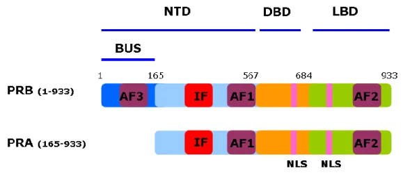
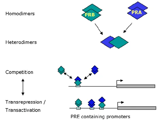
Human PR gene, located on chromosome 11 (11q2.2), codes for two receptor isoforms (PRA and PRB) by alternate initiation of transcription at internal translational start sites from two distinct estrogen regulated promoters (Kastner et al., 1990; Tetel et al., 1999). Majority of PR positive cells express equal amounts of PRA and PRB isoforms under physiological conditions. PRB is full length protein comprising of 933 amino acids (114 kDa). PRA is truncated in the N-terminal region and lacks the first 164 amino acids (94 kDa) harboring activation function 3
Figure 2. Classical structure of nuclear receptors.
The nuclear receptors are comprised of three main domains, N-terminal domain (NTD), DNA-binding domain (DBD) and ligand-DNA-binding domain (LBD). Location of Activation Function 1 (AF1), Activation Function 2 (AF2), Inhibitory Function (IF) are indicated.
6
(AF3) which is present in PRB unique sequence (also called as BUS domain) (Figure 3). Another PR isoform (PRC) has also been described which lacks first 594 amino acids. PRC lacks the N-terminal region and one zinc finger of DBD, thus unable to bind DNA and is transcriptionally inactive. PRC is nevertheless able to bind hormone and heterodimerize with full length PR, localizes in nucleus (Wei et al., 1990; Wei et al., 1996) and seems to be implicated in initiation of labor (Condon et al., 2006). Some in vitro studies show that PRC may either enhance or repress the activities of PRA and PRB (Wei et al., 1996; Wei et al., 1997), however, recent studies have questioned if any physiological role is played by PRC in progesterone signaling (Samalecos and Gellersen, 2008).
2. Mechanism of action of PR
2.1 PR a nucleo-cytoplasmic shuttling protein
Neo-synthesized PR, like other steroid hormone receptors such as glucocorticoid receptor (GR), mineralocorticoid receptor (MR), androgen receptor (AR) and estrogen receptor (ER), is assembled in an inactive multi-protein chaperone complex comprising of heat-shock protein of 90 kDa (hsp90) and hsp70 (Catelli et al., 1985; Picard, 2006). One role of these chaperone proteins is to maintain the steroid receptor in a conformation suitable for ligand binding (Picard, 2006; Pratt et al., 2004; Smith, 1993). Such complexes also protect the receptor from proteolytic damages in addition to keeping it in an inactive structure. In the absence of ligand, intracellular location of such complexes depends on the receptor type, for example, GR (DeFranco et al., 1991; Picard et al., 1990) and AR
Figure 3. Structure of progesterone receptor isoforms PRB and PRA.
PRB and PRA share identical amino acids sequence except that PRB is full length receptor and PRA is truncated in N-terminal domain and lacks first 164 amino acids also called as PRB-unique sequence (BUS) which harbors Activation Function 3 (AF3). Like other nuclear receptors, PR isoforms are comprised of three main domains, N-terminal domain (NTD), DNA-binding domain (DBD) and ligand-binding domain (LBD). Activation Function 1 (AF1), Activation Function 2 (AF2), Inhibitory Function (IF), nuclear localization signals (NLS) are shown.
7
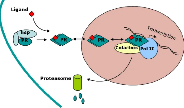
(Kemppainen et al., 1992) are predominantly found in the cytoplasm while PR (Guiochon-Mantel et al., 1991) is predominantly present in the nucleus. However, cytonuclear localization of these receptors is also influenced by the cell type. Steroid receptors shuttle between the cytoplasm and nucleus due to presence of nuclear localization signals (NLS) (Baumann et al., 1999; Dauvois et al., 1993; DeFranco et al., 1991; Guiochon-Mantel et al., 1994; Guiochon-Mantel et al., 1991; Guiochon-Mantel et al., 1989). In PR, one highly conserved constitutive NLS, comprising of 638-642 amino acids, is localized in hinge region and is responsible for hormone-independent nuclear localization since its deletion leads to preferential cytoplasmic localization of the receptor in the absence of hormone (Guiochon-Mantel et al., 1989). A second NLS is located in second zinc finger of DBD between amino acids 593-638. This NLS is ligand-dependent since ligand binding unmasks this NLS region and leads to nuclear localization of PR following its dissociation from the heat-shock proteins/chaperone complexes. Ligand binding also induces various post-translational modifications in PR (Weigel and Moore, 2007). Classically, PR like other steroid hormone receptors binds as a dimer to a specific hormone response element (HRE) located upstream of target gene promoter (Hard et al., 1990; Luisi et al., 1991; Picard and Yamamoto, 1987) (Figure 4). The DNA-bound receptor can then exert a positive or negative effect on gene transcription by recruiting either coactivators or corepressors.
Figure 4. Classical mode of action of PR
In the absence of ligand, PR is associated with heat-shock protein (hsp) chaperone complexes in the cytoplasm. Upon ligand binding, conformational changes in PR allows it to dissociate from chaperone complexes, to dimerize and translocate in the nucleus. Inside nucleus, PR binds to its response elements present in target gene promoters, recruits transcriptional cofactors and modulates the transcription. Alongwith its transcriptional activities, PR undergoes to ligand-induced proteasome-mediated rapid turnover.
8
Coactivators positively regulate transcriptional efficacy by recruiting multiprotein complexes to DNA leading to chromatin remodelling and interaction with general transcription factors. Corepressors recruited to the DNA-bound receptor facilitate chromatin condensation and silence transcription. Numerous transcriptional coactivators and corepressors have been identified whose relative and absolute expression levels vary among different cell types. During the course of transcriptional activities, PR undergoes rapid hormone-induced proteasome-mediated degradation (Lange et al., 2000). Interestingly, PR turnover is important for its transcriptional properties (Shen et al., 2001) and both of these phenomena seems to be interconnected since blocking of proteasome-mediated PR degradation impedes PR transcriptional properties (Amazit et al., 2011; Dennis et al., 2005). Likewise, inhibition of protein neosynthesis abolishes proteasome-dependent PR turnover (Kahmann et al., 1998; Khan et al., 2011) [See also Results-PAPER I) (Figure 5) suggesting involvement of neosynthesized co-regulatory proteins in these processes. Whether the same molecular partners are implicated in ligand-induced PR transcriptional activity and proteasome-mediated degradation remains to be demonstrated.
Figure 5. Coupling of PRB transcriptional activity and its down-regulation
Agonist ligand binding induces post-translational modifications in the receptor which then translocates to the nucleus and recuits transcriptional coactivators to enhance gene transcription. Receptor is thereafter degraded in the cytoplasm by proteasomes. Blockage of protein neosynthesis by cycloheximide (CHX) or proteasome-dependent protein turnover by using MG132 (MG) blocks the transcriptional activity of PR.
9
2.2 Non genomic actions of PR
Since PR is known to actively shuttle between cytoplasm and nucleus by active import and export mechanisms, recently it has been demonstrated that PR is capable of interacting with molecular partners in the cytoplasm (Boonyaratanakornkit et al., 2007). Therefore, beside classical genomic functions, PR isoforms are also capable of interacting with major cytoplasmic signaling kinases (p42/44 MAPK, c-Src/p21ras, PI3K/Akt pathways) (Carnevale et al., 2007) (Figure 6) frequently activated during carcinogenesis and resulting in progestin-inducible cell proliferation and metastasis.
The proline rich motif (PxxP) in the N-terminal domain of PR mediates interaction with Src homology 3 (SH3) domain of Src tyrosine kinase (Boonyaratanakornkit et al., 2001; Wardell et al., 2002). This explains molecular mechanism for some of the rapid non-genomic actions of progesterone. In some other studies, it has been shown that agonist-activated PRB but not PRA interacts with ER to activate Src/ras/MAPK pathway in breast cancer cells and hence could play an important role in growth-promoting effects in cancer cells (Migliaccio et al., 1998). This shows that PR isoforms might have differential impact on cytoplasmic signaling pathways. Such non-genomic pathways activated by PR influence transcription of not only PR-regulated but also PR-independent genes regulated by other transcription factors.
Figure 6. Non-genomic actions of PR
PR is a nucleo-cytoplasmic shuttling protein. Apart from its genomic effects, PR is known to interact with membrane associated receptors (MembR) and cytoplasmic proteins invloving Src-MAPKs pathways in a ligand-sensitive manner. Such interactions lead to activation of Src-MAPKs which then phosphorylate PR and other transcription factors to modulate the transcription of both PR-regulated as well as PR-independent genes regulated by other transcription factors (TF).
10
2.3 Genomic actions of PR
The genomic actions of PR are mediated either through its direct binding to specific DNA sequences or indirectly via association with other transcription factors. HRE is a consensus nucleotide sequence comprising of an inverted repeat sequence separated by three nucleotides i.e., AGAACAnnnTGTTCT called historically as glucocorticoid response element (GRE) since it was initially shown as a binding site for GR. Later on, it was found that GRE are also recognized by PR, AR and MR (Beato et al., 1989). However, transcriptional regulation by steroid receptors is not that simple as it appeared in early 1990‟s since recent thermodynamic studies have questioned this long accepted dogma, and probably rightly so knowing the fact that diverse physiological roles are played by these receptors which generally regulate different target genes. Most of our knowledge on DNA binding sites of steroid hormone receptors comes from two widely used promoters i) rat tyrosine amino transferase (TAT) promoter which harbors perfect palindromic GRE (Jantzen et al., 1987) ii) mouse mammary tumor virus long terminal repeat (MMTV-LTR) promoter which contains imperfect palindromic GRE (Hager, 1988; Lefebvre et al., 1991). These synthetic promoters were useful in understanding several aspects of transcriptional regulation by steroid hormone receptors. However, these sequences are not representative of endogenous progesterone responsive gene promoters since only 565 exact consensus GRE are present in whole human genome of which only 26 are located within 10 kb of the transcription start site of known genes (Horie-Inoue et al., 2006). Furthermore, binding of a steroid receptor to these GRE was confirmed only on 14 out of 26 perfect GRE (Horie-Inoue et al., 2006). Indeed, GRE are as abundant in PR regulated as in PR non-regulated gene promoters (Figure 7) (Jacobsen et al., 2009).
Figure 7. PRE abundance in PR regulated, non-regulated and random promoters.
In silico analysis showing PRE abundance in 12 PR-regulated or non-regulated or random promoter sequences retrieved from TRANSFAC database and compared with known PRE consensus sequence.
11
In contrast to scarcely present GRE upstream of known genes, several half-sites are present in natural PR-regulated promoters (Jacobsen et al., 2009). It has now been shown that PR monomers are competent to bind progesterone response element (PRE) half-sites and show strong cooperativity in transcriptional regulation (Connaghan-Jones et al., 2008). Another level of complexity arises from the fact that PR regulates transcription without directly binding to DNA. This indirect transcriptional regulation is achieved by PR tethering to other DNA bound transcriptional factors such as specificity protein 1 (SP1), SP4, activating protein 1 (AP1), the RelA (p65) subunit of nuclear factor B (NF- B), signal transducers and activators of transcription (STAT) (Bamberger et al., 1996; Kalkhoven et al., 1996; Owen et al., 1998; Proietti et al., 2005; Richer et al., 1998; Shatnawi et al., 2007; Tseng et al., 2003)(Figure 8). Thus PR regulates target gene transcription both directly as well as indirectly by association with other transcription factors without binding directly to promoters.
3. PR-associated Transcriptional coregulators
Initial understanding of transcriptional regulation by NR was that NRs recruit general transcription factors and RNA polymerase II at gene promoters to start gene transcription (O'Malley and Means, 1974). However, in vitro transcriptional systems consisting of purified NRs and general transcription factors did not produce efficient transcriptional activation (Klein-Hitpass et al., 1990) supporting the idea that additional factors might also be required for full ligand-induced transcriptional activation by NRs. The indirect inhibition of transcriptional properties of one NR by the activation of another overexpressed NR (Halachmi et al., 1994; Meyer et al., 1989) suggested for the existence of common
Figure 8 : Direct and indirect transcriptional regulation by PR
A schema representing transcriptional regulation by PR from promoters harboring or not PRE. Few examples of genes regulated by such mechanisms are also shown
12
coregulatory proteins that might be squelched by the overexpressed receptor. Finally, the concept of a new type of transcription factors, that do not bind to DNA, but control transcription by binding to NRs either directly or indirectly, was validated in 1995 with the discovery of the first NR coactivator, steroid receptor coactivator 1 (SRC1) (Onate et al., 1995). The following criteria were thus proposed for a protein to be categorized as a coactivator. Coactivators bind to NRs (directly or indirectly) but not directly to DNA, agonist ligands promote while antagonist ligands inhibit coactivators binding to NRs, coactivators are able to reverse receptor-receptor squelching and overexpression of coactivators greatly enhances while the expression of dominant negative mutant inhibit ligand-dependent NR function (Onate et al., 1995).
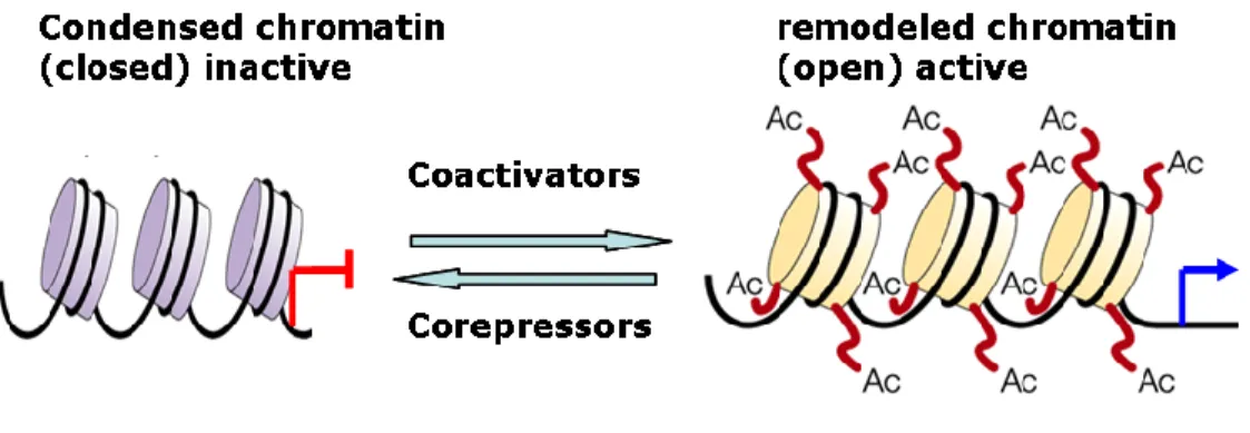
Gene transcription by NR oscillates between on and off states and is finely regulated be transcriptional coregulators (coactivators and corepressors) which play a major role in chromatin remodeling through histone amino-terminal modifications such as acetylation, methylation, phosphorylation, ubiquitylation, and sumoylation (Bhaumik et al., 2007; Kouzarides, 2007; Suganuma and Workman, 2008). The term „histone code‟ is employed to represent a specific combination of these modifications. While coactivators enhance NR- mediated transcription mostly due to associated histone acetylase activity, the primary function of corepressors is to attenuate the transcription by chromatin condensation mostly due to histone deacetylation and methylation (Figure 9).
Although the initial identification of coregulators became possible due to their interacting abilities with a given nuclear receptor, it soon became clear that most of the coregulators are not specific for a particular receptor, although preferential interaction with a particular steroid receptor depending on cell type and promoter context occurs through unclear mechanisms. Moreover, a coactivator can have highly variable effects depending on not only the cellular and promoter contexts but also the receptor subtype. For instance, a
Figure 9: Chromatin remodeling by coregulators
Corepressors by their associated enzymatic activities enhance chromatin condensation leading to gene silencing whereas enzymatic activities associated with coactivators reverse the condensed state in such a way that transcriptional initiation becomes possible. Histones are indicated by round boxes encircled by chromatin threads. Histone modification by acetylation (Ac) is shown as an example.
13
given coactivator (HBO1) can increase the transcriptional response for one receptor (PR) (Georgiakaki et al., 2006) while inhibiting transactivation properties of another receptor (AR) (Sharma et al., 2000). Depending on cell type, opposite effects of the coactivator SRC1 in regulating PR transcriptional properties was demonstrated in mouse model where SRC1 increased PR transcriptional activity in response to estrogen and progesterone in uterine stroma and myometrium while decreased PR target gene transcription in the luminal and glandular epithelial compartments (Han et al., 2005). Mechanism of action of coactivators is also variable, for example, they can stabilize the receptor or transcriptional machinery on promoter (McKenna et al., 1999; Rowan and O'Malley, 2000), participate in chromatin remodeling by covalent modifications of not only histones (Glass and Rosenfeld, 2000) but also of other coregulators (Chen et al., 1999b) or regulate the stability/turnover of regulatory proteins (Imhof and McDonnell, 1996; Nawaz et al., 1999b; Rowan and O'Malley, 2000). Furthermore, optimal transcriptional activation by specific steroid hormone receptor can employ interplay of distinct coregulators depending on the nature of ligand, promoter environment, activation/inhibition of various signaling pathways and cell type (Hermanson et al., 2002).
3.1 PR-interacting coactivators
Coactivators serve as a bridge between steroid receptor and the general transcriptional machinery, and might be considered as scaffolding proteins able to recruit other regulatory partners. These are the protein molecules which influence transcriptional response of steroid receptors on target gene transcription without binding directly to DNA. The most thoroughly characterized PR interacting transcriptional coactivators belong to p160 SRC family which comprises of three homologous proteins namely, SRC1, SRC2 and SRC3. These interact with not only several steroid receptors but also to other NR, specific and general transcription factors, and provide platform for other enzymes to facilitate the transcription by chromatin remodeling. The physiological relevance of SRC1 and SRC3 for PR signaling was demonstrated in mice by O‟Malley‟s group. SRC1 knock-out mice presented severe reproductive abnormalities particularly in the uterus (Xu et al., 1998) whereas selective ablation of SRC3 resulted in impaired mammary gland development and function (Xu et al., 2000). In agreement with the above studies, it has been recently demonstrated that physiological functions of PR in the uterus are modulated primarily by SRC1 whereas SRC3 acts as a major coactivator for normal PR functioning in mammary gland (Han et al., 2006).
3.1.1 SRC1
The SRC1, also known as NCOA1, was isolated using the PR-LBD as bait in yeast two hybrid screen of a human B-cell cDNA library and was shown to interact and enhance the transcriptional activity of several steroid hormone receptors in a ligand-dependent manner
14
(Onate et al., 1995). SRC1 exerts its action on a large number of nuclear receptors since it is capable of co-activating transcription induced by PR, GR, ER, thyroid receptor (TR), retinoic X receptor (RXR) (Onate et al., 1995), hepatocyte nuclear factor 4 (HNF4) (Wang et al., 1998), steroidogenic factor (SF1) (Crawford et al., 1997) and peroxisome proliferator-activated receptor gamma (PPAR ) (Zhu et al., 1996). Association of SRC1 with PR LBD is strictly dependent on the nature of the ligand and antagonist RU486 partially abolishes this interaction (Onate et al., 1995). Some studies have also shown the role of SRC1 in transcriptional activation of other transcription factors such as AP1 (Lee et al., 1998), serum response factor (Kim et al., 1998), NF- B (Na et al., 1998) and p53 (Lee et al., 1999). SRC1 is capable of counterbalancing the squelching phenomenon observed during transactivation by PR in the presence of ER suggesting that SRC1 is independently recruited by both of these receptors and that SRC1 recruitment by PR and ER for optimal transactivation is a limiting factor (Onate et al., 1995). Together, these studies show an important role for SRC1 in multiple intracellular signaling pathways.
Principal interacting domain of SRC1 with PR is situated in the extreme C-terminus since in vitro transient transfection experiments have demonstrated that a dominant negative mutant inhibits PR co-activation by SRC1 (Jenster et al., 1997; Onate et al., 1995). It has been shown that SRC1 interacts mainly with AF2 domain of steroid receptors, however, interaction with AF1 region has also been suggested (Ikonen et al., 1997; McInerney et al., 1996; Onate et al., 1998). Such interactions involve different domains of SRC1 and are necessary so that AF1 and AF2 can act in synergy for transcriptional activation (Benecke et al., 2000; Onate et al., 1998). Given that interaction of SRC1 with steroid receptors requires presence of AF2 amphipathic helix 12 (H12), SRC1 is considered to be interacting with AF2 (McKenna et al., 1999), in contrast to other coactivators such as steroid RNA activator (SRA) which interact more specifically with AF1 domain (Lanz et al., 1999). SRC1 has also been shown to bind the N-terminal domain of PR and AR in inverted position. Deletion of the PRB LBD led to generate a constitutive receptor which is able to recruit SRC1 suggesting an important role of PRB AF3 domain in SRC1-mediated transcriptional co-activation (Georgiakaki et al., 2006). SRC1 contains two co-activation domains AD1 and AD2 which activate the transcription of a reporter gene when fused to DNA binding domain of yeast Gal4 protein. Some in vitro studies have shown the interaction of SRC1 with general transcription factors such as TATA-binding protein (TBP) and transcription factor IIB (TFIIB) suggesting a role of SRC1 in stabilization of transcriptional machinery (Ikeda et al., 1999; Takeshita et al., 1996). We have recently shown that proteasome-mediated proteolysis of SRC1 is pivotal for PR transcriptional activity. Two degradation motifs, PEST (amino acids 2-16) and basic helix loop helix domain (amino acids 41-136) were identified and shown to control the basal expression levels as well as hormone-dependent down-regulation of SRC1.
15
3.1.2 SRC2 and SRC3
Two other homologous proteins, acting as NR coactivators have been characterized namely SRC2 (TIF2, GRIP1, NCOA2) (Hong et al., 1997; Voegel et al., 1996) and SRC3 (p/CIP, RAC3, AIB1, ACTR, TRAM1, NCOA3) (Anzick et al., 1997; Chen et al., 1997; Li et al., 1997; Takeshita et al., 1997; Torchia et al., 1997). SRC2, like SRC1, is capable of interacting with LBD of numerous steroid receptors in a hormone-dependent manner (Hong et al., 1997; Hong et al., 1996; Voegel et al., 1996). It contains two constitutive activation domains capable of stimulating transcription when fused to DBD of heterologous proteins (Hong et al., 1996; Voegel et al., 1998; Voegel et al., 1996) . Like SRC1, SRC2 restores squelching phenomenon of PR induced by ER (Voegel et al., 1996). SRC3 was first isolated from mice as a molecular partner of CREB binding protein (CBP) during transcriptional activation of CRE binding protein (CREB) (p300/CBP cointegrator-associated protein, p/CIP) (Torchia et al., 1997). Almost simultaneously, different groups characterized this transcriptional coregulator as coactivator of TR (ACTR, activator of thyroid receptor) (Chen et al., 1997) or thyroid receptor molecule 1 (TRAM1) (Takeshita et al., 1997), a coactivator overexpressed in mammary and ovarian cancers (amplified in breast cancer , AIB1) (Anzick et al., 1997), a coactivator of nuclear receptors (Receptor-associated coactivator 3, RAC-3) (Li et al., 1997) or steroid receptor coactivator 3 (SRC3) (Suen et al., 1998). In human, SRC3 acts as a transcriptional coactivator of several NRs such as PR, ER, GR, RAR, RXR, TR (Anzick et al., 1997; Chen et al., 1997; Li et al., 1997) while in mice mSRC3 presents a broader spectrum of activity since it can enhance transactivation of other transcriptional activators such as STATs and CRE-binding protein (CREB) (Torchia et al., 1997).
3.1.3 NR-coactivator interaction regions
There are three structural domains in SRCs, the most conserved amino-terminal basic helix-loop-helix-Per/ARNT/Sim (bHLH-PAS) domain which is involved in interactions with numerous other coactivators such as coiled-coil coactivator A (CoCoA) (Kim et al., 2003), GRIP1 associated coactivator 63 (GAC63) (Chen et al., 2005) and coactivator associated arginine methyltransferase 1 (CARM1) (Chen et al., 1999a; Lee et al., 2004), a central region containing three NR interacting LxxLL motifs (Darimont et al., 1998; Heery et al., 1997; Voegel et al., 1998) and a carboxyl terminus harboring two transcriptional activation domains (AD1 and AD2). AD1 is involved in interaction with CREB-binding protein (CBP) and the histone acetyltransferase p300 while the interaction with histone methyltransferases such as CARM1 and protein arginine N-methyltransferase 1 (PRMT1) is mediated via AD2 (Xu et al., 2009). The C-terminal regions of SRC1 and SRC3 also possess domains responsible for their histone acetyltransferase (HAT) activity.
16
3.2 PR-interacting corepressors
The two most widely studied transcriptional corepressors are nuclear receptor corepressor (NCoR also known as NCOR1) and silencing mediator of retinoid acid and thyroid hormone receptor (SMRT, also known as NCOR2). NCoR and SMRT were simultaneously cloned in 1995 by Rosenfeld (Horlein et al., 1995) and Evans (Chen and Evans, 1995) respectively. Using a similar approach that led to the identification of the first steroid receptor coactivator, NRs were used as a bait to isolate the interacting proteins which demonstrated transcription repressing properties. Like coactivators, corepressors such as NCoR and SMRT target not only NR but also diverse transcription factors such as TFIIB, AP1, and NF- B (Battaglia et al., 2010) reflecting multiple roles played by corepressors in pathophysiology. Altered expression and localization of NCoR and SMRT is reported in various cancers including breast, bladder and prostate cancer (Abedin et al., 2009; Banwell et al., 2006; Girault et al., 2003; Khanim et al., 2004; Kim et al., 2009; Zhang et al., 2006). In contrast to ligand dependency for coactivator‟s recruitment, initial biochemical studies elaborating the mechanisms of interaction between NR and various corepressors proposed a simple but attractive model suggesting that the open conformation of unliganded receptor allows constitutive interaction with corepressors leading to repression of target gene transcription. Specifically the hydrophobic groove in the LBD region is involved in interaction with LxxH/IIxxxI/L motif or corepressor nuclear receptor (CoRNR) box present in the corepressors (Hu and Lazar, 1999; Perissi et al., 1999). During gene activation process, these corepressors are replaced by coactivators (Figure 10).
Figure 10. Classical mechanism of coregulator recruitment by steroid receptors.
Corepressor recruited by unliganded steroid receptor maintain chromatin in condensed conformation leading to silencing of transcription. In the presence of agonist ligand, conformational change in the receptor provides platform for the recruitment of transcriptional coactivators which leads to chromatin remodelling/decondensation. Coactivators also serve as the bridge between the receptor and the general transcription machinery and serve as scaffolding protein for other coregualtors binding, ultimately activating the gene transcription.
17
Agonist ligand binding induces a conformational change in the receptor i.e. repositioning of the helix 12 of LBD in such a way that the size of hydrophobic groove is reduced which can now accommodate shorter LxxLL motif (also known as NR box) present in coactivators but not the larger CoRNR box of corepressors (Glass and Rosenfeld, 2000). This model suggests that agonist-dependent NR transactivation is due to an upshift in NR interaction with coactivators instead of corepressors which preferably interact with unliganded receptor. While agonist ligand binding to NR, facilitates recruitment of coactivators, antagonist ligand-bound NR preferably interact with corepressors. For example, in the presence of PR antagonist asoprisnil, preferential interaction of PR LBD with SMRT corepressor has been shown (Madauss et al., 2007). Although, asoprisnil-bound PR LBD preferentially recruited corepressors, interaction with coactivators SRC1, SRC2 and SRC3 was also observed (Madauss et al., 2007). On the other hand, although corepressor binding is facilitated by NR conformation in the absence of ligand, agonist ligands do not always enhance the transcriptional properties of NR and almost as many genes are repressed as activated by PR following agonist ligand treatment (Tung et al., 2006). Recent studies have revealed that certain corepressors are specifically recruited by ligand activated transcription factors leading to active silencing of target gene transcription. For example, interaction of PR and ERα with ligand-dependent corepressor (LCoR) requires hormone binding to the receptor (Palijan et al., 2009b). Indeed, LCoR is recruited to promoters of genes whose transcription is repressed following hormone treatment (Palijan et al., 2009b). Similarly, transrepression of PPARgamma on certain genes is mediated by ligand-dependent recruitment of NCoR and HDAC3 by PPARg LBD to promoter sequences (Tiefenbach et al., 2006).
Other examples of ligand-dependent corepressors include nuclear receptor-interacting protein 1 (NRIP1, also know as RIP140), preferentially expressed antigen in melanoma (PRAME), prohibitin 2 (PHB2) and transcription intermediary factor 1a (TIF1a, also known as TRIM24) which all contain LxxLL motifs responsible for their interaction with liganded NR bound to the promoter sequences. Transcriptional repression is facilitated by subsequent recruitment of other classical corepressors such as C-terminal binding protein (CtBP) and histone deacetylases (HDACs) (Augereau et al., 2006; Epping et al., 2005; Fernandes et al., 2003; Montano et al., 1999; Palijan et al., 2009a; Wei et al., 2000).
These studies suggest that transcriptional regulation by NR and coregulators is much more complex than initially thought and highly depends on the cell type, the receptor, the coregulator and the promoter context. Any model explaining such mechanisms in a generalized fashion risks resembling an iceberg and also with substantial lacunae.
18
4. PR Post-translational modifications
PR like other steroid hormone receptors undergo important post-translational modifications (PTM) including phosphorylation, acetylation, sumoylation and ubiquitylation (Abdel-Hafiz et al., 2002; Daniel et al., 2010; Lange et al., 2000; Ward and Weigel, 2009; Weigel, 1996). Several aspects of PR signaling including hormone responsiveness, subcellular localization (Qiu et al., 2003), dimerization, DNA binding (Takimoto et al., 1996), promoter selectivity (Daniel et al., 2007b), interaction with molecular partners (Wardell et al., 2010), transcriptional activity (Pierson-Mullany and Lange, 2004; Shen et al., 2001) and receptor turnover (Lange et al., 2000) are controlled by phosphorylation(s).
4.1 Phosphorylation
In PR, there are at least 14 phosphorylation sites (thirteen serine residues and one threonine residue) mostly within Serine-Proline motifs in the N-terminal domain (Beck et al., 1996a; Beck et al., 1996b; Knotts et al., 2001; Zhang et al., 1997; Zhang et al., 1994; Zhang et al., 1995) (Figure 11). Among these, six (Ser 20, 25, 81, 102, 130, 162) are only present in PRB upstream segment (BUS). In the absence of hormone, PR is basally phosphorylated at Ser 81, 162, 190 and 400 (Zhang et al., 1997), whereas ligand binding induces phosphorylation of Ser 102, 294, 345 and 400 (Zhang et al., 1995).
These residues are targeted by multiple kinases including proline-directed kinases such as cyclin dependent kinase 2 (CDK2) (Ser 25, 162, 190, 213, 400, 554, 676 and Thr 430)
Figure 11. Phosphorylation sites in PR
19
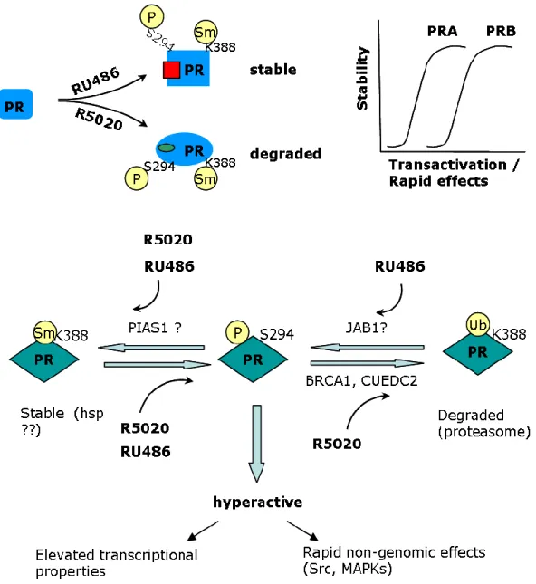
(Knotts et al., 2001; Zhang et al., 1997), MAPK (Ser 20, 294, 345) (Lange et al., 2000) and casein kinase II (Ser 81) (Zhang et al., 1994). Moreover, an individual residue might also be targeted by multiple kinases in vivo. Ser 102 and 130 and ligand-dependent Ser 294 phosphorylation is induced by unknown kinases (Lange, 2004; Qiu et al., 2003). Functional role of each of these phosphorylation sites is yet to be known. PRB serine 294 phosphorylation (pS294-PRB) has been studied extensively and shown to play a critical role in cross talk with growth factor signaling pathways. Ser 294 acts as an important sensor for growth factor inputs that affects PR functions such as nuclear translocation, DNA binding, transcriptional activity and proliferation (Daniel et al., 2007b; Dressing et al., 2009). Its role in PR degradation has also been studied by mutagenesis experiments showing that substitution of serine 294 by an alanine (S294A) led to PRB stabilization suggesting that PRB down-regulation is mainly addressed by the Ser 294 site (Lange et al., 2000). However, in stably transfected T47D cells, PRB-S294A mutant underwent ligand-induced turnover, though to lesser extent as compared to wild type PRB (Skildum et al., 2005). Furthermore, antiprogestin RU486 also induces strong Ser 294 phosphorylation. However, PRB turnover is markedly slowed down upon PR binding to RU486. This shows that mechanisms independent from S294 phosphorylation might also play a role in PR stability/turnover. [See also
Results-PAPER I].
The other relatively better studied PR phosphorylation sites include Ser 345 and 400. It has been shown that phosphorylation of S345, a MAPK consensus site, is mediated by activation of EGFR-c-Src-MAPK by progestins. Furthermore, Ser 345 phosphorylated PRB associates with SP1 to regulate transcription of selected PR target genes such as p21 and EGFR whose promoters lack canonical PRE but harbor SP1 sites (Faivre et al., 2008). The significance of Ser 400 phosphorylation in PR signaling has also been demonstrated. Ser 400 is phosphorylated in the presence of progestins or growth factors and requires CDK2 activity. Indeed, CDK2 induces Ser 400 phosphorylation even in the absence of ligand and regulates nuclear localization and transcriptional activity of both unliganded and liganded PR (Pierson-Mullany and Lange, 2004).
It has been reported that PR isoforms undergo differential phosphorylations at Ser 294 and Ser 345. PRB is rapidly and highly phosphorylated at Ser 294 residue following agonist ligand or EGF treatment while PRA undergo very weak and delayed S130 phosphorylation in response to agonist (Clemm et al., 2000) or EGF (Daniel et al., 2007a). Also basal levels of PRA S130 phosphorylation is much lower as compared to PRB S294 in cells coexpressing both PR isoforms (Clemm et al., 2000). In contrast, PRA is rapidly and more intensely phosphorylated at Ser345 residue as compared to PRB following progestin treatment (Daniel et al., 2007a).
This shows that in addition to intracellular levels of the receptor, hormone and PR-interacting proteins such as transcriptional coregulators, PR signaling is also finely controlled
20
by the activation of specific kinases and phosphatases in progesterone target cells. Functional differences in PRA and PRB might in part be related to differential PR isoforms phosphorylation pattern and kinetics [See also Results-PAPER I]
4.2 Ubiquitylation
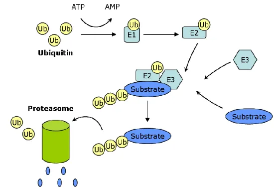
The ubiquitin/proteasome system is known for regulated protein turnover in a highly specific manner. The ubiquitin, a small protein (76 amino acids), is specifically conjugated to the target protein destined for degradation (Pickart, 2001) and also acts as an acceptor for another ubiquitin molecule resulting in a polyubiquitin chain (Hoppe, 2005). Protein ubiquitylation is a multistep process mainly involving E1-activating enzyme, E2 conjugating enzymes, and E3 ligases (Figure 12). To date, only one E1 enzyme is known while a dozen of E2 and over 200 E3 family members, responsible for substrate specificity, exist in humans (Faus and Haendler, 2006). Ubiquitin molecule is covalently attached to E1 enzyme in an ATP-dependent process. Sequential action of E2 and E3 enzymes results in the formation of an isopeptide bond between the carboxyl terminal glycine of ubiquitin and the amino group of a lysine residue in the target protein. Once ubiquitinated, the protein is recognized and degraded by a large multi-subunit protease complex called as 26S proteasome comprising of 20S core subunit and the 19S regulatory subunit (Voges et al., 1999). Similar to other post-translational modifications, ubiquitylation is a reversible process involving deubiquitylating enzymes (Nijman et al., 2005).
Figure 12. Ubiquitin-proteasome mediated protein turnover
Ubiquitin molecule is covalently attached to E1 enzyme in an ATP-dependent process. Ubiquitin is taken up by E2 conjugating enzyme and is transferred to the protein substrate to be degraded with or without assistance of E3 ligase enzyme which is responsible for protein specificity. Polyubiquitinated proteins are then recognized and degraded by 26S proteasomes.
21
Protein synthesis and degradation go side by side and the balance between these processes is critical for normal cell functioning. Despite the important role of protein degradation in cellular homeostasis, interest in understanding ubiquitin proteasome pathway is further increased by the fact that the enzymes involved in ubiquitin proteasome pathway are recruited to nuclear receptor target genes and proteolysis of nuclear receptors and coregulators is required for efficient transcriptional outcome (Nawaz and O'Malley, 2004). This coupling of the receptor transcriptional activity with its turnover was initially seamed to be puzzling and paradoxical. It was shown that recruitment of E3-ubiquitin ligase, E6-associated protein (E6-AP) and the regulatory subunits of the proteasome (Rpt6 and S1) on hormone responsive promoter is cyclical and can occur either in the absence or the presence of ligand (Kang et al., 2002). In support of these studies, several nuclear receptors (ER, PR, GR, TR, RXR, RAR) were found to be ubiquitinated and degraded in the course of their transcriptional activities (Dace et al., 2000; Deroo et al., 2002; Lange et al., 2000; Masuyama and MacDonald, 1998; Nawaz et al., 1999a; Tanaka et al., 2001; Wallace and Cidlowski, 2001; Zhu et al., 1999) [See also Results-PAPER II]. Who dictates the terms and what is the signal for determining the fate of the receptor? Neosynthesized receptors are stabilized and protected by association with chaperone heat shock protein complexes and undergo minor degradation in the absence of hormone. Dissociation of the receptor from such complexes by the use of geldamycin, a benzoquinone that blocks the addition of the chaperone assembly factors to PR-hsp complexes, results in rapid proteasome-mediated turnover even in the absence of the hormone (Lange et al., 2000). Most of the steroid hormone receptors (with the exception of AR) undergo accelerated degradation via proteasomes following hormone binding. As described above, ligand-bound receptor conformation helps it dissociate from these chaperone complexes and undergo different post-translational modifications which influence the receptor turnover. For example, in ER, a role of ligand-induced acetylation in receptor turnover has been shown (Wang et al., 2001). Similarly, PR S294A mutation reduces hormone-induced PR degradation (Lange et al., 2000).
4.3 Sumoylation
Conjugation of small ubiquitin-like modifier (SUMO) protein to steroid hormone receptors has been shown to strongly influence their transcriptional properties (Chauchereau et al., 2003; Daniel and Lange, 2009; Holmstrom et al., 2008; Le Romancer et al., 2011; Tallec et al., 2003; Yang et al., 2011b). Like ubiquitylation, sumoylation is also a three step process involving E1 activating enzyme, E2 conjugating enzyme and E3 SUMO ligases. Interplay of these enzymes leads to covalent attachment of a SUMO chain to lysine residues in target proteins. Sumoylation affects several biological functions of transcription factors ranging from subcellular localization, DNA binding or