HAL Id: hal-00514947
https://hal.archives-ouvertes.fr/hal-00514947
Preprint submitted on 3 Sep 2010
HAL is a multi-disciplinary open access
archive for the deposit and dissemination of
sci-entific research documents, whether they are
pub-lished or not. The documents may come from
teaching and research institutions in France or
abroad, or from public or private research centers.
L’archive ouverte pluridisciplinaire HAL, est
destinée au dépôt et à la diffusion de documents
scientifiques de niveau recherche, publiés ou non,
émanant des établissements d’enseignement et de
recherche français ou étrangers, des laboratoires
publics ou privés.
Enhanced diffusion due to active swimmers at a solid
surface
Gaston Miño, Thomas Mallouk, Thierry Darnige, Mauricio Hoyos, Jeremy
Dauchet, Jocelyn Dunstan, Rodrigo Soto, Yang Wang, Annie Rousselet, Eric
Clement
To cite this version:
Gaston Miño, Thomas Mallouk, Thierry Darnige, Mauricio Hoyos, Jeremy Dauchet, et al.. Enhanced
diffusion due to active swimmers at a solid surface. 2010. �hal-00514947�
The Pennsylvania State University, USA. 3 Departmento de F´ısica, FCFM, Univ. de Chile, Chile.
(Dated: September 3, 2010)
We consider two systems of active swimmers moving close to a solid surface, one being a living population of wild-type E. coli and the other being an assembly of self-propelled Au-Pt rods. In both situations, we have identified two different types of motion at the surface and evaluated the fraction of the population that displayed ballistic trajectories (active swimmers) with respect to those showing diffusive-like behavior. We studied the effect of this complex swimming activity on the diffusivity of passive tracers also present at the surface. We found that the tracer diffusivity is enhanced with respect to standard Brownian motion and increases linearly with the activity of the fluid, defined as the product of the fraction of active swimmers and their mean velocity. This result can be understood in terms of series of elementary encounters between the active swimmers and the tracers.
PACS numbers: 87.16.-b, 05.65.+b
Since the pioneering work of Wu and Libchaber [1] considerable efforts have been made to understand hy-drodynamic properties of active suspensions. Generally speaking, this is the name borne by fluids laden with self-swimming entities such as bacteria [2–5], algae [6, 7] or collections of active artificial swimmers [8]. Assemblies of microscopic motors dispersed in a fluid display emergent properties that differ strongly from passive suspensions. The momentum and energy transfer balances as well the constitutive transport properties are deeply modified by the momentum sources distributed in the bulk [2, 9]. Some of these anomalous properties have already been identified such as active diffusivity [1, 2], anomalous vis-cous response [7, 10], active transport and mixing [11] as well as the possibility to use fluctuations to extract work [12]. The presence of living and apparently gregari-ous entities also offers the possibility to move collectively and organize at the mesoscopic or macroscopic level in the form of flocks and herds [9, 14]. Similar collective effects were also identified in suspensions of self-propeled inorganic particles [15]. In the bulk, swimming bacteria with flagella such as E. coli create in the far field-limit, a force-dipole velocity field and consequently, experience a hydrodynamic attraction toward surfaces [16]. Then, it has been observed that E. coli smooth out their run-and-tumble movement and spend long times parallel to the surface undergoing circular motion as a consequence of the torque-free condition [17, 18]. When the concen-tration becomes large, the E. coli population eventually associates collectively to form a bio-film. Even in the low concentration limit, the quantitative analysis of the near surface motion increase tremendously in complex-ity, a reason being the close field hydrodynamic forces that become prevalent and require a complex treatment of the lubrication hydrodynamic fields. However, even in this frame of description, it remains unclear whether
the motion close to the surface is hydrodynamically sta-ble and if the presence of thermal noise is essential to account for the bacterium dwelling time at a surface[19]. Beyond the hydrodynamic interactions, more complex in-gredients may come into play such has the surface inter-action potentials (electrotastic or van der Waals) [20] or more refined details of the bacterium physiology such as swimming speed variations and desynchronization dur-ing bacteria cell cycle [21, 22]. From the perspective of providing a fully consistent treatment of active hydrody-namics, with important applications for understanding bacterial transfer in biological micro-vessels, microfluidic devices, or the formation of bio-films, a reliable descrip-tion of fluid activity in the vicinity of a solid surface is strongly needed. In this letter, to tackle this open and timely question, we compared the behavior of two kinds of active micrometric swimmers: wild type E. coli K12 and artificial self-propelling rods [8], with completely dif-ferent propulsion mechanisms. In both cases, we moni-tor the swimmers’ motions and their ability to activate, beyond Brownian motion, passive tracers, hence charac-terizing the active momentum transfer to the fluid.
Following the experimental procedure described in Ref. [16], wild type E. coli K12 were grown overnight in rich medium (LB). After washing, they were transferred into MMAP, a motility medium supplemented with K-acetate (0.34 mM) and polyvinyl pyrolidone (PVP: 0.005%). They were incubated for at least an hour in that medium and, in some cases, so-called “baby cells” were selected by centrifugation and resuspended in MMAP. To avoid bac-terial sedimentation (isodense conditions), Percoll was mixed with MMAP (1vol/1vol). We checked that under these conditions, the suspending fluid was still Newtonian (viscosity η = 1.28×10⁻³Pa s at 22◦C). The overall con-centration of bacteria was controlled such as to prepare suspensions between 109and 1010bact/ml. To study the
2
FIG. 1: Identification of the swimmer populations by tracking “active” swimmers (red tracks) and “diffusive” swimmers (blue tracks), ϕA is the corresponding fraction of active swimmers. Figs. 1(a,b,g) correspond to E. coli (see inset in 1c) and Figs.
1(d,e) to Au-Pt rods (see inset in 1f). The round black circles in (a,b) are 2µm latex beads; the white small circles in (d,e) are 1µm Dynal beads. Figs. (c) and (f) display the relation between the active swimmers mean velocity VAand ϕA. On Fig. 1(c),
6 independent experiments with E. coli : 1N cells (brown, red and green), mixture of 1N and 2N cells (black) and 2N cells (pink, blue). Labels (1) and (2) are for N = 100 and N = 200 cells in the observation field respectively. Fig. 1(f), for Au-Pt rods with varying proportions of inactive Au rods (2 independent experiments). Fig. 1(g), density probability of the observed bacterial tracks in the (Nc,⟨|θ|⟩) space. The colormap goes from blue for vanishing probability to red for high probability. Two clusters
are identified, centered at (0.9, 0.3) and (0.17, π/2), corresponding respectively to the “active” and “diffusive” swimmers.
effect of bacterial activity on the diffusivity of passive tracers, latex beads of 1 or 2 µm diameter (Beckman-Coulter, density ρ = 1.027 g/ml) were added to the sus-pensions. Experiments were performed in 110µm thick chambers, built with two horizontal microscope cover slips separated by a glass spacer. To avoid sticking, cover slips were coated with PVP. The biological sample con-sisted in a drop of liquid (20 µl) placed between the two slides. The suspension was observed under an inverted microscope (Zeiss-Obzerver, Z1-magnification 40X) con-nected to a digital camera. The observation field ∆V was 96×128µm2and 5µm in depth. Short video sequences of 20s duration at 20 frames/s (fps) were recorded to track the bacterial motion. In a first series of experiments, we measured the bacterial density profile through the entire height of the chamber. We obtained profiles similar to the ones published by Berke and coworkers [16], namely a flat density in the bulk and a strong density increase near the surfaces. However, the wild-type E. coli we used was significantly less attracted by the surfaces (2.5 times in-crease in density within 10µm of the surface) than a mu-tant E. coli strain that does not display tumbling motion [17, 18]. Another series of experiments was performed with bimetallic Au-Pt self-propelled rods (length 1.2 µm and diameter 0.4 µm) that are very similar in size to the
E. coli cell body (1 to 2 µm long, 0.8 µm diameter) but
have no flagella (15 µm long for E. coli) (see inserts in Figs. 1c and 1f). They also have a much higher density (ρ
= 17 g/ml). In the presence of 1 to 10% hydrogen perox-ide, these particles are propelled in the axial direction to-wards the platinum end by the catalytic decomposition of the peroxide fuel [8]. Recent experiments and simulations are consistent with self-electrophoresis as the dominant propulsion mechanism [23, 24]. While other mechanisms have been proposed, it is clear that the mode of propul-sion is intrinsically different from the flagellar propulpropul-sion of bacteria. The experiments were conducted in a sim-ilar fashion to those involving E. coli, but in an open chamber (without the upper wall), in order to allow the oxygen bubbles produced in the reaction to escape from the cell. The concentration of H2O2 was varied, as well
as the concentration of active rods (n = 3− 20 × 106
rods/ml). We also used passive tracers (1µm diameter beads, Dynal- My-one, density ρ = 1.8 g/ml) or 2 µm di-ameter latex beads (Beckman-Coulter, density ρ = 1.027 g/ml)) to follow the activation of the fluid by the Au-Pt rods. In all cases, all the particles in the suspension were localized at the bottom of the chamber as a consequence of sedimentation. Again, short videos (20s duration at 20f ps) were used to track the self-propelled rods.
In the following, we only focus on bacteria and rods moving close to the surface (less than 5µm). In both cases, we observed that not all swimmers display similar trajectories. This was expected as for wild type bacteria, the run or tumble dynamics may depend strongly on the microenvironment or on the position in the cell cycle. For
veloped a tracking program to analyze the short videos and obtained tracks for each swimmer present in the field. We identified two major types of motion: a ballistic and a diffusive one (see Fig.1(a,b,d,e)), and the swimmers that follow these motions are called “active” swimmers and “diffusive” swimmers, respectively. To discriminate sys-tematically all the tracks, two parameters were defined. The first parameter ⟨|θ|⟩ is the mean angle between two successive steps. For example, ⟨|θ|⟩ = 0 for straight tra-jectories and⟨|θ|⟩ = π/2 for a purely random walk. The second discriminating parameter is based on the minimal circle diameter L that encompasses a given trajectory of duration T . For an acquisition time δt and a mean step size δr, the number Nc = T δrLδt is computed. When Nc
is close to 1, the trajectory is associated with a straight line, whereas when Nc is small, its value points to
dif-fusive motion. Therefore, each track is associated with these two numbers and in the (Nc,⟨|θ|⟩) parameter-space,
we could identify two clusters that clearly differentiated the active and diffusive swimmers (see Fig.1(g)). Nev-ertheless, for very small or interrupted trajectories, the separation procedure remained ambiguous, so we system-atically discarded tracks shorter than 10 steps. In the case of artificial swimmers, we also managed to control
a priori the fraction of active swimmers by adding
in-active rods (made only of gold) and keeping the total number of rods at the surface constant. According to the trajectory classification, a fraction ϕAof active
swim-mers was determined. Thus, for a mean number ⟨N⟩ of swimmers, identified in the field of vision, we define a density of “active swimmers” as nA = ϕA⟨N⟩/∆V . In
Fig. 1, we display tracks during a time lag τ = 1.5 s, for two populations of swimmers (a-b, bacteria and d-e, Au-Pt rods), having different ϕA (a,d small active-fraction
and b,e high active fraction). In Figs.1(c) and 1(f), we present the mean velocities of active swimmers VA as
a function of their fraction ϕA, for different experiments
with bacteria and active rods. In the case of the bacterial suspension, we also tried several synchronization proto-cols to select bacteria, at different position in the cell cycle, showing different swimming characteristics. We were able to produce “baby-bacteria” populations (1N short cells: 1.12 µm long) which were found to have a ϕA
larger than the more mature bacteria populations (2N long cells: 2.5 µm long). We took advantage of this dif-ference to look at the influence of ϕA on VA in bacterial
suspensions. On Fig.1(c) we display 6 independent ex-periments performed with populations having a majority of 1N cells (brown,red and green), a mixture of 1N and 2N cells (black), a majority of 2N cells (pink, blue). For each sample, ϕA was taken from suspensions showing an
average of 100 or 200 bacteria in the observation field (represented by (1) and (2) on Fig. 1c, respectively). We
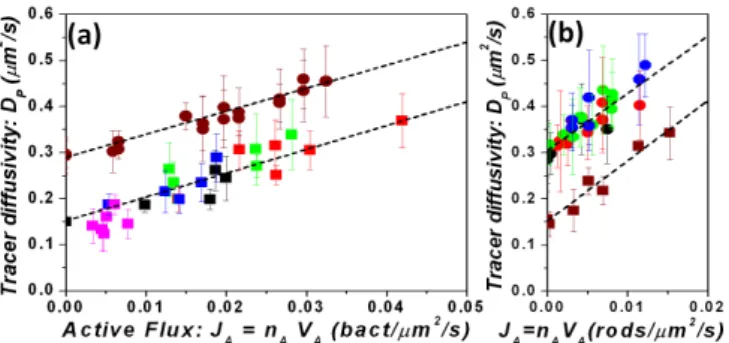
FIG. 2: Enhanced diffusivity DP of passive tracers measured
as a function of JA the “activity flux”. Squares and circles
symbols are related repsectively to tracer diameters 2µm and 1µm. Fig.2(a) corresponds to the bacterial suspensions; 1N cells (red and green) and unsynchronized (black, blue and pink). Each color defines an experiment performed over a range of bacterial dilution. Fig.2(b) corresponds to suspen-sions of Au-Pt-rods. Mixture of active and inactive Au-rods (red circles, brown squares); Various H2O2concentration from
2,5% to 20%, (green, blue, black circles). The dashed lines represent linear fits and the error bars one standard deviation.
observed that ϕA has little influence on VA but that VA
could be different according to bacteria position within the cell cycle. In the case of the artificial swimmers sus-pension, Fig.1(f) shows a stronger independence of VA
on ϕA, which is changed by varying the proportion of
inactive Au-rods.
In the following, we will relate the motion of the pas-sive tracers to the number of active swimmers and their mean velocity. Passive tracer trajectories were analyzed (about 10 tracks per long video of 300 images at 1f ps for bacteria and 40s sequences at 8f ps for Au-Pt rods). From these tracks, the mean passive tracer diffusion co-efficient DP was extracted consistently using two
inde-pendent methods. In the first case, the mean square dis-placements were measured for individual particles and in the other case, for particles as pairs, in order to eliminate residual drift. In Fig.2 the passive tracer diffusivities are displayed for all the experiments presented in Fig.1(c,f).
DP values are displayed as a function of the product
JA= nAVA, that we call the “activity flux”. For
experi-ments performed with the same tracer size, we observe a collapse of all data onto a linear curve.
DP = DBP + βJA (1)
where DPB is the Brownian diffusivity of the latex par-ticles in the vicinity of the surface, in the absence of swimmers. Note that due to lubrication forces, this value is smaller than the Brownian diffusivity expected in the bulk (DB = kBT /3πηd) and, for the parallel motion,
they are related by DB
P = αDB, where α < 1 is the
par-allel drag correction factor [25–27]. The α factor depends on the bead distance to the surface, vanishing at con-tact and going asymptotically to one at large distances. The beads are not at a fixed distance to the surface, but
4
they are distributed according to the Boltzmann’s fac-tor exp(−m∗gz/kBT ), where m∗ is the buoyant mass.
Therefore α must be averaged with this factor. In the active rods experiments we obtained α = 0.7 both for the the buoyant d = 1µm and d = 2µm latex spheres, value that agrees with the theoretical prediction given above (α = 0.64 for d = 1µm and α = 0.74 d = 2µm). In the experiments with bacteria, the suspended beads are almost isodense but they sediment anyway. The experi-mental fit gives α = 0.85. This value allows us to infer the density mismatch ∆ρ = 0.008 g/ml, which is con-sistent with the experimental preparation. The collapse holds also for bacterial populations at different matura-tion stages (1N, 2N or unsynchronized mixtures). In the expression (1), the prefactor β is a length to the fourth power. It varies from 5µm4 = (1.5µm)4 for bacteria, to 13µm4 = (1.9µm)4 for active rods, but seems to be almost independent of the passive tracer size. The en-hanced diffusivity in (1), proportional to nAand VA, can
be understood as a result of a series of elementary en-counters between active swimmers and the tracers: the number of encounters per unit time is proportional to
nAVA. On the other hand, low Reynolds dynamics points
out that the tracer displacement at each encounter is in-dependent of the swimmer velocity and depends only on geometrical factors: the impact parameter, the swimmer dimensions and weakly on the tracer size through the Fax´en correction of passive transport [28, 29]. The β fac-tor comes out from averaging the tracer’s displacements but its computation is difficult because it requires a cor-rect modeling of the near field interactions between the swimmer and the tracer, taking into account the detailled swimmer geometry and the effect of the surface.
In conclusion, we have characterized active momen-tum transfer in the vicinity of a solid surface for two active suspensions (wild type bacteria and artificial self-propelled swimmers). The effect was measured using the diffusion enhancement of a passive tracer. In spite of the a priori complexity of the hydrodynamics and es-sential differences in the propulsion modes, we demon-strated that the effect emerges quantitatively in a sim-ilar way. The resulting diffusion coefficient is the sum of the Brownian contribution near the wall and an ac-tive part, proportional to the product of the density of active swimmers and their mean velocity at the surface. The proportionality factor, scaling as the 4thpower of a micron size length, encompasses the details of momen-tum transfer for each swimmer and is found to be weakly (if at all) sensitive to the probe diameter. Importantly, discriminating between so-called “active” and “diffusive” swimmer trajectories was crucial for predicting the in-duced transport phenomenon and we have developed a protocol to make such a distinction. The functional de-pendence of the enhanced diffusivity is explained in terms of successive interactions between a single swimmer and the tracer, each one producing a net displacement. Our
result provides a justification to pursue a quantitative determination of such encounters based on simple hydro-dynamic models [28].
Acknowledgment. We thank Pr. David Grier for
dis-cussions on the tracking programs, the financial sup-port of the PGDG Foundation, the Alfa-SCAT program, Sesame Ile-de-France, Fondecyt Grants No. 1061112, No. 1100100, Fondap Grant No. 11980002, ECOS C07E08.
[1] X.-L. Wu and A. Libchaber, Phys. Rev. Lett. 84, 3017 (2000).
[2] D.T.N. Chen et al., Phys. Rev. Lett. 99, 148302 (2007). [3] D. Saintillan and M.J. Shelley, Phys. Rev. Lett. 99,
058102 (2007); Phys. Rev. Lett. 100, 178103 (2008). [4] Y. Hatwalne et al., Phys. Rev. Lett 92, 118101 (2004). [5] C. Dombrowski et al., Phys. Rev. Lett. 93, 098103(2004). [6] K.C. Leptos et al., Phys. Rev. Lett. 103, 198103 (2009). [7] S. Rafa¨ı, L. Jibuti, and P. Peyla, Phys. Rev. Lett. 104,
098102 (2010).
[8] W.F. Paxton et al., J. Am. Chem. Soc. 126, 13431 (2004).
[9] R.A. Simha and S. Ramaswany, Phys. Rev. Lett. 89, 058101(2002).
[10] A. Sokolov and I.S. Aranson, Phys. Rev. Lett. 103, 148101 (2009); B. Haines et al., Phys. Rev. E 80, 041922 (2009).
[11] N. Darnton et al., Biophys. J. 86, 1863 (2004).
[12] A. Sokolov et al., PNAS 107, 969 (2010) and refs inside. [13] R. Di Leonardo et al., PNAS 107, 9541, (2010). [14] G. Gregoire, H. Chate, and Y. Yu, Phys. Rev. E 64,
011902 (2001).
[15] M. Ibele, T.E. Mallouk, and A. Sen, Angew. Chem. Int. Ed. 48, 3308 (2009).
[16] A.P. Berke et al., Phys. Rev. Lett. 101, 038102 (2008). [17] E. Lauga et al., Biophys. J. 90, 400 (2006).
[18] P.D. Frymier et al., PNAS 92, 6195 (1995).
[19] G. Li, L.-K.Tam, and J.X. Tang, PNAS 105, 8359 (2008). [20] M.A.S. Vigeant and R.M. Ford, Appl. Environ.
Micro-biol. 63, 3474 (1997).
[21] B.M. Pr¨uβ and P. Matsumura, J. Bacteriol. 179, 5602 (1997).
[22] R. Allman, T. Schjerven, and E. Boyec, J. Bacteriol. 173, 7970 (1991)
[23] Y. Wang et al., Langmuir 22, 10451 (2006).
[24] J.L. Moran, P.M. Wheat, and J.D. Posner, Phys. Rev. E
81, 065302 (2010).
[25] H. Brenner, Chem. Eng. Sci. 16, 242 (1961); A.J. Gold-man, R.G. Cox, and H. Brenner, Chem. Eng. Sci 22, 637 (1967).
[26] P. Holmqvist, J.K.G. Dhont, and P.R. Lang, Phys. Rev. E 74, 021402 (2006).
[27] P. Huang and K.S. Breuer, Phys. Rev. E 76, 046307 (2007).
[28] J. Dunstan, M.Sc thesis, Universidad de Chile, 2010; J. Dunstan et al. (in preparation).
[29] J. Happel and H. Brenner, Low Reynolds Number
Hydro-dynamics: with special applications to particulate media,