HAL Id: hal-01112132
https://hal.sorbonne-universite.fr/hal-01112132
Submitted on 3 Feb 2015HAL is a multi-disciplinary open access archive for the deposit and dissemination of sci-entific research documents, whether they are pub-lished or not. The documents may come from teaching and research institutions in France or abroad, or from public or private research centers.
L’archive ouverte pluridisciplinaire HAL, est destinée au dépôt et à la diffusion de documents scientifiques de niveau recherche, publiés ou non, émanant des établissements d’enseignement et de recherche français ou étrangers, des laboratoires publics ou privés.
Salmonella enterica spp.: An additional tool
A. Bousquet, S. Henquet, F. Compain, N. Genel, G. Arlet, D. Decré
To cite this version:
A. Bousquet, S. Henquet, F. Compain, N. Genel, G. Arlet, et al.. Partition locus-based clas-sification of selected plasmids in Klebsiella pneumoniae, Escherichia coli and Salmonella enter-ica spp.: An additional tool. Journal of Microbiologenter-ical Methods, Elsevier, 2015, 110, pp.85-91. �10.1016/j.mimet.2015.01.019�. �hal-01112132�
ACCEPTED MANUSCRIPT
1
REVISED
Partition locus-based classification of selected plasmids in Klebsiella pneumoniae, Escherichia coli and Salmonella enterica spp: an additional tool.
Bousquet A* a,b, Henquet S a,b, Compain Fa,b, Genel Na,b, Arlet G a,b,c,d,e, Decré D a,b,e
a
Sorbonne University, UPMC Univ Paris 06 CR7, Paris, France.
b
INSERM U1135, CIMI, team E13, Paris, France.
c
AP-HP, Microbiology, Trousseau Hospital, Paris, France.
d
AP-HP, Microbiology, Tenon Hospital, Paris, France.
e
AP-HP, Microbiology, St-Antoine Hospital, Paris, France.
ACCEPTED MANUSCRIPT
2
1. Introduction
Plasmids are extrachromosomal DNA molecules capable of autonomous replication and are the main vectors of resistance and virulence genes, especially in Enterobacteriaceae. Tracing plasmids conferring drug resistance is important for analysis of evolution, epidemiology and spread of antibacterial resistance. The epidemic plasmids belong to the most frequently occurring plasmid families, however antibiotic resistance gene are not always associated with one particular replicon, transfer or partition system. Therefore is difficult to construct phylogenies of plasmids or to track the spread of particular markers.
Incompatibility (Inc) group identification has been frequently used to classify plasmids. Identification methods include the initial technique based on conjugation (Novick, 1987), hybridization with cloned replication regions (Couturier et al., 1988), PCR-based replicon typing (PBRT) (Carattoli et al., 2005) and relaxase typing (Compain et al., 2014). Moreover MLST schemes for plasmids were developed to assign plasmids to STs, in analogy to the typing developed for bacterial genomes (García-Fernández et al., 2008).
Mitotic segregation of plasmids, termed partition in bacteria, is a fundamental step of the cell cycle that ensures the transmission of the whole genome to daughter cells. It is governed by specific genetic loci named par, first identified in low-copy-number plasmids and later found to be present as homologues in most bacterial chromosomes. par loci are organized into operons encoding two proteins, an ATPase and a DNA-binding protein, and including a centromeric site. These components interact with each other to direct the subcellular localization that ensures stability of their replicons. Three types of partitioning ATPases are known (Gerdes et al., 2010): the Walker-type ATPases encoded by the par/sop gene family (type I partitioning loci which are the most common of the par systems), the actin-like ATPase encoded by the par locus of plasmid R1 (type II partitioning locus) and the tubulin-like GTPases encoded by plasmids from Bacillus sp. (type III partitioning loci). Despite their similarities in genetic organization, these three par types use entirely different molecular mechanisms (Guynet and de la Cruz, 2011).
While the acquisition of plasmids often enables bacteria to survive in the presence of antibiotics, it is possible that plasmids also confer vulnerabilities that may be exploited in tailored antibacterial therapy (Williams and Hergenrother, 2008). In order to control the spread of multiresistance plasmids, we need to determine many more variables that affect their replication, maintenance and movement.
ACCEPTED MANUSCRIPT
3 Recently we developed a multiplex PCR method called “plasmid relaxase gene typing” (PRaseT). This classification scheme is based on the relaxase, a key protein which is part of the mobilization region of transmissible plasmids. The aim of the present study was to identify different partition systems located on multiresistance plasmids and to design a multiplex PCR method here called “plasmid partition gene typing” (PAR-T). This method could further the classification of plasmids in Klebsiella pneumoniae, Escherichia coli and Salmonella enterica spp. and will constitute another option for characterising plasmids.
2. Materials and methods
2.1. Database search, primer design and in silico primer assay
An in silico analysis was carried out using GenBank BLAST (http://blast.ncbi.nlm.nih.gov/) on plasmids >40 kb from K. pneumoniae and E. coli conferring multidrug resistance. Due to a low number of plasmids IncHI, IncI1 from K. pneumoniae, the in silico analysis was extended to some plasmids from Enterobacter cloacae and Citrobacter freundii. For the eight Inc groups studied (IncF, IncA/C, IncL/M, IncN, IncHI, IncR, IncI1, IncX), the par operon was used as template; the presence of partition-specific multidomains was searched for using CD-Search (http://www.ncbi.nlm.nih.gov/Structure/cdd/wrpsb.cgi/). Multiple alignments were performed with ClustalW2 software (http://www.ebi.ac.uk/Tools/msa/clustalw2/). Primer pairs covering most sequences in each family were designed using FastPCR (http://primerdigital.com/fastpcr.html) and Primerblast (http://www.ncbi.nlm.nih.gov/tools/primer-blast) software, while minimizing codon degeneracy (Table 1). Oligonucleotide primers were tested in silico for hybridization with plasmids from Enterobacteriaceae referenced in GenBank.
ACCEPTED MANUSCRIPT
4
2.2. Bacterial strains and plasmids
For validation of the PCR assays, experiments were conducted with 136 Escherichia coli transconjugants (Tc) or transformants (Tf) of Enterobacteriaceae (Table 2). All strains carried plasmids belonging to diverse Inc groups that encoded β-lactamases conferring resistance to third-generation cephalosporins and/or carbapenems (ESBLs, acquired cephalosporinases, carbapenemases). They were part of four collections of, respectively, (i) E. coli strains isolated between 1997 and 2002 in various French university hospitals (Marcadé et al., 2009) (Branger et al., 2005)), (ii) K. pneumoniae strains from various geographical regions collected since the 1980s (D. Decré and G.Arlet, personal collection), (iii) Salmonella enterica subsp. enterica strains representing various serovars (collection of the French National Reference Center for E. coli, Shigella sp., and Salmonella sp., Institut Pasteur), (iv) E. coli, S. enterica, K. pneumoniae strains isolated during 2013 in various Argentina University Hospitals (M. Jure, personal collection). All Tc, Tf and clinical strains used in this study were analyzed in parallel with the PAR-T and PRaseT methods. The IncR plasmids which do not encode relaxases were analyzed only by using PBRT (Carattoli et al., 2005). The Tc used as positive controls in PAR-T reactions are given in Table 2. After optimization using Tc or Tf carrying replicons of various types according to PRaseT, we applied the PAR-T method to a panel of 30 clinical strains (17 E. coli, 11 K. pneumoniae and 2 S. enterica) carrying replicons of one to four different types (Table 3).
2.3. DNA extraction and PCR conditions
Lysis by boiling was used for total DNA extraction as previously described (Dallenne et al., 2010). Multiplex PCR was carried out using the Qiagen Multiplex PCR kit (Qiagen, Courtaboeuf, France). The master mix contained pre-optimized concentrations of HotStarTaq DNA polymerase and MgCl2, deoxynucleotide triphosphate and buffer. To all multiplex
PCRs, solution Q (Qiagen) that facilitates the reaction with difficult-to-amplify templates by modifying DNA melting was added. Total DNA in 2 μL of bacterial lysate was subjected to multiplex PCR in a 50 μL volume. The conditions for multiplex PCR were optimized to ensure that all targets were sufficiently amplified for amplicons to be easily visible on 1.5% agarose gels. The optimal primer concentrations are reported in Table 1. PCR conditions consisted of an initial activation at 95°C for 5 min, followed by 35 cycles of 95°C for 30 s, 57°C for 90 s and 72°C for 90 s with a final extension at 68°C for 10 min; for Multiplex I and Multiplex II annealing temperature was elevated to 60°C for 90 s. Simplex PCRs were
ACCEPTED MANUSCRIPT
5 performed in a 50 µL mix with 2U of Taq DNA polymerase (Roche Diagnostics), 10×PCR buffer/MgCl2 (Roche Diagnostics, Meylan, France), 200 µM of each deoxynucleotide
triphosphate (dNTP Mix, Eurobio, Courtaboeuf, France), 0.2 pmol/µL of each primer, 40 µL of sterile water and 2 µL of total DNA extract. PCR conditions consisted in 30 cycles [94°C for 1 min, 55°C for 40 s, 72°C for 1 min], preceded by 1 cycle at 94°C for 5 min and followed by 1 cycle at 72°C for 5 min. PCR products were separated at 100 V for 90 min (180 min for Multiplex IV) on 1.5% agarose gel electrophoresis containing ethidium bromide and visualized using GelDoc (Biorad, Marnes-La-Coquette, France). PCR products were purified using the Exosap purification kit (Illustra Exostar-1 Step, Dutscher, Brumath, France) and subjected to bidirectional DNA sequencing using the BigDye terminator v3.1 cycle sequencing kit (Applied Biosystems, Foster City, CA, USA) and an Applied Biosystems 3730 XL capillary sequencer. Sequence analysis was carried out using Sequence scanner (Applied Biosystems), GeneDoc (www.psc.edu/biomed/genedoc) and GenBank BLAST software.
3. Results and discussion 3.1. In silico analysis
We mainly focused on plasmid families previously found to be involved in the spread of resistance genes in Enterobacteriaceae (Carattoli, 2013). The majority of plasmids contain a single par locus, with the exception of some plasmids such as pR55 plasmid which include two par loci (type I parAB and type II parMR). The type I partitioning loci are the most common of the par systems (74%). The partition loci are often close to rep gene. In contrast, some of them are located at some distance, e.g. the parAB locus of pCTX-M-360 which is 30000 bp by away from RepA. Different designations are used in the annotated sequences for par loci: par, sop, stb. In silico analysis showed that some Inc groups are relatively homogeneous with respect their partition systems, such as IncL/M, N, A/C, I1 or R. The high sequence similarity within these groups (99-100%) allowed designation of a pair of primers for each group. The study of the genetic environment (500 nucleotides on either side of the par genes or their equivalent) of the same groups showed high similarity (75 to 100%). In contrast, other groups such as IncF showed more divergent par sequences. In addition, IncX plasmids from K. pneumoniae (e.g. pIncX-SHV, accession number JN247852 or pKPC-NY79, accession number JX104759) carried a parA gene encoding a Walker-type ATPase near the rep region and an annotated parB gene 25 kb apart from parA without a partition-specific multidomain. In silico analysis of IncX plasmids in E. coli showed a different
ACCEPTED MANUSCRIPT
6 organization from that found in K. pneumoniae, with parA and parB organized in an operon. In silico analysis of IncHI plasmids did not show any correlation between par loci and IncHI1 and HI2. Furthermore, the phylogram of K. pneumoniae in Fig. 1 reveals partition proteins form distinct subgroups and are connected to Inc group with the exceptions of plasmids IncF. In silico analysis led to the design of 18 pairs of primers targeting the par operons of plasmids from K. pneumoniae, E. coli and Salmonella enterica spp. belonging to Inc groups A/C, F, HI, I1, L/M, N, R. For plasmids belonging to the IncX group we decided to target only the parA gene (Table 1). The primer names for IncF (F1 to F9) and IncHI (HIa and HIb) groups were arbitrarily chosen.
3.2. Primer evaluation using transconjugants and transformants
In order to assess the sensitivity and specificity of each PCR, primers were tested using a collection of 136 recipient cells, with PRaseT as the reference method (Table 2). Each primer pair was validated using all recipient cells, first in a simplex and then a multiplex PCR and target DNA of either single cells or cell mixtures was used. PCR conditions were optimized and all amplicons were sequenced. E. coli strain J53 was used as negative control in PCR experiments to test for possible cross-hybridization with chromosomal DNA. An example of the results is shown in Fig S1. No non-specific amplification was observed. PAR-T results were largely consistent with the PRaseT results (98.6%) except for two strains which carried an IncX replicon (E. coli strains 64 and 110). These results were surprising. If the study of IncX plasmids indicates a diversity in their backbones, no data allow an explanation as to why the par loci are organized differently in K. pneumoniae and E. coli and why there are differences in the parA genes between these two species. A complete analysis of all IncX replicon present in the databases will be required (all Enterobacteriaceae).
ACCEPTED MANUSCRIPT
7
3.3. Evaluation of PAR-T using clinical strains
Thirty clinical strains, each carrying replicons of one to four different types, were tested by PAR-T to confirm the specificity of the designed primers (Table 3). An example of the results is shown in Fig S1. For 24 strains (80%) there was a perfect correlation between the results obtained with PAR-T and PRaseT. Six strains were positive with PRaseT but negative with PAR-T. Among these, five carried IncX replicons (four E. coli strains, i.e. 17, 19, 28 and 34, and one K. pneumoniae strain, KpS20) and one (E. coli strain 105) carried an IncI1 replicon. As shown by in silico analysis, par loci in IncX plasmids are organized differently between K. pneumoniae and E. coli. It is likely that our analysis targeting plasmids >40 kb of K. pneumoniae and some E. coli did not allow the study of all the par loci of IncX plasmids. A complete analysis of all IncX replicon present in the databases will be required (all the Enterobacteriaceae). The strain (E. coli 105) that carried an IncI1 plasmid was negative with PAR-T. We considered three possibilities: (i) a divergent IncI1 par locus that could not hybridize with our primers was present, (ii) the par locus was truncated or (iii) the gene locus was absent. In silico analysis has shown that the par sequences of IncI1 replicons are well conserved and form a homogen group, thus making the first possibility (i) unlikely.
Overall the specificity were very high (>90%) for all Inc groups studied except IncX.
Finally, four strains (E. coli 19 and 34, and K. pneumoniae KpS5 and KpS26) which were negative with PRaseT were found to contain IncF and IncR plasmids, respectively, when PAR-T was used. For the K. pneumoniae strains, the results were not unexpected as IncR plasmids do not encode relaxases. The two E. coli were positive with PBRT with IncFIA, IncFIB and IncFII replicons found in both. Moreover, PCR targeting the genes of the type IV secretion system was negative and the plasmids from neither strain could be transferred to a recipient cell by conjugation (Compain et al., 2014). We considered the possibility of the loss of genes for conjugative transfer.
ACCEPTED MANUSCRIPT
8
4. Conclusions
Considering the complexity of constant plasmid evolution and the unavailability of full-length plasmid sequencing in most laboratories, the combined use of several complementary classification methods should be a practical value. Our set of seven multiplex PCRs allowed classification of the most frequently encountered transmissible plasmids in Klebsiella pneumoniae, Escherichia coli and Salmonella enterica spp. by targeting their par loci, with the exception of IncX replicons. For this group, in silico analysis of all plasmids present in the databases must be carried out to design new primers and to improve the value of the method described here.
Conflicting interest
ACCEPTED MANUSCRIPT
9 References
Branger, C., Zamfir, O., Geoffroy, S., Laurans, G., Arlet, G., Thien, H.V., Gouriou, S., Picard, B., Denamur, E., 2005. Genetic background of Escherichia coli and extended-spectrum beta-lactamase type. Emerg. Infect. Dis. 11, 54–61.
Carattoli, A., 2013. Plasmids and the spread of resistance. Int. J. Med. Microbiol. IJMM 303, 298–304.
Carattoli, A., Bertini, A., Villa, L., Falbo, V., Hopkins, K.L., Threlfall, E.J., 2005a. Identification of plasmids by PCR-based replicon typing. J. Microbiol. Methods 63, 219–228.
Compain, F., Poisson, A., Le Hello, S., Branger, C., Weill, F.-X., Arlet, G., Decré, D., 2014. Targeting relaxase genes for classification of the predominant plasmids in Enterobacteriaceae. Int. J. Med. Microbiol. IJMM 304, 236–242.
Couturier, M., Bex, F., Bergquist, P.L., Maas, W.K., 1988. Identification and classification of bacterial plasmids. Microbiol. Rev. 52, 375–395.
Dallenne, C., Da Costa, A., Decré, D., Favier, C., Arlet, G., 2010. Development of a set of multiplex PCR assays for the detection of genes encoding important beta-lactamases in Enterobacteriaceae. J. Antimicrob. Chemother. 65, 490–495.
García-Fernández, A., Chiaretto, G., Bertini, A., Villa, L., Fortini, D., Ricci, A., Carattoli, A., 2008. Multilocus sequence typing of IncI1 plasmids carrying extended-spectrum beta-lactamases in Escherichia coli and Salmonella of human and animal origin. J. Antimicrob. Chemother. 61, 1229–1233.
Gerdes, K., Howard, M., Szardenings, F., 2010. Pushing and pulling in prokaryotic DNA segregation. Cell 141, 927–942.
Guynet, C., de la Cruz, F., 2011. Plasmid segregation without partition. Mob. Genet. Elem. 1, 236–241.
Marcadé, G., Deschamps, C., Boyd, A., Gautier, V., Picard, B., Branger, C., Denamur, E., Arlet, G., 2009. Replicon typing of plasmids in Escherichia coli producing extended-spectrum beta-lactamases. J. Antimicrob. Chemother. 63, 67–71.
Novick, R.P., 1987. Plasmid incompatibility. Microbiol. Rev. 51, 381–395.
Williams, J.J., Hergenrother, P.J., 2008. Exposing plasmids as the Achilles’ heel of drug-resistant bacteria. Curr. Opin. Chem. Biol. 12, 389–399.
ACCEPTED MANUSCRIPT
10 Figure 1. Phylogram of the partition proteins. Unrooted evolutionary tree showing 24 ParA and ParB proteins (or equivalent) from plasmid of K. pneumoniae, E. coli, Salmonella spp., C. freundii, E. cloacae. pKpQIL-IT include two par loci. Types I and II refer to the two types of plasmid-encoded partitioning loci shown in Fig. 1 and described in the text. The phylogram was constructed using Mega5 (Neighbor-Joining algorithm, bootstrap: 1000 replication).
ACCEPTED MANUSCRIPT
11
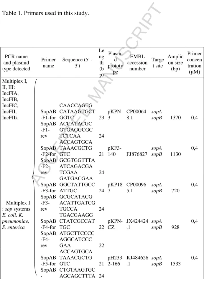
Table 1. Primers used in this study.
PCR name and plasmid type detected Primer name Sequence (5' - 3') Le ng th (b p) Plasmi d prototy pe EMBL accession number Targe t site Amplic on size (bp) Primer concen tration (μM) Multiplex I, II, III: IncFIA, IncFIB, IncFIC, IncFII, IncFIIk SopAB -F1-for CAACCAGTG CATAAGTGCT GGTC 23 pKPN 3 CP00064 8.1 sopA sopB 1370 0,4 SopAB -F1-rev ACCATACGC GTGAGGCGC TCTCAA 24 SopAB -F2-for ACCAGTGCA TAAACGCTG GTC 21 pKF3-140 FJ876827 sopA sopB 1130 0,4 SopAB -F2-rev GCGTGGTTTA ATCAGACGA TCGAA 24 SopAB -F3-for GATGACGAA GGCTATTGCC ATTGC 24 pKP18 7 CP00096 5.1 sopA sopB 720 0,4 Multiplex I : sop systems SopAB -F3-rev GCGCATACG ACATTGATCG TGCCA 24 E. coli, K. pneumoniae, S. enterica SopAB -F4-for TGACGAAGG CTATCGCCAT TGC 22 pKPN-CZ JX424424 .1 sopA sopB 928 0,4 SopAB -F4-rev ATGCTTCCCC AGGCATCCC GAA 22 SopAB -F5-for ACCAGTGCA TAAACGCTG GTC 21 pH233 2-166 KJ484626 .1 sopA sopB 1533 0,4 SopAB -CTGTAAGTGC AGCAGCTTTA 24
ACCEPTED MANUSCRIPT
12 F5_rev CGAC KP-StbAB -F2-for TTTGAAGGC GATGAGCTTC AGACC 24 pKP04 8 FJ628167 stbA stbB 1211 0,2 Multiplex II : stb systems KP-StbAB -F2-rev GCCCCACCAT TTTCGGGCTC CATCC 24 K. pneumoniae KP-StbAB -F4-for GTCACGGTAT TTGTTGTACA CTCC 24 pKpQI L-IT JN233705 stbA stbB 453 0,2 KP-StbAB -F4-rev GTTGATACG GTTTTGGATA CGCCA 24 EC-StbAB -F1-for GAACGTATA CTGCGATGAT GG 21 pEC14 _114 GQ39808 6 stbA stbB 905 0,2 EC-StbAB -F1-rev CTTTTTGCCC AAGATGGTG CCA 22 Multiplex III : stb systems EC-StbAB -F2-for CCGGAATGG TCTATGACGC TGCA 23 pKPC-LKEc KC78840 5 stbA stbB 1061 0,2 E. coli EC-StbAB -F2-rev ATCAGGAAC GGCAATCGTT CATCC 24 EC-StbAB -F3-for GCGGTCGCA AAATTGCCG AAGCTG 24 pO145 -13516 CP00626 3.1 stbA stbB 1194 0,2 EC-StbAB -F3-rev GAATTTTGCT TGTTTCCCAG AC 22 Multiplex IV : IncA/C, L/M, N ParAB -AC-for AGGCCTTTTA TCTGGCGTTA 20 pKPH S3 CP00322 5 parA parB 1817 0,4 ParAB -AC-rev GACAGTAGA CGGAACCAG AG 20 ParAB -LM-for CCACCAACA TCAAACTGG C 19 pOXA -48 JN626286 parA parB 1378 0,1 ParAB -LM-rev CAGATGCTG GACGTTCTTA C 20 StbAB C-N-TCCCGGCATT ATTGATAAA 24 pKP96 EU19544 9 stbA stbB 1093 0,1ACCEPTED MANUSCRIPT
13 for GAGTT stbC StbAB C-N-rev ACGGGTT-AAACGTCTC AGC 18 Multiplex V : IncHI ParAB -HIa-for AAGATCGCC CTCGTTGGTC AG 21 pEC-IMP EU85578 8 parA parB 2020 0,4 ParAB -HIa-rev CAACTTTTTG AGCAACCTG GAG 22 ParAB -HIb-for AAGATCGCC CTTGTCGGTC AACG 23 pMAK 1 AB36644 0 parA parB 1765 0,4 ParAB -HIb-rev TGGTAACAA ATCCATGCTC TTCCA 24 Multiplex VI : IncR, IncI1 ParAB -R-for TC-ACGA-CCAGCAAAA AGAGGAA 20 pKP91 CP00096 6 parA parB 2032 0,4 ParAB -R-rev GCTAAACTC ATAAGTCAG CGT 20 ParAB -I1-for GACGGCGAG AAGTTTTCAT T 20 Ec699 Replicols cope parA parB 1227 0,4 ParAB -I1-rev TTCAGCGTTT CTTCTGGTCT 20 Simplex VII : IncX ParA-X-for GAGCTTCAA CAGCAGAAC AG 21 pIncX-SHV JN247852 parA 633 0,2 ParA-X-rev ATTGCATCAT GTCTGGCTTG 20ACCEPTED MANUSCRIPT
14 Table 2. Recipient cells used in validation experiments. Tc : transconjugants, Tf : transformant, PRaseT : plasmid relaxase gene typing, PBRT : PCR-based replicon typing, PAR-T : plasmid partitioning genes typing. † :Tc used as a positive control in partitioning genes typing.
N° Parental strain Tc/Tf β-Lactamase Plasmid classification PRaseT (and/or PBRT) PAR-T 48 E. coli Tc TEM-52 X1 X 51 E. coli Tc TEM-3 L/M X 52 E. coli Tf TEM-52 X1 X 57 E. coli Tc SHV-12 F F 62 E. coli Tc SHV-2 FIB F 64 E. coli Tc SHV-5 X4 -
73 E. coli Tc SHV-4 FIIK FIIK
85 E. coli Tf CTX-M-1 FIA F
91 E. coli Tc CTX-M-3 N N
94† E. coli Tc CTX-M-9 FII F
98 E. coli Tc CTX-M-1 X1 X
100 E. coli Tc CTX-M-1 FIA, FII F
102 E. coli Tc CTX-M-1 HI2 HIa
104 E. coli Tf CTX-M-1 FIA, FIB, FII F
105 E. coli Tc CTX-M-1 L/M L/M 108 E. coli Tf CTX-M-1 FIA F 110 E. coli Tc CTX-M-1 X4 - 111† E. coli Tc CTX-M-1 FIA F 114 E. coli Tc CTX-M-1 L/M L/M 115 E. coli Tc CTX-M-9 FII F
118 E. coli Tc CTX-M-1 FIA, FIB F
120 E. coli Tc CTX-M-3 FIB F
122 E. coli Tc CTX-M-9 FII F
125 E. coli Tc CTX-M-1 FII F
126 E. coli Tc CTX-M-1 N N
127 E. coli Tc CTX-M-3 FIB F
Levy E. coli Tc OXA-48 L/M L/M
AD-48 E. coli Tc OXA-48 L/M L/M
AD-50 E. coli Tc OXA-48 L/M L/M
AD-17 E. coli Tc OXA-48 L/M L/M
AD-44-2 E. coli Tc NDM-1 N N
AD-2 E. coli Tc OXA-48 L/M L/M
Goe-137 E. coli Tc VIM HI2 HIa
Goe-132 E. coli Tc VIM A/C A/C
KATS E. coli Tc NDM-1, CTX-M-1 F F
M-2 E. coli Tc KPC-2 L/M L/M
M-3 E. coli Tc KPC-2 L/M L/M
M-5 E. coli Tc KPC-2 L/M L/M
Ec50 E. coli Tc TEM-52 I1 I1
ACCEPTED MANUSCRIPT
15
S3 K. pneumoniae Tc FOX-3 A/C A/C
S4 K. pneumoniae Tc DHA-1 A/C A/C
S6 K. pneumoniae Tc SHV-5 A/C A/C
S8 K. pneumoniae Tc CMY-2 A/C A/C
S9 K. pneumoniae† Tc SHV-4 FIIk F
S10 K. pneumoniae Tc ACC-1 A/C A/C
S11 K. pneumoniae Tc CMY-4 A/C A/C
S12 K. pneumoniae Tc SHV-2, DHA-1 FIIk, R F, R
S13 K. pneumoniae Tc SHV-1 A/C A/C
S14† K. pneumoniae Tc
CTX-M-15,
CMY-4, VIM-4 A/C A/C
S16 K. pneumoniae Tc CTX-M-3 A/C A/C
S18 K. pneumoniae Tc OXA-1, CTX-M-15 F F S19† K. pneumoniae Tc OXA-1, CTX-M-15 N N
S20 K. pneumoniae Tc OXA-1, SHV-2a,
CTX-M-15 F F
S21 K. pneumoniae Tc TEM-3 A/C A/C
S23 K. pneumoniae Tc CTX-M- 3 N, A/C N, A/C
S24 K. pneumoniae Tc TEM-3 A/C A/C
S26 K. pneumoniae Tc SHV-12 N N
S28 K. pneumoniae Tc DHA-1 L/M L/M
S30† K. pneumoniae Tf OXA-1, DHA-1 R R
S33 K. pneumoniae Tf DHA-1 R R
S34† K. pneumoniae Tc DHA-1 L/M L/M
S36 K. pneumoniae Tc OXA-1, CTX-M-3 FII F
S43 K. pneumoniae Tc CTX-M-3 N N S45 K. pneumoniae Tc OXA-1, CTX-M-15 L/M L/M S46 K. pneumoniae Tc CTX-M-15 L/M L/M S47 K. pneumoniae Tc CTX-M-3 L/M L/M S48 K. pneumoniae Tc CTX-M-15 L/M L/M S49 K. pneumoniae Tc CTX-M-15 L/M L/M S51 K. pneumoniae Tc SHV-12 FIIk F S53 K. pneumoniae Tc SHV-12 R R S55 K. pneumoniae Tc OXA-1, CTX-M-15 FIIk F S56 K. pneumoniae Tc OXA-1, CTX-M-15 FIIk R F, R S61 K. pneumoniae Tc OXA-1, CTX-M-15 F F
S68 K. pneumoniae Tc TEM-129 A/C A/C
S72 K. pneumoniae Tc SHV2a F F
S73 K. pneumoniae Tc SHV-4 A/C A/C
S75† K. pneumoniae Tc OXA-1, CTX-M-15 FIIk F S76 K. pneumoniae Tc TEM-12 F F S77 K. pneumoniae Tf SHV-12 R R S78 K. pneumoniae Tc CTX-M-15 FII F
ACCEPTED MANUSCRIPT
16
S79 K. pneumoniae Tc CTX-M-15 F F
S82 K. pneumoniae Tc CTX-M-15 FII F
S83 K. pneumoniae Tc TEM-3 A/C A/C
S86 K. pneumoniae Tc CMY-4 A/C A/C
S88 K. pneumoniae Tc SHV-2a FIIk F
S89 K. pneumoniae Tc TEM-21 A/C A/C
S90† K. pneumoniae Tc KPC X3 X FM1 K. pneumoniae Tc OXA-48, CTX-M-15 L/M L/M FM2 K. pneumoniae Tc OXA-48, CTX-M-15 L/M L/M
FUR-STA K. pneumoniae Tc OXA-48 L/M L/M
CLE-TN K. pneumoniae Tc OXA-48 L/M L/M
PET-TN K. pneumoniae Tc OXA-48 L/M L/M
MUR-STA K. pneumoniae Tc NDM-1 FIIk F
2966 K. pneumoniae Tc OXA-48 L/M L/M LD-1131 K. pneumoniae Tc VIM R R LD-3856 K. pneumoniae Tc OXA-48 L/M L/M Z-19760 K. pneumoniae Tc OXA-48 L/M L/M Z-45518 K. pneumoniae Tc OXA-48 L/M L/M Z-16300 K. pneumoniae Tc OXA-48 L/M L/M Z-47994 K. pneumoniae Tc OXA-48 L/M L/M Z-4359 K. pneumoniae Tc OXA-48 L/M L/M
MIKH K. pneumoniae Tc OXA-48, CTX-M9 HI1 HIb
BHR K. pneumoniae Tc VIM-4, CTX-M-1 HI1 HIb
M-4 K. pneumoniae Tc KPC-2 L/M L/M M-6 K. pneumoniae Tc KPC-2 L/M L/M M-7 K. pneumoniae Tc KPC-2 L/M L/M M-14 K. pneumoniae Tc KPC-2 L/M L/M M-20 K. pneumoniae Tc KPC-2 L/M L/M M-32 K. pneumoniae Tc KPC-2 L/M L/M M-40 K. pneumoniae Tc KPC-2 L/M L/M M-41 K. pneumoniae Tc KPC-2 L/M L/M M-50 K. pneumoniae Tc KPC-2 L/M L/M M-52 K. pneumoniae Tc KPC-2 L/M L/M M-53 K. pneumoniae Tc KPC-2 L/M L/M TNDHA
-5 K. pneumoniae Tc DHA-1, SHV-12 HI2 HIb
TNDHA
-6 K. pneumoniae Tc DHA-1, SHV-12 HI2 HIb
TNDHA
-7 K. pneumoniae Tc DHA-1, SHV-12 HI2 HIb
TNDHA
-8 K. pneumoniae Tc DHA-1, SHV-12 HI2 HIb
S00056
S. enterica
Typhimurium Tc CTX-M-2 HI2 HIa
S00319†
S. enterica
ACCEPTED MANUSCRIPT
17 S01331
S. enterica Tel
el kebir Tc CTX-M-15 HI2 HIa
S01477† S. enterica Typhimurium Tc CTX-M-1/CMY-2 HI1, I1 I1, HIb S01650
S. enterica
Brandeburg Tc CTX-M-14 FrepB F
S03207
S. enterica
Typhimurium Tf CTX-M-15 FIA, FIB F
S03663
S. enterica
Grumpensis Tc CTX-M-15 HI2 HIa
S03664 S. enterica Typhimurium Tc CTX-M-15 N N S04662 S. enterica Virchow Tc CTX-M-32 N N S05343 S. enterica
Concord Tc CTX-M-15 HI2 HIa
S07364
S. enterica
Miami Tc SHV-2 N N
S09118
S. enterica
Keurmassar Tc SHV-12 HI2, FI F, HIa
S27078 S. enterica Carmel Tc CTX-M-15 FrepB F S7917 S. enterica Derby Tc ND FIA F S7981 S. enterica Saintpaul Tc OXA-48 L/M L/M M-1 S. enterica Enteritidis Tc KPC-2 L/M L/M S1106† S. enterica Virchow Tc SHV-12 I1 I1
ACCEPTED MANUSCRIPT
18 Table 3. Plasmid partition gene typing of 30 clinical strains of Enterobacteriaceae.
N° Parental species β-Lactam ase PRase T PAR-T Multip lex I and II Multipl ex III Multipl ex IV Multipl ex V Simple x VI 3 E. coli TEM-24 A/C A/C 15 E. coli TEM-24 A/C, I1 A/C I1 17 E. coli TEM-24 A/C, F, HI2, X4 F A/C HI - 19 E. coli TEM-21 A/C, N, X1 F A/C, N - 23 E. coli TEM-24 A/C, F, I1 F A/C I1 26 E. coli TEM-24 A/C, F,
HI1 F A/C HIa
28 E. coli TEM-21 A/C, X1 A/C - 33 E. coli TEM-24 A/C A/C 34 E. coli TEM-24 A/C, N, X1 F A/C, N - 40 E. coli TEM-24 A/C, F F A/C 50 E. coli TEM-52 I1 I1 84 E. coli CTX-M-1 F HI1, N F N HIb 88 E. coli CTX-M-1 F, HI1, N F N HIa 101 E. coli CTX-M-2 HI2 Hia 105 E. coli CTX-M-1 L/M, I1 L/M - 106 E. coli CTX-M-1 F, HI1, N F N HIa 107 E. coli CTX-M-2 I1, F F I1 KpS 5 K. pneumoniae DHA-1 FIIK, L/M F L/M R KpS 19 K. pneumoniae CTX-M-15, DHA-1 FIIK, F, L/M, N F L/M, N KpS 20 K. pneumoniae CTX- M-15,SH F, X4 F -
ACCEPTED MANUSCRIPT
19 V-2a KpS 26 K. pneumoniae SHV-12 FIIK, N F N R KpS 47 K. pneumoniae CTX-M-3 HI2, L/M L/M HIa KpS 63 K. pneumoniae DHA-1 FIIK, L/M F L/M KpS 83 K. pneumoniae TEM-3 A/C, F, N A/C, N KpS 88 K. pneumoniae SHV-2a FIIK F KpS 92 K. pneumoniae CTX-M-14, VIM-1 F, FIIK, I1 F I1 FM-2 K. pneumoniae OXA-48 FIIK, L/M, N F L/M, N FM-10 K. pneumoniae OXA-48 A/C, L/M A/C, L/M S10-1477 S. enterica Typhimurium CTX-M-1, CMY-2 HI1, I1 HIb I1 S10-1526 S. enterica Typhimurium CTX-M-1, CMY-2 HI1, I1 HIb I1ACCEPTED MANUSCRIPT
20 Highlights
-Low-copy-number plasmids utilize partition systems for plasmid maintenance.
-par loci are organized into operons encoding two proteins, an ATPase and a DNA-binding protein, and including a centromeric site.
-The method called “plasmid partition gene typing” showed high specificity for the classification of resistance plasmids (IncA/C, FIA, FIB, FIC, FIIk, FII, HI1, HI2, I1, L/M, N) except for IncX replicons.