23. ISKDC. Primary nephrotic syndrome in children: clinical signifi-cance of histopathologic variants of minimal change and of diffuse mesangial hypercellularity. A report of the International Study of Kidney Disease in children. Kidney Int 1981; 20: 765–771 24. Kitamura A, Tsukaguchi H, Hiramoto R et al. A familial
childhood-onset relapsing nephrotic syndrome. Kidney Int 2007; 71: 946–951
25. Khoshnoodi J, Hill S, Tryggvason K, Hudson B, Friedman DB. Iden-tification of N-linked glycosylation sites in human nephrin using mass spectrometry. J Mass Spectrom 2007; 42: 370–379
Received for publication: 3.8.09; Accepted in revised form: 2.2.10
Nephrol Dial Transplant (2010) 25: 2976–2981 doi: 10.1093/ndt/gfq119
Advance Access publication 10 March 2010
Long-term follow-up of patients with Bartter syndrome type I and II
Elena Puricelli
1,4,**, Alberto Bettinelli
1,**, Nicolò Borsa
2, Francesca Sironi
2, Camilla Mattiello
2,
Fabiana Tammaro
1, Silvana Tedeschi
2, Mario G. Bianchetti
3and Italian Collaborative Group for Bartter Syndrome
*1
Department of Pediatrics, San Leopoldo Mandic Hospital, Largo Mandic 1, Merate, Lecco, Italy,2Laboratory of Medical Genetics, Fondazione IRCCS Ca’Granda-Ospedale Maggiore Policlinico, Mangiagalli e Regina Elena, Milan, Italy,3Department of Pediatrics, Mendrisio and Bellinzona Hospitals, University of Bern, Bern, Switzerland and4Department of Pediatrics, F.Del Ponte Hospital, Via F. Del Ponte 19, Insubria University of Varese, Varese, Italy
Correspondence and offprint requests to: Alberto Bettinelli; E-mail: [email protected]; [email protected]
*
Silvio Maringhini, Unit of Pediatric Nephrology, Civile Hospital, Palermo, Italy; Paolo Porcelli, Unit of Pediatric Endocrinology, Villa Sofia Hospital, Palermo, Italy; Marco Materassi, Department of Pediatrics, Meyer Hospital, University of Florence, Italy; Maria Renata Proverbio, Department of Pediatric Nephrology, Cardarelli Hospital, Naples, Italy; Nunzia Miglietti, Department of Pediatrics, University of Brescia, Italy; Maria Gabriella Porcellini, Department of Pediatric Nephrology, Regina Margherita Hospital, Turin, Italy; Carla Navone, Department of Pediatrics, Santa Corona Hospital, Pietra Ligure, Savona, Italy; Giuseppe Ruffa, Department of Pediatrics, Gaslini Hospital, Genoa, Italy; Aldo Rosini, Department of Pediatrics, B. Eustachio Hospital, San Severino Marche, Italy; Aurora Rossodivita, Department of Pediatrics, Gemelli Hospital, Rome, Italy.
**
These authors contributed equally to the work.
Abstract
Background. Little information is available on a long-term follow-up in Bartter syndrome type I and II. Methods. Clinical presentation, treatment and long-term follow-up (5.0–21, median 11 years) were evaluated in 15 Italian patients with homozygous (n = 7) or compound heterozygous (n = 8) mutations in the SLC12A1 (n = 10) or KCNJ1 (n = 5) genes.
Results. Thirteen new mutations were identified. The 15 children were born pre-term with a normal for gestational age body weight. Medical treatment at the last follow-up control included supplementation with potassium in 13, non-steroidal anti-inflammatory agents in 12 and gastropro-tective drugs in five patients. At last follow-up, body weight and height were within normal ranges in the patients. Glo-merular filtration rate was <90 mL/min/1.73 m2 in four patients (one of them with a pathologically increased uri-nary protein excretion). In three patients, abdominal ul-trasound detected gallstones. The group of patients with antenatal Bartter syndrome had a lower renin ratio (P < 0.05) and a higher standard deviation score (SDS) for height (P < 0.05) than a previously studied group of pa-tients with classical Bartter syndrome.
Conclusions. Patients with Bartter syndrome type I and II tend to present a satisfactory prognosis after a median
fol-low-up of more than 10 years. Gallstones might represent a new complication of antenatal Bartter syndrome.
Keywords: Bartter syndrome; cholelithiasis; growth retardation; KCNJ1 gene; SLC12A1 gene
Introduction
Bartter syndrome type I (BS I) and type II (BS II) are salt-wasting renal tubular disorders that are clinically character-ized by polyhydramnios leading to premature delivery, marked polyuria and a tendency towards nephrocalcinosis [1,2]. Loss-of-function mutations either in the furosemide-sensitive sodium–potassium–chloride cotransporter gene (SLC12A1; BS I, OMIM 601678) or in the inwardly rectify-ing potassium channel ROMK gene (KCNJ1; BS II, OMIM 241200) have been identified in the vast majority of patients with this autosomal recessive disorder [3,4]. Mutations in the CLCNKB chloride channel gene (Bartter syndrome type III also defined as classical Bartter syndrome—OMIM 607364) as well as in the BSND gene (Bartter syndrome form associated with sensorineural deafness—Bartter type IV— OMIM 602522) are also sometimes responsible for an iden-tical clinical phenotype but will not be treated in this report.
2976 E. Puricelli et al.
© The Author 2010. Published by Oxford University Press on behalf of ERA-EDTA. All rights reserved. For Permissions, please e-mail: [email protected]
Little information is currently available on the disease course of these patients [5–7]. The purpose of the current analysis was to shed additional light on clinical presenta-tion, initial diagnostic pitfalls and especially follow-up in 15 patients with bi-allelic mutations either in the SLC12A1 or in the KCNJ1 genes. The outcome in patients with an-tenatal Bartter syndrome was also compared with that ob-served in a recently published group of patients affected with classical Bartter syndrome [8].
Materials and methods
Among 34 patients with the clinical and biochemical diagnosis of antena-tal Bartter syndrome on follow-up at our institutions, 15 presented homo-zygous or compound heterohomo-zygous mutations in the SLC12A1 or KCNJ1 genes together with a clinical–biochemical follow-up of 5 years or more. Thirteen patients with antenatal Bartter syndrome presenting bi-allelic mutations and a shorter follow-up and another six patients with only one mutation detected were not further analysed in this study. The clinical features at presentation and at the last follow-up control were collected for these patients. The glomerular filtration rate was estimated from height and creatinine using the original Schwartz formula [9]. Since plasma renin (or plasma renin activity) and aldosterone were evaluated in different lab-oratories, we refer to their results either as renin ratio or aldosterone ratio. These indexes were calculated by dividing the individual renin (either plasma renin activity or active renin level) or aldosterone value by the corresponding upper reference value [8]. Renal ultrasound imaging was performed at diagnosis and at last follow-up in all patients. The last renal ecography was also evaluated for the grade of medullary nephrocalcinosis by three different paediatricians. The grading scale for medullary nephro-calcinosis was as follows: grade 1, mild increase in the echogenicity around the border of the medullary pyramid; grade 2, mild diffuse in-crease in the echogenicity of the entire medullary pyramid; grade 3, great-er or more homogeneous increase in the echogenicity of the entire medullary pyramid. In some patients, follow-up imaging included also the evaluation of liver and gallbladder.
The non-parametric two-tailed Mann–Whitney–Wilcoxon test for two independent samples was used for analysis. Statistical significance was defined as a P-value of <0.05.
Genomic DNA was extracted from peripheral white blood cells using standard methods. All coding sequences of SLC12A1 and KCNJ1 genes with their intron/exon boundaries were amplified by means of PCR us-ing specific primer pairs. Direct sequencus-ing of the purified PCR pro-ducts was then performed bidirectionally by the dye terminator cycle sequencing method (Applied Biosystem, Foster City, CA, USA) loaded on a 16-capillary ABI Prism 3100 genetic analyser (Applied Biosystem, Foster City, CA, USA) and analysed with the Sequencing Analysis 3.7 software. Mutations were discerned from polymorphisms by demonstra-tion of their absence in 100 control chromosomes. The nomenclature is based on the recommendations of the Human Genomic Variant Society starting with the nucleotide 1 of the start codon (ATG) at cDNA level. A written informed consent had been obtained from all patients and their family members.
Results
Molecular findings
The 15 patients with homozygous or compound heterozy-gous mutations considered in this study (8 female and 7 male subjects) belong to 15 non-consanguineous Italian families (Table 1). The results of direct sequencing of the SLC12A1 and KCNJ1 genes revealed 13 new muta-tions and 7 previously described mutamuta-tions. Patient 3, who carried a compound heterozygous mutation, had been previously described as a single heterozygous mutation
[10]. Among the 10 patients presenting SLC12A1 gene Ta
b le 1 . Molecu lar cha racterization of the genetic defects in the SLC12 A1 (Bartter synd rom e type I) or KCNJ1 (Bartter syndrom e type II) genes P atie nt number Gend er Bartter synd rome type Status N ucleotide change Pr edicted ef fect on prot ein Exon T ype of muta tion Reference 1 F ema le I Homoz ygous c.13 81T>C p.[Cys 461Ar g] + [C ys461A rg ] 1 0 Misse nse [10] 2 Male I Homoz ygous c.10 62delG p.[L ys35 4AsnfsX 73] + [L ys35 4Asnf sX73] 7 F rameshift [10] 3 F ema le I Compou nd heterozygous c.13 81T>C / c.1630C >T p.[Cys 461Ar g] + [P ro544S er] 10/12 Misse nse / missense [10] / n ew 4 Male I Compou nd heterozygous c.34 7G>A / c.1954G>A p.[Ar g116 His] + [Gl y652 Ser] 1/15 Misse nse / missense Ne w / ne w 5 F ema le I Homoz ygous c.90 3_904de lC p.[Ar g302 Gl yfsX2 ] + [Ar g302 Gl yfsX2 ] 6 F rameshift [3] 6 F ema le I Homoz ygous c.16 63G>A p.[Ala 555Th r] + [Ala555Thr] 12 Misse nse [10] 7 Male I Compou nd heterozygous c.55 1T>A / c.611T> C p.[Le u184G ln] + [V al20 4Ala] 2/3 Misse nse / missense Ne w / ne w 8 F ema le I Compou nd heterozygous c.11 90G>C / c.3164+1 G> A p.[Gl y397 Ala] + c.[3164 +1g>a ] 8/25 Misse nse / splice site Ne w / [7] 9 Male I Compou nd heterozygous c.14 93C>T / c.1522G> A p.[Ala 498V al] + [Ala508Th r] 11/11 Misse nse / missense Ne w / [19] 10 F ema le I Compou nd heterozygous c.90 3_904de lC / c.1493C >T p.[Ar g302 Gl yfsX2 ] + [Ala498V al] 6/11 F rameshift / missense [3] / n ew 11 F ema le II Compou nd heterozygous c.80 8C>T / c.592G >T p.[His 270Th r]+ [Ala198S er] 5 Misse nse / missense Ne w / ne w 12 F ema le II Compou nd heterozygous c.80 G>A / c.277T> G p.[T rp27X] + [P he93V al] 5 N onsense / misse nse Ne w / ne w 13 Male II Homoz ygous c.57 2C>T p.[Th r191Ile ] + [Thr 191Ile] 5 Misse nse Ne w 14 Male II Homoz ygous c.80 8C>T p.[His 270Th r] + [His27 0Thr] 5 Misse nse Ne w 15 Male II Homoz ygous c.42 2C>T p.[Th r141Ile ] + [Thr 141Ile] 5 Misse nse Ne w
mutations, 6 were compound heterozygous and 4 homozy-gous. Among the five patients presenting KCNJ1 gene mu-tations, three were homozygous and two compound heterozygous. The homozygous mutations were confirmed on both the parents of the patients in order to exclude the presence of a heterozygous deletion of corresponding exons. The genetic abnormalities described were not pres-ent in a sample of 50 healthy controls (100 chromosomes investigated).
Initial findings
History of premature delivery, polyhydramnios and poly-uria was present in all patients, but the final clinical diag-nosis of Bartter syndrome was made before the age of 26 months in 12 patients (Figure 1). Birth weight was never less than 2 standard deviation scores (SDS) below the mean for gestational age. The birth weight SDS was on the aver-age higher (P < 0.05) in BS I than in BS II patients. The di-agnosis of diabetes insipidus was initially suspected in two BS I patients who presented with hypernatraemia and that of pseudohypoaldosteronism in the two BS I patients who pre-sented with plasma potassium >6.0 mmol/L. In one of them (Patient 7), acute renal failure was also present.
The overall median plasma sodium was 137 mmol/L (range 125–156), and it was increased in three and mildly
decreased in two patients (Figure 1). The overall median plasma chloride was 98 mmol/L (range 84–107), and it was decreased in four patients. Plasma potassium was re-duced in eight but normal (n = 1) or even increased (n = 1) in BS I patients (median 2.7, range 1.9–6.3 mmol/L). On the contrary, the level of this electrolyte was mildly re-duced in one but normal (n = 1) or even increased (n = 3) in four of the five BS II patients (median 5.7, range 3.3– 6.7 mmol/L): the difference between BS I and BS II was significant (P < 0.03). Hyperbicarbonataemia (HCO3− >25 mmol/L) was similarly frequent both in patients with BS I (8 of 10 patients) as well as in patients with BS II. The overall median plasma bicarbonate was 27 mmol/L (range 25–32). No hypomagnesaemia was noted in the 15 patients, and the overall median plasma magnesium level was 2.4 mg/dL (range 1.9–2.8). Aldosterone ratio was increased in all but two patients, and renin ratio was increased in all patients. Plasma creatinine was moderately increased in 1 of the neonates affected with Bartter type I syndrome (Patient 7) and normal in the remaining 14 patients (Figure 1).
Renal ultrasound disclosed signs of medullary nephro-calcinosis in all patients with the exception of one BS II patient (Patient 11). The degree of nephrocalcinosis at diagnosis and the urinary calcium excretion were not evaluated. Age at Final Diagnosis (Months) 0 24 48 62 96 120 Gestational Age (Weeks) 26 30 34 38 Birth Weight (SDS) -2.0 -1.0 0.0 1.0 2.0 120 130 140 150 160 Plasma Na (mmol/L) 80 85 90 95 100 105 110 Plasma Cl (mmol/L) Plasma HCO3 (mmol/L) 20 25 30 35 2.0 3.0 4.0 5.0 6.0 Plasma K (mmol/L) Plasma Mg (mg/dL) 1.9 2.1 2.3 2.5 2.7 0 5 10 15 20 25 30 Aldosterone Ratio 0 5 10 15 20 Renin Ratio
Bartter syndrome type I Bartter syndrome type II P<0.05 P<0.03 Plasma Creatinine (SDS) -2 -1 0 1 2 3
Fig. 1. Age at the final diagnosis and the initial clinical and laboratory data in 15 patients with homozygous or heterozygous mutations in the SLC12A1 (BS I; closed symbols) or KCNJ1 (BS II; open symbols) genes. Plasma bicarbonate was not assessed at presentation in one patient affected with BS II. Birth weight SDS was significantly higher (P < 0.05) and plasma potassium lower (P < 0.03) in BS I than in BS II. The frames denote the reference ranges.
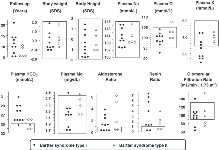
Follow-up
Clinical and biochemical data at the last follow-up are giv-en in Figure 2. None of the 15 patigiv-ents suffered from overt underweight or small stature. Plasma sodium and chloride were within the normal ranges in all patients with the ex-ception of one BS I patient who presented with mild hypo-chloraemia. The overall median plasma sodium was 139 mmol/L (range 137–145) and plasma chloride 101 mmol/L (range 93–108), respectively. Plasma potassi-um was reduced in 6 of the 10 BS I patients (median 3.1, range 2.5–4.3 mmol/L) and within the normal ranges in all BS II patients (median 3.9, range 3.6–4.1 mmol/L). How-ever, the difference in plasma potassium between these two groups did not reach significant values. Hyperbicarbona-taemia was noted in 9 of the 10 BS I patients (median 27, range 25–31 mmol/L) and in 2 of the 5 BS II patients (median 25, range 24–26 mmol/L). The plasma total mag-nesium level was never reduced in the 15 patients. Aldo-sterone ratio was normalized in seven patients: four with BS I and three with BS II. Renin ratio was normalized in four patients. Glomerular filtration rate was >90 mL/ min/1.73 m2 in 11 and mildly reduced in 4 patients, 2 patients each with BS I and BS II. Moderate glomerular proteinuria (∼1 g/day) was noted in one of the patients with a reduced glomerular filtration rate (Patient 5).
The urinary molar calcium/creatinine ratio was evaluat-ed at the last clinical control in 8 out the 10 patients with BS I and in 3 out of 5 patients with BS II: this parameter was increased (>0.60) in 10 of the 11 aforementioned patients.
As at presentation, renal ultrasound disclosed signs of nephrocalcinosis in all patients with the exception of Pa-tient 11. Nephrocalcinosis was classified as follows: five BS I patients presented with grade 2 and five with grade 3; 2 BS II patients presented with grade 1 and two with grade 3. At least one complete abdominal ultrasound was performed in 10 patients: in three patients, two with BS I (Patient 8 and 9) and one with BS II (Patient 13), asymptomatic gallstones were detected (1–3 stones per pa-tient with a diameter of≤5 mm). Long-term parenteral nu-trition, a recognized cause of gallstones, had been performed in Patient 8. The age at detection of cholelithi-asis was 2.2 and 8.7 years in BS I and 2.9 years in BS II. A persistent cholelithiasis was demonstrated on follow-up in Patient 9 and 13. No corresponding follow-up is so far available for Patient 8.
All patients had normal blood pressure, and no hearing loss during the follow-up period was detected. The neuro-developmental assessment was normal in the patients with the exception of Patient 15 who presented a mild left hemi-paresis secondary to neonatal ischaemic brain damage.
5 10 15 20 Follow up (Years) -2.0 -1.0 0.0 1.0 2.0 Body weight (SDS) -2 -1 0 1 2 Body Height (SDS) 135 137 139 141 143 145 Plasma Na (mmol/L) 90 95 100 105 110 Plasma Cl (mmol/L) 23 25 27 29 31 Plasma HCO3 (mmol/L) 0 2 4 6 Aldosterone Ratio 0 1 2 3 4 5 6 7 Renin Ratio
Bartter syndrome type I Bartter syndrome type II Plasma Mg (mg/dL) 1.7 1.9 2.1 2.3 2.5 2.7 2.9 3.0 4.0 5.0 Plasma K (mmol/L) 60 80 100 120 140 Glomerular Filtration Rate (mL/min . 1.73 m2)
Fig. 2. Clinical and laboratory features in 15 patients with homozygous or heterozygous mutations in SLC12A1 (BS I; closed symbols) or in KCNJ1 (BS II; open symbols) genes 5.0–21, median 11 years after diagnosis. No significant difference was noted between BS I and BS II syndromes. The frames denote the reference ranges.
Medical treatment at the last follow-up control included the non-steroidal anti-inflammatory agent indomethacin in 11 patients (from 0.2 to 2.2, median 0.9 mg/kg body weight daily), flurbiprofen in one patient, gastroprotective agents in five patients (ranitidine, n = 4; lansoprazole, n = 1) and spironolactone associated with hydrochlorothiazide in one patient. Supplementation with potassium chloride was administered in 13 patients (from 0.5 to 2.3 mmol/ kg body weight daily). Patient 3 developed a peptic ulcer at 6 years of age on indomethacin 2.0 mg/kg body weight daily. This agent was reintroduced 2 years later without re-currence of the peptic disease. In Patient 2, a growth hor-mone deficiency was demonstrated on follow-up, and treatment with recombinant human growth hormone was introduced during 3 years.
Comparison with classical Bartter syndrome (BS III) The findings at follow-up in our 15 patients with BS I and BS II were compared with those noted in a group of 13 patients affected with BS III recently as reported by some members of our group [8]. No differences between the two groups were noted with respect to the blood levels of sodium, potassium, chloride and bicarbonate; the aldosterone ratio; and the glomerular filtration rate. The plasma renin ratio was significantly (P < 0.05) lower in the group of patients with BS I and BS II as compared with BS III (Figure 3). The height SDS was significantly (P < 0.05) higher in BS I–II types than in BS III. No signifi-cant correlation between renin ratio and height SDS was not-ed in BS III, in BS I–II types or in the cumulatnot-ed group of patients with either BS III or BS I–II types.
Discussion
The present study describes the genetic findings, the initial clinical and laboratory characteristics, and especially, the
outcome and current management in 15 patients with ho-mozygous or compound heterozygous mutations in the SLC12A1 and KCNJ1 genes after a median follow-up of 11 years. BS I patients showed mutations that were spread all over the SLC12A1 gene; on the contrary, all BS II pa-tients showed mutations in exon 5 of the KCNJI gene, as mainly observed [3–7]. The seven new mutations in the SLC12A1 and five out of the six mutations in the KCNJ1 were missense. The 13 new missense variants are likely pathogenic, since they substitute highly conserved amino acids in both the genes and were not detected in 100 healthy chromosomes.
The p.Arg116His mutation identif ied in Patient 4, which has also been described as a single-nucleotide poly-morphism (rs34819316), was detected in another homozy-gous patient not included in this study. So far, functional studies have never been performed on this variant unequiv-ocally demonstrating its polymorphic nature.
It is assumed that patients affected by antenatal Bartter syndrome are often born prematurely or with a small for gestational age birth weight [5–7]. The present survey con-firms that these patients are born prematurely after a preg-nancy complicated by polyhydramnios but with a normal for gestational age weight, therefore excluding major intra-uterine growth retardation in these conditions (however, the birth weight SDS is lower in BS II than in BS I pa-tients). Patients with BS I and BS II present with similar clinical and laboratory findings with the exception of po-tassium level, which is often reduced in BS I but normal or even increased in BS II [5–7].
Since hypokalaemia and metabolic alkalosis, two pecu-liar biochemical findings in Bartter syndrome, are often absent at presentation in the antenatal form of this syn-drome, other conditions including diabetes insipidus (in subjects with hypernatraemia) or pseudohypoaldosteron-ism (in patients with hyperkalaemia) are initially suspected in some cases [5,7,11].
Patients with antenatal Bartter syndrome are managed with potassium chloride and non-steroidal anti-inflamma-tory agents, mostly indomethacin. The present experience confirms that, in patients affected by hypokalaemic salt-losing tubulopathies, gastrointestinal side effects of non-steroidal anti-inflammatory agents are rather rare and not severe [5,8,12]. This observation is likely related to the lower dose of indomethacin (on average 0.9 mg/kg/body weight daily) as compared with other studies [5,7,12]. Ob-viously, the risks associated with a suboptimal correction of hypokalaemia must be balance against the risks of high-dose therapy with indomethacin.
In our antenatal Bartter patients, both the neurodevelop-mental outcome and the somatic growth are almost always normal. In salt-wasting renal tubular disorders, somatic growth retardation is common and traditionally causally linked among others with extracellular fluid volume deple-tion [13]. The present data with a better growth in BS I and BS II than in BS III and with a higher renin ratio, a recog-nized marker of fluid volume depletion, in BS III than in BS I and BSII support the aforementioned link between growth retardation and fluid volume depletion in the various forms of Bartter syndrome. Potassium deficiency may also impair the metabolism of growth hormone [14]; however, the sim--4.0 -3.0 -2.0 -1.0 0.0 1.0 2.0 Body Height (SDS) 2 4 6 Renin Ratio 10 8 P<0.05 P<0.05
Bartter syndrome type I or II Bartter syndrome type III
Fig. 3. Body height SDS and renin ratio in 15 patients with BS I or BS II (closed symbols) 5.0–21, median 11 years after diagnosis, and in 13 patients affected with BS III (open symbols) 5.0–24, median 14 years after diagnosis [8]. Height SDS was significantly higher (P < 0.05) and plasma renin ratio lower (P < 0.05) in BS I and BS II syndromes than in BS III. The frames denote the reference ranges.
ilar potassium level in antenatal and in classical Bartter syn-drome may suggest that potassium does not account for growth retardation in classical Bartter syndrome.
A glomerular filtration rate of <90 mL/min/1.73 m2, sometimes associated with overt proteinuria, was noted on follow-up in approximately one quarter of our patients. Nephrocalcinosis is a possible explanation for these obser-vations. Prolonged hypokalaemia, which can lead to inter-stitial fibrosis and tubular atrophy, is a further possible cause [15]. Finally, non-steroidal anti-inflammatory agents have also been associated with reduced renal function [16]. However, in antenatal Bartter syndrome, the use of indomethacin is not related to any relevant lesion on renal biopsy [5].
We were surprised by the fact that three patients with antenatal Bartter syndrome developed gallstones, confirm-ing a recent case report [17]. Possible explanations are that these patients are born prematurely, that they present with laboratory characteristics, which may be mimicked by medication with loop diuretics, and that both prematurity and long-term administration of loop diuretics predispose to gallbladder stone formation in childhood [18]. Alterna-tively, in these patients, the process of gallstone formation might result from an altered function of either the sodium– potassium–chloride cotransporter or the channel ROMK within the hepatobiliary system.
In conclusion, our patients with homozygous or com-pound heterozygous mutations in the SLC12A1 or KCNJ1 genes managed with potassium chloride and indomethacin present a satisfactory somatic growth after a median follow-up of 11 years. Some impairment of kidney function is ob-served in approximately one quarter of the patients. Finally, a large subset of these patients develops gallbladder stones.
Acknowledgements. This study was funded by a grant from the Associa-zione per il Bambino Nefropatico, Milan, Italy.
Conflict of interest statement. None declared.
References
1. Seyberth HW. An improved terminology and classification of Bartter-like syndromes. Nat Clin Pract Nephrol 2008; 4: 560–567 2. Proesmans W. Threading through the mizmaze of Bartter syndrome.
Pediatr Nephrol 2006; 21: 896–902
3. Simon DB, Karet FE, Hamdan JM, Di Pietro A, Sanjad SA, Lifton RP. Bartter's syndrome, hypokalaemic alkalosis with hypercalciuria, is caused by mutations in the Na–K–2Cl cotransporter NKCC2. Nat Genet 1996; 13: 183–188
4. Simon DB, Karet FE, Rodríguez-Soriano J et al. Genetic heterogene-ity of Bartter's syndrome revealed by mutations in the K+channel,
ROMK. Nat Genet 1996; 14: 152–156
5. Reinalter SC, Gröne HJ, Konrad M, Seyberth HW, Klaus G. Evalu-ation of long-term treatment with indomethacin in hereditary hypo-kalemic salt-losing tubulopathies. J Pediatr 2001; 139: 398–406 6. Finer G, Shalev H, Birk OS et al. Transient neonatal hyperkalemia in
the antenatal (ROMK defective) Bartter syndrome. J Pediatr 2003; 142: 318–323
7. Brochard K, Boyer O, Blanchard A et al. Phenotype–genotype corre-lation in antenatal and neonatal variants of Bartter syndrome. Nephrol Dial Transplant 2009; 24: 1455–1464
8. Bettinelli A, Borsa N, Bellantuono R et al. Patients with biallelic mu-tations in the chloride channel gene CLCNKB: long-term manage-ment and outcome. Am J Kidney Dis 2007; 49: 91–98
9. Schwartz GJ, Haycock GB, Edelmann CM Jr, Spitzer A. A simple estimate of glomerular filtration rate in children derived from body length and plasma creatinine. Pediatrics 1976; 58: 259–263 10. Bettinelli A, Ciarmatori S, Cesareo L et al. Phenotypic variability in
Bartter syndrome type I. Pediatr Nephrol 2000; 14: 940–945 11. Bichet DG. Hereditary polyuric disorders: new concepts and
differ-ential diagnosis. Semin Nephrol 2006; 26: 224–233
12. Vaisbich MH, Fujimura MD, Koch VH. Bartter syndrome: benefits and side effects of long-term treatment. Pediatr Nephrol 2004; 19: 858–863
13. Haycock GB. The influence of sodium on growth in infancy. Pediatr Nephrol 1993; 7: 871–875
14. Flyvbjerg A, Dørup I, Everts ME, Orskov H. Evidence that potassium deficiency induces growth retardation through reduced circulating le-vels of growth hormone and insulin-like growth factor I. Metabolism 1991; 40: 769–775
15. Reungjui S, Roncal CA, Sato W et al. Hypokalemic nephropathy is associated with impaired angiogenesis. J Am Soc Nephrol 2008; 19: 125–134
16. Ulinski T, Bensman A. Complications rénales des anti-inflamma-toires non stéroïdiens. Arch Pédiatr 2004; 11: 885–888
17. Robitaille P, Tousignant K, Dubois J. Bartter syndrome and choleli-thiasis in an infant: is this a mere coincidence? Eur J Pediatr 2008; 167: 109–110
18. Wesdorp I, Bosman D, de Graaff A, Aronson P, Vander Blij F, Taminiau J. Clinical presentations and predisposing factors of cho-lelithiasis and sludge in children. J Pediatr Gastroenterol Nutr 2000; 31: 411–417
19. Vargas-Poussou R, Feldmann D, Vollmer M et al. Novel molecular variants of the Na–K–2Cl cotransporter gene are responsible for an-tenatal Bartter syndrome. Am J Hum Genet 1998; 62: 1332–1340 Received for publication: 6.12.09; Accepted in revised form: 15.2.10

![Fig. 3. Body height SDS and renin ratio in 15 patients with BS I or BS II (closed symbols) 5.0 – 21, median 11 years after diagnosis, and in 13 patients affected with BS III (open symbols) 5.0 – 24, median 14 years after diagnosis [8]](https://thumb-eu.123doks.com/thumbv2/123doknet/14888739.648185/5.918.93.402.80.321/height-patients-symbols-diagnosis-patients-affected-symbols-diagnosis.webp)
