Cite this article as: Kostron A, Horn-Tutic M, Franzen D, Kestenholz P, Schneiter D, Opitz Iet al. Repeated lung volume reduction surgery is successful in selected patients. Eur J Cardiothorac Surg 2015;48:710–715.
Repeated lung volume reduction surgery is successful
in selected patients
†
Arthur Kostron
‡, Michaela Horn-Tutic
‡, Daniel Franzen, Peter Kestenholz, Didier Schneiter,
Isabelle Opitz, Malcolm Kohler and Walter Weder*
Department of Thoracic Surgery and Division of Pulmonology, University Hospital, Zurich, Switzerland
* Corresponding author. Division of Thoracic Surgery, University Hospital, University of Zurich, Raemistrasse 100, 8091 Zurich, Switzerland. Tel: +41-444-2558802; fax: +41-442-558805; e-mail: [email protected] (W. Weder).
Received 19 July 2014; received in revised form 10 November 2014; accepted 18 November 2014
Abstract
OBJECTIVES: Lung volume reduction surgery (LVRS) improves dyspnoea, quality of life and may even prolong survival in carefully selected patients with end-stage emphysema. The benefit may be sustained for several years and vanishes with the natural progression of the disease. Data on repeated surgical treatment of emphysema are scarce. The aim of this study was to evaluate the safety, effects and out-comes of repeated LVRS (Re-LVRS) in patients no longer benefiting from their initial LVRS.
METHODS: Between June 2002 and December 2013, 22 patients (9 females) with advanced emphysema underwent Re-LVRS at a median of 60 months (25–196) after their initial LVRS. While initial LVRS was performed thoracoscopically as a bilateral procedure, Re-LVRS was performed unilaterally by a video-assisted thoracoscopic technique in 19 patients and, due to adhesions, by thoracotomy in 3 patients. Pulmonary function test (PFT) was performed at 3 and 12 months postoperatively.
RESULTS: Lung function at Re-LVRS was similar to that prior to thefirst LVRS. The 90-day mortality rate was 0%. The first patient died 15
months postoperatively. The median hospitalization time after Re-LVRS was significantly longer compared with the initial LVRS [14 days,
interquartile range (IQR): 11–19, vs 9 days, IQR: 8–14; P = 0.017]. The most frequent complication was prolonged air leak with a median
drainage time of 11 days (IQR: 6–13); reoperations due to persistent air leak were necessary in 7 patients (32%). Five patients (23%) had no
complications. Lung function and Medical Research Council (MRC) score improved significantly for up to 12 months after Re-LVRS, with
results similar to those after initial bilateral LVRS. The average increase in the forced expiratory volume in 1 s (FEV1) was 25% (a 7% increase
over the predicted value or 0.18 l) at 3 months, and the mean reduction in hyperinflation, assessed by relative decrease in RV/TLC (residual
volume/total lung capacity), was 12% at 3 months (a decrease of 8% in absolute ratios). The mean MRC breathlessness score decreased
sig-nificantly after 3 months (from 3.7 to 2.2).
CONCLUSIONS: Re-LVRS can be performed successfully in carefully selected patients as a palliative treatment. It may be performed as a bridge to transplantation or in patients with newly diagnosed intrapulmonary nodules or during elective cardiac surgery. Morbidity is acceptable and outcomes may be satisfactory with significantly improved lung function and reduced dyspnoea for at least 12 months postoperatively.
Keywords:Lung volume reduction surgery• End-stage pulmonary emphysema • Lung volume reduction • Reoperation
INTRODUCTION
Chronic obstructive pulmonary disease (COPD) is a leading global health problem and is predicted to be the third largest cause of
death worldwide by 2020 [1]. It leads to emphysematous
destruc-tion of the lung parenchyma and therefore causes dyspnoea as
well as rapid loss of physicalfitness and quality of life [2].
Lung volume reduction surgery (LVRS) is a successful and well-established palliative treatment for end-stage emphysema in care-fully selected patients. Several randomized controlled trials have
confirmed the feasibility of surgical therapy and that it is superior
to best medical treatment in long-term survival and exercise
cap-acity in a specific subgroup of patients [3–6].
The pathophysiology of severe emphysema includes
hyper-inflation and small airway obstruction through parenchymal
destruction, thereby allowing the possibility of mechanical repair. By surgically removing the most affected areas of lung
paren-chyma, hyperinflation is reduced and diaphragm and chest wall
†Presented at the 22nd European Conference on General Thoracic Surgery,
Copenhagen, Denmark, 15–18 June 2014.
‡Thefirst two authors contributed equally to this manuscript.
© The Author 2014. Published by Oxford University Press on behalf of the European Association for Cardio-Thoracic Surgery. All rights reserved. –715
mechanics are improved. Furthermore, small airway obstruction
can be reduced by an increase of elastic recoil forces [7].
The effects of LVRS may last for up to 5 years, depending on the
morphology of the emphysema [8, 9]. Surgery itself does not
modify the subsequent natural history of the disease [3] and, as
the natural history of emphysema and the annual decline in pul-monary function take their course, lung function and dyspnoea return to pre-LVRS levels after a certain period that differs from
person-to-person [4].
The only treatment that can stop the progressive course of the disease is bilateral lung transplantation. In some patients, it may
be feasible to use LVRS as a bridge to transplantation [10–12];
however, many patients with end-stage COPD do not qualify for transplantation because of advanced age or comorbidities, and therefore LVRS has been described as a successful alternative in
appropriately selected patients [11,13,14].
Recent data from the Society of Thoracic Surgeons database suggests a limited use of LVRS in the USA, with an initial increase of LVRS performed after data from the National Emphysema Treatment Trial were published but with numbers remaining
steady since 2004 [15]; data for Europe are, unfortunately, not
available, but again a limited use can be assumed. However, many patients treated by LVRS survive the duration of the positive effect and re-experience the loss of quality of life despite receiving optimal medical treatment. For those not eligible for transplant-ation, a limited range of therapeutic options remain.
Repeated LVRS (Re-LVRS) is a technically and also medically challenging procedure that targets the ongoing parenchymal destruction. Data regarding the feasibility and safety of Re-LVRS
are scarce. However, afirst case was described by Stammberger
et al. in 2000 [16], after which the beneficial effects of Re-LVRS
on pulmonary function and quality of life were shown in a
small population of 16 patients [17]. However, while it was
tech-nically feasible and led to overall improvements, the reported mortality rate was as high as 11.7%, rendering it a high-risk
operation [17].
The aim of this retrospective study was to further evaluate the
morbidity and mortality, as well as the efficacy and outcome, due
to Re-LVRS in carefully selected patients after cessation of the
beneficial effects of initial LVRS, to elucidate the role of repeated
surgery in this high-risk population.
MATERIALS AND METHODS
Patient selection
Inclusion criteria were adapted from our modified patient
selec-tion criteria for initial LVRS [7]. Patients eligible for Re-LRVS had to
have experienced a significant improvement of dyspnoea and
pul-monary function test after the initial LVRS, but had to present with a deterioration of their dyspnoea [measured by Medical Research Council (MRC) dyspnoea score] and pulmonary function tests (PFTs) to levels similar to the pre-LVRS level. In addition to severe
airflow obstruction (FEV1 < 35%), hyperinflation of the lung
(TLC > 120%, RV > 200%, RV/TLC > 65) and sustained minimum dif-fusion capacity (DLCO > 20%), the patients selected for Re-LVRS had to be highly motivated for a repeated operation.
CT scans had to reveal either heterogeneous to intermediately distributed marked areas of emphysematous destruction, or newly diagnosed intrapulmonary nodules suspicious for malignancy.
Patients with significant comorbidities, like coronary artery disease
or pulmonary hypertension, were excluded.
Preoperative assessment
Prior to the operation, rigorous testing was performed to verify operability. In addition to recent PFTs and chest CT scans with densitometry, a radionuclide lung perfusion scan was performed to identify the most affected areas. To further evaluate operability, myocardial SPECT was performed in patients with uncertain myo-cardial function (n = 7, 32%).
The MRC dyspnoea score, a simple and valid score that consists
offive categories (0–4), and which has been shown to correctly
categorize disability in COPD patients [18], was used to categorize
the physical disability of the patients due to COPD.
Decisions on reoperation were taken according to our inclu-sion/exclusion criteria by an interdisciplinary committee consist-ing of thoracic surgeons, pulmonologists and radiologists.
Surgery
Re-LVRS was performed unilaterally by video-assisted thoraco-scopy surgery (VATS) in 19 patients, and in 3 patients by thora-cotomy due to adhesions from the previous LVRS or infection; 1
patient had a bilateral Re-LVRS (Table1). The areas of pulmonary
parenchyma exhibiting greatest destruction were resected using
standard staplers (COVIDIEN Endo GIA™ Ultra Universal, ETHICON
Echelon ENDOPATH™) without buttressing (for both, initial LVRS
and Re-LVRS).
Follow-up and outcome measures
All PFTs were performed using a standard body plethysmograph and CO diffusion capacity (Zahn, Germany). The follow-up in-cluded PFT and MRC dyspnoea score at baseline and 3 and 12
Table 1: Patient specifics
Age, years (IQR) Bilateral procedure (n) Unilateral procedure (n) Additional information (n) Thoracoscopy Thoracotomy Thoracoscopy Thoracotomy
Initial LVRS 59 (55–67) 20 – 1 1 Suspicious nodule: 1
Re-LVRS 65 (62–72) – 1 19 2 Transplantation waiting list: 2
Suspicious nodules: 4 Elective cardiac surgery: 1
THO
R
A
C
months after surgery. The success of the procedure was judged by
(i) reduction of hyperinflation assessed by a decrease in residual
volume (RV), total lung capacity (TLC) and their ratio (RV/TLC), (ii) increase in forced vital capacity (FVC), forced expiratory volume in 1 s (FEV1) and diffusion capacity of the lung for carbon monoxide (DLCO), and (iii) reduction of dyspnoea assessed by the MRC score. Patients with suspected or proven malignancy had regular CT scans for the cancer follow-up.
Complete data sets of PFT are available for 22 (100%), 21 (95%) and 14 patients (64%) after LVRS and for 22 (100%), 11 (50%) and 9 patients (41%) after Re-LVRS at baseline, 3 and 12 months after the operation, respectively.
Statistical analysis
All values are displayed as the mean ± standard error or median and interquartile range (IQR), unless otherwise stated. Descriptive statistical analysis was performed comparing perioperative mor-bidity and duration of hospitalization. PFT at 3 and 12 months after either LVRS or Re-LVRS were compared with baseline values
using a two-tailed paired samplest-test. For evaluation between
the LVRS and the Re-LVRS group the Wilcoxon matched-pair
signed-rank test was used for continuous variables and McNemar’s
test for categorical variables.
A P-value of <0.05 was determined as significant (*), and
P < 0.001 as highly significant (**). All data were produced using SPSS (IBM Corp., Released 2013. IBM SPSS Statistics for Windows, Version 22.0. Armonk, NY, USA: IBM Corp.). Graphs were plotted using GraphPad Prism version 4.0c for Macintosh (GraphPad Software, San Diego, CA, USA).
RESULTS
Between June 2002 and December 2013, 22 patients (9 females)
with a median age of 65 years (IQR: 62–72) underwent repeated
LVRS (Re-LVRS) for the following reasons: (i) cessation of the bene-ficial effects of initial LVRS only (n = 15); (ii) tissue-diagnosis and
treatment of newly diagnosed intrapulmonary nodules (n = 4); (iii)
as a bridge to transplantation (n = 2) or (iv) during elective cardiac
surgery (n = 1) (also see Table1). The median time span between
initial and repeated LVRS was 60 months (range: 25–196). During
the same period (between June 2002 and December 2013) 303
patients underwent first-time LVRS, which means that 7% of all
LVRS performed during this time were Re-LVRS.
The morphology of the emphysema for initial LVRS was hetero-geneous in 17 patients (77%), intermediate in 3 patients (14%) and
homogeneous in 2 patients (9%) according to Wederet al. in 1997
[19]. CT morphology had changed in 2 patients from a markedly
heterogeneous to an intermediate type after initial LVRS. Initial LVRS was performed mostly bilaterally including upper lobes
(n = 17, 77%), while Re-LVRS was performed unilaterally in all but
1 patient and lower lobes were operated on more frequently
(Table 2). In 14 patients (64%) the same lobe was operated, 6
patients had lung resections in a different lobe (27%), and 2 patients had resections in the same and a different lobe (9%).
Severe adhesions were significantly more frequent in Re-LVRS
compared with initial LVRS (8 vs 2, P = 0.031) and no relevant
adhesions were present significantly more often during initial
LVRS (18 vs 7,P = 0.001).
The perioperative and 90-day mortality rate was 0% (Table3);
thefirst patient died 15 months postoperatively. The median
hos-pitalization time was significantly longer for Re-LVRS compared
Table 2: Operated lobes for initial LVRS and Re-LVRS. Numbers shown are absolute patient numbers.
Upper lobe Lower lobe Combined Middle lobe
Bilateral Unilateral Bilateral Unilateral 2 Ipsilateral >2 Bilateral
Left Right Left Right
Initial LVRS 15 – 2 1 – – – 4 –
Re-LVRS 1 3 6 – 5 2 4 – 1
Same lobe Different lobe Same + different lobes
Re-LVRS 14 6 2
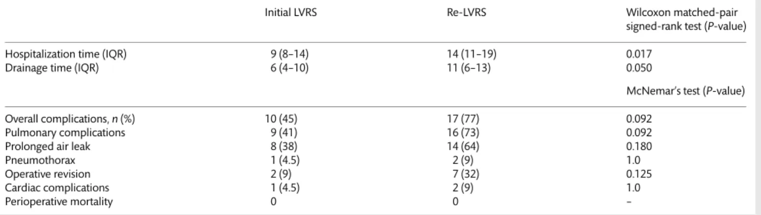
Table 3: Hospitalization specifics and perioperative morbidity and mortality for initial LVRS and Re-LVRS
Initial LVRS Re-LVRS Wilcoxon matched-pair
signed-rank test (P-value)
Hospitalization time (IQR) 9 (8–14) 14 (11–19) 0.017
Drainage time (IQR) 6 (4–10) 11 (6–13) 0.050
McNemar’s test (P-value)
Overall complications,n (%) 10 (45) 17 (77) 0.092
Pulmonary complications 9 (41) 16 (73) 0.092
Prolonged air leak 8 (38) 14 (64) 0.180
Pneumothorax 1 (4.5) 2 (9) 1.0
Operative revision 2 (9) 7 (32) 0.125
Cardiac complications 1 (4.5) 2 (9) 1.0
with initial LVRS (14 days, IQR: 11–19 vs 9 days, IQR: 8–14, P = 0.017). The overall complication rate was lower, though not
statistically significant, for LVRS compared with Re-LVRS (45 vs
77%, P = 0.092). Prolonged air leak accounted for the most
fre-quent cause of morbidity in both groups but did not reach
statis-tical significance (36 vs 64%, P = 0.092). Surgical revisions were
three times more frequent after Re-LVRS than after initial LVRS,
but did not reach statistical significance (32 vs 9% patients,
P = 0.125). Other morbidities are summarized in Table3.
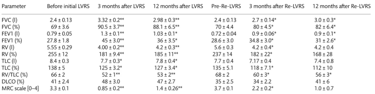
Levels of pulmonary function and subjective dyspnoea at the time of reoperation were similar to levels prior to the initial
oper-ation (Table 4). Pulmonary function tests and dyspnoea score
(MRC breathlessness scale) were assessed 3 and 12 months after
Re-LVRS, showing significant overall improvement of lung function
with improved FEV1 (Fig.1), improved diffusion capacity (Fig.2),
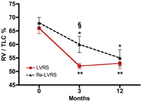
reduction in hyperinflation assessed by RV/TLC (Fig.3) as well as
reduced dyspnoea assessed by the MRC score (Fig.4) both after
initial and Re-LVRS (see Table4).
After Re-LVRS the mean increase in FEV1 was 25 and 12% from baseline (absolute increase of 7 and 3.2% of the predicted value corresponding to 0.18 and 0.18 l) at 3 months and 12 months, re-spectively, compared with 62 and 29% (absolute increase of 17.2 and 8.2% of the predicted value corresponding to 0.5 and 0.24 l)
after initial LVRS (also see Fig.1). The mean reduction in
pulmon-ary hyperinflation, assessed by the decrease in RV/TLC, was 8 and
13%, and 14 and 13% from the baseline at 3 and 12 months for Re-LVRS and initial LVRS, respectively. LVRS thereby showed a
more pronounced increase of FEV1 (P = 0.022), reduction of
hyperinflation assessed by RV/TLC (P = 0.008) as well as a lower
MRC score (P = 0.033) when compared with Re-LVRS at 3 months.
These differences were not detectable at 12 months.
DISCUSSION
In this retrospective study, we evaluated the safety, feasibility and effects of repeated LVRS (Re-LVRS) in 22 carefully selected patients.
The question of further treatment options after the beneficial
effects of an initially successful LVRS have vanished is raised fre-quently in clinical practice; even though LVRS seems to be broadly
underused in Europe as well as the USA, patient numbers are at
least stable [15]. As ongoing parenchymal destruction and the
natural course of the disease continue at individual rates, the time span towards deterioration to initial exercise ability varies between
patients and emphysema type [9], but positive effects may last
for up to 5 years. After this individual time span the patients
Table 4: Pulmonary function tests before and 3 and 12 months after initial LVRS and Re-LVRS
Parameter Before initial LVRS 3 months after LVRS 12 months after LVRS Pre-Re-LVRS 3 months after Re-LVRS 12 months after Re-LVRS FVC (l) 2.4 ± 0.13 3.32 ± 0.2** 2.98 ± 0.3** 2.4 ± 0.13 2.7 ± 0.14* 3.0 ± 0.3* FVC (%) 69 ± 3.6 90.5 ± 3.7** 88.1 ± 6.5** 70 ± 4.4 80 ± 4.5* 82 ± 6.4* FEV1 (l) 0.79 ± 0.05 1.3 ± 0.1** 1.03 ± 0.1* 0.72 ± 0.04 0.9 ± 0.06* 0.9 ± 0.1* FEV1 (%) 27.8 ± 1.8 45 ± 3.0** 36 ± 3.5* 28.6 ± 3.0 34.8 ± 3.0* 31 ± 2.6* RV (l) 5.55 ± 0.29 4.00 ± 0.2** 4.2 ± 0.3** 5.6 ± 0.3 4.2 ± 0.4* 4.2 ± 0.4 RV (%) 255 ± 12 181 ± 9.4** 185 ± 11** 237 ± 14 182 ± 22* 168 ± 28 TLC (l) 8.4 ± 0.3 7.7 ± 0.3* 7.8 ± 0.4* 7.7 ± 0.4 7.17 ± 0.4 7.4 ± 0.8 TLC (%) 138 ± 5 125 ± 3.2* 127 ± 3.4* 135 ± 5.1 118 ± 7.1* 112 ± 10 RV/TLC (%) 66 ± 2 52 ± 1** 53 ± 2** 68 ± 2 60 ± 3* 56 ± 3* DLCO (%) 41 ± 2.4 48 ± 3.0 47 ± 2.7 35 ± 2.5 34 ± 2.2 41 ± 6 MRC scale [0–4] 3.3 ± 0.1 0.85 ± 0.2** 1.4 ± 0.26** 3.7 ± 0.1 2.2 ± 0.2* 1.0 ± 0.7
DLCO: diffusion capacity of the lung for carbon monoxide; FEV1: forced expiratory volume in 1 s; FVC: forced vital capacity; RV: residual volume; TLC: total lung capacity
Mean ± standard error. *P < 0.05;
**P < 0.001 compared with preoperative values.
Figure 1:Course of FEV1 (% of predicted) preoperative (0), 3 and 12 months for initial LVRS and Re-LVRS. *P < 0.05, **P < 0.001 for 3 and 12 months compared with baseline.§P < 0.05 for LVRS vs Re-LVRS. FEV1: forced expiratory volume in
1 s.
Figure 2:Course of DLCO (% of predicted) preoperative (0), 3 and 12 months for initial LVRS and Re-LVRS. Changes at 3 and 12 months not significant com-pared with baseline.§P < 0.05 for LVRS vs Re-LVRS. DLCO: diffusion capacity of
the lung for carbon monoxide.
THO
R
A
C
re-experience the loss of physicalfitness and quality of life and, for those not eligible for transplantation, treatment options are very limited.
Re-LVRS has so far been reported in a case report as well as in
one small retrospective study; beneficial effects of Re-LVRS were
suggested in some patients [16,17]. However, high mortality and
morbidity were reported, particularly due to the high-risk popula-tion of end-stage emphysema patients in the setting of a reopera-tion, with a higher incidence of ARDS in these patients compared
with thefirst-time LVRS population [17].
In our cohort of 22 patients, the preoperative lung function values had decreased to levels similar to that before the initial LVRS, and repeated LVRS was performed after a median time of 60 months. Improvements in lung function after unilateral,
thora-coscopic Re-LVRS were statistically significant compared with
baseline and showed a similar extent at 12 months compared with
the initial LVRS. However, beneficial effects were more
pro-nounced after initial LVRS compared with Re-LVRS at 3 months.
The reduction in hyperinflation as well as subjective dyspnoea
was comparable with data published by Taccconi et al. [17].
Reoperations were technically feasible, and all but three were per-formed by VATS. Not surprisingly, overall morbidity was lower after the initial LVRS compared with Re-LVRS (operative revisions were necessary three times more frequently in the latter; also see
Table3), but these differences were not statistically significant. The
complication rate stayed within an acceptable range and was
comparable with international data after LVRS [3–6,15]. In contrast
to a previous report of Re-LVRS, we observed no perioperative mortality or ARDS in our cohort.
The 90-day mortality rate of 0% may be explained, to some extent, by rigorous selection of candidates and high surgical ex-pertise (all operations were performed by the same surgeon (Walter Weder) as well as anaesthesia and ICU management. Other important factors include optimal medical treatment as well as intensive physiotherapy and pulmonary rehabilitation
pro-grammes and a multidisciplinary approach [20].
Re-LVRS successfully treated the disabling dyspnoea in this population of 22 patients; however, the small number of patients and limited availability of complete data sets limit the validity of this study to some extent. Moreover, the retrospective design may have resulted in a selection bias. To shed more light on the
ques-tion of safety and efficacy, prospective analysis of a bigger patient
population in a prospective manner is needed. However, this small study proves that Re-LVRS is an option and should be con-sidered in selected patients.
While small numbers limit the validity of the study as a whole,
each patient represents their own control group;first, because they
had already proved to benefit from LVRS due to the specific
patho-physiological mechanisms of their disease, and secondly, because lung function had deteriorated to a pre-LVRS level despite adminis-tration of optimal medical treatment. A repeated gain in lung func-tion after Re-LVRS therefore may work by the same mechanisms as initial LVRS and might be superior to the best medical treatment in some patients. Hence, a previous LVRS should not be considered as a contraindication to perform Re-LVRS and patients should be selected according to the same criteria. For safety reasons and due
to the fact that DLCO was lower compared with thefirst
interven-tion, we decided to perform Re-LVRS unilaterally only.
End-stage emphysema remains an incurable disease and no
medical treatment has been shown to influence the course of the
disease; therefore the only definitive treatment for patients
previ-ously treated by LVRS is lung transplantation. However, consider-ing the limited donor availability and high mortality rates on the
waiting list [12], Re-LVRS can be considered as a bridge to
trans-plantation in younger patients and as afinal treatment in elderly
patients or those otherwise not eligible for transplantation.
In conclusion, ourfindings emphasize that Re-LVRS is safe to be
performed in carefully selected patients and may lead to signi
fi-cantly reduced dyspnoea and improved lung function for at least 12 months postoperatively. A previous LVRS should therefore not be considered a contraindication for LVRS, and the same patient selection criteria should be applied.
Funding
This work was supported by the following grants: Swiss National Foundation 3200-063709.00 and Zurich League and Sonnenwiese-Stiftung.
Conflict of interest: none declared.
REFERENCES
[1] Murray CJ, Lopez AD. Measuring the global burden of disease. N Engl J Med 2013;369:448–57.
[2] Minai OA, Benditt J, Martinez FJ. Natural history of emphysema. Proc Am Thorac Soc 2008;5:468–74.
Figure 4:MRC dyspnoea score for preoperative (0), 3 and 12 months for initial LVRS and Re-LVRS. *P < 0.05, **P < 0.001 for 3 and 12 months compared with baseline.§P < 0.05 for LVRS vs Re-LVRS. MRC score: Medical Research Council
score.
Figure 3:Course of RV/TLC (%) preoperative (0), 3 and 12 months for initial LVRS and Re-LVRS. *P < 0.05, **P < 0.001 for 3 and 12 months compared with baseline.§P < 0.05 for LVRS vs Re-LVRS. RV: residual volume; TLC: total lung
[3] Geddes D, Davies M, Koyama H, Hansell D, Pastorino U, Pepper Jet al. Effect of lung-volume-reduction surgery in patients with severe emphyse-ma. N Engl J Med 2000;343:239–45.
[4] Miller JD, Malthaner RA, Goldsmith CH, Goeree R, Higgins D, Cox PG et al. A randomized clinical trial of lung volume reduction surgery versus best medical care for patients with advanced emphysema: a two-year study from Canada. Ann Thorac Surg 2006;81:314–20; discussion 20–1.
[5] Wilkens H, Demertzis S, Konig J, Leitnaker CK, Schafers HJ, Sybrecht GW. Lung volume reduction surgery versus conservative treatment in severe emphysema. Eur Respir J 2000;16:1043–9.
[6] Fishman A, Martinez F, Naunheim K, Piantadosi S, Wise R, Ries Aet al. A randomized trial comparing lung-volume-reduction surgery with medical therapy for severe emphysema. N Engl J Med 2003;348:2059–73. [7] Russi EW, Stammberger U, Weder W. Lung volume reduction surgery for
emphysema. Eur Respir J 1997;10:208–18.
[8] Naunheim KS, Wood DE, Mohsenifar Z, Sternberg AL, Criner GJ, DeCamp MM et al. Long-term follow-up of patients receiving lung-volume-reduction surgery versus medical therapy for severe emphysema by the National Emphysema Treatment Trial Research Group. Ann Thorac Surg 2006;82:431–43 e19.
[9] Bloch KE, Georgescu CL, Russi EW, Weder W. Gain and subsequent loss of lung function after lung volume reduction surgery in cases of severe em-physema with different morphologic patterns. J Thorac Cardiovasc Surg 2002;123:845–54.
[10] Shigemura N, Gilbert S, Bhama JK, Crespo MM, Zaldonis D, Pilewski JMet al. Lung transplantation after lung volume reduction surgery. Transplantation 2013;96:421–5.
[11] Tutic M, Lardinois D, Imfeld S, Korom S, Boehler A, Speich Ret al. Lung-volume reduction surgery as an alternative or bridging procedure to lung transplantation. Ann Thorac Surg 2006;82:208–13; discussion 13.
[12] Backhus L, Sargent J, Cheng A, Zeliadt S, Wood D, Mulligan M. Outcomes in lung transplantation after previous lung volume reduction surgery in a contemporary cohort. J Thorac Cardiovasc Surg 2014;147: 1678–83 e1.
[13] Criner GJ. Alternatives to lung transplantation: lung volume reduction for COPD. Clin Chest Med 2011;32:379–97.
[14] Gaissert HA, Trulock EP, Cooper JD, Sundaresan RS, Patterson GA. Comparison of early functional results after volume reduction or lung transplantation for chronic obstructive pulmonary disease. J Thorac Cardiovasc Surg 1996;111:296–306; discussion 06–7.
[15] Decker MR, Leverson GE, Jaoude WA, Maloney JD. Lung volume reduction surgery since the National Emphysema Treatment Trial: Study of Society of Thoracic Surgeons Database. J Thorac Cardiovasc Surg 2014;148:2651–8.e1.
[16] Stammberger U, Thurnheer R, Schmid RA, Russi EW, Weder W. Redo lung volume reduction surgery in a patient with alpha1-antitrypsin deficiency. Ann Thorac Surg 2000;69:632–3.
[17] Tacconi F, Pompeo E, Forcella D, Marino M, Varvaras D, Mineo TC. Lung volume reduction reoperations. Ann Thorac Surg 2008;85:1171–7. [18] Bestall JC, Paul EA, Garrod R, Garnham R, Jones PW, Wedzicha JA.
Usefulness of the Medical Research Council (MRC) dyspnoea scale as a measure of disability in patients with chronic obstructive pulmonary disease. Thorax 1999;54:581–6.
[19] Weder W, Thurnheer R, Stammberger U, Burge M, Russi EW, Bloch KE. Radiologic emphysema morphology is associated with outcome after sur-gical lung volume reduction. Ann Thorac Surg 1997;64:313–9; discussion 19–20.
[20] Rathinam S, Oey I, Steiner M, Spyt T, Morgan MD, Waller DA. The role of the emphysema multidisciplinary team in a successful lung volume reduction surgery programme. Eur J Cardiothorac Surg 2014;46:1021–6; discussion 1026. THO R A C IC