1306
Patterns of Proinflammatory Cytokines and Inhibitors during Typhoid Fever
Monique Keuter, Edi Dharmana, M. Hussein Gasem, Johanna van der Ven-Jongekrijg,
Robert Djokomoeljanto, Wil M. V. Dolmans, Pierre Demacker, Robert Sauerwein, Harald Gallati, and Jos W. M. van der Meer
University Hospital, Nijmegen. Netherlands; Diponegoro University, Semarang. Indonesia;F.Hoffman-Lakoche, Basel, Switzerland
Cytokines and inhibitors in plasma were measured in 44 patients with typhoid fever. Ex vivo production of the cytokines was analyzed in a whole blood culture system with and without lipopolysaccharide (LPS). Acute phase circulating concentrations of cytokines (±SD) were as follows: interleukin (IL)-IP, <140pg/ml.;tumor necrosis factor-a (TNFa), 130 ± 50 pg/mL; IL-6, 96± 131pg/ml.;and IL-8, 278±293pg/ml.,Circulating inhibitors were elevated in the acute phase: IL-l receptor antagonist (IL-IRA) was 2304 ± 1427pg/ml, and soluble TNF receptors 55 and 75 were 4973± 2644 pgJmL and 22,865 ± 15,143 pgJmL, respectively. LPS-stimulated production of cytokines was lower during the acute phase than during convalescence (mean values: IL-IP, 2547 vs. 6576pg/ml.;TNFa,2609 vs. 6338pg/rnl.;IL-6, 2416 vs. 7713 pg/ml.),LPS-stimulated production orIL-iRA was higher in the acute than during the convales-cent phase (5608 vs. 3977 pg/mL). Inhibited production of cytokines during the acute phase may
bedue to a switch from a proinflammatory to an antiinflammatory mode.
Typhoid fever is caused by the facultative intracellular gram-negative bacillus Salmonella typhi and occasionally by Salmonella para typh i. Although salmonellae contain lipo-polysaccharide (LPS; bacterial endotoxin), the clinical pic-ture of typhoid fever differs from gram-negative sepsis, and the role of endotoxin in the pathophysiology of typhoid fever is controversial
[I].
The proinflammatory cytokines interleukin (IL)-I,8, tu-mor necrosis factor-a (TNFa; cachectin), IL-6, and IL-8 have been implicated in the pathogenesis of sepsis caused by gram-negative microorganisms [2-4]. When LPS is injected intravenously into animals or human volunteers, elevated concentrations of these cytokines can be detected, and the symptoms and signs of sepsis are mimicked [5-7]. Elevated circulating levels ofTNFa have been correlated with poor prognosis in sepsis, meningococcemia, and cerebral malaria [7-10]. In contrast, in infections with intracellular patho-gens, such as Leishmania species, Listeria monocytogenes, or mycobacteria, administration of TNFa inhibits the out-growth of the microorganisms, whereas administration of
an-Received 13 September 1993; revised 12 January 1994.
Presented in part: Third International Workshop on Cytokines, Sienna, Italy, 10-14 November 1991.
Patients gave informed consent, and guidelines for human experimenta-tion of the Dr. Kariadi Hospital, Diponegoro University, were followed.
Grant support: Scientific Research in the Tropics. Netherlands Founda-tion for Science.
Reprints or correspondence: Dr. Monique Keuter, University Hospital, Dept. of Internal Medicine. P.O. Box 910 I, 6500 HB Nijmegen, Nether-lands.
The Journal of Infectious Diseases 1994;169:1306-11 © 1994 by The University of Chicago. All rights reserved. 0022-1899/94/6906-0017$01.00
tibodies to this cytokine are detrimental [11-16], In experi-mental Salmonella typhimurium infection in mice. the role ofTNFa is similar to that in other intracellular infections [17-19). However, in calves with S. typhimurium sepsis, the cytokine pattern appears to differ from that seen after intrave-nously administered LPS. Where TNFarose 1 h after LPS administration, salmonella sepsis caused a barely detectable increase in TNFa[20].
In contrast to these animal studies, circulating cytokines
(TNFa, IL-6, and IL-l,8) were elevated in children with ty-phoid fever in Chile (21). Butler et al. [22] studied the out-come of typhoid fever in adults in Nepal and found that higher values of IL-6 and soluble TNF receptor p55 were related to poorer outcome.
In 1989, joint research on several aspects of typhoid fever was started between Nijmegen University and Diponegoro University. To obtain more insight into the pathophysiology of typhoid fever, we measured levels of circulating pyrogenic cytokines (IL-l,8, TNFa, TNF,8 [lymphotoxin], and IL-6) and concentrations of IL-8, the cytokine inhibitor IL-l re-ceptor antagonist (IL-l RA), and the soluble TNF rere-ceptors p55 and p75 (sTNF-R). In addition, we investigated the ca-pacity of blood cells to produce IL-l,8,TNFa,6, and IL-IRA ex vivo in the acute and convalescent phases of hospital-ized patients with typhoid fever.
We used the whole blood cytokine test as described by van Deuren et al. [23] and Nerad et al. [24]. This assay is simple, reproducible, and especially suitable fOT use in laboratories that are not particularly well equipped for work with cyto-kines. In addition, the method may be less artificial than is isolating mononuclear cells over a gradient and probably is a more natural mirror ofwhat happens in vivo, because plasma factors and other cells are left in situ.
JlD 1994;1 69(June) Cytokines in Typhoid Fever 1307
Table1. Characteristics of 44 hospitalized culture-proven ty-phoid fever patients.
The study was done in Dr. Kariadi Hospital, Diponegoro Uni-versity, Sernarang, Indonesia, beginning in December 1990. Blood and bone marrow cultures were done for all adult patients (> 14 years old) hospitalized with suspected typhoid fever (de-fined as fever >38.5°C and at least one of the following signs: relative bradycardia, abdominal complaints, mental changes, signs of complicated typhoid fever, enlarged liver or spleen and no apparent other disease). If blood or bone marrow cultures were positive for S.typhi or S. paratyphi A or patients were found to have perforated ilea at surgical exploration, typhoid fever was considered proven.
A total of 44 patients were studied. Patient characteristics are shown in table I. Complications of typhoid fever were defined as gastrointestinal bleeding, intestinal perforation, shock, delir-ium, stupor or coma, pneumonia, or diffuse intravascular coagu-lation.
Treatment consisted of chloramphenicol (40 mg/kg/day orally) if leukocyte counts were > 2 X 10⁹/ L.
If fever did not subside within 6 days, treatment was changed to sulfamethoxa-zole (800 mg) and trimethoprim (160 mg) twice daily or ampi-cillin (I g four times daily). Surgical patients received ampicil-lin, metronidazole, and gentamicin during and after surgery. No cyclooxygenase inhibitors were given. Only 2 patients received a single dose of 120 mg of dexamethasone, but not before blood was obtained for cytokine measurement. Most patients were dis-charged 7-10 days after defervescence, which we defined as con-valescence. No patients died.
Cytokine measurements. On admission and during convales-cence, blood was drawn for cytokine measurement. Venous blood samples were aseptically collected into sterile 4-mL tubes (Vacutainer; Becton Dickinson, Rutherford, NJ) containing EDTA. Unless stated otherwise, a total of 3 tubes of blood was drawn from each patient [23). To each tube, 250 JLL ofaprotinin (Trasylol 2500 kallikreine inactivating units [KIU]; Bayer, Le-verkusen, Germany; final concentration 625 KIU/mL) was added through the stopper by a tuberculin needle and syringe. One tube was centrifuged directly (1250g, 10 min), platelets from the supernatant plasma were removed by a second
centrifu-Patients and Methods
Characteristic No. of patients Mean age (range), years Males/females Median (range) of
days with fever before admission Leukocyte countat admission (range) Complications Pneumonia Delirium Perforation Bleeding Complicated disease 16 20 (14-34) 7/9 10.0 (4-20) 7.3 X 10⁹/L (2.6-37.0) 16 10 2 5 1 Uncomplicated disease 28 24 (14-60) 12/16 8.5(4-30) 4.4 X I09/L (1.6-7.4)
gation ( I 5,000 g, I min), and plasma was collected and stored at -20°C until assayed for cytokines. To one of the two remaining tubes, 50 JLL of LPS(Escherichia coliserotype 055:B5; Sigma, St. Louis; final concentration 10 JLg/mL) was added to stimulate cytokine production. Unstimulated samples contained apro-tinin only (no LPS). Both tubes were incubated at 37°C for 24 h. For 17 (random) patients, a fourth 4-mL tube of blood was obtained in the acute phase into which indomethacin was added (0.5 JLg/mL final concentration). For 26 (random) patients, we obtained a total of six tubes of blood. From two of these, we removed the plasma, replacing it with a like amount of PBS.
TNFawas determined by an RIA (detection level 100 pg/
ml.),described in [25). NormalTNFavalues for our laboratory (circulating concentrations and ex vivo production without LPS below the detection limit and ex vivo production after 24 h of stimulation with LPS) are 3780 ± 950pg/ml.,
IL-I,Bwas measured by RIA according to the method of Lisi et al. [26] but without chloroform extraction (detection level 140 pg/mL). Normal values for our laboratory (circulating con-centrations and ex vivo production without LPS below the de-tection limit and ex vivo production after 24 h of stimulation with LPS) are 6930 ±3160 pg/ml.,
IL-6 was measured by an ELISA as described (detection level, 20 pg/ml.) [27). Normal values for our laboratory (circulating concentrations and ex vivo production without LPS) were below the detection limit.
IL-8 was measured by ELISA quantikine (R & D Systems, Europe, Abingdon, UK). The detection limit was 45 pg/mL, and normal values were below the detection limit. We sought
TNF~(lymphotoxin) by ELISA quantikine (R & D Systems, Europe) but failed to detect any. IL-IRA was determined by an RIA according to the method of Poutsiaka et al. [28] (detection level 300 pg/mL). Normal values for our laboratory (circulating concentrations and ex vivo production without LPS below the detection limit, ex vivo production after 24 h stimulation with LPS) were 5757 ± 1060pg/ml.,
sTNF-R was measured by an enzyme-linked immunobinding assay (Hoffman-La Roche, Basel, Switzerland; detection level, 80 and 300 pg/mL for p55 and p75, respectively). Normal val-ues for circulating concentrations are 1.50 ng/rnl, (p55) and 2.51 ng/ml, (p75). All samples from the same patient were ana-lyzed in the same run in duplicatetominimize analytical errors. Statistics. When frequency distribution was parametric, we used paired and unpaired Student'sttest. When not parametric, Wilcoxon signed-rank test or Mann-Whitney U test was used. P
<.05 was considered significant.
Results
Circulating cytokines and inhibitors during acute and conva-lescent phases oftyphoid fever. Coneen trations of pyrogenic cytokines during the acute phase (IL-l,B, IL-6, TNFa, lym-photoxin) are shown in figure I. IL-I,Bconcentrations were below the detection limit in both acute and convalescent phases. IL-6 concentrations ranged from undetectable «20 pg/mL) to 600 pgjmL (median, 73). TNFaconcentrations ranged from below the detection limit to 310 pgjmL
(me-1308 Keuter et al. JID 1994;169(June)
Figure 1. Circulating concentrations of pyrogenic cytokines in-terleukin (Il,)-1{J,tumor necrosis factor (TNF)-a,Il-6, and IL-8 in patients during acute phase of typhoid fever. Patients had been ill
>I week. Horizontal continuous circles= detection limit; horizon-tal bars= median values. In comparison with normal values, IL-6, Il-8, and TNFa are slightly elevated.
600 0
E
0 Ol a. 400 0 0 0 0 0 200.L.
0 0 0 CiiXhi&LiiJJiJU 8-t-
e
8 0IL-6 TNF IL-1 B IL-B
dian, 110) in the acute phase and to 300 pg/ml,(median, below detection limit) during the convalescent phase. All lymphotoxin concentrations were below detection limits during acute and convalescent phases. IL-8 concentrations were detectable in the acute phase (median, 145; range, 47-998 pg/mL) but lower during the convalescent phase (me-dian, 46; range, 46-180 pg/mL).
Inhibitors such as IL-I RA and sTNF-R (p55 and p75) were significantly higher in the acute phase than during the convalescent phase: IL-IRA, 2304 ± 1427 pg/mL versus 469 ± 324pg/ml.;sTNF-R55, 4973± 2644 pg/mL versus 1671 ± 532 pg/mL; and sTNF-R75, 22,865
±
15,143 pg/ mL versus 5971 ±2750 pg/mL (figure 2). 1000 BOO o oCirculating cytokines and inhibitors in complicated and un-complicated disease. No differences were found in circulat-ing cytokines or inhibitors between the 16 patients with com-plicated and the 28 patients with uncomcom-plicated disease courses.
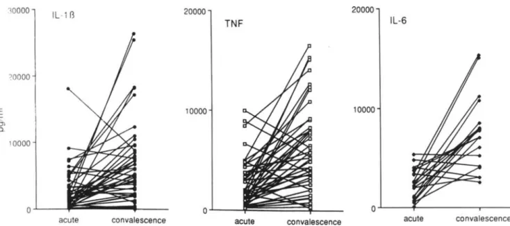
Ex vivo production ofcytokines and inhibitors during acute and convalescent phases. Unstimulated whole blood cul-tures did not have detectable IL-II3, TNFa, or IL-6 (not shown). After incubation with LPS for 24h,the supernatants contained detectable amounts of these cytokines, which were significantly lower in the acute phase than in convales-cence (IL-II3: 2547 ± 3319 vs. 6576 ± 6275 pg/ml., P< .00 I; TNFa: 2609
±
2443 vs. 6338±
4366 pg/mL, P<
.001; IL-6: 2416 ± 1531 vs. 7713 ± 3809pg/rnl.,
P= .01; figure 3).In the acute phase, there was a correlation between the LPS-stimulated production of IL-113 and TN Fa(r
=
.664), IL-l{j and IL-6(r=
.531), and TNFa and IL-6(r= .434). Such correlations were not found during convalescence.When indomethacin was added to the stimulated samples, the concentrations of TNFa and IL-I{j in the acute phase were not different from those without indomethacin (2859 ±
2630 vs. 2609 ± 2443 and 2782
±
2821 vs. 2547±
3319 pg/rnl,for TNFa and IL-II3, with and without indometha-cin, respectively). Also, removal of plasma and replacement with PBS did not change the stimulated production of TN Fa and IL-l {j in the acute phase (TNFa: 3307 ± 3920pg/ml.;u-is.
2244± 2512 pg/mL).The IL-l RA concentrations found in unstimulated cul-tures (not shown) were similar to those during the acute phase. However, the LPS-stimulated production of IL-l RA was high and reached significantly higher concentrations in the acute phase of the disease (5608 ± 1832 pg/mL) than during convalescence (3977± 1974 pg/mL;P
<
.05). sTNF-R, IL-8, and lymphotoxin were not generated in the cultures. Ex vivo production ofcytokines and inhibitors during com-plicated and uncomcom-plicated courses of disease. In the acuteconvalescent
sTNF-R p75
12sTNF-R p55
ED 10 00 8 6 40 4 ID 2 0 0convalescent acute convalescent acute
acute
Figure 2. Individual concentrations ofcirculating interleukin (Il)-l receptor antagonist(ra)and tumor necrosis factor (sTNF-R) soluble receptors 55 and 75 in patients during acute and convalescent phases of typhoid fever. Inhibitors like Il-lra and sTNF-R (p55 and p75) were significantly higher in acute phase than during convalescent phase.
JID 1994;169 (June) Cytokinesin TyphoidFever 1309 10000 IL-113 20000 TNF 20000 IL-6
o
. . l . - - - ' f - - - -- - r -- -convalescence acute 10000 convalescence acute convalescence acute ~o oooE
0> Q. '0000Figure 3. Individual production capacityof interleukin (IL)-ll3.tumor necrosisfactor(TNF)-a.and IL-6 in acute and during convales-cent phasesof typhoid fever. After incubationwith lipopolysaccharide for 24 h.supernatants contained detectable amounts of cytokines, which were significantly lower in acute phase than during convalescence.
phase, patients with complicated disease had significantly less IL-I{j production after ex vivo stimulation with LPS (1341 ± 1373
vs
,
6563± 1342pg/ml.;
P<.005) and a trend towards lowerTNFa production (1650± 1407 vs. 3064 ± 2770 pg/mL; P=
.06).Such differences were not found for the production of IL-I RA or IL-6. During the convalescent phase. the ex vivo-stimulated production of IL-I(j and TNFa did not differ for complicated and uncomplicated casesof disease.Discussion
In this study we found several signs of cytokine activation during typhoid fever. The concen trations ofcirculating inhib-itors such as IL-IRA and sTNF-R were high in the acute phase of the disease. IL-I RA is known not to be detectable in the circulation of normal subjects. Normal values for sTNF-Rare 1.50 ng/mL (p55) and 2.5I ng/mL (p75) [29. 30). We also found that the production capacity of pyrogenic cyto-kines in whole blood is depressed in the acute phase of ty-phoid fever but is restored during the convalescent phase. Although the patients in our study usually had severe ty-phoid fever, we found that those with complicated disease courses had significantly lower proinflammatory cytokine production capacity than did those with uncomplicated dis-ease.
A low production capacity of cytokines has been found in other serious conditions. such as severe postoperative infec-tion [31). sepsis [32-34), and attacks of familial Mediterran-ean fever [35. 36). In these reports. all investigators used isolated peripheral blood mononuclear cells or tissue
macro-phages. We have previously found depressed cytokine pro-duction capacity in the whole blood culture system during the acute phase of meningococcal disease and during
Pneu-mocystis carinii infection (unpublished data). From these studies and the work presented here, we conclude that the depressed cytokine production capacity is not a consequence offewer white blood cells during the acute phase ofthe infec-tion, since we found no correlation between leukocyte count and cytokine production (table I).
Many investigators [31-36) have interpreted the finding of low cytokine production capacity as exhaustion of cytokine -producing cells, whichcould be a consequence of exposure in vivo to stimuli such as endotoxin. Our finding that IL-IRA is produced in high concentrationsargues against such a hypothesis and also rules out the possibility that the de-creased production of proinflammatory cytokines is due to an enhanced lysis of producing cells or to increased inactiva-tion of LPS by lipids in the acute phase.Although we have been unable to demonstrate that the proinflammatory cyto-kines and IL-I RA are produced by thesame kind ofcells, we hypothesize that after the initial phase of infection. cytokine-producing cells switch from a balanced proinflammatory to an antiinflammatory repertoire. Our findings that patients in the acute phase of typhoid fever have high concentration of soluble sTNF-Rs in their blood is in agreement with this notion.
Since our cultures used whole blood, we investigated whether some common circulating factor could be responsi-ble for the correlated low production capacity of the cyto-kines IL-I{j,TNFa,and IL-6 in the acute phase. Cyclooxy-genase products. such as prostaglandin E2 • which inhibit
1310 Keuter et al. JIO 1994;169 (June)
production of IL-l and TNFa [37], were not responsible since addition of indomethacin to the whole blood cultures did not lead to significant changes in cytokine production. Likewise, removal of plasma and addition of saline before incubation did not overcome the suppression in the acute phase of the disease.
Itis possible that exposure in vivo to other inhibitory fac-tors will explain the low cytokine production capacity. Schindler et al. [38] demonstrated that exposure of isolated mononuclear cells to IL-6 inhibits the production of IL-l andTNFa. In the present study, we found no correlation between IL-6 concentrations in plasma and the magnitude of the production ofIL-l {1 and TNF(r
=
.041 and .035, respec-tively). Exposure to other cytokines such as, IL-4, IL-l 0, and transforming growth factor-S could, however, play a role. Vannier et al. [39] have provided evidence that exposure of cells to IL-4 suppresses the IL-l production but up-regulates the synthesis of IL-I RA.With few exceptions, patients with typhoid fever have a continuous fever. Hence, pyrogenic cytokines would be ex-pected to be present in the circulation during the acute phase of the disease. In our series of febrile patients with typhoid fever, we could not detect appreciable concentrations of the pyrogenic cytokines IL-l {1, TNFa, and lymphotoxin. The concentrations of IL-6, generally considered a relatively weak pyrogen [40], were low compared to findings with other febrile conditions [3, 41]. We did detect elevated con-centrations of IL-8, but this cytokine is considered nonpyro-genic [42].
Thus, the question of which pyrogens are responsible for the continuous fever in typhoid fever remains unanswered.
Acknowledgment
We thank James Vannice (Synergen, Boulder, CO) for sup-plying reagents for IL-l RA measurements.
References
I. Greisman SE, Hornick RB. Wagner HN Jr, Woodward WE, Wood-ward TE. The role of endotoxin during typhoid fever and tularemia in man. IV. The integrity of the endotoxin tolerance mechanisms during infection. 1 Clin Invest 1969;48:613-29.
2. Cannon lG. Tompkins RG, Gelfland JA, et al. Circulating interleukin-I and tumor necrosis factor in septic shock and experimental fever. J Infect Dis 1990; 161:79-84.
3. Hack CE, De Groot ER. Felt-Bersma R1F. et al. Increased plasma levels of interleukin-6 in sepsis. Blood 1989;74: 1704-10. 4. Mitchie HR, Spriggs DR, Manogue KR, et al. Tumor necrosis factor
and endotoxin induce similar metabolic responses in human beings. Surgery 1988; 104:280-5.
5. Okusawa S, Gelfland JA. Ikejima T, Connolly Rl, Dinarello CA.
Inter-Ieukin-I induces a shock-like state in rabbits. Synergism with tumor necrosis factor and the effect of cyclooxygenase inhibition. J Clin Invest 1988;81: 1162-72.
6. Fischer E, Marano MA. Barber AE, et al. Comparison between effects of interleukin-Iaadministration and sublethal endotoxemia in pri-mates. Am J Physiol 1991;261:R442-52.
7. Calandra T, Baumgartner lD. Grau GE. et al. Prognostic values of tumor necrosis factor/cachectin, interleukl , interferon-a. and in-terferon-v in the serum of patients with septic shock. J Infect Dis 1990; 161:982-7.
8. Damas P, Reuter A, Gysen P, Demonty J, Lamy M. Franchimont P. Tumor necrosis factor and interleukin-I serum levels during severe sepsis in humans. Crit Care Med 1989; 17:975-8.
9. Waage A. Brandtzaeg P, Halstensen A, Kierulf'P, Espevik T. The com-plex pattern of cytokines in serum from patients with meningococcal septic shock. Association between interleukin-o, interleukin-l , and fatal outcome. 1 Exp Med 1989; 169:333-8.
10. Grau GE. Fajardo LF. Piguet PF. Allet B, Lambert PH. Vassalli P. Tumor necrosis factor (cachectin) as an essential mediator in murine cerebral malaria. Science 1987;237: 1210-2.
II. Barnes PF. Chatterjee D, Brennan PJ, Rea TH, Modlin RL. Tumor necrosis factor production in patients with leprosy. Infect Immun 1992;60: 1441-6.
12. Havell EA. Production of tumor necrosis factor during murine listerio-sis. J Immunol 1987; 139:4225-31.
13. Liew FY, ParkinsonC.Millot S, Severn A, Carrier M. Tumor necrosis factor (TN F) in leishmaniasis I. TNF mediates host protection against cutaneous leishmaniasis. Immunology 1990;69:570-3. 14. Nakane A, Minagawa T. Kato K. Endogenous tumor necrosis factor
(cachectin) is essential to host resistance against Listeria
monocyte-genes infection. Infect Immun 1988;56:2563-9.
15. Silva CL, Foss NT. Tumor necrosis factor in leprosy patients. 1 Infect Dis 1989; 159:787-90.
16. Titus RG, SherryB,Cerami A. Tumor necrosis factor plays a protective role in experimental murine cutaneous leishmaniasis. 1 Exp Med 1989; 170:2097-104.
17. Nakano Y, Onuzuka K. Terada Y. Shinomiya H, Nakano M. Protective effect of recombinant tumor necrosis factor-a in murine salmonello-sis. J Immunol 1990; 144: 1935-41.
18. NaucielC.Espinasse-Maes F. Role of')' interferon and tumor necrosis factor-a in resistance to Salmonella typhimurium infection. Infect Immun 1992;60:450-4.
19. Tite lP. Dougan G. Chatfield SN. The involvement of tumor necrosis factor in immunity to Salmonella infection. J Immunol 1991;147:3161-4.
20. Peelrt,Voirol Ml. KollyC.Gobet D. Martinod S. Induction ofcircu-lating tumor necrosis factor cannot be demonstrated during septice-mic salmonellosis in calves. Infect Immun 1990;58:439-42. 21. RoineI.Herrera P. Ledermann W, Peltola H. Tumor necrosis
factor-alfa (TNF-a). interleukin-l-beta (IL-IfJ)and interleukin-6 (IL-6) lev-els in typhoid fever (TF) [abstract 299]. In: Program and abstracts of the 30th Interscience Conference on Antimicrobial Agents and Che-motherapy (Atlanta). Washington. DC American Society for Micro-biology. 1990.
22. Butler T, Ho M. Acharya G. Tiwari M, Gallati H. Interleukin-S, gamma interferon and tumor necrosis factor receptors in typhoid fever re-lated to outcome of antimicrobial therapy. Antimicrob Agents Che-mother 1993;37:2418-21.
23. van Deuren M, van der Ven-longekrijg J. Keuter M. Demacker PNM. van der Meer lWM. Cytokine production in whole blood cultures. J Int Fed Clin Chern 1993;5:216-21.
(IL-JID 1994;169 (June) Cytokines in Typhoid Fever 1311
1,6), IL-I receptor antagonist and TNFa production in whole blood. J Leukoc Bioi 1992;52:687-92.
25. van der Meer JWM, Endres S, Lonnemann G, et al. Concentrations of immunoreactive human tumor necrosis factor alpha produced by human mononuclear cells in vitro. J Leukoc Bioi 1988;43:216-23. 26. Lisi PJ, Chu CW, Koch GA. Endres S, Lonnemann G, Dinarello CA. Development and use of radioimmunoassay for human interleukin-1,6. Lymphokine Res 1987;6:229-44.
27. Barrera P, Boerbooms AMT, Janssen EM, et al. Circulating soluble TNF receptors and interleukin-2 receptors, tumor necrosis factor-a and interleukin-6 in rheumatoid arthritis. Longitudinal evaluation during methotrexate and azathioprine therapy. Arthritis Rheuma 1993;36: 1072-9.
28. Poutsiaka DD, Clark BD, Vannier E, Dinarello CA. Production of in-terleukin-I receptor antagonist and inin-terleukin-Iri by peripheral blood mononuclear cells is differentially regulated. Blood 1991 ;78: 1275-81.
29. Dinarello CA. Interleukin-I and interleukin-I antagonism. Blood 1991;77: 1627-52.
30. ShapiroL.Clark BD, Orencole SF. Poutsiaka DD. Granowitz EV. Din-arello CA. Detection of tumor necrosis factor soluble receptor p55 in blood samples from healthy and endotoxemic humans. J Infect Dis 1993; 167: 1344-50.
31. Luger A, Graf H, Schwarz HP. Stummvoll HK, Luger TA. Decreased serum interleukin I activity and monocyte interleukin I production in patients with fatal sepsis. Crit Care Med 1986; 14:458-61. 32. Simpson SQ, Modi H, Balk RA. Bone RC, Casey LC. Reduced alveolar
macrophage production of tumor necrosis factor during sepsis in mice and man. Crit Care Med 1991; 19:1060-6.
33. SrugoI.Berger A, Lapidot Z. Katz R, Pollak S. Interleukin-I secretion by blood monocytes of septic premature infants. Infection 1991J:150-4.
34. Helminen M. Interleukin-l production from peripheral blood mono-cytes in septic infections in children. Scand J Infect Dis 1991;23:607-11.
35. Rozenbaum M. Katz R, Rozner I. Pollack S. Decreased interleukin I activity released from circulating monocytes of patients with familial Mediterranean fever during in vitro stimulation by lipopolysaccha-ride. J Rheumatol 1992; 19:416-8.
36. Schattner A, Lachmi M, Livneh A, Pras M, Hahn T. Tumor necrosis factor in familial Mediterranean fever. Am J Med 1991;90:434-8. 37. Endres S. Cannon JG, Ghorbani R, et al. In vitro production ofIL-l,6,
IL-I a, TNFa and IL-2: distribution, effect of cyclooxygenase inhibi-tion and evidence of independent gene regulainhibi-tion. Eur J Immunol 1989;19:2327-33.
38. Schindler R. Mancilla J, Endres S. et al. Correlations and interactions in the production of interleukin-6 (IL-6), interleukin-I (IL-I) and tumor necrosis factor (TNF) in human blood mononuclear cells: IL-6 suppresses IL-I and TNF. Blood 1990;75:40-7.
39. Vannier E, Miller LC, Dinarello CA. Coordinated antiinflammatory effects of interleukin-4: interleukin-4 suppresses interleukin-I pro-duction but upregulates gene expression and synthesis ofinterleukin-I receptor antagonist. Proc Nat! Acad Sci USA 1992;89:4076-80. 40. Dinarello CA, Cannon JG. Mancilla J. BishaiI.Lees J, Coceani F.
Interleukin-6 as an endogenous pyrogen: induction of prostaglandin £2 in brain but not in peripheral blood mononuclear cells. Brain Res 1991 ;562: 199-206.
41. Waage A. Brandtzaeg p. Halstensen A. KierulfP, EspevikT.The com-plex pattern of cytokines in serum from patients with meningococcal septic shock. Association between interleukin-6, interleukin-I and fatal outcome. J Exp Med 1989; 169:333-8.
42. Van Damme J. Interleukin-8 and related molecules. In: Thomson AW. ed. The cytokine handbook. London: Academic Press. 1991.