Clin Rheumatol (1998) 17:62-64
9 1998 Clinical Rheumatology
Clinical
Rheumatology
Case Report
Computed Tomographic Imaging of Subcutaneous Gouty Tophi
J. C. Gerster 1, M. L a n d r y 2 and G. R i v i e r 11Service de Rhumatologie, M6decine Physique et R6habilitation and 2Service de Radiodiagnostic, CHUV, Lausanne, Switzerland
Introduction
In rheumatological practice, the differential diagnosis of subcutaneous nodules includes mainly rheumatoid nodules, gouty tophi and, exceptionally, the tophus of cholesterol [1] as well as the tophus in calcium pyrophosphate dihydrate (CPPD) crystal deposition disease [2]. The purpose of this study, involving two cases of chronic tophaceous gout, is to assess the usefulness of computed tomography (CT) imaging of subcutaneous tophi. Indeed, we have previously demon- strated that CT can reveal, within the knee joints of patients with gout, the presence of opacities that were considered to be MSU deposits [3]. We have previously measured the density of 20 opacities in nine knee joints: the mean ( • 2SD) value was 170 _+ 30 Hounsfield units (HU), which was in the same range as that of tophi in vitro [3].
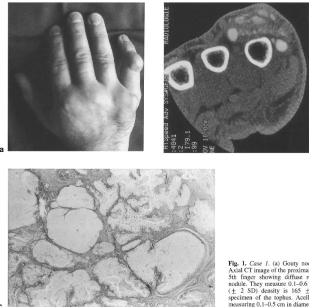
tender; subcutaneous tophi were found bilaterally over the olecranon processes and Achilles tendons and on the right little finger (Fig. la). The skin above the tophi was of normal colour and was not ulcerated. Plain radio- graphs failed to reveal bony erosions or opacities within the nodules of the hand. Helical CT imaging was performed with a GE scanner (9800 high-speed advantage) using 120 kV, 140 mA and 1 mm collimation (table speed 1 mm/s, pitch 1). Round and oval opacities were seen, measuring 2-6 mm in diameter (Fig. lb); the mean ( + 2SD) density of these opacities was 165 _+ 40 HU, i.e. in the same range as that found in the knee joints [3]. Histological examination of the tophus, which was surgically resected, showed birefringent MSU crystal deposits embedded in fibrous tissues (Fig. lc), whose diameter was 2 - 5 ram.
Case Reports
Case 1
A 42-year-old man had been suffering from tophaceous gout for 15 years. He was treated irregularly with antihyperuricaemic drugs. The diagnosis of gout was confirmed by several analyses of synovial fluid revealing negatively birefringent crystals. Except for chronic alcohol abuse and obesity, there were no other co- morbid conditions. On clinical examination in 1996, both knee and ankle joints were slightly swollen and
Correspondence and offprint requests to: J. C. Gerster, Service de
Rhumatologie, CHUV, 1011 Lausanne, Switzerland. Tel: 21-314 41 76; Fax: 21-314 14 71.
Case 2
A 70-year-old man with a 10-year history of gout (co- morbid conditions were also obesity and alcohol abuse) was admitted to our department for acute arthritis of the left ankle. A few days later, he had a severe attack of gout of the right wrist; the skin above the affected joint was hot and red and the signs of inflammation seen on the entire forearm resembled those of bacterial cellulitis. There was a bilateral chronic olecranon bursitis; small nodules were palpable around both olecranon processes and on the posterior surface of the proximal forearms. Plain radiographs of the elbows showed erosions of the ulnar olecranon but no opacities in the soft tissues; CT imaging of the right elbow revealed several subcuta- neous nodular opacities (Fig. 2). The density of these opacities was in the range of MSU deposits [3].
CT Imaging of Subcutaneous Gouty Tophi 63
a b
C
Fig, 1. Case 1, (a) Gouty nodules of the 5th finger. (b) Axial CT image of the proximal intelphalangeal joint of the 5th finger showing diffuse rounded opacities within a nodule. They measure 0.1-0.6 cm in diameter; their mean (_+ 2 SD) density is 165 _+ 40 HU. (c) Histological specimen of the tophus. Acellular foci of MSU deposits measuring 0.1-0.5 cm in diameter are seen (H and E x 10).
a
Fig, 2. Case 2. (a) Axial CT image of the right elbow. A nodule around the olecranon, limited by a curvilinear opacity and subcutaneous small opacities are seen. (b) CT sagittal image reconstruction of the same elbow. Numerous small, rounded opacities are seen in the subcutaneous tissue.
64 J.C. Gerster et al.
Discussion
Gouty tophi might, on rare occasions, be confused with other nodules, in particular with rheumatoid nodules [4]. As we have shown in a previous paper [3], MSU deposits within the knee joints may be demonstrated with CT. Their density, as measured with a collimation at least half of the diameter of the deposits, is 170 __ 30 HU (mean +_ 2SD); the density of tophi in vitro is in the same range [3]. In the present study, CT examination of subcutaneous nodules clearly revealed round and oval opacities whose density is in the same range: their size was 2-6 mm in diameter. These opacities represent MSU deposits as demonstrated in case 1 in view of an obvious pathological correlation. In case 2, the deposits were diffusely spread in the subcutaneous tissue (Fig. 2) and might be responsible for the intense skin inflammatory reactions of this patient, which are a classical feature of gouty arthritis. CT images of subcutaneous tophi can clearly delineate the areas of MSU crystal deposits and can characterise the deposits by their shape, diameter and density.
CT imaging seems to represent a simple, non-invasive procedure that clearly differentiates subcutaneous gouty tophi from other nodules in cases presenting a problem of diagnosis. However, in view of its cost, CT should be reserved for selected atypical cases.
Acknowledgements. The authors wish to thank Dr Charlotte Fontolliet,
Department of Pathology of the CHUV, for examining the histological sections.
References
1. Fam AG, Sugai M, Gertner E, Lewis A. Cholesterol "tophus'. Arthritis Rheum 1983;26:1525-8.
2. Leisen JCC, Austad ED, Bluhm GB, Sigler JW. The tophus in calcium pyrophosphate deposition disease. JAMA 1980;244:1711- 2.
3. Gerster JC, Landry M, Duvoisin B, Rappoport G. Computed tomography of the knee joint as an indicator of intraarticular tophi in gout. Arthritis Rheum 1996;39:1406-9.
4. Talbott JH, Altman RD, Yti TF. Gouty arthritis masquerading as rheumatoid arthritis or vice versa. Semin Arthritis Rheum 1978;8:77-114.
Received for publication 27 December 1996 Accepted in revised form 20 June 1997