HAL Id: hal-02333382
https://hal-amu.archives-ouvertes.fr/hal-02333382
Submitted on 25 Oct 2019
HAL is a multi-disciplinary open access archive for the deposit and dissemination of sci-entific research documents, whether they are pub-lished or not. The documents may come from teaching and research institutions in France or abroad, or from public or private research centers.
L’archive ouverte pluridisciplinaire HAL, est destinée au dépôt et à la diffusion de documents scientifiques de niveau recherche, publiés ou non, émanant des établissements d’enseignement et de recherche français ou étrangers, des laboratoires publics ou privés.
GTPase contributes to cell polarity oscillations in
bacterial motility
Jyoti Baranwal, Sebastien Lhospice, Manil Kanade, Sukanya Chakraborty,
Priyanka Rajendra Gade, Shrikant Harne, Julien Herrou, Tam Mignot,
Pananghat Gayathri
To cite this version:
Jyoti Baranwal, Sebastien Lhospice, Manil Kanade, Sukanya Chakraborty, Priyanka Rajendra Gade, et al.. Allosteric regulation of a prokaryotic small Ras-like GTPase contributes to cell polarity os-cillations in bacterial motility. PLoS Biology, Public Library of Science, 2019, 17 (9), pp.e3000459. �10.1371/journal.pbio.3000459�. �hal-02333382�
Allosteric regulation of a prokaryotic small
Ras-like GTPase contributes to cell polarity
oscillations in bacterial motility
Jyoti BaranwalID1, Se´bastien Lhospice2, Manil Kanade1, Sukanya Chakraborty1, Priyanka Rajendra Gade1, Shrikant HarneID1, Julien Herrou2, Taˆm Mignot2, Pananghat GayathriID1* 1 Indian Institute of Science Education and Research, Pune, India, 2 CNRS-Aix Marseille University,
Laboratoire de Chimie Bacte´rienne, Marseille, France *gayathri@iiserpune.ac.in
Abstract
Mutual gliding motility A (MglA), a small Ras-like GTPase; Mutual gliding motility B (MglB), its GTPase activating protein (GAP); and Required for Motility Response Regulator (RomR), a protein that contains a response regulator receiver domain, are major compo-nents of a GTPase-dependent biochemical oscillator that drives cell polarity reversals in the bacterium Myxococcus xanthus. We report the crystal structure of a complex of M. xanthus MglA and MglB, which reveals that the C-terminal helix (Ct-helix) from one protomer of the dimeric MglB binds to a pocket distal to the active site of MglA. MglB increases the GTPase activity of MglA by reorientation of key catalytic residues of MglA (a GAP function) combined with allosteric regulation of nucleotide exchange by the Ct-helix (a guanine nucleotide exchange factor [GEF] function). The dual GAP-GEF activities of MglB accelerate the rate of GTP hydrolysis over multiple enzymatic cycles. Consistent with its GAP and GEF activi-ties, MglB interacts with MglA bound to either GTP or GDP. The regulation is essential for cell polarity, because deletion of the Ct-helix causes bipolar localization of MglA, MglB, and RomR, thereby causing reversal defects in M. xanthus. A bioinformatics analysis reveals the presence of Ct-helix in homologues of MglB in other bacterial phyla, suggestive of the prevalence of the allosteric mechanism among other prokaryotic small Ras-like GTPases.
Introduction
Small Ras-like GTPases are ubiquitous in eukaryotes and perform varied functions, including cell signaling and motility. Recently, such GTPases have been identified in prokaryotes too [1,2]. The soil bacteriumMyxococcus xanthus exhibits 2 types of motility—adventurous gliding
motility and social motility, which are essential for its normal life cycle [3]. The localization of motility complexes, and thereby the direction of movement, is determined by an oscillatory system, which includes Mutual gliding motility A (MglA), a small Ras-like GTPase, and Mutual gliding motility B (MglB), its GTPase activating protein (GAP) as major components [1,4,5]. Mutual gliding motility (mgl) was identified as a locus essential for both social and
a1111111111 a1111111111 a1111111111 a1111111111 a1111111111 OPEN ACCESS
Citation: Baranwal J, Lhospice S, Kanade M,
Chakraborty S, Gade PR, Harne S, et al. (2019) Allosteric regulation of a prokaryotic small Ras-like GTPase contributes to cell polarity oscillations in bacterial motility. PLoS Biol 17(9): e3000459.
https://doi.org/10.1371/journal.pbio.3000459 Academic Editor: Ann M. Stock, Rutgers
University-Robert Wood Johnson Medical School, UNITED STATES
Received: May 29, 2019 Accepted: September 4, 2019 Published: September 27, 2019
Copyright:© 2019 Baranwal et al. This is an open access article distributed under the terms of the
Creative Commons Attribution License, which permits unrestricted use, distribution, and reproduction in any medium, provided the original author and source are credited.
Data Availability Statement: Numerical data for all
the graphs reported in the paper are given inS1 Data. The coordinates of the crystal structures reported in the manuscript have been deposited in the PDB and the accession numbers are 5YMX (MxMglA–GDP) and 6IZW (MxMglAB–GTPγS).
Funding: PG acknowledges by funding from
Department of Science and Technology (DST) Innovation in Science Pursuit for Inspired Research (INSPIRE) Faculty Research Grant
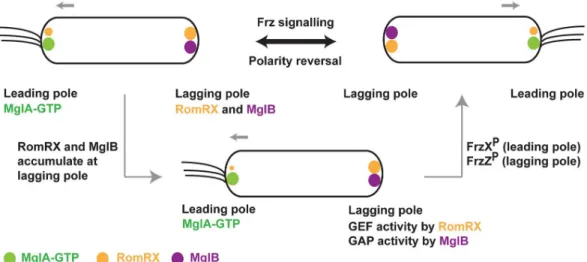
(IFA12-LSBM-adventurous motilities inM. xanthus, comprising the 2 genes mglA and mglB [1,3]. Studies on MglA and MglB showed that these proteins exhibit a mutually exclusive localization pattern [4,6,7]. MglA localizes to the leading pole presumably in its GTP-bound state [4], whereas MglB localizes at the lagging pole (Fig 1). These studies [1,3–7] established the role of the pro-karyotic small Ras-like GTPase in cell polarity determination and motility, which are functions analogous to its eukaryotic counterparts [8,9].
A remarkable feature ofMyxococcus motility is the frequent reversals in its cell polarity in
response to environmental cues [10].M. xanthus changes its direction of movement by
swap-ping the leading and lagging poles. The reversal frequency is regulated by the chemosensory-like frizzy (Frz) pathway, which relays environmental signals to the downstream MglA-MglB polarity control system [11–13] (Fig 1). High-resolution time-lapse experiments revealed that MglA and MglB relocalize sequentially during a reversal [4,7,11]. Importantly, the Frz system does not establish polarity, but its activity is required for inversion of the polarity axis. Another protein involved in the polarity control module is RomR, a protein with a response regulator receiver-like domain [14,15]. It was recently shown that RomR and its interacting partner RomX are essential for recruiting MglA-GTP to the poles [16]. Frz-signaling and RomR stimu-lates the pole-to-pole exchange of MglA and MglB [11,13–15]. All together, these proteins form a so-called gated relaxation oscillator, which functions to drive the polarity reversals in response to environmental signals [11].
Regulated reversals are centrally controlled by the MglA GTPase cycle. Though it is estab-lished that MglB functions as a GAP for MglA [4,7], proteins that function as guanine nucleo-tide exchange factors (GEFs) [17] for MglA were not identified until very recently [16]. The complex of RomR and RomX has been recently demonstrated to function as a GEF for MglA [16]. In vivo, the RomRX complex recruits MglA to the leading pole at the time of reversal. Between reversals, RomRX dissociates slowly and relocalizes to the lagging cell pole where RomR interacts with MglB [14,15]. Thus, both GAP and GEF activities accumulate at the lag-ging cell pole, preparing the next reversal event (Fig 1). However, at this stage, the cell will only reverse if signals from Frz are sufficiently strong. When this occurs, 2 response regulators FrzXP(phosphorylated FrzX) and FrzZP(phosphorylated FrzZ) bind to opposite poles, which trigger the detachment of MglA from the leading cell pole and its relocalization to the lagging cell pole. MglB is subsequently detached and relocated to the opposite pole, completing the switch.
Many questions remain unaddressed concerning the exact sequence of events that take place at the poles. Specifically, the presence of antagonizing GAP and GEF activities at both cell poles requires that this balance be regulated. The accumulation of RomRX at the lagging pole is not sufficient to drag MglA to the lagging cell pole, most likely because the MglB GAP activity predominates. Accumulation of FrzXPat the lagging pole could switch this balance in favor of the GEF and provoke the switch. In this study, we show that MglB is in fact a bifunc-tional enzyme and that MglB regulation could be key for the control of this enzymatic balance at the pole.
Previously, biochemical and structural studies with homologous proteins fromThermus thermophilus demonstrated that MglB is the GAP of MglA [4,5]. MglA and MglB fromT. ther-mophilus (TtMglA and TtMglB) share 62% and 29% sequence identities, respectively, with the M. xanthus counterparts. Unlike the majority of small Ras-like GTPases, both the active site
residues of the GTPase were found within TtMglA itself (Arg53Aand Gln82A; residue names with superscript “A” denote residues of MglA) instead of being contributed by the GAP [5]. A dimer of TtMglB interacts with a monomer of TtMglA. The structural studies revealed thatβ2
strand of TtMglA undergoes a screw-type movement during the interaction between TtMglA and TtMglB in the presence of GTP analogues [5]. Theβ-screw movement positions the active
52), Science and Engineering Research Board (SERB) extramural grant (EMR/2014/00563), SERB Women Excellence Award, Indian National Science Academy (INSA) Young Scientist Research Award, and Indo-French Centre for Promotion of Advanced Research (CEFIPRA) Collaborative Research Programme Grant. JB and MK acknowledge Council of Scientific and Industrial Research (CSIR) and INSPIRE respectively for fellowships. TM acknowledges CEFIPRA and Agence Nationale de la Recherche (ANR) “BACTOCOMPASS”. The funders had no role in study design, data collection and analysis, decision to publish, or preparation of the manuscript.
Competing interests: The authors have declared
that no competing interests exist.
Abbreviations: Ct-helix, C-terminal helix; FP,
fluorescent protein; Frz, frizzy; FrzXP,
phosphorylated FrzX; FrzZP, phosphorylated FrzZ; GAP, GTPase activating protein; GEF, guanine nucleotide exchange factor; GDP, guanosine di-nucleotide; GppNHp, guanosine-5’-[(β,γ)-imido] triphosphate; GNP, guanosine-5’-[(β,γ)-imido] triphosphate; GTP, guanosine tri-nucleotide; GTPγS, guanosine 5’-O-[gamma-thio]triphosphate; HPLC, high pressure liquid chromatography; IAA, isoamyl alcohol; LDH, lactate dehydrogenase; mant, 2’/3’-O-(N-Methyl-anthraniloyl); mgl, mutual gliding motility; MglA, Mutual gliding motility A; MglB, Mutual gliding motility B; MxMglA, M.
xanthus MglA; MxMglB, M. xanthus MglB; nG,
neonGreen; PCR, Polymerase Chain Reaction; PDB, Protein Data Bank; PFS, Perfect Focus System; Pi, inorganic phosphate; PK, pyruvate kinase; Rbl/LC7, Roadblock/LC7; RomR, Required for Motility Response Regulator; SLIC, sequence-and ligation-independent cloning; SMART, Simple Modular Architecture Research Tool; TtMglA, T.
thermophilus MglA; TtMglB, T. thermophilus MglB;
site residues favorably for GTP hydrolysis. In contrast toM. xanthus, T. thermophilus is not
known to switch polarity and appears to lack most of the other regulatory components (e.g., Frz proteins). Hence, it is imperative to study theMyxococcus proteins for a complete
molecu-lar characterization of the oscillatory system.
In this study, we report the biochemical and structural characterization ofM. xanthus
MglA and MglB (MxMglA and MxMglB). Our investigations led to the discovery of an alloste-ric pocket on the small Ras-like GTPase fold. Allostealloste-ric binding of the C-terminal helix (Ct-helix) of MxMglB on MxMglA facilitates nucleotide exchange. Thus, MxMglB plays a dual role in increasing the GTPase activity of MxMglA by orienting the catalytic residues (role of a GAP) and by facilitating exchange of GDP with GTP (role of a GEF). Furthermore, our study reveals that the Ct-helix plays an important role in regulating the mutually exclusive localiza-tion of MglA, MglB, and RomR. Accordingly, delelocaliza-tion of the helix inM. xanthus trapped
MglA, MglB, and RomR at both poles of the cell and drastically affected motility. Importantly, the study reveals the integration of GAP and GEF activities into a single interacting partner of a prokaryotic small Ras-like GTPase. Our study highlights the mechanistic features of a GEF in a prokaryotic small Ras-like GTPase family.
Results
Crystal structure of MxMglA is in the GDP-bound conformation
The crystal structure of MxMglA was determined at a high resolution of 1.3Å (Protein Data Bank [PDB] ID: 5YMX;S1 Table).Fig 2Acontains a description of the small Ras-like GTPase fold of MglA with the secondary structure elements labeled. Structure of MxMglA is similar to TtMglA with 2 additionalβ-strands (β0andβ2�), compared with the 5α-helices and 6
β-strands present in the small Ras-like GTPase fold [5]. All the residues of the small Ras-like GTPase fold as well as the C-terminal hexahistidine tag were modeled in the MxMglA struc-ture. Though GDP was not added during protein purification or crystallization, the crystal
Fig 1. Localization of cell polarity determining proteins inM. xanthus. Schematic showing the localization of MglA, MglB, and RomRX inM. xanthus. MglA (GTP-bound state) localizes to the leading pole, whereas MglB and RomRX mainly
localize at the lagging pole. A polarity reversal occurs when the proteins relocalize. A signaling cascade consisting of the Frz-signaling proteins drives the polarity reversals. MglA, MglB, and RomRX are represented as green, magenta, and orange spheres. The asymmetry in the size of orange spheres at the 2 poles denotes the asymmetric localization of RomR, with higher concentration at the lagging pole. FrzXPand FrzZPdenote phosphorylated states of FrzX and FrzZ, respectively. Frz,
frizzy; Mgl, mutual gliding motility.
structure of MxMglA contained GDP in the nucleotide-binding pocket (Fig 2A,S1A Fig). High-performance liquid chromatography (HPLC) analysis of the protein sample reconfirmed that the bound ligand is GDP (S1A Fig,S1B Fig).
Structure of MxMglAB–GTP
γS complex revealed a binding site for
MxMglB Ct-helix
Crystal structure of the complex of MxMglA and MxMglB in the presence of guanosine 5’-O-[gamma-thio]triphosphate (GTPγS, a substrate analogue) (MxMglAB–GTPγS), was deter-mined at a resolution of 2.4Å (PDB ID: 6IZW;S1 Table). In the structure, a monomer of MxMglA was bound to a dimer of MxMglB (Fig 2B). MxMglA interacts with the MxMglB dimer through an extensive hydrophobic interface involvingβ2strand of MxMglA andα2
heli-ces of MxMglB monomers (Fig 2C), an interface similar to that of TtMglAB [5].
One of the novel features of the MxMglAB–GTPγS complex is the asymmetry between the 2 protomers of MxMglB (labeled MglB1and MglB2;Fig 2B). MglB1and MglB2were
asymmet-ric at their N- and C-terminal residues in the MxMglAB–GTPγS complex. The core of MxMglB, ranging from residues 10 to 131, belongs to the Roadblock/LC7 (Rbl/LC7) domain fold [18]. In MglB1, the N-terminal amino acids (2–7) comprised aβ-strand that formed
hydrogen bonds with theβ0strand of MxMglA, thus continuing the centralβ-sheet of the
MxMglA fold (Fig 2B).α1helix of the Rbl/LC7 fold started from Glu11B(residue names with
superscript B denote residues of MglB) of MglB1. In contrast, in MglB2, residues 1 to 7 were
disordered, and theα1helix comprised ordered residues starting from Tyr8B.
Fig 2. Crystal structures ofM. xanthus MglA and MglAB–GTPγS complex. (A) Crystal structure of MxMglA bound to GDP. The secondary structure elements are labeled. GDP is shown in stick representation in light brown, and the catalytic loops switch 1 (blue) and switch 2 (red) are labeled as SW1 and SW2. The rest of the loops are shown in light pink, whereas the secondary structure elements are shown in shades of green. The C-terminal hexa-histidine tag (His)6, which is ordered in
the crystal structure, is shown in gray. (B) Crystal structure of the complex of MxMglA (green) bound to GTPγS (yellow) and MxMglB (magenta). “Front” view and a 90˚ rotated view (side view) are shown. The 2 protomers of MxMglB are labeled as MglB1and MglB2. The C-terminal helix of MxMglB (Ct-helix), the respective N- and C-terminal ends, switch 1 (blue; SW1)
and switch 2 (red; SW2), and relevant secondary structures of MxMglA are labeled. GTPγS is shown in stick representation in yellow. Dotted magenta line connects the ends on either sides of the stretch of disordered residues in MglB1. Boxed
regions in the front and side view panels highlight the major interfaces between MxMglA with the Rbl/LC7 domain of MxMglB (box with short dashes) and the Ct-helix of MxMglB (box with long dashes), respectively. (C) Residues ofβ2strand
(Phe56A, Phe57A, and Phe59A) of MxMglA (green) form a hydrophobic surface that interacts with MxMglB (magenta). The
relevant secondary structure elements are labeled, and switch 1 and 2 (SW1 and SW2) are colored in blue and red, respectively. The region corresponds to a zoomed view of the boxed region (short dashes) in the front view of panel B. (D) The interacting interface of the Ct-helix of MxMglB (magenta) and MxMglA (green). The side chains of relevant interface residues are shown in stick representation and labeled. Water molecules are represented as red spheres, and dotted yellow lines represent hydrogen bond interactions. The region corresponds to a zoomed view of the boxed region (long dashes) in the side view of panel B. (E and F) Conformational changes in switch 1 (SW1; 2 shades of blue) and switch 2 (SW2; 2 shades of red) in the presence of MxMglB illustrates the mechanism of GAP activity (by reorientation of active site residues Arg53A
and Glu82A). MxMglA–GDP conformation and MglA from the MxMglAB–GTPγS complex are shown in panels E and F,
respectively. The 2 structures from a superposed orientation are shown as separate panels. Secondary structures extending from the switch loops for MxMglA–GDP conformation and MglA from the MxMglAB–GTPγS complex are shown in brown and green, respectively. Switch 1 (or 2) of the 2 structures are labeled SW1 (or 2) and SW1 (or 2) using GDP and GSP as subscripts for the MxMglA–GDP and MxMglAB–GTPγS complexes, respectively. (G and H) Ct-helix of MxMglB (magenta) and GTP (represented by GTPγS shown in stick representation) bind to the C- and N-terminal ends of the β2strand of
MxMglA, respectively. MxMglA–GDP conformation and MglA from the MxMglAB–GTPγS complex are shown in panels G and H, respectively. The 2 structures from a superposed orientation are shown as separate panels. The unflipped and flipped states of the strand are shown (brown, MxMglA–GDP; green, MglA conformation in MxMglAB–GTPγS structure; switch 1 and switch 2 are in shades of blue and red, respectively). A schematic representation of the registry shift andβ2flip is shown
below, with the same color scheme. A 6-pointed yellow star and a 5-pointed light brown star represent GTP (labeled as “T’) and GDP (labeled as “D”), respectively. Theβ2strand is represented by an arrow. The residues that form the interface are
schematically shown by 2 circles on the bottom or top of theβ2strand representation in the flipped and unflipped states of
the strand, respectively. Ct-helix of MxMglB is schematically shown by a cylinder that is labeled “H.” Ct-helix, C-terminal helix; GAP, GTPase activating protein; GTPγS, guanosine 5’-O-[gamma-thio]triphosphate; Mgl, mutual gliding motility; MxMglA,M. xanthus MglA; MxMglB, M. xanthus MglB; Rbl, Roadblock; SW1, switch 1; SW2, switch 2.
At the C terminus, density was absent for residues 131 to146. However, residues 147 to 157 of the MglB1protomer could be modeled into the electron density (S1D Fig,S1E Fig), which
adopted a helical conformation. Henceforth, this will be referred to as Ct-helix (C-terminal helix of MxMglB). In the MglB2protomer, electron density was absent for residues beyond
130. The Ct-helix of MglB1bound to a pocket in MxMglA distal from the nucleotide-binding
site (Fig 2BandFig 2D). The binding pocket in MxMglA was formed by the residues of theα5
helix,β2strand, and the loop connectingβ2andβ3strands (β2–β3loop) of MxMglA
(inter-switch region of small Ras-like GTPases [19];Fig 2D). The correspondingβ2toβ3loop was
disordered in the TtMglAB structure [5] (S1C Fig).
The conformational features described below are consistent with the previously determined structures of the corresponding complex from TtMglAB [5] (S1C Fig). Some of the prominent features are the optimal orientation of the active site residues (Fig 2E,Fig 2F) and theβ-screw movement (Fig 2G,Fig 2H), which were also observed in the TtMglAB structures [5]. The con-formational changes between MxMglA–GDP and the MxMglA–GTPγS bound to MxMglB resulted in movements of switch 1 and switch 2 such that Arg53Aand Gln82Aof MxMglA are now oriented towards the catalytic water (Fig 2E,Fig 2F). Another interesting observation is the orientation of the residues Thr54Aand Asp58Aof MxMglA, which coordinate the Mg2+ ion bound to GTPγS (Fig 2F). Theβ-screw movement exposed hydrophobic residues of MxMglA toward the MxMglB interface and facilitated MxMglAB interaction (Fig 2C,Fig 2H).
Ct-helix allosterically affected the GTPase activity
To elucidate the functional role of the MglB1Ct-helix, we compared features of GTPase
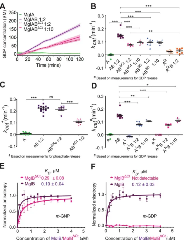
stimu-lation and MxMglA interaction with full-length MxMglB (MglB) and a truncated version of MxMglB lacking the Ct-helix (MglBΔCt;S1F Fig). The GTPase activity was monitored by esti-mation of released GDP using a coupled enzyme assay [20] or phosphate release using mala-chite green–based assay [21]. The reactions were carried out with excess GTP (substrate) and hence measured multiple turnovers of the GTPase. Both approaches for monitoring enzyme activity showed similar rates of hydrolysis. The GTPase activity of MxMglA on its own was negligibly low, whereas the activity increased in the presence of MxMglB (Fig 3A,Fig 3B,Fig 3C,S2A Fig). This confirmed the role of MglB as a GAP, consistent with published results [4,5,7]. Control experiments were performed with only MxMglB (no MxMglA) and excess of MxMglB (S2A Fig). Similar GTPase activities were observed for MxMglA in the presence of MxMglB with or without a C-terminal hexahistidine tag (Fig 3C), and hence all further experi-ments were performed using the construct with C-terminal hexahistidine tag.
We found that the GTP hydrolysis rate was approximately 2-fold lower in the presence of MxMglBΔCtcompared with MxMglB (Fig 3A,Fig 3B,Fig 3C). The GTPase activity remained low despite using 5 times excess of MxMglBΔCt(Fig 3A,Fig 3B), suggesting that the effect was not due to a reduced affinity of MxMglBΔCtto MxMglA. We compared the GTPase activities with that of the active site mutant MxMglAQ82L(referred to as MxMglAQ) in the presence of MxMglB (S2B Fig,S2C Fig;Fig 3B). The GTPase activity of MxMglABΔCtwas, however, higher than that of MxMglAQB.
Next, we proceeded to confirm that the interactions observed in the crystal structure between Ct-helix and the binding pocket of MxMglA are indeed relevant for the observed increase in GTPase activity. Analysis of the crystal structure of MxMglAB–GTPγS complex showed that Asp150B, Asp151B, and Asp152Bon the MxMglB Ct-helix formed water-mediated as well as direct interactions with MxMglA (Fig 2D). Lys181Aand Lys185Aof theα5helix of
MxMglA interacted with Phe157Band Asp154Bof the MxMglB Ct-helix, respectively (Fig 2D). Leu64Aand Ile67Aof MxMglA were on theβ2toβ3loop (interswitch region) and were within
Fig 3. Ct-helix of MxMglB allosterically regulates GTPase activity of MxMglA. (A) GTPase activities of wild-type MxMglA only
(green), and in the presence of MxMglB (dark purple; MglAB), and MxMglBΔCt(magenta; MglABΔCt). Ratio variations of 1:2 and 1:10 for MxMglBΔCtare shown in dotted and solid lines, respectively. The release of GDP was estimated using NADH-based enzyme-coupled assay. The lines represent the average of multiple repeats (at least 3), and the shaded zones represent standard error. (B) Comparison ofkcatvalues for MxMglA, MxMglAB, MxMglABΔCt, MxMglAB3D, and MxMglAQB. The release of GDP was estimated using NADH-based enzyme-coupled assay. 1:2 and 1:10 represent the molar ratio of MxMglA and MxMglB monomers. (C) Comparison ofkcatvalues for MxMglA, MxMglAB, MxMglABΔH6(MglB construct without a hexahistidine tag) and MxMglABΔCt. The release of phosphate was estimated using malachite green assay. 1:2 represents the molar ratio of MxMglA and MxMglB/
interacting distances from the MxMglB Ct-helix (Fig 2D). There was a reduction in GTPase activity in the presence of MxMglB3D(Fig 3B), a mutant of MxMglB in which Asp150B, Asp151B, and Asp152Bon the Ct-helix were replaced with alanine. Additionally, we studied the GTPase activities of the MxMglA mutants MxMglAK(a double mutant of K181A and K185A) and MxMglAL(a double mutant of L64A and I67A;S2B Fig,S2D–S2F Fig).
MxMglB-stimulated GTPase activity of the MxMglA mutants was less than the wild type (Fig 3D). The GTPase activity remained lower despite 5-fold increase in the concentration of MxMglB (Fig 3D). The above data demonstrated that mutation of residues in the helix-binding pocket affected rate of nucleotide hydrolysis, even though they were located away from the GTPase active site. This implied that MxMglB Ct-helix played an allosteric role in regulating the GTPase.
Ct-helix facilitated interaction between MxMglB and GDP-bound MxMglA
Binding affinities of MxMglA with MxMglB and MxMglBΔCtin the presence of GDP and GTP, respectively, were estimated using fluorescence anisotropy measurements of
mant-labeled nucleotides (fluorescent nucleotide analoguesm-GDP or m-GppNHp, referred to as m-GNP hereafter) bound to MxMglA. Binding studies showed that m-GNP-bound MxMglA
(MxMglA–m-GNP) bound to both MxMglB and MxMglBΔCt, respectively (Fig 3E,S2A Table). The binding affinities of MxMglB to MxMglAKand MxMglALin presence ofm-GNP were
also not affected drastically despite the mutations (S2F Fig,S2B Table). Hence, we concluded that the lower GTPase activities observed for these mutant constructs were not due to decreased affinity between the GTPase and MxMglB constructs.
Interestingly, MxMglB interacted with MxMglA in presence of eitherm-GNP or m-GDP
(Fig 3E,Fig 3F). However, MxMglBΔCtbound to MxMglA in the presence of onlym-GNP but
notm-GDP (Fig 3E,Fig 3F). The binding affinities (reported asKDvalues) between MxMglB
andm-GNP–and m-GDP–bound MxMglA (0.10 ± 0.04 μM and 0.12 ± 0.03 μM, respectively)
were comparable to the binding affinity of 0.29± 0.08 μM between MxMglBΔCtand
m-GNP-bound MxMglA (Fig 3F,S2A Table).KDbetween MxMglBΔCtandm-GDP-bound MxMglA
could not be estimated, because the binding was insignificant (Fig 3F,S2A Table).
Thus, the Ct-helix appears to be essential for stabilizing the interaction between MxMglB and GDP-bound MxMglA. In the previous study with TtMglB, the binding between TtMglB and TtMglA was observed only in the presence of GNP (or GTP analogues), a characteristic feature of a GAP. Our studies imply that nucleotide hydrolysis by MxMglA is not sufficient for disrupting the MxMglAB complex inM. xanthus. Also, interaction with the GDP-bound state
of a GTPase is a characteristic feature of a GEF [17]. Because we observed that MxMglA and MxMglB interact in the presence of GDP, we proceeded to check whether MxMglB could also perform a GEF function.
MxMglBΔH6/MxMglABΔCtmonomers. (D) Comparison ofkcatvalues for MxMglA, MxMglAK, and MxMglALin the presence of MxMglB. The release of GDP was estimated using NADH-based enzyme-coupled assay. 1:2 and 1:10 represent the molar ratios of MxMglA and MxMglB monomers. The data shown for MxMglA and MxMglAB have been duplicated from panel B for the sake of comparison. (E) Fluorescence anisotropy measurements for MxMglB (dark purple) and MxMglBΔCt(magenta) titrated against MxMglA–m-GNP, showing that both MxMglB and MxMglBΔCtbound to MxMglA in the presence ofm-GNP. (F) Fluorescence
anisotropy measurements for MxMglB (dark purple) and MxMglBΔCt(magenta) titrated against MxMglA–m-GDP, showing that
MxMglB, but not MxMglBΔCt, bound to MxMglA in the presence ofm-GDP. The mean and 95% confidence intervals (long and short
black horizontal lines, respectively) are shown for each sample in panels B, C and D.�p = 0.01–0.05,��p = 0.001–0.01, and
�� �p < 0.001. The numerical data for all the figure panels have been provided in the respective sheets inS1 Data. Ct-helix, C-terminal helix;m-GNP, 2’/3’-O-(N-Methyl-anthraniloyl)-guanosine-5’-[(β,γ)-imido]triphosphate; MxMglABΔH6, Mx MglA and MglB (without hexahistidine tag) complex; MxMglAB3D, Mx MglA and MglB D150A, D151A and D152A triple mutant); MxMglAQB, MxMglA Q82L mutant; MxMglA,M. xanthus MglA; MxMglAK, MxMglA K181A and K185A double mutant; MxMglAL, MxMglA L64A and
I67A mutant; MxMglB,M. xanthus MglB; MxMglBΔCt, MxMglB Ct-helix truncation; ns, non-significant.
Ct-helix of MxMglB contributed toward nucleotide exchange
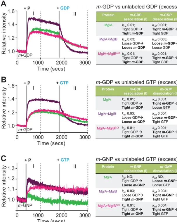
As mentioned earlier, the purified MxMglA contained GDP bound to the active site pocket (S1 Fig). To observe the loading ofm-GDP to MxMglA, fluorescence intensity of m-GDP was
monitored upon addition of MxMglA. An increase in fluorescence intensity indicated increased binding ofm-GDP. Exchange was observed to occur spontaneously upon addition
of MxMglA but was accelerated upon addition of a mix of MxMglA and MxMglB (Fig 4A, phase I). In contrast, the presence of MxMglBΔCtinstead of MxMglB did not affect the exchange kinetics (Fig 4A, phase I). The trend did not change despite addition of MxMglBΔCt in a 1:10 ratio (S2G Fig). Next, we monitored the exchange kinetics by competing the bound
m-GDP with excess of unlabeled GDP. The rate of loss of fluorescence (koff) was higher for
MxMglA+MxMglB mix compared with MxMglA or MxMglA+MxMglBΔCtmix (Fig 4A, phase II). This suggested that full-length MxMglB assisted in GDP exchange, whereas the C-terminal deletion mutant failed to stimulate GDP exchange.
We proceeded to carry out the same experiment by competingm-GDP with GTP, a
rele-vant reaction for a GEF activity. MxMglB addition affected the rate of displacement ofm-GDP
in this experiment too (Fig 4B; phase II). Loading withm-GNP and competition with
unla-beled GTP provided further information on the exchange kinetics (Fig 4C). Lack of a major increase in fluorescence indicated that binding ofm-GNP to MxMglA was very unstable (Fig 4C, phase I). This, presumably, is due to the presence of unlabeled GDP prebound to the MxMglA sample. However, addition of either MxMglB or MxMglBΔCtwith MxMglA stabi-lized the binding ofm-GNP (Fig 4C, phase I). The rate of exchange of prebound GDP (from purification of MxMglA) withm-GNP (kon;Fig 4C, phase I) for MxMglA+MxMglB mix was 3
times faster than that of MxMglA+MxMglBΔCtmix. Subsequent competition ofm-GNP with
unlabeled GTP in the MxMglA+MxMglB mix and the MxMglA+MxMglBΔCtmix showed very slow or negligible exchange (Fig 4C, phase II).
These experiments suggested that MxMglA favored binding of GDP over GTP. However, in the presence of MxMglB, GTP bound better than GDP, as evidenced by a higherkonand
higherkoffform-GDP and a higher konbut a lowerkoffform-GNP (Fig 4C). In contrast,
MxMglBΔCtdid not affect the MxMglA–GDP complex (equalkonform-GDP for MxMglA and
MxMglA+MxMglBΔCtmix;Fig 4A, phase I). However, it stabilized the GTP-bound conforma-tion of MxMglA (increased intensity form-GNP exchange;Fig 4C, phase I). This effect is con-sistent with our observations that MxMglB interacts with both MxMglA–GDP and MxMglA– GTP, whereas MxMglBΔCtinteracts only with MxMglA–GTP (Fig 3E,Fig 3F). Thus, MxMglB functions not only by stabilizing GTP-bound MxMglA but also by destabilizing the bound GDP, whereas MxMglBΔCtcontributes only toward stabilizing GTP-bound MxMglA.
Our assays measuring exchange kinetics with different combinations of labeled and unla-beled nucleotide pairs clearly implied that the Ct-helix played a role in nucleotide exchange. Consequently, we concluded that MxMglB accelerates MxMglA GTPase not only by appropri-ately orienting the catalytic residues but also by facilitating GDP release through an allosteric effect via the Ct-helix. This effect increased the overall hydrolysis rate in our GTPase assays, which are multiple turnover enzymatic reactions.
mglB
ΔCtdoes not respond to frz signaling similar to
ΔmglB
To test whether the MxMglB Ct-helix is important for MxMglB activity in vivo, we expressed MxMglB and the MxMglBΔCtvariant under the control ofmglB promoter at the Mx8 phage
attachment site in anmglB deletion mutant of M. xanthus (ΔmglB). MxMglBΔCtwas stably expressed, although its expression level appeared slightly decreased compared to MxMglB (it is also possible that this apparent decrease is because the Ct-helix is an important epitope for the
Fig 4. MglB Ct-helix facilitates nucleotide exchange of MglA. (A) Kinetic measurements of increase inm-GDP fluorescence
(phase I) upon addition of MxMglA (green), mix of MxMglA and MxMglB in 1:2 ratio (dark purple), mix of MxMglA and MxMglBΔCtin 1:2 ratio (magenta) at 400 seconds (marked by solid grey line labeled “+ P”), followed by competition ofm-GDP
(phase II) by addition of excess of unlabeled GDP at 1,800 seconds (marked by solid gray line labeled “+ GDP”). (B) Kinetic measurements of increase inm-GDP fluorescence (phase I) upon addition of MxMglA (green), mix of MxMglA and MxMglB in 1:2
ratio (dark purple), mix of MxMglA and MxMglBΔCtin 1:2 ratio (magenta) at 400 seconds (marked by solid gray line and labeled “+ P”), followed by competition ofm-GDP (phase II) by addition of excess of unlabeled GTP at 1,800 seconds (marked by solid gray
line labeled “+ GTP”). (C) Kinetic measurements of increase inm-GNP fluorescence (phase I) upon addition of MxMglA (green),
mix of MxMglA and MxMglB in 1:2 ratio (purple), mix of MxMglA and MxMglBΔCtin 1:2 ratio (magenta) at 400 seconds (marked by solid grey line labeled “+ P”), followed by competition ofm-GNP (phase II) by addition of excess of unlabeled GTP at 1,800
seconds (marked by solid gray line labeled “+ GTP”). ND for thekonandkoffvalues in panel C stands for “not determined”. For panels A to C, phase I (between solid gray line labeled “+ P” and the first dotted line) represents kinetics for association (forkon
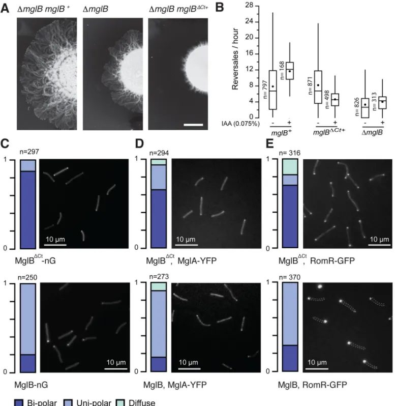
MxMglB antibody;S3A Fig,S3B Fig). MxMglBΔCtmust nevertheless carry biological activity because it induced a drastic motility phenotype and localized at the poles (see below). MglA was expressed to comparable levels in strains complemented by MxMglB or MxMglBΔCt(S3A Fig,S3B Fig). The level of MglA is slightly reduced in aΔmglB mutant because of a weak polar effect but this reduction did not create detectable phenotype [7]. The strainmglB+ comple-mented motility on an agar surface, whilemglBΔCt+led to profoundly defective motility in col-ony plate assays (Fig 5A). The motility phenotype was in fact more pronounced than for the ΔmglB mutant, suggesting that expression of MxMglBΔCtdeeply perturbs the function of MglA. However, single cells were motile, and the defect could be linked, at least partially, to aberrant cell reversals (Fig 5B,S3C Fig,S3D Fig). InΔmglB mutant cells, the polarity axis is disrupted and therefore the reversal distribution does not change depending on environmental conditions, contrarily to wild-type (WT) cells in which the reversals depend on the Frz-signal-ing state [7]. This can be shown in cells in which Frz signaling can be induced by addition of isoamyl alcohol (IAA), which increases the reversal frequency ofmglB+cells (Fig 5B) [7,22,23]. As expected, theΔmglB mutant showed a reversal frequency distribution that did not change upon addition of IAA (Fig 5B). Remarkably,mglBΔCt+cells also did not respond to the addi-tion of IAA (Fig 5B), suggesting that the polarity axis and Frz-dependent regulations are deeply affected in absence of the MxMglB Ct-helix.
MxMglA, MxMglB
ΔCt, and RomR localize to both cell poles in mglB
ΔCt+To further explore if MxMglBΔCtperturbs cell polarity, we analyzed its localization in single cells ofM. xanthus by expressing C-terminal neonGreen (nG) fused [24] to MxMglB and MxMglBΔCtinmglB deletion backgrounds. MglB-nG localized to the lagging cell pole and
oscillated from pole-to-pole with kinetics comparable to previously described yellow fluores-cent protein (YFP) and mCherry fusions (Fig 5Ctop,S3C Fig,S4A Fig) [4,5,11]. In contrast, MxMglBΔCt-nG mostly localized symmetrically at both the poles and failed to oscillate (Fig 5C
bottom,S3C Fig,S4A Fig). Thus, the Ct-helix is required for ensuring a unipolar localization of MxMglB.
We next tested how the bipolar localization of MxMglBΔCtaffected the other polarity pro-teins, MxMglA and RomR, by expressing MxMglA-YFP or RomR-GFP, respectively, in the
mglBΔCt+strain. Remarkably, both MxMglA-YFP and RomR-GFP were bipolar and static (Fig 5D,Fig 5E[top],S4B Fig,S4C Fig), similar to the localization pattern observed in theΔmglB deletion mutant [12,13]. Thus, in the presence of MxMglBΔCt, the polarity axis is disrupted, exactly as observed when MxMglB is deleted. Most strikingly, all 3 proteins MxMglA, RomR, and MxMglBΔCtco-localize at the poles.
Allosteric regulation in MglA homologues and other Ras-like GTPases
The regulation of MxMglA GTPase activity and MxMglAB interaction by MxMglB Ct-helix prompted us to explore whether analogous mechanisms exist among other prokaryotic and eukaryotic small Ras-like GTPases.
estimation), phase II (between solid line labeled “+ GDP/GTP” and final dotted line) represents kinetics for dissociation (forkoff estimation). A schematic representation of the observations and probable explanations at each phase is shown on the right side, following the same color scheme for the font as in the plots. The events directly observable through fluorescence intensity change of themant-labeled component in the experiment are highlighted in bold. The panels include representative plots of multiple repeats,
and thekonandkoffvalues (in units of s-1) represent the average values of the multiple repeats. The numerical data for all the figure panels have been provided in the respective sheets inS1 Data. Ct-helix, C-terminal helix;m-GDP,
2’/3’-O-(N-Methyl-anthraniloyl)-guanosine-diphosphate; MxMglA,M. xanthus MglA; m-GNP, 2’/3’-O-(N-Methyl-anthraniloyl)-guanosine-5’-[(β,γ)-imido]
triphosphate; MxMglB,M. xanthus MglB; MxMglBΔCt,M. xanthus MglB with Ct-helix truncated; ND, not determined.
Fig 5. MxMglB Ct-helix is required for MxMglB function in vivo. (A) Expression of MxMglBΔCtleads to motility defects. Motility on 0.5% agar is shown after 48 hours of incubation at 32˚C. Scale bar = 1 cm. (B) Reversal frequency ofM. xanthus cells of ΔmglB mutant strain and cells expressing wild-type MxMglB (mglB+) or MxMglBΔCt(mglBΔCt+). The reversal frequencies were scored in absence (−) or presence (+) of 0.075% IAA, a condition known to activate the Frz system. For each strain and condition, the number of trajectories analyzed (n), the median (vertical line), and the mean (black dot) are reported for each box-and-whisker plot.
(C) MxMglBΔCtshows a bipolar localization pattern. Representative fields of cells expressing MxMglBΔCt-nG and MxMglB-nG are shown in top and bottom images, respectively. The fractions of cells exhibiting bipolar (dark blue), unipolar (light blue), and diffuse (cyan) localization are depicted on the left for each of the field views, in which “n” represents the total number of cells analyzed. The fluorescence intensity profiles for each cell are compiled and shown inS4A Fig. (D) MxMglA shows a bipolar localization pattern in the presence of MxMglBΔCt. Representative fields of cells expressing MxMglA-YFP inmglBΔCt+andmglB+strains
Earlier, it has been shown that MglA and MglB, which exist in almost all phyla of eubac-teria, can be categorized into 5 phylogenetically related groups [25]. Genes encoding MglB-like proteins are either associated with genes encoding MglA-MglB-like protein in the same operon, called coupled MglBs, or without an associated MglA gene (orphan MglBs) [25] (S3 Table). We analyzed these sequences and found that 66 coupled and 25 orphan MglBs possessed a C-terminal extension (more than 15 amino acids beyond the Rbl/LC7 fold;S3 Table). A total of 56 of the 66 coupled MglB sequences showed a predicted helical region containing 2 to 4 aspar-tates or glutamates (D/E rich), whereas the C-terminal extensions of the remaining MglB sequences contained proline-rich stretches with no predicted secondary structure (Fig 6A,S3 Table).
Out of these 56 coupled sequences, theα5helix (which interacts with the Ct-helix) of 48
MglA sequences possessed positively charged residues (either lysines or arginines;Fig 6B). These positively charged residues were not conserved in other MglA sequences (Fig 6B). This suggested that the Ct-helix of MglB and its binding pocket on MglA have coevolved. Interest-ingly, we found MglB having the D/E rich Ct-helix to be present in all the 5 phylogenetic groups of MglA-like proteins, including a variety of bacteria and also in the archaea Methano-bacterium paludis (S5 Fig). This indicated that the mechanism of allosteric regulation of the MglA GTPase by the MglB Ct-helix could be widespread in prokaryotic Ras-like GTPases and not limited toMyxococcus. Because the allosteric regulation by full-length MglB has not been
characterized for any other prokaryotic MglA-like and MglB-like proteins, the mechanism proposed here would be relevant toward understanding their function.
Discussion
MxMglB Ct-helix facilitates nucleotide exchange allosterically
Our results revealed that the Ct-helix is essential for MxMglB to interact with MxMglA in the GDP-bound state. Deletion of the helix resulted in the formation of MxMglAB complex in the presence of only GTP but not GDP. This prompted us to identify the structural features that enable MxMglB to bind MxMglA–GDP. Theβ-screw movement (flipping of MxMglA β2
strand) appears to be essential for the interaction of MxMglB with MxMglA. This conforma-tional change exposes residues that form a major part of MxMglAB interface.
An inspection of the MxMglAB–GTPγS structure revealed that the switch 1 loop and the binding pocket of the Ct-helix are at the N- and C-terminal ends of theβ2strand of MxMglA,
respectively (Fig 2H). Hence, we propose that the flipped state of theβ2strand that favors
MxMglB binding can be achieved in 2 ways—by interaction of either theγ phosphate of GTP with the switch 1 loop or the Ct-helix with theβ2toβ3loop (Fig 2H). The flipped state achieved
by the Ct-helix interaction and the consequent stabilization by the Rbl/LC7 fold of MxMglB results in the formation of a complex with MxMglA–GDP. As a consequence of the flipped state of theβ2strand, MxMglA in the MxMglAB complex will potentially possess a
nucleotide-binding pocket that favors nucleotide-binding of GTP over GDP, consistent with our observations from the nucleotide exchange assays (Fig 4). Thus, the proposed model provides an explanation for
field views, in which “n” represents the total number of cells analyzed. The fluorescence intensity profiles for each cell are compiled and shown inS4B Fig. (E) RomR shows a bipolar localization pattern in the presence of MxMglBΔCt. Representative fields of cells expressing RomR-GFP inmglBΔCt+andmglB+
strains are shown in top and bottom images, respectively. The fractions of cells exhibiting bipolar, unipolar, and diffuse localization are depicted on the left for each of the field views, in which “n” represents the total number of cells analyzed. In the bottom panel (RomR-GFP, mglB+), dashed lines indicate bacterial contour. The
fluorescence intensity profiles for each cell are compiled and shown inS4B Fig. The numerical data for all the figure panels have been provided in the respective sheets inS1 Data. Ct-helix, C-terminal helix; Frz, frizzy; IAA, isoamyl alcohol; MxMglA,M. xanthus MglA; MxMglB, M. xanthus MglB; MxMglBΔCt, MxMglB with Ct-helix truncated; nG, neonGreen; YPF, yellow fluorescent protein.
Fig 6. Conservation of sequence features of the allosteric pocket on MglA and MglB Ct-helix. (A) Comparison of
MxMglAB–GDP complex formation and a mechanistic insight into the allosteric action of Ct-helix for facilitating nucleotide exchange (Fig 7A).
Interestingly, like MglB, many eukaryotic GEFs such as DENN-1B domain, TRAPP-I, and Mon1-Ccz1, have the Rbl/LC7 fold [26]. Furthermore, we found that the intersubunit orienta-tion in MglAB is conserved in the GTPase-GEF complexes of Rab35 with DENN-1B domain [27], Rab Ypt1p with TRAPP-I [28], and Ypt7 with Mon1-Ccz1 [29] (S6 Fig). However, unlike in MglAB complex, the mechanism of GEF action in these eukaryotic systems is based on a glutamate or aspartate residue interacting directly with switch 1 or switch 2 (S6A Fig,S6B Fig). These residues are present in extensions from the Rbl/LC7 fold or are contributed by other interacting partners that form the GEF complex [26].
Mechanism of dual GAP and GEF action by MxMglB
We propose that MxMglB increases the rate of GTP hydrolysis by MxMglA through 2 comple-mentary ways. Firstly, Rbl/LC7 domain of MxMglB stabilizes the flippedβ2strand interface,
which orients the MxMglA catalytic residues favorably. Secondly, its Ct-helix, a flexible ex-tension from the main Rbl/LC7 fold, retains the flipped state of theβ2strand post GTP
hydro-lysis and, consequently, stabilizes the MxMglAB–GDP complex. This interaction results in a nucleotide-binding pocket that accelerates release of GDP from the complex and favors GTP-binding. Thus, by facilitating GDP-to-GTP exchange MxMglB enhances the rate of GTP hydrolysis by MxMglA, increasing its potency as a GAP. Indeed, our in vitro results show that MxMglBΔCtexhibits reduced GAP activity, which we attribute to a defect in nucleotide exchange.
Role of Ct-helix and exchange activity of MxMglB in the polarity oscillation
cycle
In vivo, we propose that the MxMglB Ct-helix is critical for the balance between GAP and GEF activities at the lagging pole. In absence of the Ct-helix, all 3 proteins—MglA, MxMglBΔCt, and RomR—localize to both cell poles. Thus, the MxMglB Ct-helix is absolutely required to enable the cascade of interactions that establish polarity and permit its switch. Co-localization of both MxMglA and MxMglBΔCtsuggest that MxMglBΔCtdoes not support its GAP function in vivo, despite exhibiting only a reduction in GAP activity in vitro. In between reversals in wild-type cells, MxMglB and RomRX stably co-localize at the lagging pole [11,14,15]. MxMglA is efficiently excluded from the lagging pole, suggesting that the GAP activity of MxMglB predominates over the GEF activity of RomRX. Thus the MglB Ct-helix possibly overcomes the action of RomRX.
How could this function at the molecular level? Analysis of RomR revealed a stretch of neg-atively charged residues (Glu-rich) akin to that of MxMglB Ct-helix (S7A Fig). Thus, similar to MxMglB, the RomRX complex could facilitate GDP exchange from MxMglA by acting through the allosteric pocket on MxMglA. In this process, the C-terminal Glu-rich region of
implicated in interaction with the allosteric pocket on MglA are highlighted by “�” in the sequence conservation logo. The fractional occupancy of amino acids in the sequence alignment is shown for each amino acid position. The numbers within brackets denote the number of sequences in the alignment. The residues are colored in 20 different shades. (B) Comparison of residue conservation in theα5helix of MglA sequences coupled with MglB sequences
possessing a negatively charged C-terminal extension versus that of all coupled MglA sequences. The lysine residues that potentially interact with the Ct-helix of MglB are highlighted by “�” in the sequence conservation logo. The fractional occupancy of amino acids in the sequence alignment is shown for each amino acid position. The numbers within brackets denote the number of sequences in the alignment. Ct-helix, C-terminal helix; MglA, Mutual Gliding motility A; MglB, Mutual Gliding motility B.
RomR might thus assist nucleotide exchange, whereas RomX could assist in stabilizing the flippedβ2strand of MxMglA. We tested this hypothesis by replacing the Ct-helix of MxMglB
with the Ct-helix from RomR (S7A Fig,S7B Fig) and carrying out GTPase activity measure-ments in the presence of the chimeric MglB (MglBRhelix). Indeed, we observed that the chime-ric construct accelerated the GTPase activity of MglA similar to that of wild-type MxMglB (S7C Fig), supporting the hypothesis that the Ct-helix of RomR is capable of binding at the allosteric pocket of MxMglA. At the lagging pole, the MxMglB Ct-helix and the RomR Glu-rich helix might compete for binding at the MxMglA helix-binding pocket, which would dis-place RomRX from MxMglA. When the MglB Ct-helix is deleted, RomRX could therefore interact with MxMglA thus blocking MxMglBΔCtfrom exerting its GAP activity. This would allow RomRX to exert its GEF activity at the lagging pole too and stabilize the anchoring of MglA to both the poles.
Additional regulation of the Ct-helix must take place to determine the nucleotide specificity of MxMglB binding to MxMglA. Additional regulators in vivo will potentially determine the
Fig 7. Ct-helix regulates MxMglAB interaction by binding to an allosteric pocket of the small Ras-like GTPase fold. (A)
A schematic representation of the conditions that facilitate MxMglAB complex formation. PDB IDs 6IZW, 3T12, and 5YMX correspond to the crystal structures of MxMglAB–GTPγS, TtMglAB–GTPγS, and MxMglA–GDP complexes. (B) Proposed model for MxMglAB relocalization. Proteins that can bind in the allosteric pocket potentially compete out the MxMglB Ct-helix and dissociate the MxMglAB–GDP complex. In panels A and B, MxMglB dimer is shown in magenta, whereas MxMglA conformations with the flipped and unflipped states ofβ2are shown in green and brown, respectively. A
6-pointed yellow star and a 5-pointed light brown star represent GTP (labeled as “T”) and GDP (labeled as “D”), respectively. Theβ2strand is represented by an arrow. The residues that form the interface are schematically shown by 2
circles on bottom or top of theβ2strand representation in the flipped and unflipped states of the strand, respectively.
Ct-helix of MxMglB is schematically shown by a cylinder and labeled as “H.” Ct-Ct-helix, C-terminal Ct-helix; MxMglA–GDP, MxMglA bound to guanosine 5’-diphosphate; MxMglA,M. xanthus MglA; MxMglAB, M. xanthus MglA and MglB
complex; MglAB–GTPγS, MglA and MglB complex formed in the presence of guanosine 5’-O-[gamma-thio]triphosphate; PDB ID, Protein Data Bank identification.
accessibility of the Ct-helix and thus the localization of MxMglA and MxMglB. At the pole, the exact molecular organization of the signaling complexes is not known. Other proteins that par-ticipate in the polarity switch and that are present at the lagging pole, for example, the MglB-like protein MglC, can potentially regulate the Ct-helix availability [30]. During the switch, action of FrzXPcould, for example, block the inhibitory effect of MglB on RomRX and thus initiate the switch. This possibility could be explored in the future. Further characterization of other polarity determining components and their interactions with MxMglAB are therefore essential to discriminate the GAP and GEF-active phases of MxMglB during the polarity rever-sal cycle.
Insights gained from the prokaryotic small Ras-like GTPase MxMglA and its interacting partner MxMglB highlight the dual role of MxMglB both as a GAP and a GEF. The novel mechanism of GEF action is based on an allosteric interaction with the small Ras-like GTPase. Based on sequence analysis, it appears to be a conserved mechanism among prokaryotic small Ras-like GTPases. The study also opens up a new avenue for design of compounds or factors that can modulate the enzyme activity by targeting the newly discovered allosteric binding pocket of the universal Ras-like fold.
Methods and materials
Protein expression and purification
Protein constructs, cloning, and overexpression. Genes corresponding tomglA and mglB were amplified from M. xanthus genomic DNA (obtained from DSMZ, Germany,
cata-logue number 16526) using suitable primers (S4 Table) and cloned intopHis17 vector
(obtained from Lo¨we lab, MRC LMB, Cambridge; refer Addgene plasmid #78201 for vector backbone) between the restriction enzyme sitesNdeI (New England Biolabs Inc) and BamHI
(New England Biolabs Inc). Single-point mutants of MxMglA and deletion constructs of MxMglB were generated using a site-directed mutagenesis strategy utilizing PCR-based meth-ods followed byDpnI (New England Biolabs Inc) digestion, transformation, and screening of
positive clones. All clones were confirmed by sequencing. The list of clones and the primers used to generate them are summarized inS5andS4Tables. All chemicals were procured from Sigma Aldrich, unless otherwise mentioned.
Protein expression. Plasmids containing the gene of interest were transformed into
suit-ableEscherichia coli strains, and culture was grown at 37˚C and at 30˚C post induction. E. coli
strain BL21-DE3 was used for expressing MxMglA wild type and its mutants. The cultures were grown in LB media containing 50μg/ml of ampicillin and induced with 0.5 mM IPTG at OD600value of 0.8, whereas MxMglB and its mutants were expressed in the strain BL21-AI
and induced with 0.02% L-arabinose (SRL Chemicals, India) at OD600value of 0.6.
Seleno-methionine-labeled proteins for MxMglBL156Mand MxMglBI148Mhave been expressed by the feedback inhibition method [31].
Purification of MxMglA(His6). For purification, harvested cells were resuspended in the
lysis buffer L (50 mM Tris, 200 mM NaCl [pH 8.0], and 10% glycerol) and then spun at 39,000g for 45 minutes at 4˚C. Supernatant was loaded on to a 5-ml HisTrap (GE Lifesciences)
column because the presence of hexahistidine tag at the C-terminus facilitated the binding of overexpressed protein of interest to the column. The column was equilibrated with binding buffer (Buffer A: 50 mM Tris [pH 8.0], 200 mM NaCl) prior to loading the supernatant and the bound protein was washed and eluted with a step gradient of 2%, 5%, 10%, 20%, 50%, and 100% of Buffer B (Buffer A containing 500 mM imidazole). The fractions containing the pro-tein were pooled, concentrated, and loaded onto Superdex75, 10/300 (GE Lifesciences). The protein was eluted into 50 mM Tris (pH 8.0), 50 mM NaCl. Fractions containing the protein
were pooled, concentrated, flash frozen, and stored at−80˚C. MxMglA mutants, i.e., MglAK, MglAL, and MglAQ, were also purified using a similar protocol.
Purification of MxMglB. MxMglB(His6), MxMglBΔCt(His6), and MxMglBRhelix(His6)
were purified using the same protocol as described for MxMglA(His6). For MxMglB
con-structs without histidine tag, i.e., MxMglB, MxMglB3D, and selenomethionine-labeled MxMglBL156Mand MxMglBI148M, ion-exchange chromatography was used to purify the pro-teins. First, the cells were resuspended in the lysis buffer (50 mM Tris [pH 8.0], 200 mM NaCl, 10% glycerol), and then spun at 39,000g for 45 minutes at 4˚C. An ion-exchange column, QHP
(5 ml, GE Lifesciences) was used to purify the protein. Buffers used for binding and elution were Buffer A (50 mM Tris [pH 8.0], 50 mM NaCl), and a linear gradient of Buffer A with Buffer B (50 mM Tris [pH 8.0], 1 M NaCl), respectively, ranging from 0% to 50% Buffer B over 20 column volumes. Fractions containing the protein of interest were pooled. This protein was dialyzed into Buffer A25 (50 mM Tris [pH 8.0], 25 mM NaCl) and again spun at 39,000g at
4˚C, filtered, and loaded on MonoQ 10/100 (GE Lifesciences) to remove minor impurities present. Binding and elution buffers were the same as earlier. Fractions from the MonoQ run were checked on SDS-PAGE, and those containing protein of interest were concentrated and stored at−80˚C.
Crystallization and structure determination
MxMglA crystallization. About 500 conditions of commercially available screens
(Molec-ular dimensions, Hampton Research) were screened using Mosquito crystallization robotic system, using drop sizes consisting of 100 nl of protein at a concentration of 10 mg/ml and 100 nl of crystallization cocktail, in 96-well sitting drop plates (MRC plate, SWISS-SCI). Initial hits were obtained in many of the conditions, and, further, it was reproduced and optimized to get well-diffracting crystals. For crystallization of MxMglA, protein was diluted to a final concen-tration of 10 mg/ml in A50 buffer (50 mM Tris [pH 8.0], 50 mM NaCl). Diffraction quality crystals were obtained using the following conditions in a drop ratio of 1:1 volumes for protein and crystallization condition: (i) 0.1 M sodium cacodylate (pH 6.5), 40% v/v 2,4-methyl pen-tane diol, and 5% PEG 8000; (ii) 0.1 M imidazole (pH 8.0), 30% w/v 2,4-methyl penpen-tane diol, and 10% w/v PEG 4000; (iii) 0.1 M sodium citrate (pH 5.6) and 35% w/v tertiary-Butanol; 20% ethylene glycol was included as cryoprotectant in the parent condition during crystal freezing.
MxMglAB crystallization. MxMglA and MxMglB were mixed in a ratio of 1:1
(consider-ing monomeric molecular weight of MglB) in A50 buffer contain(consider-ing 5 mM MgCl2and 2 mM
GTPγS (Sigma). The concentration of the proteins used to crystalize was 4 mg/ml of each. Screening for crystallization hits yielded a few conditions that were further optimized to get the desirable crystals. The crystals were obtained in 2 different conditions, namely, (i) PEG 4000 (8%) and ammonium sulphate (200 mM) and (ii) PEG 3350 (12%) and ammonium sul-phate (200 mM). Diffraction quality crystals were obtained at a drop ratio of 1:1 volumes of protein to crystallization buffer. Crystallization conditions containing 20% PEG 400 were used as cryoprotectant during freezing of crystals. Crystals of MxMglA and selenomethionine-labeled MxMglB complex were obtained in the same crystallization conditions.
Structure solution and refinement. Diffraction data from the crystals were collected at
the home source using Rigaku Micromax 007 X-ray generator, and higher resolution data and anomalous data were collected at the synchrotron sources at Diamond Light Source, Harwell, UK, and ESRF, Grenoble. Data reduction was performed using IMOSFLM [32] or XDS [33], and scaling using AIMLESS [34] in CCP4 package [35]. MxMglA and MxMglB structures were solved by molecular replacement using the TtMglAB structure (PDB code: 3T12). Molec-ular replacement was performed using PHASER [36] available in CCP4 package. Refinement
was carried out using PHENIX package [37] and model building usingCoot [38]. The refined structures have been deposited in the PDB with accession numbers 5YMX (MxMglA–GDP) and 6IZW (MxMglAB–GTPγS).
In order to confirm the registry of the amino acids belonging to the Ct-helix of MxMglB, selenomethionine-labeled protein for mutant constructs of MxMglB where Ile148Band Leu156B, respectively, were mutated to methionines, were purified, and selenomethionine-labeled MxMglB was used for obtain MxMglAB crystals in the same crystallization condition. The anomalous data from the crystals were collected, and it was confirmed that the correct amino acids were modeled into the electron density. The anomalous signal from the methio-nines in these 2 mutants, respectively, confirmed the registry of the amino acids of the MxMglB Ct-helix (S1D and E Fig).
Fluorescence anisotropy experiments
Binding studies using fluorescent anisotropy measurements. Binding of
fluorescently-labeled nucleotides,mant-GDP (m-GDP) or mant-GppNHp (m-GNP) (Jena Bioscience) with
the proteins were monitored by measuring the change in anisotropy [39]. The excitation and emission wavelengths used for monitoring themant-labeled nucleotide fluorescence were 360
nm and 440 nm, respectively. The experiments were performed on Fluoromax-4 (Horiba), with a sample volume of 200μl in a cuvette, and excitation and emission slit widths of 5 nm.
Nucleotide binding of MxMglA and its mutants were estimated by measuring the change in fluorescence anisotropy of themant-nucleotide upon titration with increasing concentrations
of the protein. Protein samples (MxMglA or its mutants) were titrated against a fixed concen-tration ofmant-labeled nucleotide (100 nM). Each reading corresponds to 10 averaged single
point anisotropy values. The initial value of anisotropy of themant-labeled nucleotide was
sub-tracted from all the values. GraphPad Prism was used to plot the values against the concentra-tion of protein (concentraconcentra-tion of MxMglA) and to fit the data to the binding equaconcentra-tion for estimation ofKD. Because it is known that MxMglA has a single binding site for nucleotides,
equation for one site-specific binding, as given below, was used to fit the data points. The equa-tion is y = Bmax× x � (KD+ x), where y represents the bound fraction (fluorescence anisotropy
based readout), Bmaxis the maximum value obtained, x is the concentration of MxMglA, and
KDis the binding constant.
The values were normalized by dividing each of them by the maximum value of anisotropy, Bmax. The normalized anisotropy values were plotted to obtain a binding curve. Because
MxMglA was purified in the GDP-bound form, the measurements from this experiment are not absolute affinities of MxMglA (or its mutants) to the nucleotides but a reliable measure of exchange of the prebound GDP withm-GDP or m-GNP.
Similarly, anisotropy measurements for increasing amounts of MxMglB/MxMglBΔCtwith nucleotide-bound MxMglA provided information about the binding affinity of MxMglB to nucleotide-bound MxMglA. A mix of 400 nM ofmant-labeled nucleotide with 2 μM of
MxMglA was titrated with increasing concentrations of MxMglB/MxMglBΔCt. The same calcu-lations as described above were performed to obtain the binding affinities (KD). In this case,
the x-axis represents the concentration of MxMglB dimer. Because MxMglA binds to one dimer of MxMglB, equation for one site-specific binding was used to fit the data points.
Each data point is an average of at least 3 independent measurements, and the standard error is shown in which each data represent 3 to 5 repeats. Error bars represent standard error.
Nucleotide exchange assays. Intensity of fluorescence emission bymant-labeled
nucleo-tide (GDP or GMPPNP [GNP]; Jena Bioscience) at 440 nm was monitored after the excitation at 360 nm. All the kinetic measurements for MxMglA, MxMglA with MxMglB, and their
mutants were performed on Fluoromax-4 (Horiba), with a sample volume of 200μl in a cuvette and excitation and emission slit widths of 2 nm.m-GDP/GNP (800 nM) was added
with Buffer A50 (50 mM Tris, 50 mM NaCl, 5mM MgCl2[pH 8.0]) in a quartz cuvette (10× 2
mm path length), and the fluorescence intensity was monitored initially for 400 seconds. The protein, i.e., MxMglA or the mix of MxMglAB/MxMglABΔCt(3μM of MxMglA and 6 μM MxMglB/MxMglBΔCtmonomer), was added in the cuvette at 400 seconds after stabilization of the signal from onlymant-nucleotide. Consequently, the fluorescence was recorded for 1,400
seconds. The binding was estimated in terms of increase in fluorescence intensity. At 1,800 seconds, themant-labeled nucleotide was competed out with excess of unlabeled nucleotide
(GDP or GTP; 500μM), resulting in a decrease of fluorescence intensity because of release of
mant-labeled nucleotide from the protein. For plotting the relative intensities from the
mea-surements, each value was divided by the average of first 200 readings (400 seconds). These accumulation and decay reactions were fitted to exponential binding equations as given below to estimate thekonandkoffvalues.
P + N1! PN1+ N2! PN2+ N1, where P represents protein, N1is the labeled nucleotide,
N2is the unlabeled nucleotide, and PN denotes the protein-nucleotide complex.
For estimation ofkon, PNt= PNmax(1− e−kt).
For estimation ofkoff, PNt= PNmax− PNmin(e−kt) + PNmin
Here, PNtrepresents the amount of the complex at time t, PNmaxis the maximum amount
of the complex upon association, and PNminis the minimum amount of the complex after
dissociation.
GTP hydrolysis assay
NADH coupled assay. GTPase activity measurements were made using a coupled enzyme
based assay in which the conversion of NADH to NAD+is coupled to the utilization of GDP produced [20]. The GTPase or ATPase hydrolyses the nucleotide while pyruvate kinase (PK) uses GDP (or ADP) and phosphoenol pyruvate (PEP) to produce GTP (or ATP) and pyruvate. Lactate dehydrogenase (LDH) utilizes NADH and pyruvate to produce NAD+and lactate. Because the utilization of NADH is equivalent to the GDP produced by the GTPase reaction, the actual read out is decrease in absorbance of NADH, which in turn estimates the GDP pro-duced by the GTPase. The amount of NADH utilized in the reaction was measured by moni-toring the NADH absorbance at 340 nm using a multiplate reader, Varioskan Flash (Thermo Scientific). A master mix was prepared in Buffer A50 containing PEP (1mM; Sigma), GTP (1mM; Jena Bioscience), NADH (600μm; Sigma), and PK/LDH mix (approximately 25 U/ml; Sigma). All the components of NADH reaction were mixed in a total reaction volume of 200μl and added in a 96-well plate. The reaction was initiated by addition of MxMglA or MxMglAB mix and their relevant mutant constructs. MxMglA and its mutants were used at 10μM and MxMglB and its mutants at concentrations 20μM and 200 μM (in a molar ratio of 1:2 and 1:10 considering monomeric molecular weight of MxMglB).
Readings were recorded at the interval of 20 seconds for 2 hours. The absorbance value of buffer containing GTP was subtracted from all the values. Also, the absorbance at the 0 time point for each reaction was subtracted from all readings of the reaction. The absorbance read-ings were represented as concentration of GDP released, by using a conversion factor based on the standard graph obtained by plotting the absorbance values for known concentrations of NADH.kcatvalues were calculated and plotted using GraphPad Prism. Readings from 0 to
7,200 seconds were fitted to a line using linear regression, and the slope of the fitted line was calculated to obtain the amount of GDP released (inμM) per unit time. kcatwas calculated as
further to compare the enzyme activities among all the mutants. The significance was calcu-lated using GraphPad Prism, using one-way ANOVA with Tukey test of 95% confidence inter-val, and significance was marked on the scatter plot.
Phosphate release measurements using malachite green assay. This assay was
per-formed using MxMglA at a concentration of 10μM, and MxMglB in molar ratio 1:2 (20 μM, considering monomeric molecular weight of MxMglB) with excess of GTP, i.e., 1 mM GTP in buffer A50 (50 mM Tris, 50 mM NaCl [pH 8.0]) containing 5 mM MgCl2. All the enzyme
activity assays were performed at 30˚C. Readings were taken at different time points, i.e., 0, 15, 30, 45, 60, 75, 90, 105, and 120 minutes. To stop the reaction, the sample was heated at 65˚C and then malachite green reagent (prepared as described in [21]) was added. The absorbance was recorded at 630 nm after 20 minutes of incubation, using a plate reader (Varioskan, Thermo Scientific). Amount of Pi (inorganic phosphate) released inμM was calculated using a standard curve, obtained by mixing a set of known concentrations of NaH2PO4with malachite
green solution. The experiments were repeated multiple times, and the slope was calculated from every repeat. Readings from 0 to 120 minutes were fitted to a line using linear regression, and the slope of the fitted line was calculated to obtain the amount of Pi released (inμM) per unit time.kcatwas calculated as Pi released perμM enzyme concentration for each reaction.
kcatvalues were plotted, and significance was calculated on GraphPad Prism, using one-way
ANOVA with Tukey test of 95% confidence interval, and significance was marked on the scat-ter plot.
HPLC for bound nucleotide estimation
Purified MxMglA was diluted to 2 to 3 mg/ml concentration in buffer A50 and then heated at 65˚C. The heated protein sample was spun at 21,000g for 10 minutes at 4˚C. Then supernatant
was filtered with 0.22μm cellulose acetate filter (Corning), and the sample was loaded on the DNAPac 200 ion-exchange column (Thermo Fisher). Buffer A (2 mM Tris [pH 8.0]) was used as binding buffer. The runs were performed at a flow rate of 0.8 ml/min, with steps of 100% Buffer A for 3 minutes, followed by linear gradients of 0% to 30% Buffer B (2 mM Tris [pH 8.0], 1.25 M NaCl) for 10 minutes, and 30% to 100% B for 12 minutes. InS1B Fig, the peak profile obtained for 18μM of MglA was compared with that of 50 μM GDP solution, with 35μl of sample injection volume for both. The absorbance at 260 nm, indicative of the pres-ence of GDP, was plotted against retention time.
Thermal shift assay [
40
]
In a 25-μl reaction volume containing 2 μM of protein in buffer A50, SYPRO Orange dye (Sigma; available as a 5,000× concentration in DMSO) was added to a final concentration of 5×, in 96-well multiwell PCR plates. Biorad CFX96 Real-Time System was used to monitor the change in fluorescence of SYPRO Orange (Sigma-Aldrich). The mix was heated from 4˚C to 90˚C with an increment of 1.2˚C per minute. Readings were recorded at intervals of 0.4˚C. The melting temperature (Tm) was calculated based on the differential of the fluorescence
intensity versus temperature plot (dF/dT). This gave an estimate of the stability of the protein.
In vivo experiments
Myxococcus strains, plasmids, growth conditions, and genetic constructs. Primers,
plasmids, and strains used for this study are listed inS4 Table,S5 TableandS6 Table, respec-tively. In general,M. xanthus strains were grown at 32˚C in CYE rich media as previously
described by Bustamante et al. [23]. Plasmids were introduced inM. xanthus by