EDITORIAL
Mens sana in corpore sano revisited
Raban V. Jeger
*
Cardiology University Hospital Basel, Petersgraben 4, CH-4031 Basel, Switzerland Online publish-ahead-of-print 4 July 2013
This editorial refers to ‘The use of cholinesterase inhibitors and the risk of myocardial infarction and death: a nationwide cohort study in subjects with Alzheimer’s disease’†, by P. Nordstro¨ m et al., on page 2585
Orandum est ut sit mens sana in corpore sano.
Juvenal, Satire X
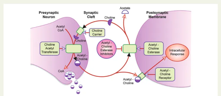
Acetylcholine is an important neurotransmitter for the central, peripheral, and autonomic nervous systems of the human body. It is synthesized in the presynaptic neuron by the enzyme choline acetyl-transferase from choline and acetyl-coenzyme A. After its release into the synaptic cleft, it binds to the acetylcholine receptors on the postsynaptic membrane that trigger the intracellular response (Figure 1). In the synaptic cleft, acetylcholine is hydrolysed by the enzyme acetylcholinesterase in order to terminate synaptic transmission.
Depending on the type of nervous system and the target tissue, acetylcholine may act in numerous ways. While it activates skeletal muscles via the peripheral nervous system, it exerts various effects in the autonomic nerve system, specifically by sympathetic and parasympathetic neurons. Originally discovered in 1921, acetylcho-line is the main neurotransmitter in the parasympathetic nervous system regulating internal organs and glands.1Last but not least, acetylcholine-dependent neurons act as the cholinergic system in the central nervous system, with important effects on cognitive func-tions specifically during waking and maintaining attention.
Patients with Alzheimer’s disease show less activity of the enzyme choline acetyltransferase in the presynaptic neuron, which leads to a decreased production of acetylcholine. Acetylcholinesterase inhibi-tors (AChEIs) block the acetylcholinesterase in the synaptic cleft, therefore inhibiting the breakdown of acetylcholine into acetate and choline and prolonging its duration of action (Figure1). Drugs such as galantamine (e.g. Nivalinw
or Razadynew
), donepezil (Ariceptw
), and rivastigmine (Exelonw
) are widely used for the treat-ment of detreat-mentia due to Alzheimer’s disease. Used in patients with mild to moderate dementia, treatment with an AChEI over 6 – 12 months has been shown to improve cognitive function and activities of daily living and behaviour.2Side effects are mainly gastrointestinal,
and include nausea, vomiting, and diarrhoea. However, although most drug effects take place in the central nervous system and the gastrointestinal tract, AChEIs may also act in the cardiovascular system due to the fact that the heart and vessels have a rich auto-nomic innervation. Potentially adverse cardiovascular side effects of AChEIs include hypertension and prolongation of the conduction time in both the sinus and atrioventricular nodes, with resulting bradycardia and reduced beat-to-beat fluctuations.3AChEI therapy is associated with increased rates of hospitalizations due to bradycar-dia and syncope.4–6However, data from randomized controlled trials show no evidence for increased rates of adverse cardiovascular side effects in patients treated with AChEIs.2Since patients with de-mentia usually suffer from co-morbid conditions such as cardiovascu-lar diseases, but most of them are not included in randomized controlled trials because they exhibit exclusion criteria,7no clear evi-dence exists on the cardiovascular side effects of AChEIs in a general population with Alzheimer’s disease.8
Nordstro¨m et al. now report data from nationwide Swedish data-bases, i.e. the Swedish Dementia Registry, the National Patient Regis-ter, and the National Register for Prescribed and Expedited Drugs, on the association on AChEI use and cardiovascular events.9After ad-justment for confounders, AChEI use was associated with lower rates of myocardial infarction [hazard ratio (HR) 0.62, 95% confi-dence interval (CI) 0.40 – 0.95] and death (HR 0.64, 95% CI 0.54 – 0.76) among 7073 subjects with newly diagnosed dementia due to Alzheimer’s disease between 2007 and 2010 over a mean follow-up time of 503 days, an effect that was even more pronounced in patients taking the highest recommended AChEI dose. However, data on bradycardia and syncope were not reported.
How can these results be put into perspective? Based on theoret-ical considerations and results from previous population-based studies, the use of AChEIs was associated with increased rather than decreased cardiovascular event rates in a population with Alzheimer’s disease, mainly due to cholinergic side effects with po-tentially higher rates of bradycardia and syncope.4–6However, the opposite was true in the study of Nordstro¨m et al.,9with a clearly lower rate of myocardial infarction and death in patients treated with AChEIs. What could be the possible mechanism for this appeal-ing findappeal-ing? Recently, a so-called cholinergic anti-inflammatory
*Corresponding author. Tel:+41 61 265 2525, Fax: +41 61 265 4598, Email:raban.jeger@usb.ch
The opinions expressed in this article are not necessarily those of the Editors of the European Heart Journal or of the European Society of Cardiology.
†doi:10.1093/eurheartj/eht182.
Published on behalf of the European Society of Cardiology. All rights reserved.&The Author 2013. For permissions please email: journals.permissions@oup.com
European Heart Journal (2013) 34, 2580–2581 doi:10.1093/eurheartj/eht244
pathway was described, i.e. a mechanism of autonomic regulation of local and systemic inflammation through the vagus nerve and its major neurotransmitter, acetylcholine.10Based on this paradigm, it might be hypothesized that the modulation of inflammation in the heart’s cholinergic system could influence degenerative processes in the vascular bed. Therefore, AChEIs, due to their proposed anti-inflammatory properties, might stabilize arteriosclerotic plaques and hamper the development of acute coronary syndromes. Other potential mechanisms such as a lower heart rate could lead to a decreased cardiac output and lower oxygen demand, which could also improve cardiovascular outcome. Finally, AChEIs may interfere with local levels of acetylcholine in the vasculature since acetylcholine stimulates the activity of the enzyme endothelial nitric oxide synthase and the release of nitric oxide via muscarinic receptors on endothelial cells. Previous data show that AChEI treat-ment increases cerebral blood flow in patients with Alzheimer’s disease and improves cognitive functions even in patients with vascular dementia.11,12
What we hear is the tale of a drug that improves both cognitive functions and cardiovascular health in a multimorbid population with dementia. But is this credible? Based on current knowledge, there is no clear evidence of whether AChEIs are beneficial or not in terms of cardiovascular events in elderly and multimorbid patients with Alzheimer’s disease. Still additional large population-based studies are needed to evaluate further the cardiovascular risk in such a cardiovascular high-risk population. Since the study of Nord-stro¨m et al. is purely observational, its results have to be interpreted with caution and currently are hypothesis generating only. However, if AChEI treatment is demonstrated to improve not only mental but also physical health, there might then be a case for a large randomized
outcome study in patients with coronary artery disease irrespective of dementia.
Conflict of interest: none declared.
References
1. Loewi O. On the background of the discovery of neurochemical transmission. J Mt Sinai Hosp NY 1957;24:1014 – 1016.
2. Birks J. Cholinesterase inhibitors for Alzheimer’s disease. Cochrane Database Syst Rev 2006;1:CD005593.
3. Masuda Y. Cardiac effect of cholinesterase inhibitors used in Alzheimer’s disease— from basic research to bedside. Curr Alzheimer Res 2004;1:315 – 321.
4. Park-Wyllie LY, Mamdani MM, Li P, Gill SS, Laupacis A, Juurlink DN. Cholinesterase inhibitors and hospitalization for bradycardia: a population-based study. PLoS Med 2009;6:e1000157.
5. Hernandez RK, Farwell W, Cantor MD, Lawler EV. Cholinesterase inhibitors and in-cidence of bradycardia in patients with dementia in the veterans affairs new England healthcare system. J Am Geriatr Soc 2009;57:1997 – 2003.
6. Gill SS, Anderson GM, Fischer HD, Bell CM, Li P, Normand SL, Rochon PA. Syncope and its consequences in patients with dementia receiving cholinesterase inhibitors: a population-based cohort study. Arch Intern Med 2009;169:867 – 73.
7. Gill SS, Bronskill SE, Mamdani M, Sykora K, Li P, Shulman KI, Anderson GM, Hillmer MP, Wodchis WP, Rochon PA. Representation of patients with dementia in clinical trials of donepezil. Can J Clin Pharmacol 2004;11:e274 – e285.
8. Malone DM, Lindesay J. Cholinesterase inhibitors and cardiovascular disease: a survey of old age psychiatrists’ practice. Age Ageing 2007;36:331 – 333.
9. Nordstro¨m P, Religa D, Wimo A, Winblad B, Eriksdotter M. The use of cholinester-ase inhibitors and the risk of myocardial infarction and death: a nationwide cohort study in subjects with Alzheimer’s disease. Eur Heart J 2013;34:2585 – 2591. 10. Rosas-Ballina M, Tracey KJ. Cholinergic control of inflammation. J Intern Med 2009;
265:663 – 679.
11. Pratt RD, Perdomo CA. Donepezil-treated patients with probable vascular demen-tia demonstrate cognitive benefits. Ann NY Acad Sci 2002;977:513 – 522. 12. Rosengarten B, Paulsen S, Molnar S, Kaschel R, Gallhofer B, Kaps M. Acetylcholine
esterase inhibitor donepezil improves dynamic cerebrovascular regulation in Alzhei-mer patients. J Neurol 2006;253:58 – 64.
Figure 1 Acetylcholinesterase inhibitors (AChEIs) in cholinergic nerve transmission. Acetylcholine is produced in the presynaptic neuron by the enzyme choline acetyltransferase from acetyl-coenzyme A and choline, and later released in the synaptic cleft where it binds to the acetylcholine receptor on the postsynaptic membrane, triggering an intracellular response. The enzyme acetylcholinesterase hydrolyses acetylcholine into acetate and choline in order to terminate synaptic transmission. Choline is transported into the presynaptic neuron by the choline carrier and serves as a substrate for the described production of acetylcholine. AChEIs inhibit the enzyme acetylcholinesterase, which in turn inhibits the break-down of acetylcholine into acetate and choline and prolongs its duration of action.