Full Terms & Conditions of access and use can be found at
http://www.tandfonline.com/action/journalInformation?journalCode=ierj20
Download by: [The University Of Melbourne Libraries] Date: 06 May 2017, At: 06:31
Expert Review of Clinical Pharmacology
ISSN: 1751-2433 (Print) 1751-2441 (Online) Journal homepage: http://www.tandfonline.com/loi/ierj20
Nanoparticles as potential clinical therapeutic
agents in Alzheimer’s disease: Focus on selenium
nanoparticles
Mustafa Nazıroğlu, Salina Muhamad & Laszlo Pecze
To cite this article: Mustafa Nazıroğlu, Salina Muhamad & Laszlo Pecze (2017): Nanoparticles as potential clinical therapeutic agents in Alzheimer’s disease: Focus on selenium nanoparticles, Expert Review of Clinical Pharmacology, DOI: 10.1080/17512433.2017.1324781
To link to this article: http://dx.doi.org/10.1080/17512433.2017.1324781
Accepted author version posted online: 02 May 2017.
Submit your article to this journal
Article views: 2
View related articles
Publisher: Taylor & Francis
Journal: Expert Review of Clinical Pharmacology DOI: 10.1080/17512433.2017.1324781
Review
Nanoparticles as potential clinical therapeutic agents in Alzheimer’s disease: Focus on selenium nanoparticles
Mustafa Nazıroğlu1, Salina Muhamad2, Laszlo Pecze3
1Neuroscience Research Center, Suleyman Demirel University, TR-32260 Isparta, Turkey 2 NANO Elec-Tronic Centre, Faculty of Electrical Engineering, Universiti Teknologi MARA,
40450 Shah Alam, Selangor, Malaysia
3Institute of Anatomy, Department of Medicine, University of Fribourg, Route Albert-Gockel 1,
Fribourg, CH-1700, Switzerland Corresponding address;
Mustafa NAZIROĞLU
Nörolojik Bilimler Uygulama ve Araştırma Merkezi (NÖROBAM), Süleyman Demirel University, TR-32260, Isparta, Turkey
Tel: +90 246 2113708 Fax: +90 246 2371165
E-mail: mustafanaziroglu@sdu.edu.tr
List of Abbreviations
AD, Alzheimer’s Disease Ag, silver
BBA, blood-brain barrier GSH, glutathione
GSH-Px, glutathione peroxidase GSSG, oxidized glutathione NAC, N acetyl cysteine NO, nitric oxide
NOS, nitric oxide synthase
PARP, poly (ADP-ribose) polymerase QDs, quantum dots
RNS, reactive nitrogen species ROS; reactive oxygen species Se, selenium
SiO2, silica dioxide
SOD, superoxide dismutase TiO2, titanium dioxide
ZnO, zinc oxide
TRP, transient receptor potential
TRPM2, transient receptor potential melastatin TRPV1, transient receptor potential vanilloid 1 VGCC, voltage gated calcium channels
Abstract
Introduction: In etiology of Alzheimer’s disease (AD), involvement of amyloid β (Aβ) plaque accumulation and oxidative stress in the brain have important roles. Several nanoparticles such as titanium dioxide, silica dioxide, silver and zinc oxide have been experimentally using for treatment of neurological disease. In the last decade, there has been a great interest on combination of antioxidant bioactive compounds such as selenium (Se) and flavonoids with the oxidant nanoparticles in AD. We evaluated the most current data available on the physiological effects of oxidant and antioxidant nanoparticles.
Areas covered: Oxidative nanoparticles decreased the activities of reactive oxygen species (ROS)
scavenging enzymes such as glutathione peroxidase (GSH-Px), superoxide dismutase (SOD) and catalase in the brain of rats and mice. However, Se-rich nanoparticles in small size (5-15 nm) depleted Aβ formation through decreasing ROS production. Reports on low levels of Se in blood and tissue samples and the low activities of GSH-Px, catalase and SOD enzymes in AD patients and animal models support the proposed crucial role of oxidative stress in the pathogenesis of AD.
Expert Commentary: In conclusion, present literature suggests that Se-rich nanoparticles appeared
to be a potential therapeutic compound for the treatment of AD.
1. Introduction
Metal-rich nanoparticles are between 1 and 200 nm in size and they possess special physicochemical characteristics [1]. In the last decade, the effects of nanoparticles on human diseases including neurological diseases such as Alzheimer’s disease (AD) and dementia has been extensively studied. The experimental animals were treated with metal-rich nanoparticles via inhalation [2], oral supplementation [3], intratracheal [4], intravenous and intraperitoneal injection [5]. The metal-rich nanoparticles such as titanium dioxide (TiO2), silica dioxide (SiO2), silver and
zinc oxide (ZnO) are absorbed into the body via the digestive tract, skin and also through inhalation (Table 1). These absorbed particles are stored in different organs but the brain is the most vulnerable to their oxidative toxic effects [6].
Brain and neurons are particularly vulnerable to oxidative stress-induced damage due to the following three reasons: (1) their high amount of oxygen consumption (The brain utilizes approximately 25% of the body's total oxygen consumption), (2) the presence of high amount of polyunsaturated fatty acids, and (3) low levels of enzymatic antioxidant activity [7,8]. Metal-rich nanoparticles enhance the production of reactive oxygen species (ROS). The main targets of reactive oxygen species (ROS) are the lipids (lipid peroxidation), proteins (protein-carbonyl formation) and nucleic acids (RNA and DNA oxidation, double- and single strand break of DNA). The ROS are scavenged by enzymatic and non-enzymatic antioxidants. Enzymatic antioxidant glutathione peroxidase (GSH-Px) contains selenium as co-factor. Hydrogen peroxide (H2O2) is
converted to water by the catalytic effects of GSH-Px. Selenium can also have direct antioxidant effect on brain and neurons [8]. Selenium forms several allotropes (red, black and gray) that interconvert with temperature changes. It has two opposite physiological features. Concentration-dependently selenium can exert either its therapeutic or toxic effects. High doses of selenium
promotes the proliferation of cancer cells and has neurotoxic effects, although low and intermediate doses inhibit cancer cell proliferation and has a therapeutic effects on neurological diseases including AD. Antioxidant and anti-inflammatory effects of selenium were observed in vitro using cell lines and in vivo in animal models [9]. In addition, there is direct relationship between the evoked effect and the type of selenium compounds administered [10]. However, the results on the effects of selenium and selenium nanoparticles for the treatment of neuronal diseases are conflicting. These conflicting results can be in part attributed to the different concentrations used in experiments, and to the different cell lines used in in vitro assays. Oxidative stress is one of the main factors involved in the pathogenesis of many neurological diseases. Therefore, therapies using natural antioxidants compounds have been developed to cure several neurodegenerative diseases [11,12]. However, the success rate of treatments with natural antioxidants is low. Therefore, there is an urgent need for development of new nanoparticles with diagnostic and/or therapeutic properties to improve the detection and treatment of neurological diseases. Accumulating evidences indicate that there is a direct relationship between deficiency of selenium in serum and hair samples and memory deficit in patients with AD [13-16]. It has been reported that selenium treatment may decrease the risk of memory deficits in animal models and in AD patients [17,18]. Recently, high interest emerged to study the role of selenium and selenoproteins in neurodegenerative diseases including AD (Table 2). Selenoproteins are proteins containing selenium in the form of amino acid, selenocysteine. Selenoproteins are mainly expressed in the human brain and most likely involved in antioxidant processes, which are the key factor in preventing the onset and progression of AD [19]. In addition, there are some newly synthesized selenoproteins and selenium nanoparticles possessing remarkable physiological properties [20,21]. Due to the high potency and low systemic toxicity of these particles, they might serve as alternatives for conventional therapeutic drugs for the treatment of neurological diseases including AD.
In this review, we aimed to summarize the oxidant and antioxidant effects of nanoparticles in the brain. We also reviewed the potential therapeutic application of selenium nanoparticles for the treatment of AD. This review is expected to be a useful resource for the scientists working in the field of neurodegenerative diseases, oxidative brain injury or selenium-based drug development.
2. Role of selenium on oxidative stress in Alzheimer disease (AD)
There are two major types of free radical species: ROS and reactive nitrogen species (RNS). ROS are chemically reactive species containing oxygen. Examples include peroxides, superoxide, hydroxyl radical, and singlet oxygen. They are produced during the physiological processes. For example, engulfed bacteria and viruses in the neutrophils are killed by the ROS. Both ROS and RNS are produced in brain as a result of exposure to environmental factors such as electromagnetic radiation and industrial element pollution [22,23]. Toxic nanoparticles, for example aluminum or manganese oxide nanoparticles, from air pollution can induce excessive ROS production and oxidative stress in the brain. They can accelerate the progression of neurodegenerative diseases such as AD, amyloid lateral sclerosis and Parkinson’s disease [23]. If the amount of ROS will not be controlled by antioxidants, they will cause significant reversible or irreversible damage to a wide range of biological molecules, including nucleic acids, lipids and proteins or any nearby molecule causing a cascade of chain reactions [24]. There is considerable interest in the ROS-induced reactions, and their relationship in the physiology and pathology of AD [10].
Oxidative stress was considered to be one of the main factors in the etiology of AD. AD is the world’s most common form of dementia. Incidence of AD is still increasing in the world and the development of new strategies is needed for the treatment of AD. Despite the high number of studies, the molecular mechanisms responsible for the initiation of AD have not been elucidated yet. There are many theories to explain the cause of Alzheimer's disease. i.) accumulation of amyloid precursor protein forming amyloid-β (Aβ) plaques, ii) hyperphosphorylation of tau protein, iii) oxidative stress, iv) alterations in cholinergic neurotransmission v) environmental pollution vi) genetic factors including mutations of amyloid precursor protein (APP) and presenilin (PSEN)
genes, vi) immune system dysfunction, etc. [17,25,26]. Pathologically, the extracellular depositions of Aβ proteins and the presence of intraneuronal neurofibrillary tangles are important markers for posthumous diagnosis of AD. Aβ is the main protein component of senile plaques in the AD brain and it is composed of spontaneously aggregating peptides of 39-43 amino acids [17,27]. It is also known that microglia is the main source of neuroinflammatory factors and neuroinflammatory factors affect the nature of AD [17]. Inflammatory processes are associated with ROS production. Both ROS and RNS are considered to play important roles in induction of AD [11,28]. Moreover, Aβ fragment 25-35 (Aβ25–35) induces activation of inducible nitric oxide (NO) synthase [29] and protein oxidation in hippocampal fibroblasts derived from AD patients [30] and they are appeared to be responsible for ROS and RNS-induced hippocampal damage. In addition, the ROS and RNS initiate other injury processes such as neuroinflammation and protein misfolding in the AD brain [31,32]. There are some reports showing that sulfur atoms are also involved in the oxidation process. For example, the oxidative stress-inducing properties of Aβ-peptide (1-42) are totally abolished by substitution of the sulfur atom of metionin at position 35 with a methylene group [33].
Selenium is an essential trace element in our body. One of the most important selenium-dependent detoxifying processes is associated with the activity of GSH-Px enzyme. GSH-Px protein contains a selenocysteine (Se-Cys) moiety in its active site. GSH-Px, catalase and SOD enzymes have synergistic functions in the removal of H2O2 and organic peroxides [8]. GSH-Px catalyzes a
reaction, in which 2 reduced monomeric glutathione (GSH) react with H2O2, and form oxidized
glutathione (GS-SG) and H2O. GSH contains thiol groups in its structure. GS-SG is reduced back to
its thiol form (GSH) by the glutathione reductase enzyme [19].
Thioredoxins are small peptides in the cytosol and mitochondria and also play an important role in maintaining a reduced environment in the cells through thiol-disulfide exchange reactions and protects cells and tissues from oxidative stress [34]. Reduction of thioredoxin is catalyzed by thioredoxin reductase-1 (Trx1) and most radicals such as hydrogen peroxide (H2O2) and nitric oxide
Besides of the neuronal injury evoked by hyperphosphorilated tau and Aβ plaques, accumulating evidences indicate the involvement of oxidative stress in apoptotic neuronal loss observed in the AD brain [35]. Since antioxidants are able to inhibit of Aβ aggregation pathways, new therapies have been developed for the treatment of AD. As it was mentioned above, thioredoxin (Trx) and glutathione (GSH) are the two major systems which play an important role in the maintenance of cellular redox homeostasis (Figure 1). It was reported that the inhibition of GSH synthesis resulted in an increase in Aβ-induced cell death and intracellular Aβ accumulation [36]. Decreased levels of selenium, GSH and GSH-Px were observed in model animals and in AD patients [37]. Correlation between the loss of cognitive function and plasma selenium levels was also observed in patients with AD. Therefore, the relevance of selenium and GSH redox system in patients with AD indicates their important roles in redox regulation [38]. Reduced selenium contents in plasma and hair were reported in the patients with AD and it was found a negative correlation between the selenium content in plasma and incidence of AD [39]. Moreover, selenium treatments decreased the levels of oxidative stress and Aβ formation in the brain of experimental animal models for AD [40,41]. Positive correlations between plasma and hair selenium deficiency and cognitive decline were reported in patients with AD [15,16]. Recently, selenium, total tau, Aβ42, GSH, lipid peroxidation and antioxidant enzyme values were analyzed in the blood samples from AD patients in South India. No correlations were reported between the levels of selenium, total tau and Aβ42 [42]. On the contrary, decreased GSH-Px enzyme activity and reduced levels of selenium were reported in erythrocytes [13] of patients with dementia and AD.
2.1. Size dependent effects of metal-rich nanoparticles in brain and AD
It is well known that the brain is the most vulnerable organ in body for oxidative stress-induced neurodegenerative injuries such as cerebral ischemia [42,43] and traumatic brain injury [44]. In most neurodegenerative diseases, these injuries are irreversible. The nanoparticles are mostly oxidant and they could induce neuronal injury and impair brain cognitive functions in AD
[39,45-48]. Recent in vivo studies in the experimental animals treated intraperitoneal or oral with nanoparticles have shown that metal-rich oxidative nanoparticles such as TiO2, ZnO, iron oxide,
SiO2, silver and gold can pass the blood-brain barrier (BBA) and accumulate in the brain [23,49,50]
(Table 1). For example, high transport rate of Ag nanoparticles from blood to brain via BBA was reported in brain of rats after injection a dose 5 mg/kg silver nanoparticles (20 and 200 nm) for 28 days. In addition, there is low rate of excretion from the brain and this leads to a gradual accumulation of nanoparticles in the brain. Contrary to intraperitoneal and oral applications, no accumulation of TiO2 nanoparticles in the brain area via nasal application was reported in mice
[51].
Apart from the oxidant nanoparticles, there are nanoparticles with antioxidant properties, for instance selenium. Metabolism of selenium nanoparticles in cells were reported in a recent paper [52] and it was also summarized in Figure 1. Mitochondria are physiologically the main source of ROS such as superoxide and hydroxyl radicals in mammalian cells during normal energy metabolisms [53].
Selenium consumed in foods and supplements exists in a number of forms including selenomethionine, selenocysteine, selenate and selenite. Reduced form of sodium selenite is selenide and it is produced in cytosol by catalytic effects of the GSH and superoxide radicals [54]. Then, the selenide is transformed to methylselenol and it is further transformed to methylated metabolites [55]. In addition, the selenide in cytosol serves as a source for selenoproteins, selenium-sugar, and elemental selenium. In a recent study, high amount of elemental selenium was indicated in human lung cancer cell line treated with sodium selenite [56].
Oxidative properties of nanoparticles are affected by physicochemical and biochemical factors such as surface properties, charge, and the adsorption rate of the nanoparticles [57]. In addition to these factors, the size of nanoparticles is an important factor which influences their biological activity. The large size of nanoparticle decreases its transport into brain and it is also constantly clearing foreign materials from the brain [45-48]. Redox regulatory and ROS scavenging
activities of the selenium nanoparticles with different sizes has been reported [58]. ROS scavenging effects of red selenium nanoparticles with small (5-15 nm), medium (20-60 nm) and large (80-200 nm) sizes were investigated in an in vitro model and the highest ROS scavenging effect was observed in the case of small-size red selenium nanoparticles [58]. Red elemental selenium nanoparticles with 20-60 nm sizes are produced from sodium selenite by the activated GSH redox system and they induced the proliferation rate of human hepatic cancer cells in vitro. They induced an increased activity of GSH-Px and Trx1 enzymes compared to sodium selenite [20]. On the contrary, Zhang and colleagues [59] reported that GSH-Px and Trx1 activities were affected neither in human hepatoma HepG2 cells nor in the mice liver by administration of selenium-rich nanoparticles in a size range of 5 to 200 nm. Similarly, chitosan nanoparticles with small size (40 and 70 nm) were more active and toxic tested on cancer cells [60]. In addition, size dependent protective effect of selenium nanoparticles against ROS production and DNA damage has been found [61]. More recently, the protective role of dextrin coated selenium nanoparticles (64 nm)-treated rats were investigated in the rat model of chronic inflammatory arthritis [56]. Increased SOD, GSH-Px and catalase activities were reported in liver, kidney and spleen of rats treated with dextrin-coated selenium nanoparticles [63,64]. According to the present literature, it seems that the small size range of selenium will ensure longer circulation time and increased accumulation in the brain of patients with AD
3. Role of nanoparticles in the brain
The presence of metals in the brain can be physiologically a necessity as well as a poison. Accumulation of the metals in brain tissues occur not only in the form of free ions, but also in the form of metalloproteins [23]. Abnormal homeostasis of metal ions in brain may induce neuronal damage if their levels move out of the normal physiological range especially due to the lack of specific metal binding proteins [39]. Biometals play significant roles in the biological systems through regulating and participating in numerous cellular processes such as induction of action
potential and co-factor of enzymes [54]. In particular, homeostasis of these elements has been demonstrated to be critical in brain, because they are involved in enzymatic activities, mitochondrial function, myelination, and neurotransmitter release [19].
The well-known metal-rich nanoparticles such as TiO2, ZnO, iron oxide, SiO2, silver (Ag)
and gold (Au) have mostly oxidative effect in the brain [23,39]. On the contrary, selenium rich nanoparticles with antioxidant properties such as red selenium and sodium selenite are used in treatment of brain diseases and they size-dependently improve cognitive functions. In the following sections we will review recent reports on metal-rich nanoparticles such as TiO2, ZnO, SiO2 and
silver.
3.1. Effects of TiO2 and silica nanoparticles on antioxidant enzyme activity in the brain: TiO2
nanoparticles are of high stability and have anticorrosion properties. They have been used for the treatment of neurological disease in experimental animals. However, potential toxic effects of TiO2
have not been clarified yet.
Decreased activities of GSH-Px, catalase and SOD in the mouse brain samples were reported after nasal application of TiO2 nanoparticles [51]. Results of another study indicated that
activities of SOD and GSH-Px are decreased in the brain cortex and hippocampus region after oral administration of TiO2 with/without lead acetate [3] and the study concluded that TiO2-induced
oxidative toxicity is supported by lead acetate. Ze and colleagues [64] investigated brain injury by nasal administration of different doses of TiO2 (2.5, 5, and 10 mg/kg) for 90 consecutive days.
They performed oxidant and antioxidant analyses and analyzed the gene expression profile of the brain samples. They observed that the levels of malondialdehyde (MDA) and protein carbonyls as markers of oxidative stress in the mouse brain were higher in the three treated group than in the control. Another study reported that inhalation of silica nanoparticles decreased GSH-Px, catalase and SOD activities in the rat corpus striatum [55]. Microglia is the main phagocytic neuronal cells in the brain and they play an important role in the process of inflammation. Recently, positive role
of silica nanoparticles on excessive ROS, RNS and cytokine production, and increased expression level of proinflammatory genes were reported in primary rat microglia [66].
3.2. Effects of ZnO on the activity of antioxidant enzymes in the brain: ZnO nanoparticles are
commonly used in many commercial products such as construction material, toothpaste, beauty material, and wall paints. The ZnO nanoparticles attract specific attention in medical area due to small size and high specific surface area. The toxic effect of ZnO nanoparticles has been well known in experimental animals and cell lines. ZnO nanoparticles are toxic for different tissues, including the brain [67].
There are limited reports on how ZnO influence GSH-Px enzyme activity in the brain or neuronal cell lines. In a study, the effects of ZnO on oxidative stress and inflammation were investigated on brain samples from adult and old male mice. Intraperitoneal injection of ZnO nanoparticles decreased the activities of SOD and GSH-Px enzymes but MDA levels were increased [68].
3.3. Effects of CuO and silver nanoparticles on the activity of antioxidant enzymes in the brain: Expression levels of antioxidant enzymes in neurons are differently affected by CuO,
manganese and silver. For example, Wang et al. [69] reported that expression of Trx1 is increased in PC12 neuronal cell line by incubation with copper but not with manganese and silver. However, GSH-Px expression in the cell line was decreased by the copper and silver nanoparticles but its expression level was not affected by manganese.
Dziendzikowska et al. [70] reported that silver nanoparticles caused neuronal oxidative damage through excessive ROS production and directly interfered with calcium responses in primary neural cells. Moreover, oxidative stress-induced GSH gene expression levels changed in caudate nucleus of the striatum, frontal cortex and hippocampus after administration of 25 nm Ag nanoparticles to mice [71]. Similarly, Hritcu and colleagues [5] reported that SOD and GSH-Px activities in temporal cortex of rat brain were decreased by intraperitoneal treatments of two doses
(23 and 29 nm) of Ag nanoparticles for 7 days, although lipid peroxidation level as MDA were increased in the brain area.
3.4. Effects of cerium dioxide (CeO2) and on the activity of antioxidant enzymes in AD: CeO2
nanoparticles as coating agent protecting from corrosion in mechanical polishing were extensively used in addition to its utilization as diesel fuel additive [72]. It was reported that activities of several antioxidant enzymes such as GSH-Px, catalase and SOD are generally increased by CeO2
nanoparticle treatments although excessive production of ROS decreased [72]. Recent reports indicated potential catalytic antioxidant role of CeO2 nanoparticles in AD [73,74]. Decreased level
of Aβ plaque formation and oxidative stress in cortical neurons was reported by CeO2 nanoparticles
through regulation of mitochondrial function [74]. Result of a recent study indicated that Cu2+
-induced Aβ aggregation and ROS production were decreased by CeNP@MnMoS4 Core-Shell nanoparticles [75]. In addition, Aβ-mediated toxicity was also decreased by CeONP@POMD nanoparticles [76]. The antioxidant enzyme-mimetic activity of CeO2 nanoparticles were attributed
to the auto-regenerative cycle of Ce3+/Ce4+ and oxygen vacancies on the surface of CeO2 [72].
Therefore, it seems that CeO2 nanoparticles have protective effects against oxidative stress in AD
supporting antioxidant enzyme activities.
4. Protective role of nanoparticles for treatment of AD
As it was mentioned above, AD is the world’s most common form of dementia characterized by the Aβ plaque accumulation, tau protein hyperphosphorylation, oxidative stress, and cell apoptosis in the brain [17,77]. Accumulating evidences indicated that Aβ polymerization may be crucial to AD pathologies [27]. Hence, there is a considerable effort to develop new drugs that inhibit Aβ fibrillation. Recently, nanoparticles such as sialic acid-modified selenium nanoparticles [41] and silver [69] nanoparticles have been investigated and their effects on AD are summarized in the following sections.
4.1. Role of selenium nanoparticles in AD: As mentioned above, trace element selenium as an
essential nutrient has important health effect in human biology [64]. Accumulating evidences indicate the important role of selenium in redox regulation because there are considerable data on positive correlation between increased cognitive decline and decreased selenium level [70]. The redox cycles of selenium (II), sodium selenate (VI) and sodium selenite (IV) forms are thought to be the most important mechanism linked to biological systems in brain by the inhibition of ROS [19]. Over the last decades, involvement of oxidative stress in neurological diseases such as dementia [18] and AD [10,41] have been extensively investigated. Different forms and sizes of selenium can influence its performance. The elemental selenium nanoparticles have size between 20 and 500 nm and they are also referred to as red or elemental selenium. However, the size of red elemental selenium formed was dependent on the amount of protein in the redox system. For example, it was reported in two recent studies that selenium nanoparticles between 20 and 60 nm in size has similar bioavailability to sodium selenite [20,21]. Over the last decade, other selenium containing antioxidant nanoparticles attracted high interest in human neurobiology due to their inhibitory effects on oxidative stress and their excellent low toxicity [20]. The effects of sialic acid-modified selenium nanoparticles in PC12 neuronal cell line were investigated by analyses of cytotoxicity, mitochondrial membrane depolarization-induced oxidative stress and apoptosis levels. The potential therapeutic application of sialic acid-modified selenium nanoparticles coated with B6 peptide has been reported. It inhibited Aβ aggregation and crossed the BBA [41]. Main phenol component of green tea is epigallocatechin-3-gallate and it has antioxidant role against oxidative stress in cells and protective role of epigallocatechin-3-gallate-stabilized selenium nanoparticles on Aβ aggregation and amyloid fibril disaggregation was reported in in vitro model [37]. Some tree species (i.e. Oroxylum indicum) and flowers produce a flavonoid chrysin (5,7- dihydroxyflavone). The protective role of chrysin loaded solid lipid nanoparticles against Aβ25–35 induced oxidative stress was recently reported in rat hippocampal region [32]. Wang and colleagues [26] investigated involvement of selenium-containing clioquinol derivatives on Cu(II)-induced Aβ oxidation and they
were observed beneficial effects of the selenium containing clioquinol derivatives on hydrogen peroxide scavenging activity, intracellular ROS production and Cu2+-induced Aβ aggregation in SH-SY5Y neuroblastoma cell line. Gupta et al. [78] investigated potential therapeutic effects of water soluble biphenyl ethers-coree shell CdSe/ZnS quantum dots on Aβ fiber in an in vitro model and they observed direct inhibitory effects of the dots on Aβ fiber formation.
4.2. Role of silver nanoparticles in AD: There is limited report on how Ag-rich nanoparticles
influence redox status. Huang et al. [79] investigated the toxic effects of Ag-rich nanoparticles on induction and progression of AD in three neuronal cell lines (murine brain ALT astrocytes, murine microglial BV-2 cells and mouse neuroblastoma Neuro-2a). After the exposure, these neural cells had increased inflammatory cytokine secretion and signs of oxidative stress. Additionally, this study found Aβ plaque formation deposited in neural cells after Ag-rich nanoparticles treatment.
5. Expert commentary
Selenium compounds are potent neuroprotective agents, with modest effect on normal tissues and clinically well tolerated. The exact mechanism of their neuroprotective action has not been solved completely, though numerous mechanisms have been proposed depending on compound and system examined. Low selenium levels in blood, hair and tissue samples from AD patients were reported. Diagnosis of AD is difficult because it can be confused with other neurological diseases. Therefore, there is need for new diagnostic markers and low selenium level and GSH-Px activity might serve as diagnostic markers of AD.
Apart from the diagnosis, there are six main future directions and expert’s recommendation for this issue. There are only few reports on the application of selenium nanoparticles for the treatment of AD. Additional studies should be performed using nanoparticles as potential therapeutic neuroprotective agents in cell lines and experimental animals.
In addition, there are limited reports on interactions between selenium-rich nanoparticles and neuronal cells and the available reports are conflicting (Tables 1). Hence, effects of the selenium nanoparticles on the activity of GSH-Px should be investigated by further experiments.
Third, there are not enough reports on bio-distribution of oxidant and antioxidant nanoparticles in different brain areas. It is well known that each brain area is responsible for different physiological functions and hippocampus region is initially affected in AD. Therefore, clarifying the third subject is important to understand the etiopathogenesis of oxidative stress-induced neurological diseases.
Forth, changing the physicochemical structures of metal-rich nanoparticles may decrease their oxidative properties, resulting in their reduced oxidative toxicity in brain and neurological disease. The subject should be investigated in future studies by using different size of oxidant nanoparticles including ZnO and silver.
Fifth, the potential therapeutic effects of selenium nanoparticles on AD should be tested properly in animal models and in clinical trials.
Sixth, interactions between selenium nanoparticles and calcium ion (Ca2+) homeostasis
should be investigated in neurons. The Ca2+ plays a crucial physiological role in physiological and pathophysiological functions such as apoptosis and mitochondrial functions [53,81]. Alteration in the function of Ca2+ entry channels on the plasma membrane including voltage gated calcium channels (VGCC) and transient receptor potential (TRP) cation channels play also an important role in oxidative stress-induced apoptosis and mitochondrial overload of Ca2+ [82]. Some transient TRP
channels such as TRP melastatin 2 (TRPM2) and TRP vanilloid 1 (TRPV1) are modulated by different stimuli, including altered extracellular ionic milieu and metal cations [83]. A growing amount of evidence has shown that increased levels of Ca2+ entry through TRPM2 and TRPV1 channels induces excessive ROS production via increased depolarization membrane rate of mitochondria [12]. Since neurons express higher levels of TRPV1 channels compared to epithelial or cancer cells, neurons are more vulnerable [84,85]. Recently, we observed a modulator role of
selenium on excessive ROS production and increased mitochondrial membrane depolarization via inhibition of TRPM2, TRPV1 channel activity in hippocampus of dementia and diabetes-induced rats [18,80]. Contrary to our reports, increased intracellular Ca2+ concentration was reported in primary cultures of rat hippocampal neurons by CdSe quantum dots (nanocrystals) through activation of VGCC [81]. In the conversion of selenide into elemental selenium, ROS plays an important role [52]. Overload Ca2+ entry via TRPM2, TRPV1 and VGCC may induce mitochondrial membrane depolarization and excessive ROS production increasing the levels of elemental selenium. This, in turn, may induce an increase in the production of GSH-Px enzyme leading to a decrease of ROS in neurons (negative feedback mechanism). This issue should be clarified by a study on neuronal cell lines or primary neuronal culture.
6. Five year review
ROS production occurs during the normal physiological function of our body. Physiological levels of ROS are not toxic if they are controlled by antioxidants enzymes and proteins such as GSH-Px. Etiology of AD has not been clarified although oxidative stress can be an important factor in this process. In addition, diagnosis of AD with continuous monitoring of dementia is not easy. Therefore, plasma selenium level and GSH-Px activity might be a diagnostic indicator of old patients with AD. Nanoparticles are used for commercial purposes such as leather production, cosmetics, microelectronics, and drug carriers and accumulating evidences indicated their environmental toxic effects [86]. In addition of their application in the commercial area, the nanoparticles are used in last decade for the treatment of diseases such as cancer and neurological diseases. Most metal-rich drug nanoparticles such as ZnO and TiO2 have been extensively
investigated in cancer research and they are used as a therapeutical agent for cancer treatment [87]. Some metal-rich nanoparticles are proposed to be used in the treatment of neurological diseases, including AD but they influence brain functions. The threatening effects of the nanoparticles are drived from their ability to stimulate of ROS production and generate inflammation. In addition, the
excessive accumulation of cytosolic metal-rich nanoparticles in the brain is another problem originated from high transport rate of the nanoparticles via BB [88,89]. In order to inhibit the oxidant effects of nanoparticles, studies with antioxidant covered nanoparticles such as selenium and flavonoid-covered selenium nanoparticles have been already introduced for the treatments of AD [26,32]. Selenium-rich nanoparticles inhibited formation and oxidation of Aβ in experimental animals and cell lines with AD. Yet, there is no human study on Aβ-formation although they are several studies on other metal-rich nanoparticles such as ZnO and TiO2. Hence, there is a need to
study the effects of selenium-rich nanoparticles on human samples. The metal-rich nanoparticles have beneficial effects via excessive ROS production in cancer cells but selenium might have a therapeutic effect formation on in vitro models of AD via inhibition of excessive ROS production and Aβ formation. Hence, decreased activity of antioxidant enzymes such as GSH-Px, SOD and catalase are reported by the metal-rich nanoparticles, whereas the enzyme activities are increased by selenium rich nanoparticles treatments. It seems that selenium and flavonoid enriched nanoparticles are important candidates for drug development to treat AD [26,32]. However, results of the limited studies are not enough to clarify the role of selenium-rich nanoparticles. Therefore, the putative correlations between improvement in AD symptoms and treatment with selenium nanoparticles require further studies. Normally, sizes of selenium rich nanoparticles are between 1-200 nm. scavenging effectiveness of selenium-rich nanoparticles is affected by size and highest ROS-scavenging activity was found in the case of small size nanoparticles (5 and 15 nm).
7. Key issues
• Investigation of selenium rich nanoparticles is important for drug development in AD. • There are serious unmet needs in the examination of the neuroprotective role of
selenium-rich nanoparticles in biological samples from experimental animal models and patients with AD.
• The bio-distribution of selenium nanoparticles in brain areas should be investigated. • Further experiments are needed to clarify the main role of GSH-Px in AD patients.
• Selenium nanoparticles differing in physicochemical properties and size should be tested focusing their inhibitory effect on oxidative stress.
• Interactions of oxidative stress-related TRP channels such as TRPM2 and TRPV1 and selenium- rich nanoparticles can be also an important topic.
Funding
This manuscript was not funded.
Declaration of Interest
The authors have no relevant affiliations or financial involvement with any organization or entity with a financial interest in or financial conflict with the subject matter or materials discussed in the manuscript. This includes employment, consultancies, honoraria, stock ownership or options, expert testimony, grants or patents received or pending, or royalties.
Acknowledgements
The authors wish to thank Bilal Çiğ (Department Neuroscience, Health Science Institute, Suleyman Demirel University, Isparta, Turkey) for preparation of the graphical abstract. The authors wish to thank Janine Wörthmüller (Freiburg University, Switzerland) for polishing the English. Results of the study was presented as invited speaker in ICLSR-2016 (International Conference of Life Sciences Revaluations), 29-30 November 2016, Shah Alam, Malaysia by MN and abstract of the manuscript was published in abstract book of the congress.
References
1 Song B, Zhang Y, Liu J, Feng XL, Zhou T, Shao LQ. Is neurotoxicity of metallic nanoparticles the cascades of oxidative stress? Nanoscale Res Lett 11, 291, (2016).
2 Parveen A, Rizvi SH, Sushma, et al. Intranasal exposure to silica nanoparticles induce alterations in pro-inflammatory environment of rat brain, Involvement of oxidative stress. Toxicol Ind Health. pii, 0748233715602985 (2015).
3 Zhang R, Niu YJ, Li YW, et al. Acute toxicity study of the interaction between titanium dioxide nanoparticles and lead acetate in mice. Environ Toxicol Pharmacol 30, 52-60 (2010). 4 Shinohara N, Oshima Y, Kobayashi T et al. Pulmonary clearance kinetics and extrapulmonary
translocation of seven titanium dioxide nano- and submicron materials following intratracheal administration in rats. Nanotoxicology 9(8), 1050-1058, (2015).
5 Hritcu L, Stefan M, Ursu L, et al. Exposure to silver nanoparticles induces oxidative stress and memory deficits in laboratory rats. Cen Euro J Biol 6(4), 497–509, (2011).
6 Feng XL, Chen AJ, Zhang YL, Wang JF, Shao LQ, Wei LM. Central nervous system toxicity of metallic nanoparticles. Int J Nanomed 10, 4321-4340, (2015).
7 Halliwell B. Oxidative stress and neurodegeneration, where are we now? J Neurochem. 97(6), 1634-1658, (2006).
8 Nazıroğlu M. Role of selenium on calcium signaling and oxidative stress- induced molecular pathways in epilepsy. Neurochem Res 34, 2181–2191, (2009).
9 Nogueira CW, Rocha JB. Toxicology and pharmacology of selenium, emphasis on synthetic organoselenium compounds. Arch Toxicol. 2011)85(11),1313-1359.
10 Weekley CM, Harris HH. Which form is that? The importance of selenium speciation and metabolism in the prevention and treatment of disease. Chem Soc Rev. 42,8870-8894, (2013). 11 Nazıroğlu M. TRPM2 cation channels, oxidative stress and neurological diseases, where are
we now? Neurochem Res. 36,355-366, (2011).
12 Nazıroğlu M, Demirdaş A. Psychiatric disorders and TRP channels, Focus on psychotropic drugs. Curr Neuropharmacol. 13(2),248-57, (2015).
13 Ceballos-Picot I, Merad-Boudia M, Nicole A, et al. Peripheral antioxidant enzyme activities and selenium in elderly subjects and in dementia of Alzheimer's type--place of the extracellular glutathione peroxidase. Free Radic Biol Med. 20(4),579-587, (1996).
14 Vural H, Demirin H, Kara Y, Eren I, Delibas N. Alterations of plasma magnesium, copper, zinc, iron and selenium concentrations and some related erythrocyte antioxidant enzyme activities in patients with Alzheimer's disease. J Trace Elem Med Biol. 24(3),169-173, (2010). 15 Rita Cardoso B, Silva Bandeira V, Jacob-Filho W, Franciscato Cozzolino SM. Selenium
status in elderly, relation to cognitive decline. J Trace Elem Med Biol. 28,422-426, (2014). 16 Koç ER, Ilhan A, Zübeyde Aytürk, Acar B, et al. A comparison of hair and serum trace
elements in patients with Alzheimer disease and healthy participants. Turk J Med Sci. 45,1034-1039, (2015).
17 Kosik KS. Alzheimer’s disease, a cell biological perspective. Science 256,780-783 (1992). 18 Balaban H, Nazıroğlu M, Demirci K, Övey İS. The Protective role of selenium on
scopolamine-induced memory impairment, oxidative stress, and apoptosis in aged rats, The ivolvement of TRPM2 and TRPV1 channels. Mol Neurobiol. doi,10.1007/s12035-016-9835-0, (2016)
19 Schweizer U, Bräuer AU, Köhrle J, Nitsch R, Savaskan NE. Selenium and brain function, a poorly recognized liaison. Brain Res Brain Res Rev 45,164-178, (2004).
20 Zhang J, Gao X, Zhang L, Bao Y. Biological effects of a nano red elemental selenium. BioFactors 15,27-38, (2001).
21 Fernandes AP, Gandin V. Selenium compounds as therapeutic agents in cancer. Biochim Biophys Acta. 1850(8),1642-1660, (2015).
22 Kahya MC, Nazıroğlu M, Çiğ B. Selenium reduces mobile phone (900 MHz)-induced oxidative stress, mitochondrial function, and apoptosis in breast cancer cells. Biol Trace Elem Res. 160,285-293, (2014).
23 Heusinkveld HJ, Wahle T, Campbell A, Westerink RH, Tran L, Johnston H, Stone V, Cassee FR, Schins RP. Neurodegenerative and neurological disorders by small inhaled particles. Neurotoxicology 56,94-106, (2016).
24 Nazıroğlu M. New molecular mechanisms on the activation of TRPM2 channels by oxidative stress and ADP-ribose. Neurochem Res. 32,1990-2001, (2007).
25 Bush AI, Pettingell WH, Multhaup G, et al. Rapid induction of Alzheimer A beta amyloid formation by zinc. Science 265,1464-1467, (1994).
26 Wang Z, Wang Y, Li W, et al. Design, synthesis, and evaluation of multitarget-directed selenium-containing clioquinol derivatives for the treatment of Alzheimer's disease. ACS Chem Neurosci. 5,952-962, (2014).
27 Yamin G, Ono, K, Inayathullah, M, Teplow, DB. Amyloid-protein assembly as a therapeutic target of Alzheimer’s disease. Curr Pharm Des. 14,3231-3246, (2008).
28 Ajith TA, Padmajanair G. Mitochondrial pharmaceutics, A new therapeutic strategy to ameliorate oxidative stress in Alzheimer's Disease. Curr Aging Sci. 8,235-240, (2015).
29 Tran MH, Yamada K, Olariu A., et al., Amyloid beta peptide induces nitric oxide production in rat hippocampus, association with cholinergic dysfunction and amelioration by inducible nitric oxide synthase inhibitors. FASEB J. 15,1407–1409, (2001).
30 Choi J, Malakowsky CA, Talent JM, et al. Anti-apoptotic proteins are oxidized by Abeta25-35 in Alzheimer's fibroblasts. Biochim Biophys Acta. 1637,135-141, (2003).
31 Zhao QF, Yu JT, Tan L. S-Nitrosylation in Alzheimer's disease. Mol Neurobiol. 51,268-280, (2015).
32 Vedagiri A, Thangarajan S. Mitigating effect of chrysin loaded solid lipid nanoparticles against Amyloid β25-35 induced oxidative stress in rat hippocampal region, An efficient formulation approach for Alzheimer's disease. Neuropeptides 58,111-125, (2016).
33 Durazo SA, Kompella UB. Functionalized nanosystems for targeted mitochondrial delivery. Mitochondrion 12,190-201, (2012).
34 Bonda DJ, Wang X, Perry G, et al. Oxidative stress in Alzheimer disease, A possibility for prevention. Neuropharmacology 59,290-294, (2010).
35 Jazvinšćak Jembrek M, Hof PR, Šimić G. Ceramides in Alzheimer's Disease, Key mediators of neuronal apoptosis induced by oxidative stress and Aβ accumulation. Oxid Med Cell Longev. 2015,346783, (2015).
36 Hayes JD, Flanagan JU, Jowsey IR. Glutathione transferases. Annu. Rev. Pharmacol. Toxicol. 45, 51–88, (2005).
37 Zhang J, Zhou X, Yu Q, et al. Epigallocatechin-3-gallate (EGCG)-stabilized selenium nanoparticles coated with tet Appl. Mater. Interfaces 6,8475-8487, (2014).
38 Loef M, Schrauzer GN, Walach H. Selenium and Alzheimer’s disease, a systematic review. J Alzheimers Dis 26,81-104, (2011).
39 González-Domínguez R, García-Barrera T, Gómez-Ariza JL. Homeostasis of metals in the progression of Alzheimer's disease. Biometals 27,539-549, (2014).
40 Ishrat T, Parveen K, Khan MM, et al. Selenium prevents cognitive decline and oxidative damage in rat model of streptozotocin-induced experimental dementia of Alzheimer's type. Brain Res. 1281,117-27, (2009).
41 Yin T, Yang L, Liu Y, Zhou X, Sun J, Liu J. Sialic acid (SA)-modified selenium nanoparticles coated with a high blood-brain barrier permeability peptide-B6 peptide for potential use in Alzheimer's disease. Acta Biomater. 25,172-83, (2015).
42 Krishnan S, Rani P. Evaluation of selenium, redox status and their association with plasma amyloid/tau in Alzheimer's disease. Biol Trace Elem Res. 158,158-165, (2014).
43 Kumar VS, Gopalakrishnan A, Nazıroğlu M, Rajanikant GK. Calcium ion—the key player in cerebral ischemia. Curr Med Chem. 21,2065-2075, (2014).
44 Yürüker V, Nazıroğlu M, Şenol N. Reduction in traumatic brain injury-induced oxidative stress, apoptosis, and calcium entry in rat hippocampus by melatonin, Possible involvement of TRPM2 channels. Metab Brain Dis. 30,223-231, (2015).
45 Ashraf GM, Tabrez S, Jabir NR, et al. An overview on global trends in nanotechnological approaches for Alzheimer therapy. Curr Drug Metab. 16(8):719-727 (2015).
46 Soursou G, Alexiou A, Ashraf GM, Siyal AA, Mushtaq G, Kamal MA. Applications of nanotechnology in diagnostics and therapeutics of Alzheimer's and Parkinson's disease. Curr Drug Metab. 16(8):705-712 (2015).
47 Kamal MA. Global trends in nanotechnological approaches for various health issues - Volume II. Curr Drug Metab. 16(8):598-601 (2015).
48 Ali A, Sheikh IA, Mirza Z, et al. Application of proteomic tools in modern nanotechnological approaches towards effective management of neurodegenerative disorders. Curr Drug Metab. 16(5):376-388 (2015).
49 Ahmad MZ, Ahmad J, Amin S, et al. Role of nanomedicines in delivery of anti-acetylcholinesterase compounds to the brain in Alzheimer's disease. CNS Neurol Disord Drug Targets 13(8):1315-1324 (2014).
50 Alam Q, ZubairAlam M, Karim S et al. A nanotechnological approach to the management of Alzheimer disease and type 2 diabetes. CNS Neurol Disord Drug Targets 13(3):478-486 (2014).
51 Jeon YM, Park SK, Lee MY. Toxicoproteomic identification of TiO2 nanoparticle-induced protein expression changes in mouse brain. Anim Cells Syst 15(2),107-114, (2011).
52 Bao P, Chen Z, Tai RZ, Shen HM, Martin FL, Zhu YG. Selenite-induced toxicity in cancer cells is mediated by metabolic generation of endogenous selenium nanoparticles. J Proteome Res. 14,1127-1136, (2015).
53 Carrasco C, Rodríguez AB, Pariente JA. Melatonin as a stabilizer of mitochondrial function, role in diseases and aging. Turk J Biol 39,822-831, (2015).
54 Rayman MP. The importance of selenium to human health. Lancet 356,233-241, (2000). 55 Suzuki KT, Kurasaki K, Suzuki N. Selenocysteine beta-lyase and methylselenol demethylase
in the metabolism of Se-methylated selenocompounds into selenide. Biochim Biophys Acta 1770,1053-1061, (2007).
56 Weekley CM, Aitken JB, Vogt S et al. Metabolism of selenite in human lung cancer cells, X-ray absorption and fluorescence studies. J Am Chem Soc. 133,18272-18279, (2011)
57 Verma A, Stellacci F. Effect of surface properties on nanoparticle-cell interactions. Small 6,12–21, (2010).
58 Huang B, Zhang J, Hou J, Chen C. Free radical scavenging efficiency of Nano-Se in vitro. Free Radic Biol Med. 35,805-813, (2003).
59 Zhang J, Wang H, Bao Y, Zhang L. Nano red elemental selenium has no size effect in the induction of seleno-enzymes in both cultured cells and mice. Life Sci. 75,237-244, (2004). 60 Qi L, Xu Z, Chen M. In vitro and in vivo suppression of hepatocellular carcinoma growth by
chitosan nanoparticles. Eur J Cancer. 43,184-193, (2007).
61 Peng D, Zhang J, Liu Q, Taylor EW. Size effect of elemental selenium nanoparticles (Nano-Se) at supranutritional levels on selenium accumulation and glutathione S-transferase activity. Inorg. Biochem. 101,1457-1463, (2007).
62 Malhotra S, Welling MN, Mantri SB, Desai K. In vitro and in vivo antioxidant, cytotoxic, and anti-chronic inflammatory arthritic effect of selenium nanoparticles. J Biomed Mater Res B Appl Biomater. 104,993-1003, (2016).
63 Amin KA, Hashem KS, Alshehri FS, Awad ST, Hassan MS. Antioxidant and hepatoprotective efficiency of selenium nanoparticles against acetaminophen-induced hepatic damage. Biol Trace Elem Res. [Epub ahead of print]. DOI 10.1007/s12011-016-0748-6 (2016).
64 Ze Y, Hu R, Wang X, et al. Neurotoxicity and gene expressed profile in brain-injured mice caused by exposure to titanium dioxide nanoparticles. J Biomed Mater Res Part A 102(2),470–478, (2014).
65 Parveen A, Rizvi SHM, Mahdi F, et al. Silica nanoparticles mediated neuronal cell death in corpus striatum of rat brain, implication of mitochondrial, endoplasmic reticulum and oxidative stress. J Nanopart Res 16(11),15, (2014).
66 Choi J, Zheng QD, Katz HE, Guilarte TR. Silica-based nanoparticle uptake and cellular response by primary microglia. Environ Health Perspect 118, 589-595, (2010).
67 Li CH, Shen CC, Cheng YW, Huang SH, Wu CC, Kao CC, Liao JW, Kang JJ. Organ biodistribution, clearance, and genotoxicity of orally administered zinc oxide nanoparticles in mice. Nanotoxicology. 2012)6,746-756.
68 Tian L, Lin BC, Wu L, et al. Neurotoxicity induced by zinc oxide nanoparticles, age-related differences and interaction. Sci Rep 5,12, (2015).
69 Wang JY, Rahman MF, Duhart HM, Newport GD, Patterson TA, Murdock RC et al Expression changes of dopaminergic system-related genes in PC12 cells induced by manganese, silver, or copper nanoparticles. Neurotoxicology 30(6),926-933, (2009).
70 Dziendzikowska K, Gromadzka-Ostrowska J, Lankoff A, et al. Time-dependent biodistribution and excretion of silver nanoparticles in male Wistar rats. J Appl Toxicol. 32,920-928, (2012).
71 Rahman MF, Wang J, Patterson TA, et al. Expression of genes related to oxidative stress in the mouse brain after exposure to silver-25 nanoparticles. Toxicol Lett. 187,15-21, (2009). 72 Yang Y, Mao Z, Huang W, et al. Redox enzyme-mimicking activities of CeO(2)
nanostructures: Intrinsic influence of exposed facets. Sci Rep. 6, 35344 (2016).
73 Busquets MA, Sabaté R, Estelrich J. Potential applications of magnetic particles to detect and treat Alzheimer's disease. Nanoscale Res Lett. 9(1), 538 (2014).
74 Dowding JM, Song W, Bossy K, et al. Cerium oxide nanoparticles protect against Aβ-induced mitochondrial fragmentation and neuronal cell death. Cell Death Differ. 21(10), 1622-1632 (2014).
75 Guan Y, Gao N, Ren J, Xiaogang Q. Rationally designed CeNP@MnMoS4 Core-Shell nanoparticles for modulating multiple facets of Alzheimer’s disease. Chem Eur J. 22, 14523-14526 (2016).
76 Guan Y, Li M, Dong K, Gao N, Ren J, Zheng Y, Qu X. Ceria/POMs hybrid nanoparticles as a mimicking metallopeptidase for treatment of neurotoxicity of amyloid-β peptide. Biomaterials 98,92-102 (2016).
77 Du X, Wang C, Liu Q. Potential roles of selenium and selenoproteins in the prevention of Alzheimer's disease. Curr Top Med Chem. 16,835-848, (2016).
78 Gupta S, Babu P, Surolia A. Biphenyl ethers conjugated CdSe/ZnS core/Shell quantum dots and interpretation of the mechanism of amyloid fibril disruption. Biomaterials 31(26), 6809-6822 (2010).
79 Huang CL, Hsiao IL, Lin HC, Wang CF, Huang YJ, Chuang CY. Silver nanoparticles affect on gene expression of inflammatory and neurodegenerative responses in mouse brain neural cells. Environ Res. 136, 253-263, (2015).
80 Kahya MC, Nazıroğlu M, Övey İS. Modulation of diabetes-induced oxidative stress, apoptosis, and Ca2+ entry through TRPM2 and TRPV1 channels in dorsal root ganglion and
hippocampus of diabetic rats by melatonin and selenium. Mol Neurobiol. [Epub ahead of print]. DOI 10.1007/s12035-016-9727-3, (2016).
81 Tang M, Wang M, Xing T, Zeng J, Wang H, Ruan DY. Mechanisms of unmodified CdSe quantum dot-induced elevation of cytoplasmic calcium levels in primary cultures of rat hippocampal neurons. Biomaterials 29(33), 4383-4391 (2008).
82 Pecze L, Blum W, Schwaller B. Routes of Ca2+ Shuttling during Ca2+ Oscillations: Focus on the role of mitochondrial Ca2+ handling and cytosolic Ca2+ buffers. J Biol Chem. 290(47), 28214-28230 (2015)
83 Pecze L, Blum W, Henzi T, Schwaller B. Endogenous TRPV1 stimulation leads to the activation of the inositol phospholipid pathway necessary for sustained Ca(2+) oscillations. Biochim Biophys Acta 1863(12), 2905-2915 (2016).
84 Pecze L, Winter Z, Jósvay K, et al. Divalent heavy metal cations block the TRPV1 Ca(2+) channel. Biol Trace Elem Res. 151(3), 451-461 (2013).
85 Pecze L, Jósvay K, Blum W et al. Activation of endogenous TRPV1 fails to induce overstimulation-based cytotoxicity in breast and prostate cancer cells but not in pain-sensing neurons. Biochim Biophys Acta 1863(8), 2054-2064 (2016).
86 Nel A, Xia T, Mädler L, Li N. Toxic potential of materials at the nano level. Science 311(5761),622-627, (2006).
87 Juillerat-Jeanneret, L. The targeted delivery of cancer drugs across the blood-brain barrier, chemical modifications of drugs or drug-nanoparticles? Drug Discovery Today 13,1099-1106, (2008).
88 Halamoda Kenzaoui B, Chapuis Bernasconi C, Guney-Ayra S, Juillerat-Jeanneret L. Induction of oxidative stress, lysosome activation and autophagy by nanoparticles in human brain-derived endothelial cells. Biochem J. 441,813-821, (2012).
89 Tang J, Xiong L, Zhou G, et al. Silver nanoparticles crossing through and distribution in the blood-brain barrier in vitro. J Nanosci Nanotechnol. 10(10),6313-6317, (2010).
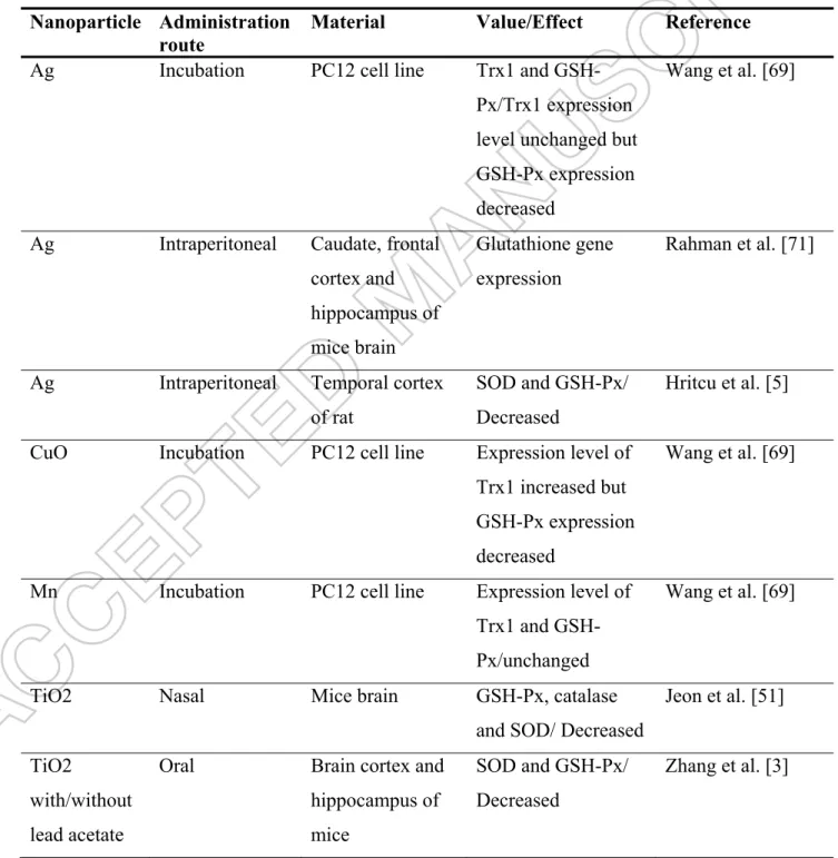
Table 1. Effects of nanoparticles on antioxidant enzymes in brain of experimental animals. Nanoparticle Administration
route
Material Value/Effect Reference
Ag Incubation PC12 cell line Trx1 and
GSH-Px/Trx1 expression level unchanged but GSH-Px expression decreased
Wang et al. [69]
Ag Intraperitoneal Caudate, frontal
cortex and hippocampus of mice brain Glutathione gene expression Rahman et al. [71]
Ag Intraperitoneal Temporal cortex
of rat
SOD and GSH-Px/ Decreased
Hritcu et al. [5]
CuO Incubation PC12 cell line Expression level of
Trx1 increased but GSH-Px expression decreased
Wang et al. [69]
Mn Incubation PC12 cell line Expression level of
Trx1 and GSH-Px/unchanged
Wang et al. [69]
TiO2 Nasal Mice brain GSH-Px, catalase
and SOD/ Decreased
Jeon et al. [51] TiO2
with/without lead acetate
Oral Brain cortex and
hippocampus of mice
SOD and GSH-Px/ Decreased
ZnO Intraperitoneal Brain of adult and old male SOD and GSH-Px/ Decreased Vedagiri and Thangarajan [32]
CAT; Catalase, GSH-Px; glutathione peroxidase, SOD; superoxide dismutase, Trx1; thioredoxin reductase 1,
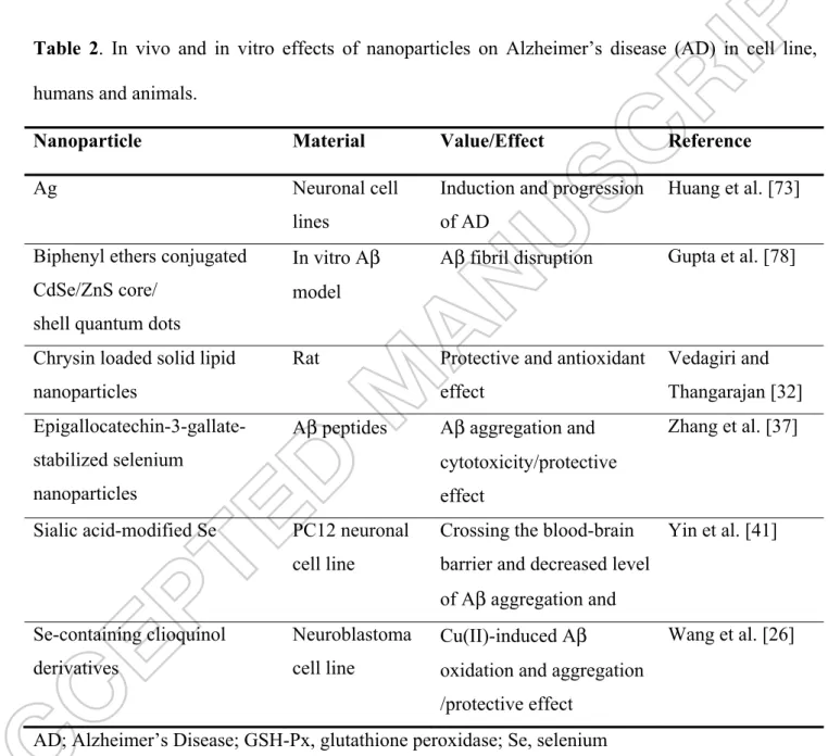
Table 2. In vivo and in vitro effects of nanoparticles on Alzheimer’s disease (AD) in cell line,
humans and animals.
Nanoparticle Material Value/Effect Reference
Ag Neuronal cell
lines
Induction and progression of AD
Huang et al. [73] Biphenyl ethers conjugated
CdSe/ZnS core/ shell quantum dots
In vitro Aβ model
Aβ fibril disruption Gupta et al. [78]
Chrysin loaded solid lipid nanoparticles
Rat Protective and antioxidant
effect Vedagiri and Thangarajan [32] Epigallocatechin-3-gallate-stabilized selenium nanoparticles
Aβ peptides Aβ aggregation and cytotoxicity/protective effect
Zhang et al. [37]
Sialic acid-modified Se PC12 neuronal cell line
Crossing the blood-brain barrier and decreased level of Aβ aggregation and Yin et al. [41] Se-containing clioquinol derivatives Neuroblastoma cell line Cu(II)-induced Aβ
oxidation and aggregation /protective effect
Wang et al. [26]
Figure formati oxygen pathoph (ZnO), importa The GS hydroge contains thiol for 1. Possible ion in bra species (R hysiology o silver (Ag) ant selenium SH-Px with en peroxide s thiol grou rm (GSH) b e pathways ain of pati ROS) occu of AD. Bloo and titaniu m-dependen catalase an e (H2O2) an ups in its str by the gluta of nanopar ents Alzhe urs in brain od-brain ba um dioxide nt detoxifyin nd superoxid nd leads to ructure and athione redu rticles on o eimer’s dis n affected arrier (BBA (TiO2) indu ng processe de dismutas the format it is synthe uctase (GR) oxidative st sease (AD) by AD. R A) permeab uces ROS p es is glutath se (SOD) p ion of oxid etized from ) enzyme. R ress and am . Excessive ROS have le nanopart production i hione perox lays a centr dized glutath GSH. GS-S Reduction of myloid beta e productio important ticles such in brain. On xidase (GSH ral role in t thione (GS-SG is reduc f thioredoxi a (Aβ) plaq on of react roles in as zinc ox ne of the m H-Px) syste the removal -SG). GSH-ced back to in is catalyz que tive the xide most em. l of -Px its zed
by thioredoxin reductase-1 enzyme (Trx1) and many radicals such as H2O2 and nitric oxide are
scavenged by Trx. Elemental selenium (Se0) from selenium rich nanoparticles is produced by catalytic effects of reduced GSH and mitochondrial ROS. Excessive Ca2+ entry through oxidative stress-activated TRP channels induce mitochondrial membrane depolarization and excessive ROS production. Interactions between TRP channel and selenium nanoparticles in AD should be investigated by future studies.