Article
J. Braz. Chem. Soc., Vol. 25, No. 12, 2329-2338, 2014. Printed in Brazil - ©2014 Sociedade Brasileira de Química
0103 - 5053 $6.00+0.00
A
http://dx.doi.org/10.5935/0103-5053.20140241*e-mail: [email protected]
Anionic Iron(III) Porphyrin Immobilized on/into Exfoliated Macroporous Layered
Double Hydroxides as Catalyst for Oxidation Reactions
Shirley Nakagaki,*,a Kelly A. D. F. Castro,a Geani M. Ucoski,a Matilte Halma,a,b Vanessa Prévot,a Claude Foranob and Fernando Wypycha
aLaboratório de Química Bioinorgânica e Catálise; Universidade Federal do Paraná (UFPR),
CP 19081, CEP 81531-990 Curitiba-PR, Brazil
bInstitut de Chimie de Clermont-Ferrand,Clermont Université, Université Blaise Pascal, BP 10448,
F-63000 Clermont-Ferrand, CNRS, UMR6296, ICCF, BP 80026, F-63171 Aubiere, France
A oxidação de substratos orgânicos é uma importante classe de reações explorada visando a produção de insumos industriais tais como epóxidos, álcoois e cetonas. As metaloporfirinas são compostos com reconhecida atividade catalítica que mimetizam processos de oxidação que ocorrem em seres vivos. A sua imobilização em diferentes sólidos robustos e inertes permite a recuperação e reuso do catalisador em processos catalíticos heterogêneos. Hidróxidos duplos lamelares (LDHs) são materiais inorgânicos constituídos de hidróxidos de metais di e trivalentes, resultando em lamelas bidimensionais carregadas positivamente. Neste trabalho reportamos a preparação de catalisadores baseados na imobilização de ferroporfirina (FeP) em LDH macroporoso (LDHM) obtido pelo método de co-precipitação usando poliestireno como template, reconstrução de óxidos e esfoliação (LDHME). A imobilização da FeP no LDH intercalado com ânions nitrato obtido pelo método de co-precipitação também é reportada. Os sólidos obtidos foram caracterizados e investigados como catalisadores na oxidação do cicloocteno e cicloexano.
The oxidation of organic substrates via catalytic routes is an important class of reactions to produce industrial input materials such as epoxides, alcohols, and ketones. Metalloporphyrins display recognized catalytic activity that mimics oxidation processes in living organisms. Their immobilization in different inert supports allows their recovery and reuse in heterogeneous catalytic processes. Layered double hydroxides (LDHs) are inorganic materials consisting of di- and trivalent metal hydroxides that afford bidimensional positively charged layers. This work reports on the preparation of the solid based on macroporous LDHs (LDHMs) by the co-precipitation method, which involved the use of polystyrene as template, oxides reconstruction, and exfoliation, to furnish LDHME. We also describe the immobilization of an iron(III) porphyrin (FeP) in LDHME and in LDH intercalated with nitrate anions, obtained by the co-precipitation method. Application of the immobilized catalysts in (Z)-cyclooctene and cyclohexane oxidation will help to assess their catalytic activity.
Keywords: porphyrin, macroporous LDH, oxidation, biomimetic reaction, heterogeneous
catalysis
Introduction
The family of enzymes collectively known as cytochrome P450 monooxygenases bear a heme prosthetic group and participate in different catalytic processes, mainly oxidative metabolism of endogenous/exogenous
products in mammals.1-5
Over the last years, researchers have made great efforts to develop routes that can generate robust synthetic
metalloporphyrins (MPs),6-10 aiming to mimic biological
enzymes such as cytochrome P450 (biomimetic approach). Indeed, MPs can efficiently and selectively catalyze
hydrocarbon oxidation.11 Some of these MPs are based
on the structure of meso-tetraphenylporphyrin [H2(TPP)].
The high efficiency and selectivity of MPs have motivated the proposition of technological catalytic systems based on this versatile family of oxidation catalysts.
Guo et al. developed and patented a process based on MP and atmospheric dioxygen to oxidize cyclohexane under
mild conditions,12 as cited by Guan Huang et al..13 Although
information about this process is scarce in the literature, its successful implementation in the industry took place in 2003.13
The first MP-based catalytic system relied on the Fe(III) complex [Fe(TPP)Cl] as catalyst and iodosylbenzene
(PhIO) as oxidant;14 it mimicked cytochrome P450 in
many reactions. Since then, the use of MPs, specially
FePs and MnPs, in oxidative systems6-11,14-21 has attracted
considerable attention, because these complexes can effectively and selectively catalyze a series of oxidation reactions in homogeneous medium, among which
epoxidation and hydroxylation stand out.15-21
Despite their efficiency in homogeneous systems, the use of this family of complexes in solution has raised concerns. In homogeneous media, secondary reactions (e.g., destructive oxidation of the complex and MP dimerization,
among others) can deactivate the catalytic species.10
Another difficulty posed by homogeneous catalysts is their recovery, reuse, and recycling, which could prevent the
design of a technological process.8,15,18 To minimize such
problems, researchers have turned to the synthesis of new
robust and resistant porphyrin structures10,22 as well as to
immobilization of these catalysts in different inorganic supports.22-33
Catalyst immobilization can facilitate catalyst recovery from the reaction medium, to enable their reuse and
recycling.17,33-39 This is particularly attractive from an
economical and environmental viewpoint. In this context, layered double hydroxides (LDHs) have emerged as
interesting supports to immobilize a variety of MPs.18,23,40-44
LDHs are synthetic layered compounds that contain divalent and trivalent metal cations [M(II) and M(III)] in a structure derived from the mineral brucite. In LDHs, M(III) metal ions replace part of the M(II) metals in the brucite-like structure, to give excess positive charge in the layers,
counterbalanced by intercalating hydrated anions.24,40,44-49
Some hydrogen-bonded water molecules may occupy the free space that remains in the interlayer region, which stabilizes the structure.
Chemical modification of the support or the MP with suitable organic groups can confer enhanced stability to the catalyst-support assembly. Some kind of interaction involving the modifier on the support and/or on the MP might occur, which shall not only increase the rate of MP
immobilization,40 but also provide a new solid catalyst with
unprecedented efficiency and selectivity.8,18,50
There has been a recent surge in scientist’s interest in nanostructured LDHs. This type of solid can be achieved
by controlling the textural properties of the material in terms of morphology, particle size, specific area, and porosity. A colloidal crystal template method has produced
tridimensional ordered macroporous MgAl-LDHs.41,51-53
Our group has already intercalated different anionic FePs in macroporous LDHs, to obtain more efficient and selective catalysts for heterogeneous hydroxylation and epoxidation
reactions than their homogeneous counterparts.18
Co-precipitation using a template like polystyrene spheres furnishes an LDH structure bearing macropores, which is
suitable for immobilization of countless FePs.41
In this work, we have prepared two solids by immobilizing the [Fe(TDCSPP)Cl] (Figure 1) - FeP - in different LDH supports, namely LDH containing intercalated nitrate
anions (LDH-NO3) and exfoliated macroporous LDH
(LDHME) (the iron porphyrin [Fe(TDCSPP)Cl] will be abbreviated as FeP by simplification; in this representation
charges and counter ion Cl1− are omitted). Because these
solids have the same composition, but different structures, we investigated their catalytic activity in the oxidation of two model substrates, (Z)-cyclooctene and cyclohexane, by PhIO.
Experimental
Materials
All the chemicals used in this study were purchased from Aldrich, Sigma or Merck and were of analytical grade. Iodosylbenzene (PhIO) was synthesized by hydrolysis
of iodosylbenzene diacetate,54 and the obtained solid
was carefully dried under reduced pressure and kept at
5 °C. LDH-NO3 and the macroporous LDH (LDHM)
were synthesized as described previously by our research group.40-42
Figure 1. Schematic representation of the molecular structure
of FeP ([FeIII(TDCSPP)Cl] employed in this work, where (TDCSPP) = 5,10,15,20-tetrakis(2,6-dichloro-3-sulfonate phenyl) porphyrin. The sulfonate groups are deprotonated in solution.
The free-base porphyrin
[5,10,15,20-tetrakis(2,6-dichloro-3-sulfonate phenyl)porphyrin], or [H2(TDCSPP)],
was synthesized, purified, and characterized as previously
described.55,56 The corresponding FeP, [FeIII(TDCSPP)Cl],
was obtained by inserting iron ion from ferrous chloride tetrahydrate into the free-base porphyrin ligand in dimethylformamide (DMF), as described by Adler, Longo,
and Kobayashi.57,58 It is expected that the FeIIP complex is
oxidized to FeIIIP by air. The FeP was purified by column
chromatography on Sephadex; deionized water was used as eluent. The metalloporphyrin was characterized by UV-Vis and electron paramagnetic resonance (EPR) spectroscopies. UV-Vis: [Fe(TDCSPP)Cl] (deionized water) 390 nm (ε = 6.4 × 10⁴ L mol−1 cm−1).
Preparation of the solid LDHME and intercalation of the FeP
To obtain LDHME, the previously synthesized LDHM40,41
was subjected to exfoliation.23,40 Shortly, 10 mL of formamide and 25 mg of solid LDHM were mixed in an Erlenmeyer flask, and the mixture was stirred in an ultrasound bath for 2 h. An almost translucent milky suspension emerged, which suggested the formation of macroporous LDH by exfoliation
(solid LDHME). The solid anionic FeP (3.8 × 10⁻⁶ mol)
was added to the suspension, and the mixture was kept under magnetic stirring for 24 h. A reddish brown solid (FeP-LDHME) arose. This solid was separated from the solution by centrifugation and extensively washed with water, until a colorless washing solution was achieved. The supernatants resulting from the washing process were collected and quantitatively analyzed by UV-Vis spectroscopy, to determine the FeP loading in the support.
Immobilization of FeP in LDH-NO3
LDH-NO3 (Mg/Al at a 3:1 molar ratio) was prepared
according to a methodology previously described by our
group.40 FeP immobilization was conducted by dispersing
LDH-NO340 (about 30 mg) in water (10 mL), which was
followed by addition of the FeP (about 3.9 × 10⁻⁶ mol).
The suspension was refluxed and stirred for 2 h. After that, the resulting solid was filtered and washed with water. The supernatant was analyzed by UV-Vis spectroscopy, to quantify the FeP that could have been removed from the matrix by leaching. The bright brown solid labeled as
FeP-LDH-NO3 was dried at 60 °C for 48 h.
Catalytic oxidation reactions
Catalytic oxidation reactions were carried out in a 2 mL thermostatic glass reactor equipped with a magnetic
stirrer, placed inside a dark chamber. The oxidation of (Z)-cyclooctene (previously purified on alumina column) and cyclohexane by PhIO was accomplished in the presence
of the catalyst FeP-LDH-NO3 or FeP-LDHME. In a
standard experiment, the solid catalyst (FeP-LDH-NO3 or
FeP-LDHME) and the oxidant (FeP/PhIO molar ratio 1:50) were suspended in 400 μL of solvent (dichloromethane/ acetonitrile 1:1 mixture, v/v) and degassed with argon for 15 min, inside a 2 mL vial. The reaction started after addition of the substrate (FeP/substrate molar ratio 1:5000); the oxidation reaction was performed under magnetic stirring for 1 h. At the end of the reaction, sodium sulfite acetonitrile saturated solution (50 µL) was added to the reaction mixture, to eliminate excess PhIO. The supernatant containing the reaction products was separated from the solid catalyst by centrifugation and transferred to a volumetric flask. The solid catalyst was washed several times with dichloromethane and acetonitrile, to extract any substrate and reaction products that might have remained adsorbed onto the solid catalyst. The washing solutions were added to the previously separated reaction supernatant, and the products and reagents content in these combined solutions was analyzed by gas chromatography,
using n-octanol (acetonitrile solution, 1.0 × 10⁻² mol L−1)
of high purity degree (99.9%) as internal standard. Product yields were based on the mass of PhIO added to each reaction. Control reactions were carried out using this same procedure, as follows: (a) substrate only, (b) substrate +
PhIO, and (c) substrate + PhIO + LDH-NO3 or LDHME
(supports without FeP). A similar procedure was adopted to test the FeP as homogeneous catalyst.
All the heterogeneous catalysts were exhaustively washed and dried for reuse in further reactions using the same procedure described above.
Characterization techniques
Scanning electron microscopy (SEM) characteristics of the samples were imaged on either a JEOL 5190 microscope operated at 15 keV or a JEOL JSM-6360LV operating 15 keV.
X-ray powder diffraction patterns (XRPD) were recorded in the reflection mode on a Shimadzu XRD-6000 diffractometer operating at 40 kV and 40 mA; CuKα
radiation (λ = 1.5418 Å) and a dwell time of 1° min−1 were
employed.
Electronic spectra (UV-Vis) were obtained on a Cary-Varian 100 Bio and Shimadzu UV-2501PC spectrophotometer, in the 200-800 nm range.
EPR measurements of the powder materials were accomplished on an EMX microX spectrometer (standard
concavity: 4102-SP and 9.5 GHz X band frequency), at
room temperature or at 77 K (in liquid N2).
Products from the catalytic oxidation reactions were quantified on a gas chromatograph Agilent 6850 (FID detector) equipped with a capillary column DB-WAX (J&W Scientific). Quantitative analyses were based on internal standards.
Results and Discussion
Preparation of LDH-NO3 and LDHM relied on
the co-precipitation methodology without previous intercalation/functionalization of the inorganic matrix with organic molecules, as detailed by our group in recent
publications.40,41 In particular, the synthesis of LDHM
involved co-precipitation of divalent (Mg2+) and trivalent
(Al3+) metal ions during the synthesis of LDH; polystyrene
beads (PS) served as template.41 After synthesis of the
LDH-NO3 solid, its calcination removed the polystyrene
template that remained in the double oxides resulting from the calcined LDH. In the presence of dodecyl sulfate (DDS)
solution, the lamellar structure of LDH-NO3 re-emerged,
whilst DDS intercalated within the macropores.
Immobilization of the FeP in LDH-NO3 (FeP-LDH-NO3) or
exfoliated macroporous LDH (FeP-LDHME)
FeP immobilization in LDH-NO3 resulted in a brown
solid with spectroscopic properties similar to those
previously reported by us.40,46
Exfoliation of LDHM in the presence of formamide afforded a white colloidal (milky) suspension, designated LDHME. After an ion exchange reaction between this suspension and the anionic FeP, a reddish brown solid arose (FeP-LDHME). This solid was further characterized by X-ray diffraction (XRD), Fourier transform infrared (FTIR), UV-Vis, and SEM. The exfoliation process generated individual lamella (or monolamella), which could facilitate chemical modification or exchange of the
anion present in the lamellar space.40,59 The formamide
employed during the exfoliation process solvated the anions and intercalated between the lamella, which caused a rupture in the lamellar structure and consequently exfoliated the compound in the form of individual lamella.
Indeed, Hibino59 described that formamide constitutes a
good solvent for this purpose and dismisses the need for heating or reflux.
As expected, the anionic FeP successfully anchored in
both LDHME and LDH-NO3: the negative charges in the
structure of this FeP (Figure 1) effectively interacted with the positively charged layers of the solids. Obviously, this
process did not exhaust the anionic exchange capacity of the solid matrixes, so part of the original anions still existed in the material.
To quantify the FeP loading in LDH-NO3 and LDHME,
we measured the amount of non-immobilized FeP in the combined solutions from the washings of the solids
FeP-LDH-NO3 and FeP-LDHME by UV-Vis spectroscopy.
The FeP loadings on LDH and LDHME were 1.53 × 10⁻⁴
and 9.23 × 10⁻⁵ mol FeP g−1 of matrix, respectively.
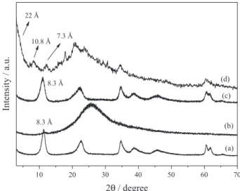
Figure 2 shows the XRD patterns of the solids before and after FeP immobilization. The solid resulting from FeP
immobilization in LDH-NO3 (Figure 2c) exhibited an X-ray
diffraction pattern similar to that obtained for LDH-NO3
alone (Figure 2a), with a basal distance of approximately 8.3 Å. This distance corresponded to the presence of intercalated nitrate anions, indicating that the immobilized FeP did not replace these ions and probably localized on
the surface of the layered crystals.10
LDHM (figure not shown) displayed reflections (00l and hkl) characteristic of hydrotalcite-like compounds, with a basal distance of 25.5 Å. These data agreed with literature values for DDS intercalated into LDHs. The XRD analysis of LDHME as a slurry revealed a halo in the 2θ region of 18 to 40° after exfoliation, typical
of amorphous compound in colloidal suspension.41 The
phase obtained after ion exchange with the anionic FeP had basal spacing of approximately 22 Å, calculated from the higher order diffraction peak at 2θ values of 12.15° (7.28Å × 3 = 21.85Å ca. 22 Å) (Figure 2d). Although not evident as indicated by an arrow in Figure 2, other two harmonic peaks related with the same basal distance (8.22° = 10.76 Å (10.76 × 2 = 21.5 Å); 12.15° = 7.28 Å
Figure 2. XRD of the solids (a) LDH-NO3; (b) LDHME suspension; (c) FeP-LDH-NO3; and (d) FeP-LDHME. The arrow indicates the peak region at 2θ values.
(7.28 × 3 = 21.85 Å)) are a clear evidence that another peak occurs at 4.04° (2θ) (21.87Å). This basal distance is close to the value reported in the literature when porphyrins are
intercalated into LDH-NO3 (Figure 2c). Therefore, we
cannot exclude FeP intercalation in the interlayer space of LDHME, but asserting that intercalation indeed took place in this case is difficult because intercalated DDS anions also exist in the matrix and produce similar basal spacing. Interestingly, DDS bilayers and FeP species exhibit high chemical and structural compatibility, so co-immobilization of this species could also occur, as recently described for
the immobilization of this same FeP in LDHM.41 Besides
this diffraction peak, the characteristic peak of LDH-NO3 in
the region of 60° (2θ) appeared, indicating that exfoliated layers restacked (Figure 2d).
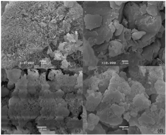
In general, the SEM images of LDHs predominantly intercalated with inorganic ions show particles agglomerated in the hexagonal form, known as “pink sand”. However, if the LDH contains intercalated organic anions, the SEM
images reveal layered crystals with round corners,40,60
which was the case with LDH-NO3 (Figure 3a), a solid that
contained submicrometric layered crystals.
Because FeP immobilization in both LDH-NO3
and LDHME happened under rash conditions, the crystals should lose their original morphology and form agglomerates without any apparent order (Figure 3b and 3d). In the case of LDHM (Figure 3c), part of the
image showed the macroporous structure that resulted from removal of the polystyrene sphere, evidencing a honeycomb-like morphology. After exfoliation and restacking, even these structures disappeared, to reveal particles arranged in the form of a “house of cards”, typical
of exfoliated and restacked LDHs.61 Obviously, some of
the layers remained stacked, to give the original crystals, as attested by XRD (Figure 2d).
The FTIR technique was inconclusive to ascertain the presence of the FeP on the surface or intercalated in the
supports. Indeed, FeP-LDH-NO3 and FeP-LDHME (in the
Supplementary Information (SI) section) display the typical
bands of FePs (region between 1200 and 1020 cm−1, typical
of the νsym.S-φ and νasym.S-φ symmetric and asymmetric
vibrations of the φ-SO3 groups.62,63 The large intensity
of the bands of the support in the region of 3000 cm−1
(surface hydroxyl groups and also water molecules in the
interlamellar space), 1628 cm−1 (water molecules present
in the interlayer space), and 1385 and 843 cm−1 (symmetric
and asymmetric vibrations of intercalated nitrate, respectively) allied with the low FeP concentration in the support probably made difficult to observe the FeP bands.
These bands occur between 1600-1370 cm−1, due to νC=C
phenyl and around 640 cm−1, attributed to out-of-the-plane
C-H vibrations.40,63 For the solid FeP-LDHME, other bands
were observed in the region of 2960-2840 cm−1 (νC-H);
around 1700 cm−1 (νC=O) and 1600 cm−1 symmetric
angular deformation in the plane (NH2) attributed the presence of formamide used in the exfoliation process.
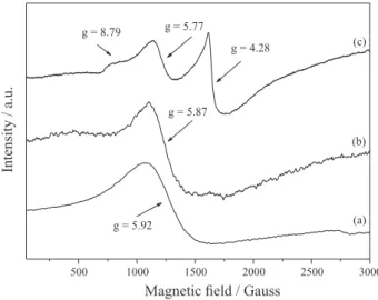
For a system containing high-spin Fe(III), the combination of five unpaired electrons results in three Kramer doublets: ±1/2, ±3/2, and ±5/2. The extent to which these energy levels are occupied depends on the separating field and temperature. In a strong crystal field of tetragonal symmetry, the deployment parameter of the field is large, and only the transitions ±1/2 are viewable (g⊥ and g// = 6.0 and 2.0, respectively).64-66 However, if the local symmetry reduces from tetragonal to orthorhombic, for example, which distorts the porphyrin plane, other signs arise. Systems with maximum distortion display only one signal at g = 4.3. The field separation can be described in terms of two parameters: D (axial separation) and E (rhombic splitting). The ratio between D and E may have values between 0 (axial symmetry) and 0.33 (rhombicity
maximum) with different values of factor g.64-66
Figure 4 illustrates the EPR spectra of the solids obtained after FeP immobilization. The free FeP (Figure 4a) presented a signal in g ca. 6.0 (axial symmetry), typical of a high-spin 5/2 FeP complex. FeP immobilization in LDH and LDHME (Figures 4b and 4c) gave the characteristic signal of high-spin Fe(III) (S = 5/2), typical of axially
symmetric FePs.17,52,67 A typical signal of Fe(III) with
rhombic distortion (g = 4.3) arose, which is usual in
immobilized FeP systems.10,68 The estimated D/E ratio was
0.20, which enabled assignment of the observed signals to transitions of the states + −1/2 + and −3/2, with g values of 8.79, 5.77, and 4.28.
The intense signal at g = 4.28 detected for solid FeP-LDHME originated from the greater distortion undergone by the FeP during the immobilization process. Such
distortion may be associated with the necessity of the FeP structure to settle as close as possible to the matrix, to maximize its interactions with the positively charged
layers of the support.17 Hence, EPR analysis confirmed the
presence of FeP in the matrixes (Figures 4b and 4c) after the immobilization process and evidenced FeP distortions (Figure 4c).
UV-Vis analysis of the solid samples also attested that FeP existed in the support (Figure 5).
The spectra of the solids FeP-LDH-NO3 and
FeP-LDHME contained the Soret band characteristic of Fe(III) Ps in the region of 400 nm. Other three bands appeared between 500 and 700 nm (Q bands).
Comparison of the spectra of the immobilized FeP with that of the free FeP revealed that immobilization promoted
a blue shift of the Soret band.9,18,69 Such shifts may result
from (i) FeP intercalation into the support, which may distort the structure of the complex, or (ii) interactions between the support surface and the FeP, which can cause steric constraints. Regardless of the immobilization mode,
in both cases (FeP-LDH-NO3 and FeP-LDHME), the blue
shift may have stemmed from interactions between the FeP plane and the support in an attempt of the complex to acquire a more planar conformation and maximize the
electrostatic interaction.29 The Soret band of FeP-LDH-NO
3 shifted less markedly, leading to the conclusion that the FeP was less constrained in this support (Figure 5d).
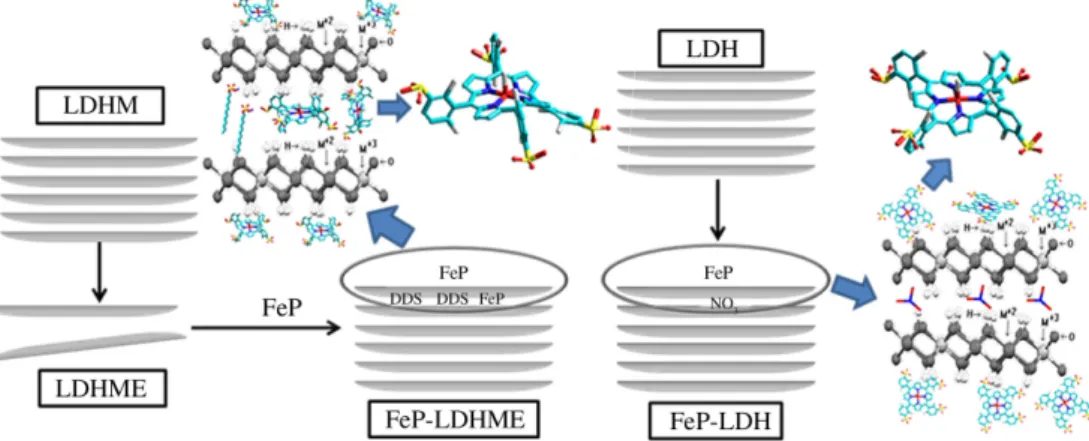
Because we immobilized the FeP in previously prepared
LDH-NO3, and on the basis of XRD analysis, we inferred
that the FeP bound at the surface of the layered crystals. In the case of LDHME, the FeP was added to a suspension of exfoliated macroporous LDH, so many individual layers were available to interact with the anionic FeP, which localized
Figure 4. EPR spectrum of [Fe(TDCSPP)Cl] before and after FeP
immobilization on LDHs: (a) [Fe(TDCSPP)Cl]; (b) FeP-LDH-NO3; and (c) FeP-LDHME.
Figure 5. UV-Vis spectra of (a) LDH-NO3; (b) LDHME; (c) FeP; (d) FeP-LDH-NO3; and (e) FeP-LDHME.
closer to the positive charges on the matrix (Figure 6). These results were in line with the EPR data, which had suggested
that FeP-LDH-NO3 displayed less rhombic distortion than
FeP-LDHME (Figures 4b and 4c, respectively). Catalytic oxidation reactions
The use of FePs as catalysts promotes C–H bond activation by oxidation under biomimetic mild conditions and in the presence of a number of oxygen atom donors such as iodosylbenzene,8,11,14,16,28,70 hydrogen peroxide,71 and
dioxygen,72,73 among others. Here, we decided to employ
PhIO as oxygen donor because MP/PhIO is a classical biomimetic system that can produce the same intermediate
catalytic species regardless of the FeP.6,11
The oxidation of (Z)-cyclooctene by MP/PhIO systems produces epoxide as the sole oxidation product, with
no traces of allylic alcohol or ketone.74 For this reason,
(Z)-cyclooctene is the substrate of choice when testing the activity of biomimetic catalytic systems involving MPs, and we also selected it to assay the efficiency and stability of the anionic FeP immobilized in both of the investigated supports. By employing this substrate, we also obtained initial information about accessibility to the activated iron site.
The activation of the highly inert C–H bonds of cyclic alkanes, to obtain hydroxylated products, is one of the most remarkable reactions that natural and synthetic systems
can accomplish.75 Indeed, C–H bond activation in alkanes
calls for more drastic conditions than those necessary for alkene functionalization, which in turn allows one to differentiate between the performances of an MP catalyst in solution and the same MP catalyst immobilized in a solid support. Cyclohexane is also a very useful substrate to test the activity of FeP/PhIO systems as its major oxidation products, cyclohexanol and cyclohexanone provides information about catalyst selectivity.
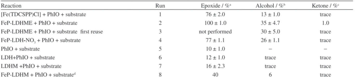
Table 1 presents the results from the oxidation of (Z)-cyclooctene and cyclohexane by PhIO catalyzed by
FeP-LDH-NO3 and FeP-LDHME. We also evaluated the
free FeP for comparison.
The immobilized FeP afforded excellent product yields for both (Z)-cyclooctene oxidation and cyclohexane hydroxylation, which were even better than the yields obtained with the free FeP in homogeneous solution (Table 1 runs 1, 2, and 4). All the reactions that involved FeP-based catalysts (runs 1 to 4) furnished considerably higher product percentages than the control reactions (runs 5 to 7), which confirmed that the catalytic activity of the studied catalysts was really due to the FeP.
The low solubility of anionic porphyrins like
[Fe(TDCSPP)] in a CH2Cl2/CH3CN 1:1 solvent mixture
certainly underlay the lower yields obtained with the free FeP in homogeneous medium (run 1) as compared with the immobilized FeP (runs 2 and 4). Another explanation for the difference between the catalytic performances of the FeP in homogeneous and heterogeneous media might be that the charged catalyst hinders the approach of the apolar substrates to the active metal center. Indeed, immobilization
of the anionic FeP in the LDH-NO3 and LDHME supports
by electrostatic interaction may have minimized this effect in the case of the heterogeneous process, to improve the catalytic activity of the FeP.40,41
Interestingly, FeP-LDHME presented better catalytic
results as compared with FeP-LDH-NO3 (runs 2 and 4,
respectively) a probable consequence of the macroporous structure of the former support. In fact, the strategy employed during catalyst immobilization can tailor catalyst efficiency for different substrates. The morphology and textural properties of the support can direct the substrates to the catalytically active center in a different way.18,36,50,76-78
Besides that, FeP immobilization protects the catalyst from the oxidative attack of another catalytically activated FeP, which avoids destruction of the catalytic species. In the
specific case of FeP-LDHME, the microporous structure of the support may have conferred better protection to the FeP, whereas the immobilized FeP remained on the surface
of the layered crystals in FeP-LDH-NO3, as indicated by
XRD analysis (Figure 2c).
Immobilized catalysts offer a number of advantages: control of the reaction medium, prevention of catalyst degradation, low cost (depending on the support), stability, and possibility of catalyst reuse and/or recycling.
Indeed, we have performed the catalysts reuse reactions that surprisingly afforded the best result in a new cyclohexane oxidation reaction (FeP-LDHME; run 3). Also, the obtained product yield was similar to that achieved with the freshly prepared solid catalyst (run 2). Therefore, this solid catalytic is potentially reusable in oxidation reactions, favoring heterogeneous catalysis over the homogeneous route.
The monitoring of the reaction solvent or the washing solutions resulting from the filtration of the catalyst FeP-LDHME was performed and no traces of porphyrin were detected by UV-Vis analysis. This fact allowed us to conclude that there was no leaching of the FeP after the first use or during the washing process and the reaction is truly performed in heterogeneous media.
In addition, FeP-LDH-NO3 and FeP-LDHME (spectra
FTIR) after catalysis reactions, the solids presented characteristic bands observed in the fresh catalysts, which seems to be a convincing evidence that the catalysts preserved its structure after the reaction, washing process and reuse. As the catalysts consist of very fine powder, after each reaction, around 10% of the solids is lost by manipulation, washing and drying.
Recently, we have verified that highly distorted immobilized MPs and MPs in solution afford better
catalytic results.22 This is because the presence of bulkier
substituents in the porphyrin macrocycle can affect ring
symmetry and distort the structure.22 This may influence
the formation and stabilization of the active catalytic oxo-species in such a way that the more distorted porphyrin
structure performs better than the less distorted one.22 In
fact, distortions in the porphyrin skeleton can destabilize the macrocycle π systems, thereby modulating their oxidation
potentials.79-87 Hence, distinct structural configurations
could also determine the catalytic outcome.78,79
The catalytic results achieved with the solid FeP-LDHME
were also better than those reported by Halma et al.41 for
FeP-LDHM (Table 1, run 8).41 In the former catalyst,
the FeP acquired a more distorted arrangement due to exfoliation associated with the structure and macroporous morphology of LDHME, as evidenced by SEM.
Conclusions
This work described the preparation of two solid catalysts via immobilization of the anionic [Fe(TDCSPP)] in LDH supports, with the aim to compare the influence of supports with the same composition but different structures in the catalytic activity of the iron complex during the oxidation of two model substrates.
We selected a second generation ironporphyrin10
because the presence of electron-withdrawing groups on the phenyl substituents of the porphyrin ring made the catalyst more resistant to oxidative degradation as compared with simpler metalloporphyrins like the first-generation iron(III)
tetraphenylporphyrin [Fe(TPP)] employed by Groves.11,14
We successfully immobilized the ironporphyrin on both LDHs (one LDH was obtained by co-precipitation; the other was prepared by using polystyrene beads as template). After calcinations to eliminate polystyrene, the latter solid presented a macroporous structure of oxides that regenerated the LDH structure after hydration. Additionally, this same FeP was also immobilized on LDH of Mg/Al containing nitrate anions intercalated. Characterization of
Table 1. (Z)-cyclooctene and cyclohexane oxidation by PhIO results catalyzed by FeP in homogeneous and heterogeneous media
Reaction Run Epoxide / %a Alcohol / %b Ketone / %c
[Fe(TDCSPP)Cl] + PhIO + substrate 1 76 ± 2.0 13 ± 1.0 trace
FeP-LDHME + PhIO + substrate 2 100 ± 1.0 35 ± 4.7 1.0
FeP-LDHME + PhIO + substrate first reuse 3 not performed 30 ± 5.0 trace
FeP-LDH-NO3 + PhIO + substrate 4 77 ± 1.1 26 ± 1.1 trace
PhIO + substrate 5 10 ± 1.0 − −
LDH+PhIO + substrate 6 12 ± 1.0 trace trace
LDHM +PhIO + substrate 7 16 ± 2.3 trace trace
FeP-LDHM + PhIO + substrated 8 40 6 trace
The reaction yield of acyclooctene/cyclooctene oxide; bcyclohexane/cyclohexanol; and ccyclohexane/cyclohexanone were calculated on the basis of the amount of PhIO used in each reaction. The results represent an average of at least triplicate reactions. Reaction conditions: 1 h, room temperature, acetonitrile/dichloromethane (1:1 v/v) as solvent, inert atmosphere, FeP/PhIO/substrate (cyclooctene or cyclohexane) molar ratio = 1:50:5000; dresults from reference 41. Catalytic reaction performed with the same FeP immobilized in LHDM under reaction conditions similar to those adopt in this study, except for the FeP/PhIO/ substrate molar ratio = 1:10:1000.
the solids obtained after immobilization/intercalation of the ironporphyrin by UV-Vis and FTIR spectroscopies and XRD analysis confirmed the presence of the iron complex in the LDH. The solids displayed good catalytic activity in heterogeneous catalysis; they furnished yields higher than those achieved in homogeneous medium.
Supplementary Information
Supplementary information (spectra FTIR of all the compounds prepared) is available free of charge at http:// jbcs.sbq.org.br.
Acknowledgements
The authors are grateful to Conselho Nacional de Desenvolvimento Científico e Tecnológico (CNPq), Coordenação de Aperfeiçoamento de Pessoal de Nível Superior (CAPES), Fundação Araucária, Fundação da Universidade Federal do Paraná (FUNPAR), Universidade Federal do Paraná (UFPR), and Université Blaise Pascal for financial support. The authors are also grateful to Centro de Microscopia Eletrônica from UFPR for the SEM analysis.
References
1. Montellano, P. R. O.; Cytochrome P450: Structure, Mechanism,
and Biochemistry, 3rd ed.; Kluwer Academic/Plenum Publishers:
New York, USA, 2005.
2. Lohmann, W.; Anal. Bioanal. Chem. 2008, 391, 79.
3. Rittle, J.; Green, M. T.; Science 2010, 330, 933.
4. Guengerich, F. P.; Sohl, C. D.; Chowdhury, G.; Arch. Biochem.
Biophys. 2011, 507, 126.
5. Rendic, S.; Guengerich, F. P.; Chem. Res. Toxicol. 2012, 25, 1316. 6. Mansuy, D.; C. R. Chimie 2007, 10, 392.
7. Ricoux, R.; Raffy, Q.; Mahy, J.-P.; C. R. Chimie 2007, 10, 684. 8. Nakagaki, S.; Castro, K. A. D. F.; Machado, G. S.; Halma, M.;
Drechsel, S. M.; Wypych, F.; J. Braz. Chem. Soc. 2006, 17, 1672. 9. Kadish, K.; Smith, K.; Guillard, R.; The Porphyrins Handbook,
Academic Press: New York, USA, 1999.
10. Dolphin, D.; Traylor, T. G.; Xie, L. Y.; Acc. Chem. Res. 1997,
30, 251.
11. Groves, J. T.; J. Inorg. Biochem. 2006, 100, 434.
12. Guo, C.; Liu, Q.; Liu, Y.; CN1405131-A 2003 and CN1191218-C 2005, available at http://worldwide.espacenet.com/ and http:// www.google.com/patents/CN1405131A?cl=en, accessed in October 2014.
13. Huang, G.; Luo, Z.-C.; Hu, Y.-D.; Guo, Y.-A.; Jiang, Y.-X.; Wei, S.-J.; Chem. Eng. J. 2012, 195-196, 165.
14. Groves, J. T.; Nemo, T. E.; Myers, R. S.; J. Am. Chem. Soc.
1979, 101, 1032.
15. Ucoski, G. M.; Castro, K. A. D. F.; Ciuffi, K. J.; Ricci, G. P.; Marques, J. A.; Nunes, F. S.; Nakagaki, S.; Appl. Catal., A 2011,
404, 120.
16. Doro, F. G.; Smith, J. R. L.; Ferreira, A. G.; Assis, M. D.; J. Mol.
Catal. A: Chem. 2000, 164, 97.
17. Machado, G. S.; Castro, K. A. D. F.; de Lima, O. J.; Nassar, E. J.; Ciuffi, K. J.; Nakagaki, S.; Colloids Surf., A 2009, 349, 162. 18. Halma, M.; Castro, K. A. D. F.; Taviot-Gueho, C.; Prévot, V.;
Forano, C.; Wypych, F.; Nakagaki, S.; J. Catal. 2008, 257, 233. 19. Moreira, M. S. M.; Martins, P. R.; Curi, R. B.; Nascimento,
O. R.; Iamamoto, Y.; J. Mol. Catal. A: Chem. 2005, 233, 73. 20. Latifi, R.; Tahsini, L.; Karamzadeh, B.; Safari, N.; Nam, W.; de
Visser, S. P.; Arch. Biochem. Biophys. 2011, 507, 4.
21. Santos, J. S. D.; Faria, A. L.; Amorin, P. M. D. S.; Luna, F. M. L.; Caiado, K. L.; Silva, D. O. C. E.; Sartoratto, P. P. C.; Assis, M. D.; J. Braz. Chem. Soc. 2012, 23, 1411.
22. Castro, K. A. D. F.; Simões, M. M. Q.; Neves, M. G. P. M. S.; Cavaleiro, J. A. S.; Wypych, F.; Nakagaki, S.; Catal. Sci.
Technol. 2014, 4, 129.
23. Nakagaki, S.; Halma, M.; Bail, A.; Arízaga, G. G. C.; Wypych, F.; J. Colloid Interface Sci. 2005, 281, 417. 24. Nakagaki, S.; Wypych, F.; J. Colloid Interface Sci. 2007, 315,
142.
25. Nakagaki, S.; Ramos, A. R.; Benedito, F. L.; Peralta-Zamora, P. G.; Zarbin, A. J. G.; J. Mol. Catal. A: Chem. 2002, 185, 203. 26. Traylor, T. G.; Byun, Y. S.; Traylor, P. S.; Battioni, P.;
Mansuy, D.; J. Am. Chem. Soc. 1991, 113, 7821. 27. Bedioui, F.; Coord. Chem. Rev. 1995, 144, 39.
28. Martinez-Lorente, M. A.; Battioni, P.; Kleemiss, W.; Bartoli, J. F.; Mansuy, D.; J. Mol. Catal. A: Chem. 1996, 113, 343. 29. Cady, S. S.; Pinnavaia, T. J.; Inorg. Chem. 1978, 17, 1501. 30. Benedito, F. L.; Nakagaki, S.; Saczk, A. A.; Peralta-Zamora,
P. G.; Costa, C. M. M.; Appl. Catal., A 2003, 250, 1.
31. Ucoski, G. M.; Nunes, F. S.; Defreitas-Silva, G.; Idemori, Y. M.; Nakagaki, S.; Appl. Catal., A 2013, 459, 121.
32. Ferreira, G. K. B.; Castro, K. A. D. F.; Machado, G. S.; Ribeiro, R. R.; Ciuffi, K. J.; Ricci, G. P.; Marques, J. A.; Nakagaki, S.;
J. Mol. Catal. A: Chem. 2013, 378, 263.
33. Silva, M.; Azenha, M. E.; Pereira, M. M.; Burrows, H. D.; Sarakha, M.; Forano, C.; Ribeiro, M. F.; Fernandes, A.; Appl.
Catal., B 2010, 100, 1.
34. Machado, G. S.; Castro, K. A. D. F.; Wypych, F.; Nakagaki, S.;
J. Mol. Catal. A: Chem. 2008, 283, 99.
35. Machado, G. S.; Groszewicz, P. B.; Castro, K. A. D. F.; Wypych, F.; Nakagaki, S.; J. Colloid Interface Sci. 2012, 374, 278.
36. Halma, M.; Bail, A.; Wypych, F.; Nakagaki, S.; J. Mol. Catal. A:
Chem. 2006, 243, 44.
37. Moghadam, M.; Mirkhani, V.; Tangestaninejad, S.; Mohammdpoor-Baltork, I.; Kargar, H.; J. Mol. Catal. A: Chem.
38. Huang, G.; Liu, S.-Y.; Guo, Y.-A.; Wang, A.-P.; Luo, J.; Cai, C.-C.; Appl. Catal., A 2009, 358, 173.
39. Nakagaki, S.; Benedito, F. L.; Wypych, F.; J. Mol. Catal. A:
Chem. 2004, 217, 121.
40. Castro, K. A. D. F.; Bail, A.; Groszewicz, P. B.; Machado, G. S.; Schreiner, W. H.; Wypych, F.; Nakagaki, S.; Appl. Catal., A
2010, 386, 51.
41. Halma, M.; Castro, K. A. D. F.; Prévot, V.; Forano, C.; Wypych, F.; Nakagaki, S.; J. Mol. Catal. A: Chem. 2009, 310, 42. 42. Wypych, F.; Bubniak, G. A.; Halma, M.; Nakagaki, S.; J. Colloid
Interface Sci. 2003, 264, 203.
43. Kovanda, F.; Jindová, E.; Lang, K.; Kubát, P.; Sedláková, Z.;
Appl. Clay Sci. 2010, 48, 260.
44. Rives, V.; Angeles Ulibarri, M. A.; Coord. Chem. Rev. 1999,
181, 61.
45. Crepaldi, E. L.; Valim, J. B.; Quim. Nova 1998, 21, 300. 46. Inacio, J.; Taviot-Guého, C.; Forano, C.; Besse, J. P.; Appl. Clay
Sci. 2001, 18, 255.
47. Li, F.; Zhang, L.; Evans, D. G.; Forano, C.; Duan, X.;
Thermochim. Acta 2004, 424, 15.
48. Stanimirova, T.; Hibino, T.; Appl. Clay Sci. 2006, 31, 65. 49. Ishikawa, T.; Matsumoto, K.; Kandori, K.; Nakayama, T.;
Colloids Surf., A 2007, 293, 135.
50. Machado, G. S.; Arízaga, G. G. C.; Wypych, F.; Nakagaki, S.;
J. Catal. 2010, 274, 130.
51. Rafqah, S.; Chung, P. W.-W.; Forano, C.; Sarakha, M.;
J. Photochem. Photobiol., A 2008, 199, 297.
52. Abello, S.; Medina, F.; Tichit, D.; Perez-Ramirez, J.; Cesteros, Y.; Salagre, P.; Sueiras, J. E.; Chem. Commun. 2005, 11, 1453. 53. Géraud, E.; Prévot, V.; Leroux, F.; J. Phys. Chem. Solids 2006,
67, 903.
54. Sharefkin, J. G.; Saltzmann, H.; Org. Synth. 1973, 5, 660. 55. Lindsey, J. S.; Schreiman, I. C.; Hsu, H. C.; Kearney, P. C.;
Marguerettaz, A. M.; J. Org. Chem. 1987, 52, 827. 56. Turk, H.; Ford, W. T.; J. Org. Chem. 1991, 56, 1253. 57. Adler, A. D.; Longo, F. R.; Kampas, F.; Kim, J.; J. Inorg. Nucl.
Chem. 1970, 32, 2443.
58. Kobayashi, H.; Higushi, T.; Kaizu, Y.; Osada, H.; Aoki, M.;
Bull. Chem. Soc. Jpn. 1975, 48, 3137.
59. Hibino, T.; Kobayashi, M.; J. Mater. Chem. 2005, 15, 653. 60. Leroux, F.; Gachon, J.; Besse, J.-P.; J. Solid State Chem. 2004,
177, 245.
61. Frost, R. L.; Zhu, J.; He, H.; Yuan, P.; Tao, Q.; Shen, W.; Bostrom, T.; J. Colloid Interface Sci. 2008, 319, 498. 62. Halma, M.; Wypych, F.; Drechsel, S. M.; Nakagaki, S.;
J. Porphyrins Phthalocyanines 2002, 6, 502.
63. Nakamoto, K.; Infrared and Raman Spectra of Inorganic and
Coordination Compounds, 5th ed.; John Wiley & Sons, Inc.:
Chicago, USA, 1997.
64. Whittaker, J. W.; Lipscomb, J. D.; Kent, T. A.; Munck, E.;
J. Biol. Chem. 1984, 259, 4466.
65. Tsai, R.; Yu, C. A.; Gunsalus, I. C.; Peisach, J.; Blumberg, T. W.; Orme-Johnson, W. H.; Beinert, H.; Proc. Natl. Acad. Sci.
U. S. A. 1970, 66, 1157.
66. Holman, T. R.; Juarez-Garcia, C.; Hendrich, M. P.; Que, L.; Munck, E.; J. Am. Chem. Soc. 1990, 112, 7611.
67. Krzystek, J.; Ozarowski, A.; Telser, J.; Coord.Chem. Rev. 2006,
250, 2308.
68. Castro, K. A. D. F.; Halma, M.; Machado, G. S.; Ricci, G. P.; Ucoski, G. M.; Ciuffi, K. J.; Nakagaki, S.; J. Braz. Chem. Soc.
2010, 21, 1329.
69. Guo, C.-C.; Huang, G.; Zhang, X.-B.; Guo, D.-C.; Appl.
Catal., A 2003, 247, 261.
70. Nam, W.; Ryu, Y. O.; Song, W. J.; J. Biol. Inorg. Chem. 2004,
9, 654.
71. Rebelo, S. L. H.; Gonçalves, A. R.; Pereira, M. M.; Simões, M. M. Q.; Neves, M. G. P. M. S.; Cavaleiro, J. A. S.; J. Mol.
Catal. A: Chem., 2006, 256, 321.
72. Lane, B. S.; Burgess, K.; Chem. Rev. 2003, 103, 2457. 73. Lyons, J. E.; Ellis Jr, P. E.; Metalloporphyrins in Catalytic
Oxidations; Sheldon, R. A., ed.; Marcel Dekker: New York,
USA, 1994.
74. Appleton, A. J.; Evans, S.; Smith, J. R. L.; J. Chem. Soc., Perkin
Trans. 2 1996, 2, 281.
75. Berkowitz, J.; Ellison, G. B.; Gutman, D.; J. Phys. Chem. 1994,
98, 2744.
76. Machado, G. S.; Wypych, F.; Nakagaki, S.; J. Colloid Interface
Sci. 2012, 377, 379.
77. Thomas, J. M.; Raja, R.; Sankar, G.; Bell, R. G.; Acc. Chem.
Res. 2001, 34, 191.
78. Nakagaki, S.; Xavier, C. R.; Wosniak, A. J.; Mangrich, A. S.; Wypych, F.; Cantão, M. P.; Denicoló, I.; Kubota, L. T.; Colloids
Surf., A 2000, 168, 261.
79. Durval, H.; Bulach, V.; Fischer, J.; Renner, M. W.; Fajer, J.; Weiss, R.; J. Biol. Inorg. Chem. 1997, 2, 662.
80. Barkigia, K. M.; Chantranupong, L.; Smith, K. M.; Fajer, J.;
J. Am. Chem. Soc. 1988, 110, 7566.
81. Barkigia, K. M.; Berber, M. D.; Fajer, J.; Medforth, C. J.; Renner, M. W.; Smith, K. M.; J. Am. Chem. Soc. 1990, 112, 8851. 82. Barkigia, K. M.; Renner, M. W.; Furenlid, L. R.; Medforth, C. J.;
Smith, K. M.; Fajer, J.; J. Am. Chem. Soc. 1993, 115, 3627. 83. Renner, M. W.; Barkigia, K. M.; Zhang, Y.; Medforth, C. J.;
Smith, K. M.; Fajer, J.; J. Am. Chem. Soc. 1994, 116, 8582. 84. Takeuchi, T.; Gray, H. B.; Goddard, W. A.; J. Am. Chem. Soc.
1994, 116, 9730.
85. Takeda, J.; Sato, M.; Chem. Lett. 1995, 24, 939. 86. Spiro, T.; J. Am. Chem. Soc. 1996, 118, 9452.
87. Tronrud, D. E.; Schmid, M. F.; Matthews, B. W.; J. Mol. Biol.
1986, 188, 443.
Submitted on: August 7, 2014 Published online: October 14, 2014
![Figure 1. Schematic representation of the molecular structure of FeP ([Fe III (TDCSPP)Cl] employed in this work, where (TDCSPP) = 5,10,15,20-tetrakis(2,6-dichloro-3-sulfonate phenyl) porphyrin](https://thumb-eu.123doks.com/thumbv2/123doknet/14084558.463950/2.892.507.745.563.790/schematic-representation-molecular-structure-employed-tetrakis-sulfonate-porphyrin.webp)