M A J O R A R T I C L E
Origin of Minority Drug-Resistant HIV-1
Variants in Primary HIV-1 Infection
Karin J. Metzner,1aAlexandra U. Scherrer,1,aBenjamin Preiswerk,1Beda Joos,1Viktor von Wyl,1Christine Leemann,1 Philip Rieder,1Dominique Braun,1Christina Grube,1Herbert Kuster,1Jürg Böni,2Sabine Yerly,3Thomas Klimkait,4 Vincent Aubert,5Hansjakob Furrer,6Manuel Battegay,7Pietro L. Vernazza,8Matthias Cavassini,9Alexandra Calmy,10 Enos Bernasconi,11Rainer Weber,1and Huldrych F. Günthard,1the Swiss HIV Cohort Studyb
1
Division of Infectious Diseases and Hospital Epidemiology, University Hospital Zurich;2Swiss National Center for Retroviruses, University of Zurich;
3
Laboratory of Virology and AIDS Center, University Hospital Geneva;10Division of Infectious Diseases, University Hospital Geneva;4Institute for Medical Microbiology;7Division of Infectious Diseases and Hospital Epidemiology, University Hospital Basel, University of Basel;5Division of Immunology;
9
Infectious Diseases Service, University Hospital Lausanne;6Department of Infectious Diseases, Bern University Hospital and University of Bern;
8
Division of Infectious Diseases, Cantonal Hospital St Gallen; and11Division of Infectious Diseases, Regional Hospital Lugano, Switzerland
Background. Drug-resistant human immunodeficiency virus type 1 (HIV-1) minority variants (MVs) are present in some antiretroviral therapy (ART)–naive patients. They may result from de novo mutagenesis or trans-mission. To date, the latter has not been proven.
Methods. MVs were quantified by allele-specific polymerase chain reaction in 204 acute or recent seroconvert-ers from the Zurich Primary HIV Infection study and 382 ART-naive, chronically infected patients. Phylogenetic analyses identified transmission clusters.
Results. Three lines of evidence were observed in support of transmission of MVs. First, potential transmitters were identified for 12 of 16 acute or recent seroconverters harboring M184V MVs. These variants were also detected in plasma and/or peripheral blood mononuclear cells at the estimated time of transmission in 3 of 4 potential trans-mitters who experienced virological failure accompanied by the selection of the M184V mutation before transmis-sion. Second, prevalence between MVs harboring the frequent mutation M184V and the particularly uncommon integrase mutation N155H differed highly significantly in acute or recent seroconverters (8.2% vs 0.5%; P < .001). Third, the prevalence of less-fit M184V MVs is significantly higher in acutely or recently than in chronically HIV-1–infected patients (8.2% vs 2.5%; P = .004).
Conclusions. Drug-resistant HIV-1 MVs can be transmitted. To what extent the origin—transmission vs spora-dic appearance—of these variants determines their impact on ART needs to be further explored.
Keywords. HIV-1; primary HIV-1 infection; drug resistance; transmission; drug-resistant HIV-1 minority vari-ants; prevalence; allele-specific real-time PCR.
Drug resistance testing is recommended before first-line antiretroviral therapy (ART) [1]. Routine genotypic resistance testing is based on population sequencing,
thus, missing minority variants (MVs) at levels <20%– 25% of the virus population [2,3]. Although the im-pact of drug-resistant human immunodeficiency virus type 1 (HIV-1) MVs on the success of ART is still debated [4], a substantial number of ART-naive pa-tients harbor such variants, which can lead to rapid se-lection of drug-resistant viruses and subsequent treatment failure especially in the context of regimens with a low genetic barrier to resistance [5–7].
Drug-resistant HIV-1 MVs can be frequently detected in acutely or recently HIV-1–infected patients [8–13]. They may appear due to de novo mutagenesis [14] or result from transmission, as assumed by others and our group [11,12,15,16], Transmission of MVs, however, has not been proven so far.
Received 13 November 2012; accepted 26 March 2013; electronically published 11 July 2013.
Presented in part: International Workshop on HIV & Hepatitis Virus Drug Resis-tance and Curative Strategies, Sitges, Spain, 5–9 June 2012; 20th Conference on Retroviruses and Opportunistic Infections, Atlanta, Georgia, 3–6 March 2013.
a
K. J. M. and A. U. S. contributed equally to this work.
Correspondence: Karin J. Metzner, MD, University of Zurich, University Hospital Zurich, Department of Medicine, Division of Infectious Diseases and Hospital Epi-demiology, Rämistrasse 100, CH-8091 Zurich, Switzerland ([email protected]). The Journal of Infectious Diseases 2013;208:1102–12
© The Author 2013. Published by Oxford University Press on behalf of the Infectious Diseases Society of America. All rights reserved. For Permissions, please e-mail: [email protected].
In this study we analyzed large data sets of drug-resistant HIV-1 MVs in acutely or recently and chronically HIV-1–infected pa-tients, compared their frequencies in and within both groups, and performed phylogenetic transmission cluster analyses providing several lines of evidence that transmission of MVs occurs.
METHODS
Patients and Study Design
Between March 2002 and February 2011, plasma samples from 204 patients from the Zurich Primary HIV Infection (ZPHI) cohort were obtained from ethylenediaminetetraacetic acid– treated blood samples collected at the earliest available time point during acute or recent infection and before initiation of ART. The ZPHI study is an observational, open label, nonrandomized, single center study (www.clinicaltrials.gov; ID NCT00537966) [9,
17,18]. Acute and recent HIV-1 infection were defined as de-scribed in detail elsewhere [18]. In addition, 382 samples from treatment-naive, chronically infected patients from the Swiss HIV Cohort Study (SHCS) were analyzed. Plasma samples were collected between October 1994 and June 2008. The SHCS has been approved by ethical committees from all participating insti-tutions, and written informed consent has been obtained from all individuals [19]. Peripheral blood mononuclear cells (PBMCs) were obtained from potential transmitter-recipient pairs. Viral Load and Resistance Testing
The plasma HIV-1 viral load was quantified using the Cobas AmpliPrep/Cobas TaqMan HIV-1 Test, versions 1 and 2.0 (Roche Diagnostics) with detection limits of 40 and 20 HIV-1 RNA copies/mL plasma, respectively. Genotypic resistance testing was performed by population sequencing (ViroSeq version 1 [PE Biosystems]; ViroSeq version 2 [Abbott], and vir-coTYPE HIV-1 Assay [Virco Laboratory]) and in-house methods [20]. Drug resistance mutations were defined as
rec-ommended by the International Antiviral Society–USA Drug Resistance Mutations Group and the surveillance drug resis-tance mutations list [21,22]. Proviral DNA copy numbers were determined by quantitative polymerase chain reaction (PCR) amplification of part of the HIV-1 gag gene and the single copy gene CCR5 (CC chemokine receptor 5) and then calculated per 106genomic equivalents.
Allele-Specific Real-Time PCR for Quantification of K103N, Y181C, M184V, and N155H Drug-Resistant HIV-1 MVs
Viral RNA and DNA from 0.5–1 mL plasma and PBMCs, re-spectively, were analyzed by allele-specific PCR (AS-PCR) quantifying MVs. The AS-PCR assays for the K103N, Y181C, and M184V mutations have been described elsewhere [7,10,
23], and the N155H AS-PCR is described in theSupplementary data. The PCR conditions, amplicon purification, and data
analysis are described elsewhere [9,10, 23], and the K103N
AS-PCR has been further validated in a blinded, multicenter comparison of sensitive methods for detecting MVs [24]. Each AS-PCR has a dynamic range of 6 logs and a detection limit of 10 HIV-1 DNA copies per reaction. The discriminatory abilities were 0.01% for the K103N-, 0.2% for the Y181C- and M184V-, and 0.3% for the N155H mutation. However, to compare the prevalences of those MVs in different patient groups, we chose a similar cutoff of 0.3% for all MVs. A detailed description of the establishment and validation of the AS-PCR assays, including their discriminatory abilities and the calculation of the limit of de-tection in samples with viral loads <8334 HIV-1 RNA copies/mL (ie, not reaching the cutoff of 0.3%) are given inSupplementary Figures 1 and 2andSupplementary Table 1.
C2-V3-C3 Loop Sequencing
Clonal sequencing of C2-V3-C3env fragments was performed as described elsewhere [18], and population sequencing of C2-V3-C3 env fragments was performed using the same conditions.
Phylogenetic Analyses
All available HIV-1 pol sequences (in total 18 586 sequences obtained from the SHCS drug resistance database [25]) were aligned using the profile HMM method (hmmalign, HMMER version 3.0;http://hmmer.janelia.org[26]) and used to calculate a distance matrix with DNADIST software employing the F84 model with a transition-transversion ratio of 2.0 (PHYLIP Phy-logeny Inference Package version 3.69; distributed by J. Felsenstein, Department of Genetics, University of Washing-ton, Seattle). A total of 2206 sequences were selected, based on their genetic distance <1.5% from any of the ZPHI sequences, and used to search for potential transmission clusters. Neigh-bor joining phylogenetic trees were inferred by the MEGA4 Tamura Nei 6-parameter model [27] and bootstrapping (1000 replications), and HIV-1HXB2was used as the reference strain.
Statistical Analyses
Prevalences of mutations were compared with Fisher exact test, and the Mann–Whitney U test was used for group comparison; both tests were performed with Stata 11 SE software (Stata-Corp). AllP values are 2 sided, and the level of significance was set atP < .05.
RESULTS
Patient Characteristics
From January 2002 through February 2011, a total of 265 pa-tients were included in the ZPHI cohort. Genotypic resistance testing was performed by population sequencing before ART in all but 1 patient. Of the 3 RT drug resistance mutations tested with AS-PCR, the Y181C and M184V mutations were not de-tected in any patients, and the K103N mutation was dede-tected in
2, who were excluded from the K103N MV analysis. Drug-resistant HIV-1 MVs were retrospectively quantified by AS-PCR in 204 patients, of whom 181 were infected with HIV-1 subtype B (88.7%). Sixty-one of the 265 patients were not included for the following reasons: too many mismatches in the primer binding sites of the AS-PCR assays (31 patients; HIV-1 non-B subtype), no plasma sample available (14 patients), dropping out of the study (7 patients), inability to confirm acute or recent HIV-1 infection at the time of study entry (8 patients), and un-detectable viral load in 1 elite controller. The AS-PCR assays failed repeatedly in 10 (K103N), 25 (Y181C), 8 (M184V), and 11 (N155H) of 204 patients for unexplained reasons. The M184V AS-PCR assay was not applicable in 1 patient owing to too many primer mismatches. Baseline samples were available for the M184V and K103N determinations for all patients (n = 204) but not anymore for 26 patients (Y181C) and 8 pa-tients (N155H).
Chronically HIV-1–infected patients were included from the SHCS when they fulfilled the following criteria: (1) chronic HIV-1 infection, as defined by the presence of a first positive HIV test≥6 months before sampling; (2) plasma sample avail-able before first ART with a viral load of >1000 HIV-1 RNA copies/mL; (3) no detection of K103N, Y181C, or M184V/I mutation by routine genotypic resistance testing before first ART (in 2 patients, the K103N mutation was retrospectively de-tected as the major virus population in the plasma sample used
for AS-PCR, and these patients were excluded from the K103N MV analysis). Plasma samples from 382 patients were analyzed with AS-PCR; 93.7% of those patients were infected with HIV-1 subtype B. The AS-PCR assays failed repeatedly in 47 (K103N), 10 (Y181C), 15 (M184V), and 23 (N155H) patients for unexplained reasons. The AS-PCR assays were not applica-ble in 9 (K103N), 4 (Y181C), and 4 (M184V) patients because of too many primer mismatches. Baseline samples were avail-able for the M184V and K103N determinations for all patients (n = 382) but not anymore for 33 patients (Y181V) or 213 pa-tients (N155H).
Transmission Clusters and Possible Transmission of Drug-Resistant MVs
Transmission clusters were discovered by phylogenetic analysis of 446 HIV-1 pol sequences from 262 ZPHI patients together with 1760 related sequences, which were selected by screening the entire SHCS drug resistance database containing all routine genotypic resistance testing by population sequencing [25]. Sixteen ZPHI patients harbored M184V MVs, and 12 patients could be localized in clusters together with potential transmit-ters. These were further explored, and in 4 clusters the most likely transmitters had a history of virological failure and selec-tion of M184V viruses before presumed transmission. All 4 re-cipients were classified as acutely HIV-1 infected (Fiebig stages IV, IV, V/VI, and VI [28]; estimated time of infection,≤90 days
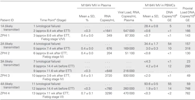
Table 1. M184V-Harboring HIV-1 MVs in Plasma and PBMCs From Pairs of Recipients and Potential Transmitters
Patient ID Time Pointa(Stage)
M184V MV in Plasma
Viral Load, RNA, Copies/mL Plasma M184V MV in PBMCs Proviral Load DNA, Copies/106 GE Mean ± SD, % RNA Copies/mL Mean ± SD, % DNA Copies/106 GE 1A (likely transmitter) 1 (virological failure) . . . 28.4 ± 0.8 5 19
2 (approx 8.4 wk after ETT) <0.3 <1641 547 000 <0.6 <1 166 ZPHI 1 3 (approx 8.6 wk after ETT;
Fiebig stage V/VI)
0.4 ± 0.0 349 87 300 <0.7 <1 143
2A (likely transmitter)
4 (virological failure) . . . 34.4 ± 1.7 54 157
5 (approx 7.4 wk after ETT) 0.4 ± 0.0 676 169 000 3.0 ± 0.3 10 318 ZPHI 2 6 (approx 8 wk after ETT;
Fiebig stage IV)
0.4 ± 0.0 204 51 100 <0.8 <1 130
3A (likely transmitter)
7 (virological failure) . . . <4.3 <1 23
8 (approx 14.4 wk before ETT) . . . 4.2 ± 0.4 12 280 9 (approx 11.6 wk after ETT) <0.3 <648 216 000 . . . . ZPHI 3 10 (approx 3.6 wk after ETT;
Fiebig stage IV)
0.4 ± 0.1 3720 930 000 <2.0 <1 49
4A (likely transmitter)
11 (virological failure) . . . 93.6 ± 0.5 55 59
12 (approx 14.4 wk before ETT) <0.3 <780 260 000 1.9 ± 0.1 14 748 ZPHI 4 13 (approx 11 wk after ETT;
Fiebig stage VI)
0.7 ± 0.1 3290 470 000 <0.3 <2 782
Abbreviations: approx, approximately; ellipses (. . .), not performed; ETT, estimated time of transmission; GE, genomic equivalents; HIV-1, human immunodeficiency virus type 1; MV, minority variant; PBMCs, peripheral blood mononuclear cells; ZPHI, Zurich Primary HIV Infection study.
aTime points 1–13 as indicated in Figure 2.
before sampling; Table1). Individual subtrees clipped from the neighbor joining tree, including a total of 2206 sequences, show the phylogenetic analysis of HIV-1 pol sequences of those 4 clusters (Figure1A). The likely transmitters were confirmed by
phylogenetic analysis of their HIV-1env C2-V3-C3 sequences combined with clonal sequences isolated from the recipients (Figure1B).
Other criteria were also used to define the most likely trans-mitters. None of the potential transmitters were receiving treat-ment at the estimated time of transmission (Figure2), and all potential transmitters had a high viral load at the time of trans-mission (Figure 2). Potential transmitter-recipient pairs be-longed to the same risk group (men who have sex with men) and originated from the same geographic regions.
In the following analysis, we focused on the 4 clusters that in-cluded most likely transmitters with a history of virological failure and the potential to transmit preselected drug-resistant
variants as MVs. All 4 potential transmitters experienced ART failure and selected the M184V mutation as major virus popu-lation 0.5 to 9.4 years before transmission (Figure2). By popu-lation sequencing, the M184V mutation was not detectable any more during the time window of likely transmission (Figure2); AS-PCR was then performed on plasma samples as close as possible to the time of transmission. The M184V mutation was detected in 1 potential transmitter at similar levels as in the recipi-ent (0.4%; Table1). In the remaining 3 potential transmitters, the M184V mutation was not detectable (<0.3%) in the plasma virus population at the estimated time point of transmission.
Next, proviral DNA was analyzed by AS-PCR to investigate the presence of the M184V mutation in the PBMC compart-ment, which can retain previously replicating variants. Notably, PBMCs from 3 of the 4 potential transmitters harbored the M184V mutation as MVs in frequencies of 1.9%–4.2% (Table 1). The M184V mutation was not present in proviral Figure 1. Phylogenetic trees of human immunodeficiency virus type 1 (HIV-1) pol (A) and C2-V3-C3 env (B) sequences of acutely HIV-1–infected patients and their potential transmitters who selected the M184V mutation before transmission. A, Individual subtrees extracted from a large neighbor joining tree inferred from 2206 pooled HIV-1 pol sequences of all Zurich Primary HIV Infection (ZPHI) cohort patients and of all cohort patients harboring closely related virus (genetic distance <1.5%). Potential transmitters are shown in blue, recipients in red. Open circles represent additional HIV-1–infected patients in the clusters. HIV-1 pol sequences were obtained by routine genotypic resistance testing using population sequencing. Bar denotes 1% nucleotide divergence. B, Neighbor joining tree constructed with clonal HIV-1 env C2-V3-C3 sequences isolated from plasma of ZPHI cohort patients and respective bulk sequenc-es from the probable transmitters. Bootstrap valusequenc-es corrsequenc-esponding to 1000 replications are indicated bsequenc-eside the prsequenc-esumed ancsequenc-estral nodsequenc-es. HIV-1HXB2was
DNA from the potential transmitter 1A or in any of the 4 recip-ients at the estimated time point of transmission (Table1).
M184V vs N155H Mutations as MVs in Acutely or Recently HIV-1 Infected Patients
The M184V mutation was detected as a MV in 16 of 195 acutely or recently HIV-1–infected patients (8.2%), Y181C was detected in 4 of 153 (2.6%), K103N in 4 of 192 (2.1%), and N155H in 1 of 185 (0.5%) (Figure3A), at frequencies of 0.4%–
8.3%, 0.5%–0.7%, 0.83%–3.76%, and 0.9% (Figure4A),
respec-tively. The difference between the very commonly detected mu-tation M184 V and the particularly uncommon integrase mutation N155H was significant (P < .001). This was also the
case when the analysis was restricted to only patients who were infected with HIV-1 subtype B and belonged to the risk group of men who have sex with men (P < .001; Figure 3B). Both
groups of acutely or recently HIV-1–infected patients—those harboring and not harboring drug-resistant HIV-1 MVs— showed no differences in CD4+T cell count, viral load, HIV-1 subtype, sex, risk group, Fiebig staging, or time between esti-mated time of infection and sampling (Table2).
M184V Mutation in Acutely or Recently vs Chronically HIV-1 Infected Patients
The detection of MVs containing the K103N, Y181C, M184V, or N155H mutation was compared in 204 acutely or recently Figure 2. Time course analysis of potential transmission pairs. Viral load kinetics of the potential transmitter (blue circles) and the recipient (red circles) are presented over time, and the estimated time of transmission is indicated (gray shaded area). Treatment history of the potential transmitter is depicted (black and green lines represent antiretroviral therapy without or with lamivudine or emtricitabine, respectively) as well as thefirst-line antiretroviral therapy of the recipient (red lines). Genotypic resistance testing by population sequencing was performed at several time points before and during the time of transmission in the potential transmitter (black arrows); the results regarding codon 184 of the reverse transcriptase (RT) are labeled M and V for M184 and M184V, respectively. Minority M184V–comprising human immunodeficiency virus type 1 (HIV-1) variants were assessed by allele-specific polymerase chain reaction in plasma and peripheral blood mononuclear cells at the indicated time points 1–13. These results are provided inTable 1. Abbreviations: 3TC, lamivudine; ABC, abacavir; ATV, atazanavir; ATV/r, ritonavir-boosted ATV; AZT, zidovudine; d4T, stavudine; ddC, zalcitabine; ddI, didanosine; DRV/r, ri-tonavir-boosted darunavir; EFV, efavirenz; FPV/r, riri-tonavir-boosted fosamprenavir; FTC, emtricitabine; IDV, indinavir; LPV, lopinavir; LPV/r, riri-tonavir-boosted LPV; SQV, saquinavir; TNV, tenofovir.
and 382 ART-naive, chronically HIV-1–infected patients. The latter group harbored drug-resistant HIV-1 MVs before first ART as follows: K103N in 10 of 324 patients (3.1%) at frequen-cies of 0.76%–22.14%, Y181C in 16 of 325 (4.9%) at frequenfrequen-cies of 0.5%–13.7%, M184V in 9 of 363 (2.5%) at frequencies of 0.9%–3.2%, and N155H in none of 146 patients (Figure3A and
Figure4B).
In 16 of 195 acutely or recently HIV-1–infected patients (8.2%), M184V MVs were present, compared with 9 of 363 chronically HIV-1–infected patients (2.5%), a significant diffe-rence (P = .004; Figure3A). The K103N and Y181C mutations
were similarly present in both patient groups (2.1% vs 3.1% and 2.6% vs 4.9%, respectively;P > .05). The N155H integrase mu-tation was not detected in chronically HIV-1–infected patients (Figure3A).
As expected, the 2 groups—acutely or recently and chroni-cally HIV-1–infected patients—differed significantly in terms of CD4+T cell count, which was lower in chronically infected patients, and viral load, which was higher in acutely or recently infected patients (Table2). They also differed significantly with
regard to the interdependent parameters of sex, risk group, and HIV-1 subtype (Table2). This shows that the chosen group of chronically infected patients well represents ART-naive patients within the nationwide Swiss HIV Cohort Study [29]. To exclude any bias derived from these parameters, only men who have sex with men and infected with HIV-1 subtype B were in-cluded in a sensitivity analysis. Again, the numbers of patients harboring the M184V mutation as MVs differed significantly between acutely or recently and chronically HIV-1–infected pa-tients (P = .03; Figure3B).
As shown for those infected acutely or recently, chronically HIV-1–infected patients harboring or not harboring drug-re-sistant HIV-1 MVs showed no significant differences in the pa-rameters CD4+T cell count, viral load, HIV-1 subtype, sex, risk group, Fiebig staging, and time between estimated time of in-fection and sampling (Table2). Notably, the percentages of pa-tients with a viral load≥8334 HIV-1 RNA copies/mL plasma were similar in all groups (Table2); that is, drug-resistant HIV-1 MVs can be detected in those patients to the defined cutoff of 0.3% without any restrictions due to too low viral loads. Thus, a higher rate of false-negative results can be excluded in chroni-cally infected patients despite the lower median of the viral loads.
DISCUSSION
This study provides several lines of strong evidence that trans-mission of drug-resistant HIV-1 MVs can occur, a phenome-non often assumed but not proved so far. First, potential transmitters of 4 transmitter-recipient pairs experienced ART failure and selected the M184V mutation 0.5 to 9.4 years before transmission. During the time of transmission, the M184V mu-tation was no longer detected in plasma by population sequenc-ing, but in 1 of those potential transmitters it was present as a MV in the plasma virus population. In 3 potential transmitters, M184V-harboring HIV-1 MVs were detected at frequencies of up to 4.2% in PBMCs. This compartment contains ancestral viral strains that are more likely to be transmitted than contem-porary viral variants [30]. Thus, the PBMC compartment Figure 3. Differences in prevalence of drug-resistant human
immuno-deficiency virus type 1 (HIV-1) minority variants (MVs) within acutely or re-cently vs chronically HIV-1–infected patients. The K103N, Y181C, M184V, and N155H drug resistance mutations were measured by allele-specific polymerase chain reaction in acutely or recently (red bars) and chronically (blue bars) HIV-1–infected patients. Numbers of patients harboring these MVs and numbers of tested patients per mutation are given. Statistical analysis was performed with 2-sided Fisher exact tests; significant differ-ences are shown for the prevaldiffer-ences of different drug-resistant HIV-1 MVs within acutely or recently HIV-1–infected patients (red) and for the M184V mutation in acutely or recently vs chronically HIV-1–infected patients (black). A, Analysis including all patients. B, Analysis restricted to patients being infected with HIV-1 subtype B and belonging to the risk group of men who have sex with men (MSM).
might reflect the source of transmitted viruses better than the plasma virus population does. Notably, one limitation of our study is that we cannot prove without any doubt that the recipi-ents were infected from the potential transmitters.
Still, the question arises how it is possible that MVs were transmitted, because it was shown that only 1 transmitted virus
serves as the founder virus in 60%–80% of patients infected via sexual transmission [17,31–33]. In our ZPHI cohort, approxi-mately 11% of acutely or recently HIV-1–infected patients show a diversity of >1% in the C2-V3-C3 region ofenv, sug-gesting a heterogeneous founder virus population [17]. The 4 recipients mentioned above did not belong to this group. One Figure 4. Detection of drug-resistant human immunodeficiency virus type 1 (HIV-1) minority variants (MVs) in plasma and peripheral blood mononuclear cells (PBMCs) from acutely or recently and chronically HIV-1–infected patients and reproducibility/accuracy of the allele-specific polymerase chain reaction (AS-PCR) assays for K103N, Y181C, M184V, and N155H. The K103N, Y181C, M184V, and N155H drug resistance mutations were measured with AS-PCR in plasma of acutely or recently (A) and chronically (B) HIV-1–infected patients, as was the M184V mutation in PBMCs from the 4 potential transmitter-re-cipient pairs (C). The values of the samples below the detection limit (dotted line, 0.3%) are represented as gray dots. The open circles represent samples with values >0.3% but for which the corresponding viral load is too low to verify the presence of drug-resistant HIV-1 variants. Black dots represent the samples that contained drug-resistant HIV-1 MVs. D, Each sample processing and AS-PCR run included 13 plasma samples from patients and a wild-type as well as a 1% mutant virus control. The wild-type control contained virus particles from a HIV-1NL4–3virus stock with a virus titer of 10 000 RNA copies in 1 mL
of plasma from an HIV-1 negative donor. The 1% mutant control was a mixture of HIV-1NL4–3virus stock (9900 RNA copies) and the respective mutant virus
stock HIV-1NL4–3_K103N, HIV-1NL4–3_Y181C, HIV-1NL4–3_M184V, and HIV-1NL4–3_N155H(100 RNA copies) in 1 mL of plasma from an HIV-1 negative donor. Data
rep-resent results of 28–78 independent experiments in which each sample was assayed in duplicate; median and interquartile ranges are given. Abbreviation: dl, detection limit.
Table 2. Demographic Data and Clinical Baseline Parameters in Acutely or Recently and Chronically, ART-Naive HIV-1–Infected Patients Harboring or Not Harboring Drug-Resistant HIV-1 MVs Data Acutely or Recently HIV-1–Infected Patients (n = 204) Chronically HIV-1–Infected Patients (n = 382) P Value Acutely or Recently HIV-1–Infected Patients Not Harboring MVs (n = 181) Acutely or Recently HIV-1–Infected Patients Harboring MVs (n = 23) P Value Chronically HIV-1–Infected Patients Not Harboring MVs (n = 352) Chronically HIV-1–Infected Patients Harboring MVs (n = 30) P Value
Age, median (range), y 34 (18–70) 38 (22–71) <.001a 34 (18
–70) 40 (23–64) .02a 38 (22
–71) 37 (23–57) NSa
CD4+T cell count, median
(range), cells/µL blood
399 (87–1304) 232 (0–800) (n = 379) <.001a 397 (87 –1304) 423 (150–1041) NSa 232 (0 –800) (n = 349) 212 (0–740) NSa
HIV-1 RNA load, median (range), log10copies/mL plasma
5.3 (2.7–8.0) 4.9 (3.0–7.0) <.001a 5.3 (2.7–8.0) 5.6 (3.2–7.5) NSa 4.9 (3.0–7.0) 4.6 (3.8–6.9) NSa
Patients, No. (%)
VL≥8334 copies/mL plasmab 188 (92.2) 345 (90.3) NSc 168 (92.8) 20 (87.0) NSc 316 (89.8) 29 (96.7) NSc
VL <8334 copies/mL plasmad 16 (7.8) 37 (9.7) 13 (7.2) 3 (13.0) 36 (10.2) 1 (3.3)
HIV-1 subtype B 181 (88.7) 358 (93.7) .04c 158 (87.3) 23 (100) NSc 328 (93.2) 30 (100) NSc
HIV-1 subtype non-B 23 (11.3) 24 (6.3) 23 (12.7) 0 24 (6.8) 0
Male 194 (95.1) 301 (78.8) <.001c 172 (95.0) 22 (95.6) NSc 274 (77.8) 27 (90.0) NSc Female 10 (4.9) 81 (21.2) 9 (5.0) 1 (4.4) 78 (22.2) 3 (10.0) MSM 167 (81.9) 197 (51.6) <.001c,e 147 (81.2) 20 (87.0) NSc,e 178 (50.6) 19 (63.3) NSb,c Heterosexual 33 (16.2) 118 (30.9) 30 (16.6) 3 (13.0) 113 (32.1) 5 (16.7) IDU 1 (0.5) 56 (14.7) 1 (0.6) 0 50 (14.2) 6 (20.0) IDU or heterosexual 2 (1.0) 11 (2.9) 2 (1.1) 0 11 (3.1) 0 Needle stick 1 (0.5) 0 1 (0.6) 0 0 0 Fiebig stage I–IV 80 (44.2) 12 (52.2) NSc,f V–VI 94 (51.9) 10 (43.5) Unknown 7 (3.9) 1 (4.4)
Acute infection (≤90 d after ETI)g 148 (81.8) 21 (91.3) NSc
Recent infection (>90 to≤180 d after ETI)g
33 (18.2) 2 (8.7)
Time of sampling after ETI, median (range), wk
6.3 (2.1–26.6) 6.0 (1.7–18.1) NSa
Abbreviations: ART, antiretroviral therapy; ETI, estimated time of infection; heterosexual, heterosexual transmission; HIV-1, human immunodeficiency virus type 1; IDU, injection drug use; MSM, men who have sex with men; MVs, minority variants; NS, not significant; VL, viral load.
a
Mann–Whitney U test.
b
For the detection of MVs to levels down to 0.3%, the viral load has to be≥8334 HIV-1 RNA copies/mL plasma.
c
Fisher exact test. d
In samples with a viral load <8334 HIV-1 RNA copies/mL plasma, the cutoff for allele-specific polymerase chain reaction has to be individually calculated. e
MSM vs non-MSM. Fiebig stage I–IV vs V–VI.
T ransmission of Minority V ariants
•
JID 2013:208 (1 October)•
1109likely possibility is that with the previously chosen cloning ap-proach the resolution may have been too low to detect minority species. We postulate that >1 virus is transmitted and that each virus within the viral quasispecies has a certain probability of being transmitted. As a theoretical example, assume that a re-cipient is exposed to 1000 viruses, of which 10 cross the mucosal barrier, and assume that 2% of the viruses carry a drug-resistance mutation. Then the probability is 16.8% that 1 of those 10 transmitted viruses contain a drug-resistance muta-tion, given that transmission is a stochastic process. One of those viruses, or sometimes more, generates the founder virus population. Because drug resistance mutations are often associ-ated with a decrease of viralfitness, it is unlikely that the drug-resistant virus variant will be the founder virus. However, it is assumable that the transmitted drug-resistant HIV-1 MV repli-cates at very low frequencies, as seen in our 4 recipients. In previ-ous longitudinal analyses of viral populations in chronically HIV-1–infected patients, we have shown that drug-resistant HIV-1 viruses can replicate at low levels in the absence of ART and within a major virus population of drug-sensitive viruses [16,23]. Although we could just include 4 transmission-recipient pairs, we were able to analyze plasma and cell samples from both the potential transmitters and the recipients very close to the estimated time point of transmission. In 1 case report, a mother-to-child-transmission of a drug-resistant HIV-1 MV is assumed [15], but uncertainty remains because the analyzed child’s and mother’s samples were taken approximately 3 and 6.5 years, respectively, after the estimated time of transmission.
Second, the prevalence of drug-resistant HIV-1 MVs in acutely or recently HIV-1–infected patients mirrors the preva-lence of those mutations in ART-experienced patients, a group of potential transmitters [34], and acute seroconverters in Swit-zerland [35] as monitored by population sequencing. The very common mutation M184V was the most frequently detected drug resistance mutation, followed by the NNRTI mutations Y181C and K103N. The so-far particularly uncommon inte-grase mutation N155H was almost absent in acutely or recently HIV-1–infected patients. Of note, during the investigated time period, selection pressure on integrase was virtually absent in the Swiss HIV-1–infected population because the first integrase inhibitor was approved for salvage therapy only in 2008 [36]. Thus, any detection of the N155H mutation might reflect the level of de novo mutagenesis. Note that biases due to different assay sensitivities can be excluded because the same cutoff was chosen for all AS-PCR assays.
If sporadic appearance alone was the reason for the emer-gence of drug-resistant HIV-1 MVs in ART-naive, acutely or recently HIV-1–infected patients, a more similar distribution of these mutations would be expected, provided that transitions occur 2–3-fold more often than transversions, especially during the early phase of infection [37]. The M184V and Y181C muta-tions are transimuta-tions, and the K103N and N155H mutamuta-tions are
transversions. However, the observed 16-fold difference in the prevalences of N155H and M184V mutations as MVs cannot be explained by preferential transitions. In addition, a higher rate of Y181C mutation and a lower rate of K103N mutation would have been expected. Thus, sporadic appearance alone cannot explain the observed pattern in the prevalence of drug-resistant HIV-1 MVs in acutely or recently HIV-1–infected pa-tients.
HIV-1 RNA structure and other sequence characteristics could potentially favor the sporadic appearance of certain mu-tations as it was shown for the K65R mutation in HIV-1 subtype C [38]. However, to our knowledge this has not yet been described for the M184V mutation. In addition, this po-tential explanation is not in concordance with our observation that the prevalence of the M184V mutation is significantly lower in chronically than in acutely or recently HIV-1–infected patients.
Hypothetically, ineffective pre- and/or postexposure prophy-laxis could explain the higher prevalence of the M184V muta-tion compared to the N155H mutamuta-tion, because most of the prophylaxis regimens contain either 3TC or FTC but not an in-tegrase inhibitor; thus, a failing prophylaxis would lead to the selection of the M184V mutation. The use of preexposure pro-phylaxis can be excluded in our patient cohort, because almost all patients were included before its success was reported at the end of 2010 [39], and it has not yet been introduced in Switzer-land. Postexposure prophylaxis can also be ruled out in our pa-tients. So far, only 2 of the 204 patients were undergoing postexposure prophylaxis during the time of transmission, as verified by systematic interviews. In both patients, drug-resis-tant HIV-1 MVs were not detected.
Third, the prevalence of drug-resistant HIV-1 MVs in chroni-cally HIV-1–infected patients and the comparison with acutely or recently HIV-1–infected patients reflect exactly what is ex-pected to follow after transmission of such MV drug-resistant viruses. The minor population harboring the M184V mutation vanishes over time due to its lower replication capacity [40], and the K103N and Y181C mutations remain unchanged owing to their negligible impact on viralfitness [41].
In summary, sporadic appearance of drug-resistant HIV-1 MV might occur, but our observations cannot be explained solely by de novo mutagenesis. Thus, we show for thefirst time that drug-resistant HIV-1 MVs can be transmitted. The origin of drug-resistant HIV-1 MVs may have clinical implications, because it is conceivable that transmitted MVs might be able to establish a pool of latently infected cells more easily and pro-foundly than sporadically appearing MVs and that transmitted MVs might not tend to disappear as readily as sporadically ap-pearing ones. The latter phenomenon has been observed in 1 of our ZPHI patients, who harbored the K103N mutation as a MV during primary HIV-1 infection, a variant that reappeared and persisted during treatment interruption of early ART [16].
Thus, the origin of drug-resistant HIV-1 MVs might help to explain the observed and still puzzling different clinical out-comes in ART-naive patients harboring those MVs and begin-ning ART.
Supplementary Data
Supplementary materialsare available atThe Journal of Infectious Diseases online (http://jid.oxfordjournals.org/). Supplementary materials consist of data provided by the author that are published to benefit the reader. The posted materials are not copyedited. The contents of all supplementary data are the sole responsibility of the authors. Questions or messages regarding errors should be addressed to the author.
Notes
Acknowledgments. We are grateful to our patients for their commit-ment and participation in the ZPHI study and in the SHCS. We thank B. Hasse, U. Karrer, R. Oberholzer, L. Aceto, R. Laffer, U. von Both, M. Huber, K. Thierfelder, E. Presterl, Y. Flammer, S. Kuster, J. Nemeth, M. Frei, T. Frey, and M. Flepp for excellent patient care, F. Burgener and D. Klimpel for technical help, and I. Nievergelt and C. Vögtli for adminis-trative assistance.
The members of the SHCS are V. Aubert, J. Barth, M. Battegay, E. Bernasconi, J. Böni, H. C. Bucher, C. Burton-Jeangros, A. Calmy, M. Cavassini, M. Egger, L. Elzi, J. Fehr, J. Fellay, H. Furrer (chairman of the Clinical and Laboratory Committee), C. A. Fux, M. Gorgievski, H. Günthard ( president of the SHCS), D. Haerry (deputy of the“Positive Council”), B. Hasse, H. H. Hirsch, I. Hösli, C. Kahlert, L. Kaiser, O. Keiser, T. Klimkait, H. Kovari, R. Kouyos, B. Ledergerber, G. Martinetti, B. Martinez de Tejada, K. Metzner, N. Müller, D. Nadal, G. Pantaleo, A. Rauch (chairman of the Scientific Board), S. Regenass, M. Rickenbach (head of Data Center), C. Rudin (chairman of the Mother & Child Substudy), P. Schmid, D. Schultze, F. Schöni-Affolter, J. Schüpbach, R. Speck, P. Taffé, P. Tarr, A. Telenti, A. Trkola, P. Vernazza, R. Weber, S. Yerly.
Financial support. This work has been supported in the framework of the SHCS, supported by the Swiss National Science Foundation (grant 33CS30_134277) and the SHCS research foundation. Further support was provided by the Swiss National Science Foundation (grants 324700–120793 to H. F. G. and K. J. M. and 320000-116035 and 324730-130865 to H. F. G.), the SHCS ( project 605; to H. F. G. and K. J. M.), the University of Zurich’s Clinical Research Priority Program “Viral Infectious Diseases: Zurich Primary HIV Infection Study” (to H. F. G), the European Commun-ity’s Seventh Framework Programme (grant FP7/2007–2013), under the Collaborative HIV and Anti-HIV Drug Resistance Network (CHAIN) (grant 223131to H. F. G.); an unrestricted research grant from the Vontobel Stiftung (to H. F. G. and K. J. M.), and one from Gilead (to H. F. G.).
Potential Conflict of Interest. K. J. M. received travel grants and hono-raria from Gilead Sciences, Roche Diagnostics, Tibotec, Bristol-Myers Squibb, and Abbott; the University of Zurich has received research grants from Gilead, Roche, and Merck Sharp & Dohme for studies for which K. J. M. serves as principal investigator and advisory board honoraria from Gilead Sciences. B. P. has received a travel grant from Roche. S. Y. has been an adviser and/or consultant for Bristol-Myers Squibb and has received travel grants and honoraria from Gilead Sciences, ViiV, and Merck Sharp & Dohme. T. K. served as advisor for Bristol-Myers Squibb and Pfizer and has received travel grants from Abbott and Pfizer. The institution of H. F. has received payments for participation in advisory boards and/or unrestricted educational grants and/or travel grants from Abbott, Bristol-Myers Squibb, ViiV Healthcare, Roche, Gilead, Merck Sharp & Dohme, Boehringer-Ingel-heim, and Tibotec-Janssen and unrestricted research support from Gilead, Merck Sharp & Dohme, and Roche. M. B has been an advisor for Janssen-Tibotec and has received educational and research grants from Boehringer-Ingelheim, Bristol-Myers Squibb, Abbott, Janssen-Tibotec, ViiV Health-care, Gilead, and Merck Sharp & Dohme. P. L. V. and his institution have
received travel grants and speaker fees or advisory board honoraria from Abbott, Bristol-Myers Squibb, Gilead, Janssen, Merck Sharp & Dohme, Roche, and ViiV Healthcare. M. C. received unrestricted research grant from Gilead and Merck Sharp & Dohme and received travel grants from Boehringer-Ingelheim, Bristol-Myers Squibb , Gilead, and Merck Sharp & Dohme; his institution received advisory board honorarium from Bristol-Myers Squibb, Gilead, Merck Sharp & Dohme, Janssen-Cilag and Viiv. A. C. has received research grants from Abbott, Janssen-Cilag, and Gilead and travel grants from Janssen Cilag, and Gilead. R. W. has received travel grants from Abbott, Boehringer Ingelheim, Bristol-Myers Squibb, Gilead Sciences, GlaxoSmithKline, Merck Sharp & Dome, Pfizer, Roche, TRB Chemedica, and Tibotec. H. F. G. has been an adviser and/or consul-tant for GlaxoSmithKline, Abbott, Gilead, Novartis, Boehringer Ingelheim, Roche, Tibotec, Pfizer and Bristol-Myers Squibb, and has received unre-stricted research and educational grants from Roche, Abbott, Bristol-Myers Squibb, Gilead, Astra-Zeneca, GlaxoSmithKline, and Merck Sharp & Dohme (all money went to the institution). All other authors declare no conflicts of interest.
All authors have submitted the ICMJE Form for Disclosure of Potential Conflicts of Interest. Conflicts that the editors consider relevant to the content of the manuscript have been disclosed.
References
1. Thompson MA, Aberg JA, Hoy JF, et al. Antiretroviral treatment of adult HIV infection: 2012 recommendations of the International Anti-viral Society-USA panel. JAMA2012; 308:387–402.
2. Gunthard HF, Wong JK, Ignacio CC, Havlir DV, Richman DD. Com-parative performance of high-density oligonucleotide sequencing and dideoxynucleotide sequencing of HIV type 1 pol from clinical samples. AIDS Res Hum Retroviruses1998; 14:869–76.
3. Schuurman R, Demeter L, Reichelderfer P, Tijnagel J, de Groot T, Boucher C. Worldwide evaluation of DNA sequencing approaches for identification of drug resistance mutations in the human immunodefi-ciency virus type 1 reverse transcriptase. J Clin Microbiol 1999; 37:2291–6.
4. Gianella S, Richman DD. Minority variants of drug-resistant HIV. J Infect Dis2010; 202:657–66.
5. Li JZ, Paredes R, Ribaudo HJ, et al. Low-frequency HIV-1 drug resis-tance mutations and risk of NNRTI-based antiretroviral treatment failure: a systematic review and pooled analysis. JAMA 2011; 305:1327–35.
6. Bansal V, Metzner KJ, Niederost B, et al. Minority K65R variants and early failure of antiretroviral therapy in HIV-1-infected eritrean immi-grant. Emerg Infect Dis2011; 17:1966–8.
7. Metzner KJ, Giulieri SG, Knoepfel SA, et al. Minority quasispecies of drug-resistant HIV-1 that lead to early therapy failure in treatment-naive and -adherent patients. Clin Infect Dis2009; 48:239–47. 8. Gianella S, Delport W, Pacold ME, et al. Detection of minority
resis-tance during early HIV-1 infection: natural variation and spurious de-tection rather than transmission and evolution of multiple viral variants. J Virol2011; 85:8359–67.
9. Metzner KJ, Rauch P, von Wyl V, et al. Efficient suppression of minori-ty drug-resistant HIV minori-type 1 (1) variants present at primary HIV-1 infection by ritonavir-boosted protease inhibitor-containing antire-troviral therapy. J Infect Dis2010; 201:1063–71.
10. Metzner KJ, Rauch P, Walter H, et al. Detection of minor populations of drug-resistant HIV-1 in acute seroconverters. AIDS2005; 19:1819–25. 11. Nicot F, Saliou A, Raymond S, et al. Minority variants associated with
resistance to HIV-1 nonnucleoside reverse transcriptase inhibitors during primary infection. J Clin Virol2012; 55:107–13.
12. Peuchant O, Thiebaut R, Capdepont S, et al. Transmission of HIV-1 minority-resistant variants and response to first-line antiretroviral therapy. AIDS2008; 22:1417–23.
13. Toni TA, Asahchop EL, Moisi D, et al. Detection of human immunode-ficiency virus (HIV) type 1 M184 V and K103N minority variants in
patients with primary HIV infection. Antimicrob Agents Chemother 2009; 53:1670–2.
14. Coffin JM. HIV population dynamics in vivo: implications for genetic variation, pathogenesis, and therapy. Science1995; 267:483–9. 15. Machado ES, Afonso AO, Nissley DV, et al. Emergency of primary
NNRTI resistance mutations without antiretroviral selective pressure in a HAART-treated child. PLoS ONE2009; 4:e4806.
16. Metzner KJ, Leemann C, Di Giallonardo F, et al. Reappearance of mi-nority K103N HIV-1 variants after interruption of ART initiated during primary HIV-1 infection. PLoS ONE2011; 6:e21734.
17. Rieder P, Joos B, Scherrer AU, et al. Characterization of human immu-nodeficiency virus type 1 (HIV-1) diversity and tropism in 145 patients with primary HIV-1 infection. Clin Infect Dis2011; 53:1271–9. 18. Rieder P, Joos B, von Wyl V, et al. HIV-1 transmission after cessation
of early antiretroviral therapy among men having sex with men. AIDS 2010; 24:1177–83.
19. Schoeni-Affolter F, Ledergerber B, Rickenbach M, et al. Cohort profile: the Swiss HIV Cohort study. Int J Epidemiol2010; 39:1179–89. 20. Yerly S, Vora S, Rizzardi P, et al. Acute HIV infection: impact on the
spread of HIV and transmission of drug resistance. AIDS 2001; 15:2287–92.
21. Hirsch MS, Gunthard HF, Schapiro JM, et al. Antiretroviral drug resistance testing in adult HIV-1 infection: 2008 recommendations of an International AIDS Society-USA panel. Clin Infect Dis 2008; 47:266–85.
22. Bennett DE, Camacho RJ, Otelea D, et al. Drug resistance mutations for surveillance of transmitted HIV-1 drug-resistance: 2009 update. PLoS ONE2009; 4:e4724.
23. Metzner KJ, Bonhoeffer S, Fischer M, et al. Emergence of minor popu-lations of human immunodeficiency virus type 1 carrying the M184 V and L90M mutations in subjects undergoing structured treatment in-terruptions. J Infect Dis2003; 188:1433–43.
24. Halvas EK, Aldrovandi GM, Balfe P, et al. Blinded, multicenter com-parison of methods to detect a drug-resistant mutant of human immu-nodeficiency virus type 1 at low frequency. J Clin Microbiol 2006; 44:2612–4.
25. von Wyl V, Yerly S, Boni J, et al. Emergence of HIV-1 drug resistance in previously untreated patients initiating combination antiretroviral treatment: a comparison of different regimen types. Arch Intern Med 2007; 167:1782–90.
26. Eddy SR. Profile hidden Markov models. Bioinformatics 1998; 14:755–63.
27. Tamura K, Dudley J, Nei M, Kumar S. MEGA4: Molecular Evolution-ary Genetics Analysis (MEGA) software version 4.0. Mol Biol Evol 2007; 24:1596–9.
28. Fiebig EW, Wright DJ, Rawal BD, et al. Dynamics of HIV viremia and antibody seroconversion in plasma donors: implications for diagnosis and staging of primary HIV infection. AIDS2003; 17:1871–9. 29. von Wyl V, Kouyos RD, Yerly S, et al. The role of migration and
do-mestic transmission in the spread of HIV-1 non-B subtypes in Switzer-land. J Infect Dis2011; 204:1095–103.
30. Redd AD, Collinson-Streng AN, Chatziandreou N, et al. Previously transmitted HIV-1 strains are preferentially selected during subsequent sexual transmissions. J Infect Dis2012; 206:1433–42.
31. Keele BF, Giorgi EE, Salazar-Gonzalez JF, et al. Identification and char-acterization of transmitted and early founder virus envelopes in primary HIV-1 infection. Proc Natl Acad Sci U S A2008; 105:7552–7. 32. Kearney M, Maldarelli F, Shao W, et al. Human immunodeficiency
virus type 1 population genetics and adaptation in newly infected indi-viduals. J Virol2009; 83:2715–27.
33. Kouyos RD, von Wyl V, Yerly S, et al. Ambiguous nucleotide calls from population-based sequencing of HIV-1 are a marker for viral diversity and the age of infection. Clin Infect Dis2011; 52:532–9.
34. von Wyl V, Yerly S, Boni J, et al. Long-term trends of HIV type 1 drug resistance prevalence among antiretroviral treatment-experienced pa-tients in Switzerland. Clin Infect Dis2009; 48:979–87.
35. Yerly S, von Wyl V, Ledergerber B, et al. Transmission of HIV-1 drug resistance in Switzerland: a 10-year molecular epidemiology survey. AIDS2007; 21:2223–9.
36. Scherrer AU, von Wyl V, Fux CA, et al. Implementation of raltegravir in routine clinical practice: selection criteria for choosing this drug, vi-rologic response rates, and characteristics of failures. J Acquir Immune Defic Syndr 2010; 53:464–71.
37. Salazar-Gonzalez JF, Bailes E, Pham KT, et al. Deciphering human im-munodeficiency virus type 1 transmission and early envelope diversifi-cation by single genome amplifidiversifi-cation and sequencing. J Virol 2008; 82:3952–70.
38. Coutsinos D, Invernizzi CF, Xu H, Brenner BG, Wainberg MA. Factors affecting template usage in the development of K65R resistance in subtype C variants of HIV type-1. Antivir Chem Chemother2010; 20:117–31.
39. Grant RM, Lama JR, Anderson PL, et al. Preexposure chemoprophylax-is for HIV prevention in men who have sex with men. N Engl J Med 2010; 363:2587–99.
40. Back NK, Nijhuis M, Keulen W, et al. Reduced replication of 3TC-resis-tant HIV-1 variants in primary cells due to a processivity defect of the reverse transcriptase enzyme. EMBO J1996; 15:4040–9.
41. Nicastri E, Sarmati L, d’Ettorre G, et al. Replication capacity, biological phenotype, and drug resistance of HIV strains isolated from patients failing antiretroviral therapy. J Med Virol2003; 69:1–6.