Isolation and characterization of human orthologs of yeast CCR4-NOT complex subunits
9
0
0
Texte intégral
(2) 810. Nucleic Acids Research, 2000, Vol. 28, No. 3. genes can act as suppressors of a mutant srb4 allele (16). ySRB4 was identified as a cofactor of the C-terminal domain of the largest subunit of pol II (17), and is essential for pol II transcription (18). Moreover, yCCR4 was recently reported to be associated with yPAF1, yHPR1 and yCDC73 in a pol II holoenzyme form that is biochemically distinct from a SRBcontaining holoenzyme (19,20). While the genetic analyses of CCR4–NOT functions in yeast showed that these proteins are involved in diverse transcriptional responses, the underlying molecular mechanisms are poorly understood. It has been suggested that NOT proteins may inhibit or sequester factors more specifically required for TATA-less core promoters (5). Interestingly, whereas NOT proteins work negatively on TC function, a number of TBPassociated factors (TAFs) like yMOT1, yTAFII19/40/67 and yTAFII130 have been demonstrated to stimulate TC-directed HIS3 transcription (21–23). Thus, the CCR4–NOT complex appears to act at the cross-roads of intermingling signaling pathways towards TBP/TFIID functions, and might be one of several factors that contribute to global gene regulation by modulation of TBP activity (for review see 24). Similar to TBP/TFIID, a functional and structural conservation of the CCR4–NOT complex throughout evolution seems very likely, and it is therefore of general interest if this complex exists in mammalian cells and if its functions are conserved. In this study we report the cloning of cDNAs encoding human (h) NOT2, hNOT3, hNOT4 and hCALIF (CAF1-like factor). We describe a multitude of two-hybrid interactions among these proteins and, in addition, interactions of hCALIF with yCCR4. The primary structure of hCALIF identifies it as a close relative of yeast, mouse and human CAF1. Endogenous hCALIF co-immunoprecipitates with hNOT3, indicating a physical association of the two proteins in vivo. We show further that upon expression of the isolated hNOT3 and hNOT4 cDNAs in yeast cells the phenotypes of mutant yeast not3 and not4 alleles are partially overcome, thereby demonstrating the heterocomplementation ability of these human cDNAs. Taken together, these results strongly suggest the existence of a human equivalent of the yeast CCR4–NOT complex. MATERIALS AND METHODS Sequences of hNOT2, hNOT3, hNOT4-N and hCALIF have been deposited at the GenBank database, with accession numbers AF180473 (hNOT2), AF180474 (hNOT3), AF180475 (hNOT4-N) and AF180476 (hCALIF). Human CCR4–NOT cDNA clones The following EST cDNA clones were obtained from either the I.M.A.G.E. consortium (IMC) or Human Genome Sciences, Inc. (HGS): hNOT2 AA679466 (IMC) and AA452758 (IMC), hNOT3 HFCBT83 (HGS) and HHFGH78 (HGS), hNOT4 HWIAL87 (HGS), HAMGL76 (HGS), HDPKC88 (HGS), AA376235 (IMC) and W39207 (IMC), and hCALIF HOUDP20 (HGS) and HLYBL69 (HGS). The hNOT1 partial cDNA used in this study (hNOT1C) is based on a partial hNOT1 EST clone HWAAW48 (HGS) that was 5′extended by a cDNA obtained from Dr V.J. Bardwell (University of Minnesota, Minneapolis). All cDNAs were sequenced on both strands, using either a T7-sequencing kit (Pharmacia) or an automated ABI310 sequencer (Perkin-Elmer). To introduce. 13 missing nucleotides into hNOT2 EST clone AA679466, two oligonucleotides with sequences 5′-TCCAGGTGTTACCTGATGGTCGGGTTACTAACATTCCTC-3′ and 5′-TAACCCGACCATCAGGTAACACCTGGATCCCTTTTTTCTG-3′ were made (missing nucleotides are underlined). In a first PCR reaction with clone AA679466 as template, these oligonucleotides were used in combination with forward and reverse outward primers to amplify the 5′ and 3′ sequences of hNOT2, respectively. The resulting products were used in a second overlap PCR reaction as templates along with the initial outward primers to generate a full-length hNOT2 sequence. Finally, this sequence was verified by DNA analysis. We find some minor differences to the partial hNOT2 protein sequence described in Benson et al. (25) (at amino acid residues 444, 451, 510, 519, and residues 530–534, respectively). To create a complete hNOT4 cDNA in EST clone HWIAL87, a direct PCR on a λgt11 fetal retina cDNA library was performed. A 5 µl aliquot of the library (containing ~4 × 10⁷ p.f.u.) was incubated for 5 min at 70°C to disrupt phage particles, and subjected to PCR with the oligonucleotide 5′-GATATCTCGAGATGTCTCGCAGTCCTGATG-3′ (with the hNOT4 start codon in bold) as forward primer and an oligonucleotide 5′-GACACGTACACTAGCCAAATG-3′ (nucleotide positions 295–315 in hNOT4) as reverse primer. The PCR product was digested with EcoRI and ligated to an EcoRI-digested fragment of HWIAL87. The resulting hNOT4 cDNA was verified by sequencing. We find differences of this hNOT4-N sequence to the hNOT4-S and hNOT4-L variants (accession nos U71267 and U71268, respectively) at hNOT4-N nucleotide positions 1062 (C versus T), 1073 (one extra C) and 1202 (one G missing). The 3′ parts of the three hNOT4 sequences differ from nucleotide position 1254 (all numbers refer to the translation start of hNOT4-N which was set as position +1). DNA manipulations and plasmids construction All DNA manipulations were done by standard methods (26). To create yeast plasmids expressing hCCR4–NOT fusion proteins with either the DNA binding domain of the bacterial LexA protein (residues 1–202) or the B42 acidic activation domain, the full-length coding sequences of these cDNAs were inserted into plasmids pEG202 and pJG4-5 (27) in-frame with LexA- or B42-sequences. For cloning purposes, a SmaI site was introduced immediately upstream of the coding sequence of hNOT2 and hCALIF by PCR, and the SmaI-digested PCR products were then cloned into the blunt-ended EcoRI sites of either pEG202 or pJG4-5, respectively. For the complementation analysis hNOT4-N and hNOT3 cDNAs were inserted into yeast high-copy expression plasmids. A XhoI-digested hNOT4-N full-length cDNA was cloned into the XhoI site of plasmid pGEN (28). To create chimera of hNOT3 and yNOT3 or yNOT5, a MluI site was created within these genes at the end of the conserved region. The following oligonucleotides for introducing the MluI site into the hNOT3 coding sequence were made: 5′-GGAGAACGCGTTTCTCTACG-3′ and 5′GAGAAACGCGTTCTCCTCG-3′ (MluI sites are indicated in bold). These oligonucleotides were used in combination with forward and reverse primers to amplify hNOT3 5′sequences and 3′-sequences by PCR, using the cloned fulllength hNOT3 cDNA as template. The hNOT3 PCR amplified 5′- and 3′-sequences were subcloned into pUCBM21, digested with SalI and MluI (hNOT3 5′-sequence) and MluI and NotI.
(3) Nucleic Acids Research, 2000, Vol. 28, No. 3 (hNOT3 3′-sequence), and finally the entire gene was reassembled by a three-piece ligation into plasmid pRS415 (29) digested with SalI and NotI. For creating the MluI sites within yNOT3 and yNOT5 coding sequences, the same approach was taken. The following oligonucleotides were used to amplify yNOT3 and yNOT5 5′- and 3′-sequences, respectively: 5′-TCAAACGCGTCATAAATAGTTTC-3′, 5′-TTATGACGCGTTGAATTTACAGAG-3′, 5′-CATTTACGCGTACATGGGTTGC-3′ and 5′-CCATGTACGCGTAAATTGTATCG-3′. Plasmids pRS316-yNOT3 and pRS316-yNOT5 (7) were used as templates in the PCR reactions. Northern blot analysis For labeling cDNA probes with [32P]dCTP, a random prime labeling system (Rediprime, Amersham Pharmacia Biotech) was used. Labeled probes were hybridized to a multiple tissue northern blot (Clontech) at various temperatures for at least 2 h in ExpressHyb hybridization solution (Clontech). Following hybridization, the blot was washed as recommended by the manufacturer and exposed to X-ray film with intensifying screen at –70°C.. 811. GENNT buffer (5% glycerol, 5 mM EDTA, 0.2% NP-40, 150 mM NaCl, 50 mM Tris–HCl pH 8.0, 0.5 mM PMSF, freshly added 2 µg/ml each of pepstatin A, leupeptin and aprotinin, 10 mM sodium fluoride and 2 mM sodium orthovanadate). Lysate was scraped off plates, put through a 27-gauge needle several times and subjected to centrifugation at 10 000 g for 15 min at 4°C. For preclearing, 500 µl of the supernatant was incubated with 100 µl of a 10% solution of protein A–agarose beads in GENNT buffer and tumbled for at least 2 h at 4°C. Immunoprecipitations were performed with 100 µl of 10% protein A–agarose beads coated with 5 µl of polyclonal anti-CALIF or polyclonal anti-ERK2 for 4 h at 4°C. The beads were washed three times with GENNT buffer and boiled with 2× SDS loading buffer. Of the eluted material, 30 µl was loaded onto 12.5% gels and analyzed by SDS–PAGE. After transfer to a nitrocellulose membrane (Protran, Schleicher & Schuell), blots were blocked in Blotto (2% dry milk, 0.2% BSA in TBST), incubated for 1 h with primary antibody and for 30 min with HRP-conjugated secondary antibody in TBST (10 mM Tris–HCl pH 8.0, 150 mM NaCl, 0.05% Tween 20), and developed using ECL solutions (Renaissance, NEN).. Yeast strains, culture and assays The yeast strains used in this study are listed in Table 1 and were generated by standard genetic techniques. Yeast cells were grown at 30°C in either liquid YPD rich medium or in complete minimal dropout media lacking the appropriate markers for selection. Yeast cells were transformed by standard lithium acetate method (26). Quantitative determination of βgalactosidase levels in yeast transformants was essentially done as described by Bourne et al. (30). Table 1. Strain list Strain. Genotype. Source. EGY48. a trp1 ura3 his3 LEU2::pLexAop1-LEU2. (44). MY27. a ura3-52 trp1-∆1 leu2::PET56 gal2 gcn4-∆1 not2-1. (6). MY508. a ura3-52 trp1-∆1 leu2::PET56 gal2 gcn4-∆1 not3::URA3. (6). MY1729. a ura3-52 trp1-∆1 leu2::PET56 gal2 gcn4-∆1 caf1::LEU2. (15). YOU484. α ura3-52 trp1-∆1 leu2::PET56 gal2 gcn4-∆1 not5::URA3. (7). YOU555. a ura3-52 trp1-∆1 leu2::PET56 gal2 gcn4-∆1 not5::LEU2. (7). YOU578. isogenic to MY508 except not5::URA3. (7). YOU637. isogenic to YOU555 except not4::LEU2 and pRS316-NOT5. (7). Immunological analyses Polyclonal rabbit antisera were raised against a recombinant His6-tagged hCALIF full-length protein or against an Nterminal fragment of hNOT3 (residues 1–102) fused to GST as described (31). For co-immunoprecipitation experiments, C33A cells were grown in DMEM medium containing 10% fetal calf serum to near-confluence on 20 cm culture dishes, washed with cold PBS and lysed on ice with 1 ml of cold. RESULTS Identification and isolation of cDNAs encoding human CCR4–NOT subunits Searching DNA sequence databases revealed the existence of various human EST cDNA clones homologous to yeast CCR4–NOT complex components, i.e. homologs matching highly conserved parts of yNOT1, yNOT2, yNOT3, yNOT4 and yCAF1. We obtained several cDNAs for all of them from different sources (see Materials and Methods) and determined their DNA sequences. This analysis revealed that EST clones for hNOT1 were only partial; isolation and characterization of the full-length hNOT1 cDNA will be reported elsewhere (manuscript in preparation). To identify the human counterpart of yNOT2, two human EST clones (GenBank accession nos AA679466 and AA452758) were obtained. Both of them showed a high degree of homology to the yNOT2 C-terminal sequence. Sequence analyses revealed that the larger clone (AA679466) contained an almost full-length open reading frame (ORF), whereas the smaller clone (AA452758) contained only the C-terminal part of the hNOT2 sequence. Careful inspection of clone AA679466 and comparison to other human EST clones in the public domain showed that this EST clone harbors an internal deletion of 13 nt (at nucleotide position 1010), resulting in a frameshift at this position. In order to restore the hNOT2 cDNA sequence, we inserted the missing nucleotides by a PCR approach (see Materials and Methods). Conceptual translation of the restored hNOT2 full-length ORF results in a protein of 540 amino acids that shows 26% overall identity to the much smaller (191 amino acids) yNOT2 protein (Fig. 1). Within the region of homology, a highly conserved domain of 80 amino acids (residues 444–524 in hNOT2) is present that shows 43% identity to the corresponding region of yNOT2. Comparison of hNOT2 to a NOT2-like ORF in the nematode Caenorhabditis elegans, and to the previously described Drosophila melanogaster NOT2 homolog Regena (32) revealed 26 and 32% overall identities on amino acid level, respectively. NOT2.
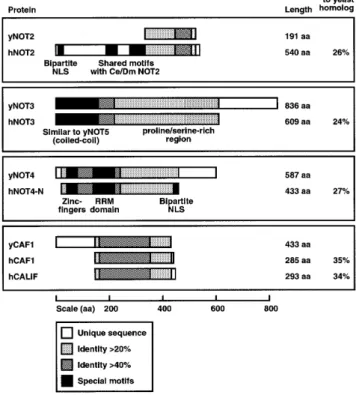
(4) 812. Nucleic Acids Research, 2000, Vol. 28, No. 3. Figure 1. Schematic presentation of conserved regions shared between S.cerevisiae yeast (y) and human (h) CCR4–NOT proteins. The percentage of identity between two proteins was calculated using the ALIGN algorithm (global alignment with no short-cuts). Different shadings denote different percentages of identity as indicated in the bottom panel; the percentage of the overall identity between two proteins is shown to the right. Special motifs and hallmarks are indicated by black boxes. For details see text (supplementary information can be obtained at http://ruummc.med.uu.nl/publications/publtxp.htm ).. proteins from these species harbor an N-terminal extension with two additional short regions of homology (coordinates 258–285 and 333–365 in hNOT2) which are interspersed by a spacer of 48 and 52 residues in hNOT2 and NOT2 in C.elegans; in the Regena protein, this spacer is much larger (157 residues) and significantly rich in glycine residues (32%). Notably, in the non-conserved N-terminal region of hNOT2 we find a sequence KRNYQVTNSMFGASRKK (residues 12–28) which matches to the consensus sequence of a bipartite nuclear localization signal (NLS) that is found in a variety of transcription factors (33,34). Two EST clones (HFCBT83, HHFGH78) showing homology to the reported yNOT3 sequence were obtained. Sequencing revealed that both cDNAs contain a common 3′ untranslated region (UTR), suggesting that the transcripts originated from the same gene. Whereas clone HFCBT83 did not encompass a full-length ORF, we found an uninterrupted open reading frame of 609 codons in clone HHFGH78. Comparison of this protein sequence to yNOT3 (836 amino acids) revealed 24% overall identity, with 41% identity in the first 232 amino acids. The recently identified yNOT5 protein is highly similar to yNOT3 in its N-terminal region, and this homology is reflected by the partial functional redundancy of yNOT3 and yNOT5 (7). Comparison of the human polypeptide to yNOT5 (560 amino acids) demonstrated 24% overall identity and 39% identity in the N-terminal 212 residues,. numbers very similar to the ones obtained for yNOT3. Thus it is difficult to assess conclusively whether we have identified a human homolog of yNOT3 or yNOT5. We will refer to this protein as hNOT3 because its sequence shows a closer relationship to yNOT3 than to yNOT5. Expression of this cDNA in mammalian cells resulted in a protein migrating as a 120 kDa species in SDS–PAGE which is identical to the mobility of the endogenous protein in HeLa cell extracts as detected by hNOT3-specific polyclonal antibodies (data not shown). We note that hNOT3 harbors in its conserved N-terminus a putative coiled-coil motif of the spectrin-repeat type (35) (Fig. 1). Whereas this region of hNOT3 contains a high proportion of acidic residues (24 aspartic acids, 26 glutamic acids), the C-terminal domain from residues 232 to 609 is extremely rich in prolines (17% content) and serines (18% content) (proline/serine-rich region, see Fig. 1). Aside from these features, no other structural motifs of the hNOT3 protein are apparent. We obtained several EST clones showing homology to the reported yNOT4 sequence and determined their DNA sequences. One clone, HWIAL87, was revealed to contain a hNOT4 ORF starting at codon 21. To obtain a complete ORF, a PCR approach was taken (see Materials and Methods). After sequence verification, the PCR fragment encompassing the 20 missing N-terminal codons of hNOT4 was rebuilt into clone HWIAL87. This restored hNOT4 ORF encodes a polypeptide of 433 amino acids. Comparison of the hNOT4 sequence to its yeast counterpart reveals a striking degree of conservation in the first 230 amino acids, with 44% identity of hNOT4 to yNOT4. In contrast, the C-terminal region of NOT4 is far less conserved. Within the highly conserved N-terminus two putative zinc-fingers of the Cys2/Cys2-type are located, with the consensus sequences C-X2-C-X13-C-X-C (residues 14–33 in hNOT4) and C-X2-C-X11-C-X2-C (residues 38–56), where C denotes a cysteine and X indicates any amino acid. In addition, we note a previously unrecognized RNA recognition motif (RRM; residues 111–194 in hNOT4, see Fig. 1) that is commonly found in RNA-binding proteins, but also in a number of proteins reported to bind to DNA (for reviews see 36,37). While this paper was in preparation, we noticed that two cDNAs encoding putative hNOT4 proteins were deposited at the GenBank database (accession nos U71267 and U71268). In contrast to the hNOT4 cDNA isolated by us, these clones contain a common 5′ UTR of 281 nt and two differently sized 3′ UTRs. Conceptual translations of these clones yield products of 575 amino acids (hNOT4-S, short variant) and of 642 amino acids (hNOT4-L, long variant), respectively. The sequences of all three putative hNOT4 proteins are identical until residue 357 and differ in their C-terminal parts. From residue 419, the protein sequence of our isolated clone shows no significant homology to neither the sequence of hNOT4-L nor hNOT4-S, but ends at residue 433. In this part, we find a consensus bipartite NLS sequence RKALADLTEPIERKR (residues 412–426) (33,34). Therefore, we will refer to our hNOT4 sequence as hNOT4-N, for NLS-containing variant. Two human EST clones (HOUDP20, HLYBL69) homologous to yCAF1 were obtained. Sequence analysis revealed that both of them contained a full-length ORF of 293 codons. Interestingly, the deduced amino acid sequence is highly similar, albeit clearly not identical (75% identity) to the one reported for hCAF1 (38). Hence, we identified a second human homolog of.
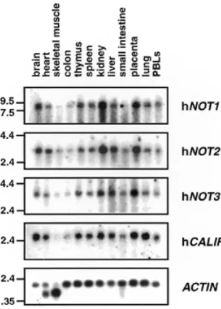
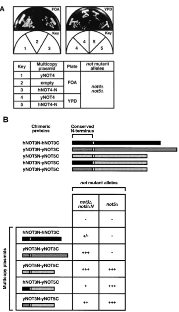
(5) Nucleic Acids Research, 2000, Vol. 28, No. 3. 813. also hybridized to the 1.6 and 1.8 kb mRNAs of α- or γ-actin present in these tissues (Fig. 2). Hence, the observed variations in hNOT1, hNOT2, hNOT3 and hCALIF mRNA levels are not due to unequal loading or degradation of RNAs but rather reflect a tissue-specific expression profile that is common to these genes. Complementation analysis of not mutations in yeast. Figure 2. Expression of CCR4–NOT mRNAs in various human tissues. A multiple human tissue northern blot was hybridized with probes specific for hNOT1, hNOT2, hNOT3, hCALIF and, as a control, with a β-ACTIN specific probe. Molecular sizes (in kb) are indicated to the left.. yCAF1. We named this cDNA hCALIF or human CAF1-like factor. Comparative analysis of the protein sequences of hCALIF, hCAF1 and yCAF1 revealed 34% overall identity of hCALIF with yCAF1 and 35% identity of hCAF1 with yCAF1 (Fig. 1). From these numbers alone it cannot be concluded whether hCALIF or hCAF1 is the true human ortholog of yCAF1. Tissue-specific expression of hCCR4–NOT genes If the gene products of the isolated human CCR4–NOT cDNAs form a protein complex, one would expect a significant overlap in their expression profiles. Therefore, we performed a northern blot analysis of poly(A)+ RNA isolated from different human tissues to investigate the expression of the isolated hCCR4–NOT genes (Fig. 2). Using a DNA probe from the isolated partial hNOT1 cDNA, we found a major hNOT1 transcript of ~8 kb. Previous analysis of the yeast NOT1 transcript, giving rise to a polypeptide of 2108 amino acids, indicated a size of 7 kb (5). The highest levels of the hNOT1 transcript were observed in RNA from brain, kidney and placenta, whereas skeletal muscle and colon showed the lowest expression levels. We found a very similar expression pattern for hNOT2 and hNOT3 mRNAs, with transcript lengths of 3.2 and 3.3 kb, respectively. Using a probe specific for the isolated hCALIF revealed a major transcript of 2.5 kb. Strikingly, like for hNOT1 to hNOT3, transcript levels of hCALIF were also high in brain, kidney and placenta, and very low in skeletal muscle and colon (Fig. 2). This common expression pattern is different from the one reported for hCAF1, with the highest levels of hCAF1 transcripts observed in heart, skeletal muscle, testis, ovary and pancreatic tissue, and low levels in liver, kidney, prostate and peripheral blood cells (38). Reprobing the multiple tissue northern blot with a β-actin control probe revealed roughly equal expression levels of a 2.0 kb β-actin mRNA, except for heart and skeletal muscle where the probe. Whereas NOT1 function is essential for viability in yeast (5), disruption or loss-of-function mutations of the other yeast CCR4–NOT genes result in viable cells with characteristic phenotypic alterations, such as growth defects and increased 3aminotriazole resistance for not2, not3, not4 and not5 mutations (6,7), and increased sensitivity to cell wall defects for mutant not1, not2, not4, ccr4 and caf1 alleles (15). To examine the functional conservation of the human CCR4–NOT genes isolated by us, we tested their ability to act as high-copy suppressors of mutant alleles in yeast. The human cDNAs were cloned in yeast expression plasmids (see Materials and Methods). The hNOT2 cDNA was tested for complementation of the temperature-sensitive phenotype conferred by the not2-1 mutation (5), and the hCALIF cDNA was tested for complementation of the caffeine-sensitive phenotype conferred by the caf1 null mutation (15) as well as for the very slow growth of this mutant on minimal medium (our unpublished observation). Neither hNOT2 nor hCALIF could complement yeast not2 or caf1 mutant phenotypes. The hNOT4-N cDNA was tested for complementation of the synthetic lethality conferred by the not4 null mutation to a not5 null strain (not4∆/not5∆) (7). This was done by transforming a multicopy plasmid containing hNOT4-N or an empty control plasmid into the not4∆/not5∆ double mutant strain bearing a URA3 plasmid which contained the yNOT5 gene. Transformants were then purified on FOA, a drug that kills cells carrying a functional URA3 gene, and therefore selects for cells which can lose the URA3 plasmid. Figure 3A shows that cells containing the hNOT4-N cDNA but not the empty control plasmid were able to grow on FOA. This result clearly points out that we have isolated a cDNA encoding a hNOT4 protein that can complement for a loss of its yeast counterpart, and demonstrates the conservation of NOT4 function in yeast and humans. Testing complementation by hNOT3 in yeast is complicated by the fact that yNOT3 function can be taken over in part by other proteins, namely by yNOT5 (7), and that deletion of yNOT3 (not3∆) does not result in a temperature-sensitive phenotype. However, an additional deletion of the conserved yNOT5 N-terminus (not5∆N) in the not3∆ strain creates a temperature-sensitive phenotype. We tested suppression in this not3∆/not5∆N double mutant by expression of hNOT3 and found that it barely suppressed (Fig. 3B). In contrast, both yNOT3 or yNOT5 fully complemented the not3∆/not5∆N strain. Therefore, we created chimeric constructs which fuse the non-conserved C-terminus of yNOT5 (yNOT5C) to the N-terminus of either hNOT3 (hNOT3-N) or yNOT3 (yNOT3-N). Both hNOT3N–yNOT5C and yNOT3N–yNOT5C weakly suppressed the temperature-sensitive phenotype of the double disruptant strain. Interestingly, these chimeras also suppressed a deletion of the complete yNOT5 gene (not5∆), in contrast to hNOT3N– hNOT3C and yNOT3N–yNOT3C (Fig. 3B). Taken together, these results indicate that (i) the function of the N-terminus of.
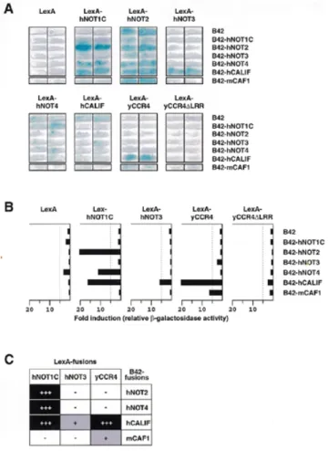
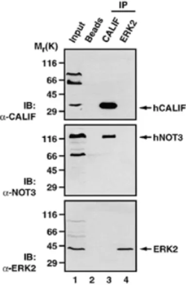
(6) 814. Nucleic Acids Research, 2000, Vol. 28, No. 3. Figure 3. Complementation of mutant not alleles in yeast by human cDNAs. (A) Complementation of not4::LEU2 by the hNOT4-N cDNA. Strain YOU637 was transformed with pGEN-hNOT4-N, pRS314-yNOT4 or pRS314 alone. Transformants were streaked on FOA plates to determine for loss of the pRS316-yNOT5 plasmid essential in this genetic background. Both pRS314-yNOT4 and pGEN-hNOT4-N transformants could grow on FOA (left). Growth of the FOA-resistant transformants was then compared on YPD rich medium (right). (B) Strains YOU578 (not3∆/not5∆N) and YOU555 (not5∆) were transformed with multicopy plasmids expressing the indicated chimeric proteins. Equivalent expression levels for the proteins examined were confirmed by western blot (data not shown). Human and yeast chimeric proteins recombined the natural sequences found within the native NOT3 and NOT5 proteins, but incorporated a few residues in frame that were necessary for creation of the protein junctions (indicated by white border lines in illustrations). Transformants were tested for growth at 37°C. Wild-type growth, (+++); slower than wild-type growth, (+); barely detectable growth, (+/–); no growth, (–).. hNOT3 is more conserved than that of its C-terminal domain and that (ii) it is most likely that we have isolated a human homolog of yNOT3. Two-hybrid interactions among human CCR4–NOT proteins To obtain evidence for interactions between human CCR4–NOT proteins, we used the yeast two-hybrid assay (39). Since. yNOT1 seems to serve as a scaffold protein that provides an indispensible interaction surface for the other complex components in yeast (M.Collart, unpublished observation), we first tested interactions of the isolated human cDNAs with the fulllength yNOT1. We found interactions of LexA-yNOT1 with B42-hNOT2, B42-hNOT4 and B42-hCALIF (data not shown). Next, we included a partial hNOT1 cDNA in this assay (hNOT1C, see Materials and Methods). hNOT1C yields an Nterminally truncated protein of ~1300 amino acids that covers two-thirds of an expected complete hNOT1 ORF (manuscript in preparation). Clear and reproducible two-hybrid interactions between LexA-hNOT1C and B42-hNOT2, B42-hNOT4 and B42-hCALIF were detected (Fig. 4A), and mirrored the ones observed with LexA-yNOT1. The C-terminus of hNOT1 present in hNOT1C seems to be sufficient for these interactions. In addition, we also found an interaction of LexA-hNOT3 and B42-hCALIF (Fig. 4A and summarized in 4C). LexA-hNOT2 strongly activated transcription on its own, resembling the previously reported activation capacity of its yeast counterpart (6,25); to a minor degree, LexA-hNOT4 and LexA-hCALIF also activated transcription of the lacZ reporter gene (Fig. 4A and data not shown). Due to this self-activation of LexA-hNOT2, LexA-hNOT4 and LexA-hCALIF, none of these proteins were used for quantitative determination of β-galactosidase activity (Fig. 4B). In addition to this evolutionarily conserved interaction between hCALIF and NOT1, an even stronger interaction was observed between hCALIF and the yeast CCR4 full-length protein. This interaction is dependent on the presence of the leucine-rich repeat (LRR) region of yCCR4, since it was completely abolished by deletion of this domain (residues 391–455) in LexA-yCCR4∆LRR (Fig. 4B). As a control we included a B42 fusion with the murine (m) CAF1 protein that has been previously shown to interact with LexA-yCCR4 (11). Quantification indicates that hCALIF interacts stronger with yCCR4 than mCAF1 (Fig. 4B). The hCAF1 protein, differing from mCAF1 only at a single residue (asparagine versus serine 282, see 11,38), has not been studied for interaction with CCR4 so far, but we expect that it behaves identically to its mouse counterpart. Co-immunoprecipitation of hCALIF and hNOT3 To confirm the observed two-hybrid interactions by a complementary approach and to obtain evidence for association of hCCR4–NOT proteins in vivo, we performed co-immunoprecipitation experiments. Lysates of human C33A cells (Fig. 5) and HeLa cells (data not shown) prepared in moderate salt concentrations were exposed to a polyclonal hCALIFspecific antiserum, the bound proteins were recovered and analyzed by SDS–PAGE. The presence of hCALIF protein in the lysate was assessed by an anti-CALIF immunoblot (Fig. 5, top, lane 1). Substantial amounts of the endogenous protein were detected after immunoprecipitation with the hCALIF antibody, whereas in control immunoprecipitates the omission of antibody in the binding reaction (lane 2) or inclusion of an unspecific polyclonal antibody (anti-ERK2, lane 4) failed to recover hCALIF. Notably, the hCALIF-specific antiserum immunoprecipitated endogenous non-overexpressed hNOT3 (Fig. 5, middle, lane 3), thereby confirming the observed twohybrid interaction for these two proteins..
(7) Nucleic Acids Research, 2000, Vol. 28, No. 3. Figure 4. Human CCR4–NOT proteins interact in the yeast two-hybrid assay. (A) Yeast cells of strain EGY48 were transformed with the indicated LexAand B42-fusion expression plasmids together with a lacZ reporter gene containing one LexA operator. Interactions were assayed by blue staining of transformants that were streaked on Xgal-containing selective plates, and incubated for 16–20 h at 30°C. (B) Relative β-galactosidase activities of EGY48 derivatives expressing the indicated proteins. Yeast extracts from EGY48 transformants selected by trp+his+ura+ prototrophy were assayed for β-galactosidase activity. Three to four independent experiments were performed for each combination and the results of one representative set of experiments are shown, with similar results obtained for the other independent sets of experiments. Measured β-galactosidase activities (in RLUs) were normalized for the amount of total protein in extracts and expressed as fold inductions relative to the β-galactosidase activity of cells expressing the B42 activation domain only. A table summarizing the observed two-hybrid interactions is presented in (C).. Taken together, these results establish interactions of human CCR4–NOT proteins, suggesting that they can potentially form a complex in human cells in vivo. In addition, the results indicate that we have isolated a new bona fide component of the human CCR4–NOT complex, namely hCALIF. DISCUSSION The present study predicts the existence of a CCR4–NOT complex in mammalian cells. We describe the identification and isolation of four components of a putative human CCR4–NOT complex, i.e. hNOT2, hNOT3, hNOT4 and hCALIF. All of these proteins reveal striking similarities to their homologs of various species, indicating that this transcriptional regulator complex is conserved in higher eukaryotic organisms. The observed two-hybrid interactions most likely reflect the potential of. 815. the human proteins to form a complex very similar to a yeast CCR4–NOT ‘core’ complex, consisiting of NOT proteins, CCR4 and CAF1 (15). We have isolated a human cDNA encoding a hNOT2 protein of 540 amino acids that shows significant homology to yeast NOT2 proteins in both Saccharomyces cerevisiae and Schizosaccharomyces pombe, and to NOT2 homologs in C.elegans and D.melanogaster. While this paper was in preparation, Benson et al. (25) reported a partial hNOT2 amino acid sequence encompassing the most C-terminally located 95 residues. Comparison of our hNOT2 cDNA to that isolated by Benson and colleagues revealed that both cDNAs indeed encode the same protein, i.e. hNOT2 and that the sequence reported in the present study is correct (J.D.Benson, personal communication). Notably, NOT2 proteins in yeast are much smaller than their counterparts in metazoans. In addition, metazoan NOT2 proteins share short regions of homology in their extended N-terminal domains, suggesting that NOT2 acquired an additional function in these organisms. In this respect it is interesting to note that the Drosophila NOT2 (Regena) protein has been identified as suppressor of a position-effect-variegation phenotype (32), suggesting a role in the organization of chromatin. It is tempting to speculate that the N-terminal domain in hNOT2 links CCR4–NOT functions to chromatin functions. The finding that expression of the isolated full-length hNOT2 cDNA did not suppress a yeast not2-1 null allele might be explained by the supposition that the non-conserved N-terminal domains of yNOT2 and hNOT2 serve different functions. This is supported by the finding that the unique yNOT2 N-terminal region (residues 1–106) fused to the hNOT2 C-terminus in a chimeric protein resulted in suppression of the temperaturesensitive not2-1 phenotype, as reported in (25). Thus, NOT2 proteins in yeast and human indeed seem to harbor two distinct modules, with an evolutionary conserved C-terminal domain that is functionally separable from the divergent N-terminal domains. In further support of this, the integrity of the unique N-terminal region of yNOT2 is required for full transcriptional activation and interaction with yeast ADA2, whereas CCR4associated functions of yNOT2 require a functional C-terminus (25). The isolated hNOT3 cDNA described in this study yields a protein that shows striking similarity to the N-terminal regions of both yNOT3 and yNOT5. Expression of this cDNA did not complement the tight temperature-sensitive phenotype of a yeast not5 null mutation. However, when expressed as fusion with the C-terminus of yNOT5, the conserved N-terminus of hNOT3 showed full complementation of the phenotype of a yeast not5∆ null allelle, and partial complementation of a yeast not3∆/not5∆N double mutant, indicating that the function of its N-terminus is well conserved. In contrast to the functional redundancy of NOT3 and NOT5 in yeast, we have found no indications for a yNOT5-like protein in human cells. The presence of a putative coiled-coil domain in the N-terminus of hNOT3 as well as in yNOT3 and yNOT5 most likely reflects an intrinsic ability of these proteins to hetero- or homodimerize. This region shows resemblance to a spectrin repeat, a repetitive module present in various cytoskeletal proteins (for a review see 35). Tertiary structure models have been proposed for the folding of the spectrin repeat into a triple-helical bundle or coiled-coil (39). Interestingly,.
(8) 816. Nucleic Acids Research, 2000, Vol. 28, No. 3. Figure 5. hCALIF and hNOT3 interact in vivo. C33A cell lysates were subjected to immunoprecipitation with protein A–agarose beads alone (lane 2), beads coupled to anti-CALIF antibody (lane 3) or to a control antibody (anti-ERK2, lane 4). The input (lane 1) contains one-tenth the quantity of extract used for each immunoprecipitation. Protein extraction and binding conditions are as described in Materials and Methods. The upper panel shows an immunoblot (IB) with anti-CALIF, the middle panel with anti-NOT3 and the lower panel with anti-ERK2. The arrows indicate the proteins found in the immunoprecipitates. Molecular weight markers are indicated on the left.. the repeats in the β-chain of spectrin are followed by a proline/ serine-rich region and a C-terminal serine-rich region which most likely are targets for regulatory phosphorylation events affecting the mechanical stability of spectrin (35). We find prolines and serines to be the predominant residues in the C-terminal region of hNOT3. One can speculate about the significance of these structural similarities in two proteins serving presumably completely different functions. Notably, NOT3 in yeast is a target for modification by kinases (U.Oberholzer and M.Collart, unpublished observation). The presence of the proline/serine-rich C-terminal region in hNOT3 might be an indication that this protein is also a phosphoprotein. Extensive post-translational modifications like hyperphosphorylation might also explain the aberrant migration in SDS–PAGE as observed for endogenous hNOT3. In yeast, NOT3 seems to directly interact with yNOT4 and yNOT5, as revealed by two-hybrid as well as genetic interactions (6,7). In the two-hybrid system as well as in co-immunoprecipitation experiments, we have found an interaction between hNOT3 and hCALIF, but no interaction between hNOT3 and hNOT4. This difference might be attributed to a different composition of the complex in yeast and humans (e.g. the lack of a human NOT5 counterpart); likewise, it might reflect an unstable or weak association of hNOT3 with the human CCR4–NOT complex. Yet, northern blot analysis of hNOT3 mRNA in various human tissues revealed both quantitatively and qualitatively a striking similarity to the expression patterns of the other hCCR4–NOT genes tested. Taken together, (i) the common expression pattern, (ii) the physical interaction between hNOT3 and hCALIF as revealed by two-hybrid analysis and co-immunoprecipitation and (iii) the conserved function of the N-terminus of hNOT3 observed in the complementation assay. suggest that hNOT3 is a subunit of the human CCR4–NOT complex. The ability of the isolated hNOT4 cDNA to complement the synthetic lethal phenotype of a double not4∆/not5∆ disruption clearly demonstrates the evolutionary conservation of NOT4 function. A modular design of NOT4, like that for NOT2, with a conserved N-terminal half and a non-conserved C-terminal region seems very likely. In their N-terminal region NOT4 proteins of various species harbor a remarkable combination of two structural motifs, namely zinc-fingers and a RRM domain. These structural signatures have been implicated in the mediation of DNA- and RNA-binding of a variety of proteins (for reviews see 36,40,41). It is tempting to speculate that this feature of NOT4 proteins provides a ‘targeting’ function for the CCR4–NOT complex. In agreement with this postulated targeting function is the observation that overexpression of the conserved N-terminus of yNOT4 can have a dominant-negative effect on HIS3 transcription in yeast (M.Collart, unpublished observation). We are currently testing this NOT4 targeting hypothesis in a number of experiments. Notably, isoforms of hNOT4 cDNAs different to the one described here have been reported. These isoforms might be products of alternative splicing events that fuse different 3′-exons to a common 5′-region. In support of this, comparison of the three hNOT4 variants (hNOT4-N, -S and -L) reveals that their amino acid sequences are identical from residues 1 to 357. From residues 358 to 400, our isolated hNOT4-N variant is almost identical to a murine NOT4 protein (with only one difference at position 361). Careful inspection and sequence comparison in this region revealed three differences at the nucleotide level between hNOT4-N, hNOT4-S and hNOT4-L cDNA sequences (see Materials and Methods). Incorporation of these differences into the cDNA sequences of hNOT4-S and hNOT4-L results in an amino acid sequence from residues 358 to 400 that is identical to the one found in hNOT-N. Moreover, these three nucleotide changes are also found in a cDNA encoding a partial hNOT4 ORF (GenBank accession no. AF091094), thereby suggesting that our hNOT4-N sequence is correct in this region. From residues 401 to 418 the sequences of hNOT4-N, -S, -L and mNOT4 are identical, but differ again from residue 419. In this most C-terminal region of hNOT4-N we find a bipartite NLS sequence. At present it is unclear if there is any functional significance of these different isoforms of hNOT4. However, the presence of a nuclear targeting signal in our isolated hNOT4 variant points to the possibility of a regulation of the subcellular localization of hNOT4 by use of alternative splice variants. Interestingly, a number of potential phosphorylation sites for kinases like CKI, CKII, GSK3 and PKC are found proximal to this C-terminal NLS signal. Regulation of the nuclear transport of transcription factors through phosphorylation at sites close to NLS signals is a common mechanism in eukaryotic cells by which the level of nuclear accumulation of these factors is controlled (for a review see 42). Interactions of the isolated hCALIF with both human and yeast NOT and CCR4 proteins strongly support our hypothesis that hCALIF is a novel subunit of the human CCR4–NOT complex. The presence of two highly related CAF1-like proteins in human cells suggests a partition of yCAF1 functions; it might also explain the inability of either mCAF1 (11) or hCALIF to complement a caf1 disruption in yeast. Both proteins are much.
(9) Nucleic Acids Research, 2000, Vol. 28, No. 3. shorter than yCAF1 which harbors an N-terminal, non-conserved extension of ~140 residues. Interestingly, partial deletion of this non-conserved yCAF1 region (residues 1–80) results in a protein that is also unable to complement the caf1 temperaturesensitive phenotype at 37°C (11). Hence, a function present in this N-terminal region of yCAF1 is presumably lacking in hCALIF and mCAF1. Since the lack of this region does not impair association of either hCALIF or mCAF1 with yCCR4, this function of yCAF1 seems to be separable from CCR4 functions. Indeed, such CCR4-independent functions of yCAF1 have already been postulated (11,43). We did not test hCAF1 (which is almost identical to mCAF1) in the twohybrid assay for interactions with hCCR4–NOT proteins. However, a striking difference emerged when we compared hCALIF with mCAF1 in this assay: whereas hCALIF showed strong interactions with hNOT1C and hNOT3, mCAF1 did not. In this respect, it is interesting to note that neither mCAF1 nor hCAF1 were initially identified as proteins interacting with NOT proteins; instead, mCAF1 was obtained in a screen as yCCR4 interacting factor (hence its name) and hCAF1 in a screen for proteins interacting with BTG1 (B cell translocation gene product 1) (11,38). Whereas yCAF1 is capable of interacting with NOT proteins (15), this characteristic feature has to our knowledge not as yet been tested for mCAF1 or hCAF1. Notably, association of yCCR4 with the NOT proteins in yeast is strictly dependent on the presence of yCAF1 since caf1 disruption effectively removes yCCR4 from the NOT complex (15). Thus, the present study describes the first CAF1-like protein identified in human cells, namely hCALIF, that is able to interact not only with yCCR4 but also with NOT proteins. By providing a physical link between NOT proteins and CCR4 function, hCALIF fulfils the major requirement of a true yCAF1 ortholog. The picture that has emerged from data obtained in yeast is that the CCR4–NOT complex is one among several other global transcriptional regulators like SAGA, MOT1 and NC2, to which TBP function might be parceled out (for a review see 24). How global gene expression is regulated through the interplay of TBP and these complexes is largely unknown. Future studies aiming at biochemical purification of the human CCR4–NOT complex, determination of its exact composition and assaying its associated functions should uncover important aspects of this fundamental process. ACKNOWLEDGEMENTS We thank C. L. Denis for the gift of the LexA-yCCR4, LexAyCCR4∆LRR and B42-mCAF1 plasmids, and J. D. Benson for communication of unpublished results. We also thank V. J. Bardwell and K. D. Huynh for providing us with a partial hNOT1 cDNA. We thank members of our laboratory for discussions, and U. Fiedler and M. de Ruwe for critical reading of this manuscript. T.K.A. received financial support from a postdoctoral fellowship of the Dutch Organization for Scientific Research-Medical Sciences (NWO-MW), H.Th.M.T. from a Pionier grant of the same organization and M.A.C. from grants of the Swiss National Science Foundation (31-39690.93 and 31-49808.96). This work was also supported by funds of the European Community, TMR network ERB-FMRX-CT96-0064.. 817. REFERENCES 1. Hampsey,M. (1998) Microbiol. Mol. Biol. Rev., 62, 465–503. 2. Bjorklund,S., Almouzni,G., Davidson,I., Nightingale,K.P. and Weiss,K. (1999) Cell, 96, 759–767. 3. Mahadevan,S. and Struhl,K. (1990) Mol. Cell. Biol., 10, 4447–4455. 4. Iyer,V. and Struhl,K. (1995) Mol. Cell. Biol., 15, 7059–7066. 5. Collart,M.A. and Struhl,K. (1993) EMBO J., 12, 177–186. 6. Collart,M.A. and Struhl,K. (1994) Genes Dev., 8, 525–537. 7. Oberholzer,U. and Collart,M.A. (1998) Gene, 207, 61–69. 8. Denis,C.L. (1984) Genetics, 108, 833–844. 9. Denis,C.L. and Malvar,T. (1990) Genetics, 124, 283–291. 10. Draper,M.P., Liu,H.Y., Nelsbach,A.H., Mosley,S.P. and Denis,C.L. (1994) Mol. Cell. Biol., 14, 4522–4531. 11. Draper,M.P., Salvadore,C. and Denis,C.L. (1995) Mol. Cell. Biol., 15, 3487–3495. 12. Sakai,A., Chibazakura,T., Shimizu,Y. and Hishinuma,F. (1992) Nucleic Acids Res., 20, 6227–6233. 13. Toyn,J.H., Araki,H., Sugino,A. and Johnston,L.H. (1991) Gene, 104, 63–70. 14. Liu,H.Y., Toyn,J.H., Chiang,Y.C., Draper,M.P., Johnston,L.H. and Denis,C.L. (1997) EMBO J., 16, 5289–5298. 15. Liu,H.Y., Badarinarayana,V., Audino,D.C., Rappsilber,J., Mann,M. and Denis,C.L. (1998) EMBO J., 17, 1096–1106. 16. Lee,T.I., Wyrick,J.J., Koh,S.S., Jennings,E.G., Gadbois,E.L. and Young,R.A. (1998) Mol. Cell. Biol., 18, 4455–4462. 17. Thompson,C.M., Koleske,A.J., Chao,D.M. and Young,R.A. (1993) Cell, 73, 1361–1375. 18. Holstege,F.C., Jennings,E.G., Wyrick,J.J., Lee,T.I., Hengartner,C.J., Green,M.R., Golub,T.R., Lander,E.S. and Young,R.A. (1998) Cell, 95, 717–728. 19. Shi,X., Chang,M., Wolf,A.J., Chang,C.H., Frazer-Abel,A.A., Wade,P.A., Burton,Z.F. and Jaehning,J.A. (1997) Mol. Cell. Biol., 17, 1160–1169. 20. Chang,M., French-Cornay,D., Fan,H.Y., Klein,H., Denis,C.L. and Jaehning,J.A. (1999) Mol. Cell. Biol., 19, 1056–1067. 21. Collart,M.A. (1996) Mol. Cell. Biol., 16, 6668–6676. 22. Moqtaderi,Z., Bai,Y., Poon,D., Weil,P.A. and Struhl,K. (1996) Nature, 383, 188–191. 23. Moqtaderi,Z., Keaveney,M. and Struhl,K. (1998) Mol. Cell., 2, 675–682. 24. Lee,T.I. and Young,R.A. (1998) Genes Dev., 12, 1398–1408. 25. Benson,J.D., Benson,M., Howley,P.M. and Struhl,K. (1998) EMBO J., 17, 6714–6722. 26. Ausubel,F.M., Brent,R., Kingston,R.E., Moore,D.D., Seidmann,J.G., Smith,J.A. and Struhl,K. (1992) Short Protocols in Molecular Biology. Greene Publishing Associates and John Wiley & Sons, New York, NY. 27. Gyuris,J., Golemis,E., Chertkov,H. and Brent,R. (1993) Cell, 75, 791–803. 28. Shpakovski,G.V., Acker,J., Wintzerith,M., Lacroix,J.F., Thuriaux,P. and Vigneron,M. (1995) Mol. Cell. Biol., 15, 4702–4710. 29. Christianson,T.W., Sikorski,R.S., Dante,M., Shero,J.H. and Hieter,P. (1992) Gene, 110, 119–122. 30. Bourne,Y., Watson,M.H., Hickey,M.J., Holmes,W., Rocque,W., Reed,S.I. and Tainer,J.A. (1996) Cell, 84, 863–874. 31. Timmers,H.T.M. and Sharp,P.A. (1991) Genes Dev., 5, 1946–1956. 32. Frolov,M.V., Benevolenskaya,E.V. and Birchler,J.A. (1998) Genetics, 148, 317–329. 33. Dingwall,C. and Laskey,R.A. (1991) Trends Biochem. Sci., 16, 478–481. 34. Robbins,J., Dilworth,S.M., Laskey,R.A. and Dingwall,C. (1991) Cell, 64, 615–623. 35. Pascual,J., Castresana,J. and Saraste,M. (1997) Bioessays, 19, 811–817. 36. Burd,C.G. and Dreyfuss,G. (1994) Science, 265, 615–621. 37. Nagai,K., Oubridge,C., Ito,N., Avis,J. and Evans,P. (1995) Trends Biochem. Sci., 20, 235–240. 38. Bogdan,J.A., Adams-Burton,C., Pedicord,D.L., Sukovich,D.A., Benfield,P.A., Corjay,M.H., Stoltenborg,J.K. and Dicker,I.B. (1998) Biochem. J., 336, 471–481. 39. Parry,D.A., Dixon,T.W. and Cohen,C. (1992) Biophys. J., 61, 858–867. 40. Nelson,H.C. (1995) Curr. Opin. Genet. Dev., 5, 180–189. 41. Papavassiliou,A.G. (1995) Anticancer Res., 15, 891–894. 42. Jans,D.A. and Hubner,S. (1996) Physiol. Rev., 76, 651–685. 43. Hata,H., Mitsui,H., Liu,H., Bai,Y., Denis,C.L., Shimizu,Y. and Sakai,A. (1998) Genetics, 148, 571–579. 44. Zervos,A.S., Gyuris,J. and Brent,R. (1993) Cell, 72, 223–232..
(10)
Figure

+3
Documents relatifs