1370
The 30-bp Deletion Variant of Epstein-Barr Virus – Encoded Latent Membrane
Protein-1 Prevails in Acute Infectious Mononucleosis
Christoph Berger, Cathy McQuain, John L. Sullivan, LINK Laboratories (Cancer Center) and Department of Pediatrics, University of Massachusetts Medical Center, Worcester, Massachusetts; David Nadal, Peter J. Quesenberry, and Hans Knecht
Infectious Diseases Unit, University Children’s Hospital Zurich, Zurich, Switzerland To assess the frequency of malignancy-associated 30-bp deletion variants of the latent membrane
protein 1 (LMP-1) in benign conditions, a comparative sequence analysis was done using samples from 20 American children with acute infectious mononucleosis and 16 Swiss children with chronic tonsillar hyperplasia. The 30-bp deletion variant (LMP-1-del) was present in 66% of patients (12/ 20 with infectious mononucleosis and 12/16 with tonsillar hyperplasia). Two additional patients had a 3-bp deletion and an inframe insertion of 18 nucleotides, respectively. All but 1 isolate had numerous nonsilent point mutations. These data identify a hypervariable region within the C-terminus of LMP-1, in a domain required for maximal stimulation of NF-kB activity. These data demonstrate that LMP-1-del variants are frequent in acute infectious mononucleosis and tonsillar hyperplasia and identical to those observed in Epstein-Barr virus – associated AIDS-related lymphoma.
Epstein-Barr virus (EBV) causes infectious mononucleosis boxy-terminal NF-kB activation domain of LMP-1 has been associated with enhanced transforming potential compared with (IM) and is consistently associated with nasopharyngeal
carci-noma (NPC) and lymphomas, especially in the immunocom- the oncoprotein prototype in vitro [7]. To further assess in vivo the relevance of this variant, we analyzed the LMP-1 C-promised host. EBV infection is ubiquitous; the virus persists
lifelong in latent form and can be detected in B lymphocytes terminus in DNA samples from American children with acute IM and Swiss children with EBV-associated chronic tonsillar and in oropharyngeal secretions [1].
Latent membrane protein-1 (LMP-1) is considered a virus hyperplasia (TH). oncogene due to its ability to transform rodent fibroblasts in
vitro and to render them tumorigenic in nude mice. It
trans-Material and Methods forms human epithelial cells and inhibits their differentiation,
induces DNA synthesis, up-regulates bcl-2 expression, and is
Samples. Twenty children with acute IM defined by positive engaged in cellular signalling processes, including NF-kB [2] heterophilic antibodies, peripheral blood lymphocytosis, and and the TNF receptor family. LMP-1 is expressed in IM and lymphadenopathy were identified at University of Massachusetts in the tumor cells of most EBV-associated malignancies (re- Medical Center between April 1988 and October 1992. Peripheral
viewed in [1]). blood mononuclear cells (PBMC) were collected during the acute
phase of the disease. Sixteen EBV-seropositive children (IgG
A naturally occurring carboxy-terminal 30-bp deletion
vari-against viral capsid antigen) had undergone tonsillectomy for
hy-ant (LMP-1-del) is found in persons with AIDS-related
perplastic tonsils at the University Children’s Hospital in Zurich
lymphoma (72% [3 – 5]), NPC [6], clinically aggressive
Hodg-from December 1992 to April 1994. At this time, tonsil tissue was
kin’s disease, and atypical lymphoproliferations [4]. This
vari-harvested and frozen until extraction of genomic DNA.
ant oncoprotein with a distinct 30-bp deletion within the
car-Polymerase chain reaction (PCR). LMP-1 genomes were identified by PCR. Three different primer sets, specific for the C-terminal domain, were used as previously described [4]. PCR conditions and amplification strategy have been described in detail Received 14 March 1997; revised 27 May 1997.
[4]. Independent triplicate amplification was performed for each Presented in part: annual meeting of the American Society of Hematology,
Orlando, Florida, 9 December 1996 (Blood 1996; 88:202 [abstract]). sample. In the samples with LMP-1-del, an additional PCR using Informed consent was obtained from patients and their parents. All proce- one primer (5*-TCATAGTCATGATTCCGGCC-3*) located dures were done in accordance with the approved institutional guidelines for within the 30-bp deletion was performed, in order to detect a low the conduct of clinical research.
copy number of additional wild type LMP-1. Financial support: Swiss National Foundation (grant no. 31-37727.93 and
DNA sequencing. Double-stranded PCR products coding pediatric postdoctoral research fellowship to C.B.); La Recherche Suisse Contre
Le Cancer (grant AKT 540). for the LMP-1 C-terminus were obtained by the primer pair Reprints or correspondence: Dr. Hans Knecht, Division of Hematology/ 5*-AGCGACTCTGCTGGAAATGAT-3*/5*-TGATTAGCTAAG-Oncology, University of Massachusetts Medical Center, 55 Lake Avenue N.,
GCATTCCCA-3* (primer pair 9/11 from [4]) or CP3/MS7 Worcester, MA 01655-0246.
(5*-TGCTCTCAAAACCTAGGCGCA-3/5*-TCATCATCT-The Journal of Infectious Diseases 1997; 176:1370 – 3
CCACCGGAACCA-3*, positions 168609–168589/168200–
q 1997 by The University of Chicago. All rights reserved.
se-1371 JID 1997; 176 (November) Concise Communications
Figure 1. Mutational hot spots within carboxy-terminus of latent membrane protein-1 (LMP-1) in children with acute infectious mononucleo-sis (IM) and tonsillar hyperplasia (TH). Point mutations with following amino acid change are underlined ; silent mutations are not underlined. Absence of letter indicates nucleotide identical to wild-type sequence. Dashed lines mark deletion of 30 bp and 3 bp, respectively. Dotted line plus bracket indicates sequence not read.l stands for insertion of 18 nucleotides [5*-TAGTCTAGACTAGGTGAC-3*] between nt 168321 and 168322. IM, infectious mononucleosis; TH, tonsillar hyperplasia.
quenced with35S-labeled dATP using a Sequenase kit (Amersham
deletion (positions 168289 – 186291) and numerous point
muta-Life Sciences, Arlington Heights, IL). Sequencing primers for tions. An insertion between position 168321 and 168322 con-LMP-1 were MS1 (5*-ACAATTGACGGAAGAGGTTGA-3*, po- taining 18 nucleotides (TAGTCTAGACTAGGTGAC) was sitions 168358 – 168338) or primer 9 for the coding strand and identified in sample TH-2, which otherwise corresponded to the MS7 or primer 11 for the noncoding strand. Sequencing from
B95.8 sequence. Of interest, 13 bp within this insertion had
independent PCR products from 25% of samples showed identical
complete homology to a measle virus nucleoprotein gene
(TCT-results. In 36% of samples, both strands were sequenced.
AGACTAGGTG), another 13 were homologous to the human Statistical analysis. The uncorrected x2
test was used to assess
herpesvirus-6 gene encoding the Rep protein
(TAGACTAGG-the association of LMP-1-del between samples from healthy
carri-TGAC), and an additional 13- to 14-bp sequence to a coat protein
ers and from persons with AIDS-related lymphomas, IM, and TH.
gene of poliovirus Sabin 1 strain (TCACCTATTCTAGA). Only 1 isolate (TH-1) corresponded exactly with the prototype B95.8.
Results Most nucleotide changes were characterized by different amino
acid substitutions, defining them as mutational ‘‘hot spots.’’ Sequencing results are summarized in figure 1. All deletions
Independent triplicate amplification of every sample consistently and most point mutations occurred within the 100 bp from
posi-showed PCR products of identical size. In none of the patients tions 168324 to 168225, within the NF-kB activation domain
were both LMP-1 variants found. A second sequencing of inde-of LMP-1. Mutations were identified in ú75% of isolates at
pendent amplification products was performed in 9 cases (includ-positions 168225, 168257, 168258, 168266, 168295, and
ing the 7 cases with the highest number of point mutations) to 168308; 76% of isolates had a point mutation at position 168357
further exclude sequence errors due to base misincorporation. that was no longer in the NF-kB activation domain. Between
This internal control revealed complete sequence homology with position 168224 and the carboxy-terminal end (168160), only
the initial results. Overall, four patterns of mutations were ob-one single point mutation (IM-7) was identified; 60% of the IM
served in patients with IM and TH: an insertion in 1 isolate, samples and 75% of the TH samples showed a 30-bp deletion
1372 Concise Communications JID 1997; 176 (November)
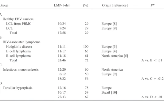
Table 1. Frequency of latent membrane protein 1-del in healthy carriers and in persons with HIV-associated lymphoma, infectious mononucleosis, and tonsillar hyperplasia.
Group LMP-1-del (%) Origin [reference] P*
A
Healthy EBV carriers
LCL from PBMC 10/34 29 Europe [8] LCL 7/24 29 Europe [9]
Total 17/58 29 B
HIV-associated lymphoma
Hodgkin’s disease 11/11 100 Europe [3] B cell lymphoma 11/17 65 Europe [4] B cell lymphoma 11/18 61 North America [5]
Total 33/46 72 A vs. Bõ .01
C
Infectious mononucleosis 12/20 60 North America 6/12 50 Europe [9]
18/32 56 A vs. CÅ .012
D
Tonsillar hyperplasia 12/16 75 Europe 10/17 59 Brazil [10]
22/33 67 A vs. Dõ .01
NOTE. LMP-1 del, 30-bp deletion variant of LMP-1; LCL, lymphoblastoid cell line; PBMC, peripheral blood mononuclear cells.
* Differences between groups B, C, and D are not significant (Pú .1).
deletion but an accumulation of point mutations in 7 isolates, in vivo relevance of these in vitro tumor-promoting effects. However, the high frequency of LMP-1-del in persons with and the 30-bp deletion plus point mutations in 24 isolates.
AIDS-related lymphoma, IM, and TH but not in asymptomatic The comparison of data from recent studies [3 – 5, 8 – 10]
carriers is consistent with the hypothesis that this evolving with our data (table 1) reveals a significant accumulation of
LMP-1 variant may arise only in the absence of T cell surveil-LMP-1-del in IM and TH, as observed in AIDS-related
lance. Several findings are in support of this hypothesis. lymphoma and in contrast with asymptomatic EBV carriers.
First, our data identify LMP-1-del as a part of a hypervariable region different from a simple viral polymorphism. The pres-ence of mutational hot spots, 3- and 30-bp deletions and an Discussion
insertion, favors the generation of viral variants. Aware of the The LMP-1 variant with a C-terminal 30-bp deletion and possibility that variants with and without deletion might be 6 clustered point mutations (LMP-1-del) has been detected present in the same patient [4, 13, 14], we were not able to predominantly in AIDS-related lymphoma and Asian NPC [3 – detect more than one variant by a very sensitive PCR approach 6]. Experimental studies with LMP-1 variants from biopsies in this collection. Viral inter- or intrastrain recombination dur-from persons with NPC (carrying LMP-1-del) indicated this ing EBV replication has been shown in oral hairy leukoplakia variant to be more tumorigenic [11] and less immunogenic [12] and NPC [6, 14] and fits with the frequent detection of LMP-than the LMP-1 prototype (B95.8) in mouse model systems. 1-del in persons with hyperplastic tonsils and in HIV-infected In particular, Li et al. [7] recently demonstrated that the deletion patients, in whom enhanced viral replication occurs. Alterna-of these 30 bp in the LMP-1 prototype mediated enhanced tively, the accumulation of LMP-1 variants in situations with oncogenic potential in vitro. In vivo, the sequence variation of ongoing germinal center activity, particularly as seen in persons LMP-1-del in a region critical for the protein’s half-life (by with TH, IM, and AIDS-associated lymphoproliferations, is prolongation of half-life) might lead to an accumulation of consistent with the generation of LMP-1 variants in germinal oncoprotein [2, 4], consistent with an enhanced transforming center reactions, where physiologically somatic hypermutations activity in the infected cell. and isotype switching of immunoglobulin genes occur [15].
This sequence analysis of LMP-1 indicates the prevalence Second, the role of cellular immunity in controlling latent of LMP-1-del in 36 children with IM and HT. Although the EBV infection is well established and, in particular, is main-total number of persons is epidemiologically limiting, the high tained after primary infection by specific clones of cytotoxic frequency of LMP-1-del in IM and HT, both benign and self- T lymphocytes targeted against latent EBV proteins, including LMP-1 [1]. Moreover, cellular immunodeficiency results in limiting processes, is surprising and may raise doubts on the
1373 JID 1997; 176 (November) Concise Communications
4. Knecht H, Raphael M, McQuain C, et al. Deletion variants within the
NF-reactivation of oropharyngeal EBV replication, occurrence of
kB activation domain of the LMP1 oncogene prevail in AIDS-related
variant virus strains, and expansion of the pool of EBV-infected
large cell lymphomas and HIV-negative atypical lymphoproliferation.
lymphocytes. As seen in AIDS-related or posttransplant Blood1996; 87:876 – 81.
lymphoma [3 – 5], in which LMP-1-del is strongly expressed, 5. Kingma DW, Weiss WB, Jaffe ES, Kumar S, Frekko K, Raffeld M.
the in vitro observed enhanced oncogenic potential may mani- Epstein-Barr virus latent membrane protein-1 oncogene deletions: corre-lations with malignancy in Epstein-Barr virus – associated
lymphopro-fest in the T cell – deficient host.
liferative disorders and malignant lymphomas. Blood1996; 88:242 – 51.
Third, the identification of LMP-1-del in the healthy
popula-6. Hu LF, Zabarovsky ER, Chen F, et al. Isolation and sequencing of the
tion (table 1) emphasizes the sustained infectivity and
transmis-Epstein-Barr virus BNLF-1 gene (LMP1) from a Chinese
nasopharyn-sion, as well as the persistence of this variant in peripheral geal carcinoma. J Gen Virol1991; 72:2399 – 409.
blood lymphocytes in healthy carriers. The accumulation of 7. Li SN, Chang YS, Liu ST. Effect of a 10-amino acid deletion on the oncogenic activity of latent membrane protein 1 of Epstein-Barr virus.
LMP-1-del in hyperplastic tonsils from Swiss and Brazilian
Oncogene1996; 12:2129 – 35.
children [10], in oral hairy leukoplakia [15], and in NPC [6]
8. Sandvej K, Gratama J, Hamilton-Dutoit S. Sequence analysis of the
Ep-suggests a higher incidence of such LMP-1 variants in the
stein-Barr virus (EBV) latent membrane protein-1 gene and promoter
oropharynx, where EBV might differ from its latent form in region: identification of four variants among wild type EBV isolates lymphocytes. However, in persons with acute IM, the fre- [abstract E8.2]. In: Program and abstracts of the 7th international EBV symposium (Hong Kong). Chapel Hill, NC: International Association
quency of LMP-1-del is significantly higher than in healthy
for Epstein-Barr Virus Research,1996.
EBV carriers if only North American and European persons
9. Khanim F, Yao QY, Niedobitek G, Sihota S, Rickinson AB, Young LS.
and PBMC samples are compared. In contrast to the situation
Analysis of Epstein-Barr virus gene polymorphisms in normal donors
during latent EBV infection, during acute IM the high virus
and in virus-associated tumors from different geographic locations.
load in PBMC and the not yet functionally developed specific Blood1996; 88:3491 – 501.
cellular immune response may contribute to this difference. 10. Chen WC, Chen YY, Bacchi MM, Bacchi CE, Alvarenga M, Weiss LM. Genotyping of Epstein-Barr virus in Brazilian Burkitt’s lymphoma and
In summary, the high percentage of LMP-1 30-bp deletion
reactive lymphoid tissue. Am J Pathol1996; 148:17 – 23.
variants in samples from persons with acute IM and TH
indi-11. Chen ML, Tsai CN, Liang CL, et al. Cloning and characterization of the
cates that their in vitro – enhanced oncogenicity may not be
latent membrane protein (LMP) of a specific Epstein-Barr virus variant
relevant in the healthy host in vivo. However, these variants derived from the nasopharyngeal carcinoma in the Taiwanese popula-may manifest their in vitro – reported oncogenic potential in tion. Oncogene1992; 7:2131 – 40.
12. Trivedi P, Hu LF, Chen F, et al. Epstein-Barr virus (EBV) – encoded
mem-settings characterized by lack of cellular immune surveillance,
brane protein LMP1 from a nasopharyngeal carcinoma is
non-immuno-as demonstrated in persons with AIDS-related lymphoma.
genic in a murine model system, in contrast to a B cell – derived homo-logue. Eur J Cancer1994; 30A:84 – 8.
References 13. Walling DM, Clark NM, Markovitz DM, et al. Epstein-Barr virus
coinfec-tion and recombinacoinfec-tion in non – human immunodeficiency virus – associ-1. Rickinson AB, Kieff E. Epstein-Barr virus. Fields BN, Knipe DM, Howley
ated oral hairy leukoplakia. J Infect Dis1995; 171:1122 – 30. PM, et al., eds. Virology. Philadelphia: Lippincott-Raven,1996:2397 –
14. Palefsky JM, Berline J, Penaranda ME, Lennette ET, Greenspan D, 446.
Greenspan JS. Sequence variation of latent membrane protein-1 of Ep-2. Mitchell T, Sugden B. Stimulation of NF-kB – mediated transcription by
stein-Barr virus strains associated with hairy leukoplakia. J Infect Dis mutant derivatives of the latent membrane protein of Epstein-Barr virus.
1996; 173:710 – 4. J Virol1995; 69:2968 – 76.
15. Liu YJ, Malisan F, de Bouteiller O, et al. Within germinal centers, isotype 3. Santon A, Manzanal AI, Campo E, Bellas C. Deletions in the
Epstein-switching of immunoglobulin genes occurs after the onset of somatic Barr virus latent membrane protein-1 oncogene in Hodgkin’s disease.