Plant & CcUPhysiol. 18: 735-741 (1977)
Critical study of coleoptile elongation controlled by IAA
and ABA II. Epidermal surfaces analysed by SEM
Rose-Marie Hofer, Claude Schlienger and PauUEmile Pilct
Institute of Plant Biology and Physiology, University of Lausanne, 1005 Lausanne, PI. de la Riponne, Switzerland
(Received January 31, 1977)
Cell wall surface! of wheat coleoptiles treated with IAA and ABA were observed using SEM. On in situ and in vitro cultured material, formation of some "cracks" was observed after stimulated elongation. Sizes and distribution of these "cracks" were related to longitudinal stress, which increased with increased cell wall elongation.
It is now possible to study the surfaces of plant tissues using Scanning Electron Microscopy (SEM) (10, 11, 15). Such a technique allows clear discernment of changes in cell wall structure (3, 12, 16). On the other hand, the growth of a plant organ is usually tested only by measurement of its axial length (13). And it is well established that wheat coleoptile elongation may be controlled by exogenous indolyl-3-acetic acid (IAA) (8, 9, 19) and by abscisic acid (ABA) (1, 20). Consequently, it was of interest to analyse—using SEM—the changes occuring in cell wall surfaces during growth of wheat coleoptiles treated or not with IAA and ABA (17).
Material and methods
Coleoptiles of Triticum vulgare, cv. Probus, were cultured for 72 hr (dark; 25 ±1°C), and seedlings with 20 ± 1 mm coleoptiles were selected. As previously described (17), two series of assays were performed; 1) in situ experiments using coleoptiles of intact seedlings on filter paper with active solutions and 2) in vitro experiments using 5 mm segments excised at 5 mm from the tip (subapical segments) and immersed in active solutions (17).
For SEM, two methods of sample preparation were used.
1. After fixation (6 hr) in a 4% buffered gliitaraldehyde solution, dehydration in a graded series of acetone and drying treatments (critical point drying apparatus with liquid COa), the specimens were coated with a metallic gold layer (about 40 nm thick) and observed at 15 keV (11, 18).
2. Fresh material was immediately frozen in liquid nitrogen on the sample holder, and placed in the microscope at — 150°C for direct observation at 5 keV, without any coating (7).
Results and discussion
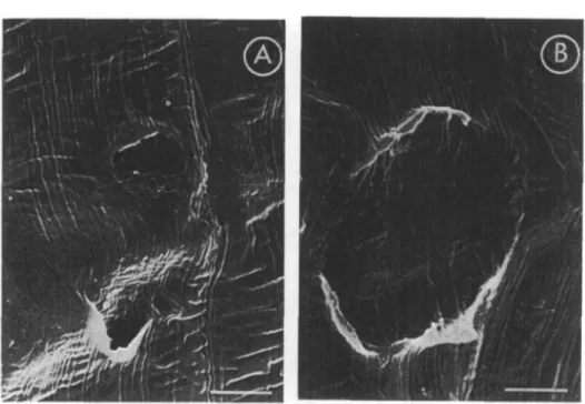
The epidermis of coleoptiles cultured in siti} in the presence of several concen-trations of IAA showed some "cracks" (Fig. 1 A), when no "cracks" could be detected in either the control (Fig. IB) or in ABA-treated coleoptiles (Fig. 1C).
Fig. 1. SEM micrographs of wheat ccleoptilt surfaces. A: Effect of IAA (10~4 M) after 24 hr of in situ
culture. B: Control after 24 hr of in situ culture. C: Effect of ABA (3.8 x 10"8 u) after 24 hr of
in situ culture. Scale bars: 10 fim.
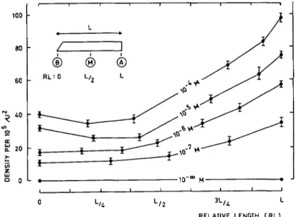
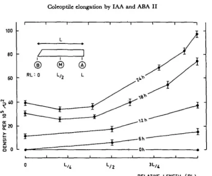
In order to study the formation of these "cracks", 5 mm segments excised at 5 mm from the tip, an area especially sensitive to IAA and ABA treatments (17), were cultured in vitro. As can be seen in Fig. 2, the apical end (A) of such a seg-ment, characterized by the greatest growth velocity (17), showed the largest "cracks". Their density was also maximal at this end of the segment (Fig. 3). The length and incidence of "cracks" increased with increasing IAA concentrations, as can be seen in Fig. 2 and 3 on one hand, and in Fig. 4A and 4B on the other. The sizes of the "cracks" (Fig. 5), as well as their density (Fig. 6) were also enhanced when
Coleoptile elongation by IAA and ABA II 737 80 70 60 50 40 30 20 J 10 • o z i 1 r i i @ ® RL: 0 L, 0 L o-RELATIVE LENGTH (RL)
Fig. 1. Variations of the length (in fim±standard error) of "cracks" along the subapical segments of wheat
coleoptiUs treated 24 hr with IAA at several concentrations (0 to 10~K M).
the incubation was longer. For the control and for ABA treated segments, no "cracks" could be detected after 24 hr.
RELATIVE LENGTH (RL)
Fig. 3. Changes in the density of the "cracks" (expressed by the number of "cracks" per llfiimfi ^standard error)
Fig. 4. SEM micrographs of wheat coleoptile surfaces. A: Effect of IAA (10"* M) after 24 hr of culture. B: Effect of IAA (10"6 M) after 24 hr of culture. Scale b a n : 5 pm.
It should be noted that all these results were obtained on samples analyzed after fixation, dehydration and drying. But quite similar "cracks" could be also observed
RELATIVE LENOTH ( RL)
Fig. 5. Variations of the length (in /imj^standard error) of "cracks" along the subapical segments of wheat
Coleoptile elongation by IAA and ABA II 739
0 L/4 L/ 2 3 L/ *
RELATIVE LENGTH (RL)
Fig. 6. Changes of the density of the "cracks" (see Fig. 3) along the subapical segments of wheat coleoptiles in
relation with time (0 to 24 hr) of culture in theprtsena of IAA (10-* M).
when employing frozen material without any coating (Fig. 7A). In contrast, for the control and for the ABA treated segments no "cracks" could be detected (Fig. 7B).
Fig. 7. SEM micrographs offroztn wheat coleoptile surfaces. A: Effect of IAA (10~* M) after 24 hr of culture. B: Control after 24 hr of culture. Scale ban: 20!/im.
Relations between the formation of these "cracks" and the extension of the coleoptile segments seem clearly established in the present material. The number of cells of the outer epidermis of the coleoptile remains constant from the "embryo" phase to maturity (2), but a slight increase in cell number for the other layers takes place before the coleoptile is 18 mm long. In the tested segments, growth is directly proportional to the elongation of all the cells, while the epidermal cells already present the largest elongation velocity. Exogenous IAA induces a growth sti-mulation which seems to be correlated to proton excretion, promoting cell wall loosening (6~). The longitudinal stress increasing within the walls during cell ex-tension (5) may act on the polylamellate structure of the wall. In such a structure, successive lamellae show a mainly longitudinal orientation of microfibrils alternating with lamellae mainly transversally oriented (4) and promote the formation of "cracks".
It is quite easy to believe that such longitudinal stress, when it is strong enough, could be the source of the "cracks". In fact, these "cracks" were only observed, on the epidermal surface when the longitudinal growth was significantly stimulated. In contrast, they were never found in the control even after 24 hr of incubation or, much less, on the coleoptile segments treated with ABA at a concentration for which significant inhibition of elongation was obtained (14, 17).
The several steps of "crack" induction and the physical changes in the micro-fibril structure during the "crack" formation are now under study.
References
( / ) Addicott, F. T.: Biochemical aspects of the action of abscisic acid. In Plant Growth Substances. Edited by D. J. Carr. p. 272-280. Springer Vcrlag, Berlin, 1972.
(2) Avery, G. S., Jr. and P. R. Burkholder: Polarized growth and cell studies on the Avena
coleo-ptile, phytohormone test object. Bull. Torrey Bot. Club 63: 1-15 (1936).
(3) Burgess, J. and P. J. Linstead: Scanning electron microscopy of cell wall formation around
isolated plant protoplasts. Planta 131: 173-178 (1976).
(4) Chafe, S. C. and A. B. Wardrop: Fine structural observations on the epidermis. I. The
epidermal cell wall. ibid. 107: 269-278 (1972).
( 5 ) d e l a n d . R . : Cell wall extension. Ann. Rev. Plant Physiol. 22: 197-222 (1971).
(6") Qeland, R.: Auxin-induced hydrogen ion excretion: correlation with growth, and control by external pH and water stress. Planta 127: 233-242 (1975).
( 7 ) Dumas, C : Le stigmate et la secretion stigmatique. These Urnvtrsiti Claude-Bemard-Lyon (1975).
(8) Goring, H., H.-P. M6ller und W. Bleiss: Kurzzcitkinetilc des Streckungswachstums und des
Younschen Moduls von Weizenkoleoptilen nach IES-Applikation und Wasserpotential-anderungen. Biochem. Physiol. Pftanztn 168: 411^20 (1975).
( 9) Hertel, R. and R. Flory: Auxin movement in corn coleoptiles. Planta 82: 123-144 (1968).
(10) Heslop-Harrison, Y. and J. Heslop-Harrison: Scanning electron microscopy of leaf surfaces.
In Proc. 2nd Annual Scanning Electron Microscopy Symposium p. 119-126, 1969.
(//) Homes, J.: Scanning electron microscopy of carrot cells and embryoids growing in vitro under various culture conditions. In Proc. Workshop on SEM and Plant Sci. Chicago, p. 335-342, 1974.
(12) Jones, M. G. K. and V. H. Droplrin: Scanning electron microscopy of syncytial transfer cells
induced in roots by cyst-nematodes. Physiol. Plant Pathol 7: 259-263 (1975).
(13) Pilet, P. E.: L'action des auxines tur la croisxance des cellules. In Handbuch der PJlanzenphysio-logu Edited by W. Ruhland. Vol. XIV: p. 784-806. Springer Verlag, Berlin, 1961.
Coleoptile elongation by IAA and ABA II 741 (14) Rchm, M. M. and M. G. Cline: Rapid growth inhibition of Avena coleoptile icgments by
absdsic acid. Plant Physiol. 51: 93-96 (1973).
(15) Reicosky, D. A. and J. W. Hanover: Seasonal changes in leaf surface waxes of Picta pungent. Amer. J. Bot. 63: 449-456 (1976).
(16) Robards, A. W., H. L. Payne and B. E. S. Gunning: Isolation of the endodcrmis using wall-degTading enzymes. Cytobiologie 13: 85-92 (1976).
(17) Schlienger, C., R.-M. Hofer and P. E. Pilet: Critical study of coleoptile elongation controlled by IAA and ABA. I. Growth kinetics and distribution. Plant & Cell Physiol. 18: 729-733 (1977).
(18) Schlienger, C , R.-M. Hofer and P. E. Pilet: Hormone-dependent dynamics of wall extension in Tritiaan coleoptile segments. In Electron Microscopy 1976 VoL II Edited by Y. Ben-Shaul. p. 489-491, 1976.
(19) Truelsen, T. A. and A. W. Galston: Changes in growth, auxin- and ribonudeic acid metabolism in wheat coleoptile sections following pulse treatment with indolyl-3-acetic acid. Physiol. Plant.
19: 167-176 (1966).
(20) Wright, S. T. C : Multiple and sequential roles of plant growth regulators. In Biochemistry and Physiology of Plant Growth Substances. Edited by F. Wightman and G. Setterfield. p. 521-542. Runge Presj, Ottawa, 1968.