APPLICATIONS AND FUNDAMENTAL STUDIES OF SELF-ASSEMBLED MONOLAYERS: CONDUCTING POLYMER DEPOSITION AND
CHEMICAL FORCE MICROSCOPY by
Lawrence Francis Rozsnyai B. S., Chemistry
University of California, Berkeley (1991)
Submitted to the Department of Chemistry in Partial Fulfillment of the Requirements
for the Degree of
DOCTOR OF PHILOSOPHY at the
MASSACHUSETTS INSTITUTE OF TECHNOLOGY May, 1995
@ 1995, Massachusetts Institute of Technology
Signature of Author S.. Department of Chemistry May 26, 1995 Certified by Mark S. Wrighton Thesis Supervisor Accepted by Dietmar Seyferth Chairnnman, Departmental Committee on Graduate Students
ARCHIVES MASSACHUSETTS INSTITUTE 0: TFCt4NOI.OrY
JUN 1
21995
I IRRARIFS ~i3This doctoral thesis has been examined by a Committee of the Department of Chemistry as follows:
Professor Alan Davisor,
Chairman
Professor Mark S. Wrighton
Professor Stephen J. Lippard
Thesis Supervisor
V
'U
___ __ __ · _
APPLICATIONS AND FUNDAMENTAL STUDIES OF SELF-ASSEMBLED MONOLAYERS: CONDUCTING POLYMER DEPOSITION AND
CHEMICAL FORCE MICROSCOPY
by
Lawrence Francis Rozsnyai
Submitted to the Department of Chemistry on May 26, 1995, in partial fulfillment of the requirements for the
Degree of Doctor of Philosophy in Chemistry
ABSTRACT
Chapter One An Introduction to Self-Assembled Monolayers
A brief summary of the formation, structure, and characterization of self-assembled monolayers is given. The use of aryl azide as a photosensitive terminal group in self-assembled monolayers to form surface patterns and the motivations for the studies presented in future chapters are also presented.
Chapter Two Selective Deposition of Conducting Polymers via Monolayer Photopatterning
Upon electropolymerization of the corresponding monomer, patterns of polyaniline, poly(3-methylthiophene), or polypyrrole replicate the pattern formed by selective irradiation of a photosensitive monolayer confined to an electrode surface. Irradiation of monolayers of di- 11-(4-azidobenzoate)- 1 -undecyl disulfide, I, on Au in the presence of various primary or secondary amines results in the attachment of the amine in very high yields. Irradiation of Au-I substrates through a mask results in a patterned monolayer. Electrodeposition of aniline from aqueous solution, or 3-methylthiophene or pyrrole from CH3CN, results in polymer deposition preferentially on the non-irradiated regions of the
surface. Polymer patterns are characterized by optical microscopy and by stylus profilometry. Cyclic voltammetry and X-ray photoelectron spectroscopy are used to measure deposition contrast, and show that under certain electrochemical conditions the ratio of polymer deposition on non-irradiated monolayer regions to irradiated monolayer regions is greater than 1000:1. Cyclic voltammetry of the monomers, Fe(CN)63-/4-, and
N,N,N',N'-tetramethyl-1,4-phenylenediamine 2+/+/0 shows that electron transfer rates are attenuated in monolayers of Au-I compared to bare Au, and are decreased further in monolayers of I irradiated in dibutyl- or dioctylamine. Differences in electron transfer through these monolayers agree with the polymer patterning results and are shown to be the principal reason for selective polymer deposition on these photopatterned monolayer substrates.
Chapter Three Controlling the Adhesion of Conducting Polymer Films with Patterned Self-Assembled Monolayers
Polyaniline and poly(3-methylthiophene) films may be selectively transferred from metal electrodes to flexible, insulating supports to form high-resolution polymer patterns. Irradiation of self-assembled monolayers (SAMs) of di- 11-(4-azidobenzoate)- 1-undecyl disulfide, I, on Au in the presence of various primary or secondary amines results in the attachment of the amine in very high yields. Irradiation of Au-I substrates through a mask results in a patterned monolayer. Electrodeposited polymers of aniline and 3-methylthiophene initially replicate the SAM pattern, and coat the entire substrate as electropolymerization continues. The interfaces between the polymer film and distinct regions of the SAM pattern differ in adhesion and correlate with the surface free energy of the monolayer terminal group, as determined by adhesive tape peel tests. Peeling adhesive tape from polymer films deposited on patterned SAM samples results in transfer of the original polymer pattern from the electrode to the tape. The transferred polymer films are conducting (a = 10-4 Q-1/cm for polyaniline) and features on the near-micron scale can be transferred successfully.
Chapter Four An Introduction to Chemical Force Microscopy
The principals of adhesion and friction forces between solids are reviewed, and a brief introduction to atomic force microscopy (AFM) is given. The ideas behind chemical force microscopy (CFM) are then presented. The placement of known molecules on the AFM probe tip and sample makes it possible to use CFM to study the chemical basis of adhesion and friction, to probe quantitatively the interactions between specific molecular groups, and to acquire high-resolution "chemical maps" of heterogeneous surfaces. Chapter Five Functional Group Imaging by Chemical Force Microscopy
The first example of functional group imaging by chemical force microscopy is presented. Using a force microscope, the adhesive and friction forces have been measured between molecularly modified probe tips and organic monolayers terminating in a lithographically defined pattern of distinct functional groups. The strength of adhesive interactions between CH3/CH3, CH3/COOH and COOH/COOH tip/sample
functional groups follows the trend expected chemically, and correlate directly with friction images of surfaces patterned with these groups. Thus, by monitoring the friction between a specifically functionalized tip and sample, it is possible to produce friction images which display predictable contrast and correspond to the spatial distribution of functional groups on the sample surface. Applications of this chemically sensitive imaging technique are discussed.
Chapter Six Chemical Force Microscopy: Using Chemically-Modified Tips to Quantify Adhesion, Friction, and Functional Group
Distributions in Self-Assembled Monolayers
A detailed account of chemical force microscopy is given. Chemical force microscopy (CFM) has been used to measure adhesion and friction forces between probe tips and substrates covalently modified with self-assembled monolayers (SAMs) that terminate in distinct functional groups. Probe tips have been modified with SAMs using a procedure that involves coating commercial Si3N4cantilever/tip assemblies with a thin
layer of polycrystalline Au followed by immersion in a solution of a functionalized thiol. This methodology provides a reproducible means for endowing the probe with different chemical functional groups. The spring constants and radii of the chemically modified cantilever/tip assemblies have been characterized to allow for quantitative friction and adhesion measurements. Au-coated Si substrates have been treated with functionalized thiols to produce SAM-coated substrates terminating in different chemical groups. A force microscope has been used to characterize the adhesive interactions between probe tips and substrates that have been modified with SAMs which terminate with COOH and CH3 functional groups in EtOH. Force vs. distance curves recorded in EtOH show that
the adhesion force between tip/sample functional groups decreases as follows: COOH/COOH > CH3/CH3 > COOH/CH3. The measured adhesion forces agree well
with predictions of the Johnson, Kendall, and Roberts (JKR) theory of adhesive contact, and show that the observed adhesion forces correlate with the surface free energy of the molecular groups in EtOH. The friction force between tips and samples modified with COOH and CH3 groups has also been measured as a function of applied load. The
magnitude of the friction force, and the friction coefficient, were found to decrease with different tip/sample functionalities: COOH/COOH > CH3/CH3 > COOH/CH3. Friction
forces between different chemical functional groups thus correlate directly with the adhesion forces between these same groups. Specifically, high friction is observed between groups that adhere strongly, while low friction is observed between weakly interacting functional groups. The dependence of friction forces on the tip and sample functionality is shown to be the basis of CFM in which lateral force images are interpreted in terms of the strength of both adhesive and frictional interactions between different chemical groups.
Chapter Seven Chemical Force Microscopy: Measuring Adhesion, Friction, and Functional Group Distributions in Aqueous Media
Chemical force microscopy (CFM) has been used to probe the interactions between acid and base groups in water. The chemical groups are localized on the probe tip and substrate via thiol or silane monolayers. Adhesion forces between CH3/CH3,
COOH/COOH, and COOH/NH2 tip/sample functional groups have been measured.
Force vs. distance curves recorded between COOH groups on the tip and sample as a function of ionic strength show that the interaction strength decreases with increasing salt concentration. The adhesion force was also measured as a function of solution pH. The observed trend is used to support a recently proposed structural model of COOH-terminated SAMs on Au. In most cases, adhesion between tip/sample functional groups in water correlates with friction. A friction image of a sample patterned with COOH and NH2 groups taken with a tip terminating in COOH groups is presented. Specific
intermolecular interactions account for contrast in this image demonstrating the ability of CFM to generate functional group images in water for the first time.
Thesis Supervisor: Mark S. Wrighton
The leading future of American science, however, and that which most distinctly characterizes it, is its utilitarianism....It may readily be conceded that the man who discovers nothing himself, but only applies useful purposes the principles which others have discovered, stands upon a lower plane than the investigator. But when the investigator becomes himself the utilizer; when the same mind that made the discovery contrives also the machine by which it is applied to useful purposes,-the combined
achievement must be ranked as superior to either of its separate results. from "The Future of American Science" Science 1883, 1, 1.
The value the world sets upon motives is often grossly unjust and inaccurate. Consider, for example, two of them: mere insatiable curiosity and the desire to do good. The latter is put high above the former, and yet it is the former that moves one of the most useful men the human race has yet produced: the scientific investigator. What actually urges him on is not some brummagem idea of Service, but a boundless, almost pathological thirst to penetrate the unknown, to uncover the secret, to find out what has not been found out before. His prototype is not the liberator releasing slaves, the good Samaritan lifting up the fallen, but a dog sniffing tremendously at an infinite series of rat-holes.
"The Scientist" Henry Louis Mencken, 1919. I_ ~_
ACKNOWLEDGMENTS
Since arriving at MIT, I have felt it both a challenge and a privilege to work for Professor Wrighton. I thank Mark for providing me with a well-equipped, sometimes even over-funded laboratory in which to work, a wealth of intriguing research interests from which to choose, and a well-trained though curious collection of students from whom to learn. Over the past years Mark has given me an appreciation of the written and spoken word, as well as a heightened awareness of detail. Most importantly, he has given me tremendous freedom in my graduate studies, with just enough guidance to keep me afloat, and has never doubted in my ability to succeed on my own.
Working in the Wrighton group was seldom easy, and I credit three people in particular for their early contribution to my learning and well-being: Eric Wollman showed me (whether I wanted him to or not) how to do things in lab-from running columns to performing simple reactions (everything in THF?) to preparing my first research reports and group meetings-in an exceedingly practical way. Cantankerous and short on explanation ("Why are we doing this, Eric?" "Why does a dog lick himself?" he would reply), Eric took it upon himself to give me the supervision I needed. For this chore, I am grateful. If Eric taught me how to do things, Dan Frisbie taught me how to think about them. In Dan's mind, no experiment was ever a waste. "Nothing worked today, but I feel good because now we know that something is wrong" seemed to be his maxim in the event of apparent failure. Dan's optimism countered my realism (well, pessimism perhaps) and did well to make me realize that all efforts in the lab should be meaningful. His research style and ability to ask probing, insightful questions were genuinely inspiring. Working together in the lab, we would call ourselves the kludge-masters, letting nothing stand in our way and reducing complex set-ups to simple concoctions using often only what was at arm's reach. Such was the spirit of conceptual experiments that exercised our intellect and imagination, and formed the basis of an active collaboration a few years later. I also thank Yoko Kato, who has been a good friend for the past four years. In my first winter, I was almost overcome by the desire to flee MIT's gray, cramped, industrial, burned-out surroundings and return to glorious California. Luckily I met Yoko, and spring had arrived. I value our friendship, admire her ability to cultivate the fine and simple points in life, and hope our friendship remains strong despite our geographic and cultural distance.
Without help and companionship from members of the Wrighton Group, both past and present, I wouldn't have made it through alive. (I was going to write "sane" but I suppose that point is arguable.) Of the odd but inspiring characters I first met in the group, I remember most Tayhas Palmore, and credit (and blame) her for encouraging me to join. Unfortunately she all too efficiently graduated, completed a postdoc at a little-known school down the street, and returned to sunny California. I consider her footsteps as ones to follow in, and only hope I can go as far as she has. Eric Lee, my first and only bench-mate, helped me initially to get to a working speed in the lab, and taught me the details of the daily grind. Though our collaborative project never got off the ground, I'll remember and appreciate his level-headed outlook, cool style, and nuggets of sound, practical advice. To past members and visiting scientists, I thank you for your periodic, well-timed encouragement and tireless technical advice. To Mike Wolf for his practicality, efficiency, and spontaneous stories (especially about RB); Ivan Lorkovic for his scientific analysis, taste in music, scores on vits, and ...uh...um...; Dave Albagli for his teachings both at the chalkboard and on the ice; Chris McCoy for his electrochemistry monologues, unequaled caffeine tolerance, and demonstrations of strapping it on; and Ron Duff for his lingo, dedication to the physical condition and organization of the lab, and cheerful amusement and tunes during the most rancid moments, I salute you all.
Many a time I have felt like the little kid on the block, and I will always appreciate and remember the help you all gave me throughout my graduate years.
The expertise of MIT's technical staff and the ability of MIT to attract truly talented people makes it an excellent place to do research. I especially thank Rich Perilli for his unstoppable help and corny jokes, and Felice Frankel for attending my departmental seminar, giving my work its much wanted artistic advantage, and landing us a cover picture. Frequently Christine Graves, as Mark's assistant for most of his tenure as Provost, helped to squeeze me into his schedule and warn us of his whereabouts. On a daily basis, Dena Groutsis has helped with group administration and finance, and has generally made it very easy to get things done with a minimum amount of hassle or delay (but often with a requisite amount of confusion).
As conspirators in research, bewilderment, and fun, I thank wholeheartedly Dan Frisbie, Alex Noy, and Dmitri Vezenov for a rich, stimulating, and rewarding collaborative research project. In the labs supervised by professor Lieber at Harvard (to whom I am also grateful), long hours, harsh words, giddy grins of disbelief, and countless sandwiches from "the Pain" went towards creating chemical force microscopy. Dan, Alex, and Dmitri all contributed greatly to Part Two of this Thesis, and their help and contribution cannot be underestimated. Collaborations should always be this good! To you all, I owe a big thinks!
The community in academic chemistry is well known to be close. Fittingly, I have shared an apartment with fellow graduate students for the last three years and between one bathroom for the four of us, a separate hot and cold faucet for all but the past few months, and an unexplainable amount of dust, it's a wonder that things worked out so well. Although we don't overlap for more than five minutes a week, I thank Linda Doerrer, who has always been attune to my moods, for never hesitating to giving me a pat on the back when she was probably in need the need of at least two (and for her late-night, almost autonomic lectures about X-ray diffraction, not to mention for putting away the dishes at 5:30 every morning). Jonathan Wilker and I compose a weird study in contrasts-we are in perpetual counterbalance with an eerie mutual understanding. I appreciate his advice about paying attention to the simple joys of everyday life, his pleas to relax and be happy, and will always remember him for being, among chemists, the silliest, most gaudy, and perpetually cheerful ham I ever knew. James White has also given me a new perspective. With an excellent CD collection and the largest assortment of random facts lodged in his brain, he has been enjoyable company-as long as he's not watching that damned TV!
After clinching a postdoctoral position on-of all places-an airplane, it occurred to me that good fortune has been with me at several pivotal moments in my professional life. Call it the influence of God, good luck, or a butterfly's wings in the Sahara; to Him, Her, or It, I am humbly thankful for the past and eagerly hopeful for the future.
I finally and most strongly thank my parents, Jean and Balazs, for their support throughout the years. Though they naturally guided me towards the sciences in my childhood, they pushed me to do my best in whatever I chose to do as I climbed the educational ladder. I hope that I followed their examples well. For their unending encouragement and love, it is to them that I dedicate this Thesis.
TABLE of CONTENTS
A B ST R A C T ... 3 ACKNOWLEDGMENTS ... 7 LIST of FIGURES ... .... ... ... 11 LIST of SCHEMES ... ... ... 18 LIST of TABLES ... 20PART ONE
An Application of Self-Assembled Monolayers:... 21Selective Electrodeposition of Conducting Polymers CHAPTER ONE An Introduction to Self-Assembled Monolayers ... 22
Text ... 23
References (with titles) ... 54
CHAPTER TWO Selective Deposition of Conducting Polymers via ... 63
Monolayer Photopatterning Introductio n... ... . 64
Experimental Section ... . 67
Results and Discussion ... ... 70
Conclusions ... 99
R eferences... ... 100
'TER THREE Controlling the Adhesion of Conducting Polymer Films ... 103
with Patterned Self-Assembled Monolayers Introduction... ... 104
Experim ental Section ... ... 105
Results and Discussion... 107
C onclusions ... ... .. 115
References ... 116
PART TWO
Fundamental Studies of Self-Assembled ... 118Monolayers: Measuring Adhesion and Friction at the Molecular Scale CHAPTER FOUR An Introduction to Chemical Force Microscopy ... 119
Text ... .. 120
References (with titles)... 132
CHAPTER FIVE Functional Group Imaging by... 135
Chemical Force Microscopy Introduction... ... 136
Results and Discussion... 138
C onclusions ... 149
References ... ... 151
CHAPTER SIX Chemical Force Microscopy: Using Chemically-Modified ... 155
Tips to Quantify Adhesion, Friction, and Functional Group Distributions in Self-Assembled Monolayers Introduction... ...156
Experimental Section ... 160
Results and Discussion... 163
C onclusions ... 190
Appendix...192
R eferences ... 195
CHAPTER SEVEN Chemical Force Microscopy: Measuring Adhesion, Friction,..201
and Functional Group Distributions in Aqueous Media Introd uctio n ... . 202
Experimental Section ... 204
Results and Discussion... 205
C onclusions ... .. 221
R eferences ... 222
PUBLICATIONS ... .. 225
BIOGRAPHICAL NOTE ... 226
LIST of FIGURES
PART ONE CHAPTER ONE
F igure 1.1 .. ... 33 A photograph of polyaniline selectively electrodeposited on a large Au-coated Si substrate. The polyaniline pattern (dark green) replicates the pattern initially generated on a photosensitive SAM.
F igu re 1.2 ... 44 XPS spectra of the N is regions of (a) Au-I and (b) Au-I irradiated in the presence of [CH3(CH2)312NH. Complete loss of the high binding energy peak upon irradiation
indicates complete loss of azide. The ratio of total peak area before and after irradiation can be used to estimate the yield of reaction, which is -80% on this sample.
F igure 1.3 .. ... 47 An example of electrochemical SAM analysis. Cyclic voltammograms of an Au electrode coated with a SAM of I taken (a) before and (b) after irradiation in the presence of an amine attached to ferrocene are shown. Integration of the anodic wave yields a coverage of 3.3 x 10⁻¹⁰ mol/cm2, roughly a monolayer of ferrocene groups, indicating
almost 100% photoattachment of amine on this sample.
F igu re 1.4 ... 50 An optical micrograph of a patterned Au-I substrate irradiated through a lithographic mask in the presence of (HOCH2CH2)2NH. As the sample is cooled in a humid
environment, water droplets spread and coalesce on irradiated, OH-terminated areas of the SAM pattern. Condensed water forms beads on the surrounding unirradiated, hydrophobic regions. The hydrophilic lines in the condensation image are 100 tm wide; the features in the middle of the image are 50 jtm wide.
CHAPTER TWO
F igu re 2.1 ... 72 Optical micrographs of polyaniline, poly(3-methylthiophene) and polypyrrole patterns formed by oxidizing the monomers on patterned SAM substrates, as described in the text. The dark regions correspond to polymer deposited on non-irradiated SAMs of I. The light regions contain little or no polymer and correspond to Au-I SAMs irradiated in the presence of [CH3(CH2)712NH. The microstructures in the bottom micrographs are 4 .tm
F igure 2.2 ... ... 74 Stylus profilometry traces of (three upper traces) the low resolution and (three lower traces) high resolution microline features. The line profiles are taken from the same
samples shown in Figure 2.1.
Figure 2.3 .. ... . 77 A comparison of N is and Au 4f XPS spectra of a polyaniline pattern: (a) a polyaniline-coated region showing one N peak and strongly attenuated Au peaks; (b), (c) a region without polyaniline showing N and Au peaks, consistent with a monolayer of irradiated aryl azide, respectively. The Au 4f peak of the polyaniline region is completely attenuated and is not shown.
F igure 2.4 .. ... 81 Cyclic voltammetric plots of (a) polyaniline, (b) poly(3-methylthiophene), and (c) polypyrrole patterns. Solid lines (-) are voltammograms from regions of Au-I SAMs; dashed lines ( - - ) are voltammograms from regions of Au-I SAMs irradiated in the presence of [CH3(CH2)712NH.
F igure 2.5 ... ... ... 85 Current vs. potential curves for aniline (0.11 M in aqueous 0.85 M H2SO4, 0.25 M
NaHSO4). Each point (taken from a linear sweep voltammogram at 200 mv/s, 0.0-1.2 V
vs. SCE) is the average of at least four measurements from freshly prepared samples. Curves are meant to guide the eye only.
F igu re 2 .6 ... 87 Current vs. potential curves for 3-methylthiophene (0.10 M in CH3CN, 0.1 M
[n-Bu4N]PF6). Each point (taken from a linear sweep voltammogram at 200 mv/s, 0.0-1.7 V
vs. Ag wire) is the average of at least two measurements from freshly prepared samples. Curves are meant to guide the eye only.
F igu re 2 .7 ... 89 Current vs. potential curves for pyrrole (0.14 M in CH3CN, 0.1 M [n-Bu4N]PF6). Each
point (taken from a linear sweep voltammogram at 200 mv/s, 0.0-1.2 V vs. Ag wire) is the average of at least two measurements from freshly prepared samples. Curves are meant to guide the eye only.
F igure 2.8 ... ... 91 Current vs. potential plots for Fe(CN)63-/4- (1.0 mM in aqueous polymerization solution,
200 mV/s) on bare Au and various homogeneous SAM surfaces.
F igure 2.9 ... ... 93 Current vs. potential plots for TMPD2+/+/o (1.0 mM in CH3CN polymerization solution,
200 mV/s) on bare Au and various homogeneous SAM surfaces. -- ~--i---
----Figure 2.10 ... ... . 98 Current vs. potential curves for aniline (0.11 M in aqueous 0.85 M H2SO4, 0.25 M
NaHSO4) on various SAM surfaces. Each point (taken from a linear sweep
voltammogram at 200 mv/s, 0.0-1.2 V vs. SCE) is the average of at least four measurements from freshly prepared samples. Curves are meant to guide the eye only.
CHAPTER THREE
Figure 3.1 ... . . 112 Micrographs of polyaniline on patterned Au electrodes. Patterns were made by irradiating an Au-I SAM in the presence of (HOCH2CH2)2NH for two minutes. The
square-shaped features are 10 x 10 .m2 and terminate in OH. The sample to the left
shows polymer selectively depositing on the unirradiated regions of the sample, and beginning to deposit on the irradiated regions. Continued electropolymerization results in complete polymer coverage of these areas. Adhesive tape is applied to the sample to selectively remove polymer from the unirradiated regions, as shown in the sample at the right.
F igure 3.2 ... ... 114 Patterned Au and adhesive tape samples containing (left) patterned polyaniline and (right) poly(3-methylthiophene) films. The SAM patterns were made using (HOCH2CH2)2NH
(see text). The mask used in fabrication of the sample was originally designed for use in the fabrication of microelectrodes; the small features range in size from 2-100 jim. The SAMs in these areas terminate in OH and are hydrophilic. Transfer of polymer film from hydrophobic regions of the Au sample to the adhesive tape is successful for both polymers. Optical microscopy has identified features replicating those of the mask of
near-micron resolution (-2 jtm) on both adhesive tape samples.
PART TWO CHAPTER FOUR
F igure 4.1 ... ... 126 A schematic illustration of an AFM. The sample rests on a piezoelectric crystal which is used to control the separation between the sample and the tip, and to scan the sample laterally. The tip rests at the end of a long, flexible cantilever. Normal forces (height) and lateral forces (friction) are detected by monitoring the deflection of a light beam that is reflected from the backside of the cantilever into a position-sensitive photodiode.
CHAPTER FIVE
Figure 5.1 ... .... 140 (A) Schematic of the surface modification procedure and a condensation image corresponding to a typical sample. Patterned sample surfaces containing lithographically defined hydrophilic and hydrophobic regions were prepared as previously described.27
Briefly, Au-coated Si(100) substrates were immersed in 1 mM methylcyclohexane solutions of di-ll-(4-azidobenzoate)-1-undecyl disulfide to yield a photosensitive monolayer. Irradiation of these substrates with UV-light (. > 260 nm) through a mask and a thin film of ethyl 4-aminobutyrate yielded ethyl ester termination in the irradiated regions of the sample. The sample was then irradiated a second time through a quartz plate and thin film of dioctylamine to yield methyl termination in the previously unreacted areas of the sample. Lastly, the ethyl ester groups were converted to COOH groups by hydrolysis. The condensation image illustrating the spatial pattern of hydrophobic (CH3) and hydrophilic (COOH) terminated regions in the sample was
recorded with an optical microscope while observing the condensation of water vapor onto the cooled sample surface.28 The water preferentially wets the hydrophilic (COOH)
terminated regions of the sample surface. (B) Modification of the cantilever/tip assembly with a specific functional group. Gold-coated Si3N4 cantilever/tip assemblies18 were
immersed in 1 mM ethanolic solutions of either octadecylmercaptan or 11-mercapto-undecanoic acid for 2 h to form covalently bound monolayers that were terminated with either hydrophobic (CH3) or hydrophilic (COOH) functional groups. The specific case of
a tip terminating with COOH groups is shown. (C) Schematic views of the experiment. The inset illustrates the interactions between a tip terminating in COOH groups and patterned sample terminating in both CH3 and COOH groups.
F igure 5.2 ... . . 142 Typical force vs. displacement curves recorded between a COOH-terminated tip and sample, a CH3-terminated tip and COOH-terminated sample, and a CH3-terminated tip
and sample in EtOH using a Nanoscope III LFM.
Figure 5.3 ... . . ... 146-148 (A) Optical condensation image of a patterned sample. The bright, raised areas in this image correspond to liquid H20 that has condensed on the hydrophilic
(COOH-terminated) regions of the sample. (B) Force microscope image of the sample topography of the same surface pattern as in (A) recorded using a probe-tip terminated with hydrophobic (CH3) functional groups. As expected, there is little difference in the
topography observable between the CH3- and COOH-terminated regions of the sample.
Similar topographic images were also recorded using COOH-terminated tips. (C) Image of the friction force recorded simultaneously with the topography in (B). The bright regions correspond a high friction force, while the dark regions correspond to a lower friction force. In this image, which was recorded with a CH3-terminated tip, high friction
is observed over CH3-terminated regions of the sample. (D) Image of the friction force recorded between a COOH-terminated tip and a similar region of the sample as in (A-C). In this image, high friction is observed over COOH-terminated regions of the sample. Images (B)-(D) were acquired under EtOH with an applied load of 4 nN and a scan rate ~·____~_~1 I·_L_·
of 3 Hz. Similar results have been obtained using scan rates between 1 and 10 Hz and applied loads from 3-10 nN.
CHAPTER SIX
F igure 6.1 ... ... 159 Schematic drawing of a CFM setup. The sample rests on a piezoelectric tube which can be finely moved laterally (x and y) and vertically (z). A laser beam is reflected from the backside of the tip into a photodiode to measure two types of tip-surface interactions: As the sample is rastered under the tip, the tip will move up and down in response to surface topography, resulting in the atomic force (height) signal. The tip will also rock back and forth in response to friction, yielding the lateral force (friction) signal. The inset illustrates the chemically specific interactions presented in this report. An Au-coated COOH-terminated tip contacts the boundary of CH3- and COOH-terminated region of the
sample.
F igu re 6.2 ... 165 Scanning electron microscopy images of an Au-coated force microscopy probe tip. The inset shows the high magnification image of the area near the end of the tip.
Figure 6.3 ... 167
Plot of the load mass vs. 1/(2rv)2 of tip/cantilever assemblies using adsorbed W balls of varying size. The force constants are 0.07 and 0.12 N/m for native and Au-coated cantilevers, respectively.
F ig u re 6 .4 ... ... 7 0 Representative force versus displacement curves recorded for CH3/COOH, CH3/CH3,
and COOH/COOH tip/sample termini using sharp tips (R = 60nm.). All data were obtained in EtOH solution using the fluid cell.
F igu re 6.5 ... 172 Histograms showing the number of times that a given adhesion force was observed in repetitive measurements using functionalized samples and "sharp" tips (-60 nm radii) terminating in (a) COOH/COOH, (b) CH3/CH3, and (c) CH3/COOH. Each histogram
represents more than 300 repetitive tip-sample contacts for one functionalized tip. All measurements were made in EtOH solution.
F igure 6.6 ... ... . . . .... 177 A typical friction loop recorded on a COOH-terminated sample using COOH-modified probe tip in EtOH solution. The applied load was 17 nN.
F igure 6.7 ... ... 179 Summary of the friction force vs. applied load data recorded for functionalized samples and tips terminating in COOH/COOH, CH3/CH3and COOH/CH3in EtOH.
Figure 6.8 ... 183 Condensation image of H20 on a photopatterned SAM substrate showing the repeat
pattern of 10 x 10 gm2 squares terminating in COOH on a background terminating in
CH3. H20 condenses only on the hydrophilic COOH-terminated regions of the sample. F igure 6.9 ... ... 185 Force microscopy images on one square of a photopatterned sample identical to that shown in Figure 6.8 of (A) topography, (B) friction force using a tip modified with a COOH-terminated SAM, and (C) friction force using a tip modified with a CH3
-terminated SAM. The apparent stretch in vertical direction observed on image (C) is due to the strain of the O-ring in the fluid cell during imaging.
F igu re 6.10 ... 188 Histograms of force vs. displacement curves recorded between functionalized samples terminating in COO- groups and tips terminating in NH3+ in (upper histogram) deionized
H20 (18 Mf.cm, pH = 6.5) and (lower histogram) 0.3 M NaCl aqueous solution (pH = 6.5).
CHAPTER SEVEN
Figure 7.1 ... ... ... 208 The adhesion force between COOH groups on the tip and surface is plotted as a function of salt concentration. The lower graph shows that the adhesion force scales linearly with the square root of salt concentration (proportional to reciprocal of the Debye length) for salt concentrations up to 0.05 M.
F igu re 7 .2 ... 2 11 Adhesion between COOH groups on the tip and sample as a function of pH using buffered (0.01 M total salt concentration) aqueous solutions. Each adhesion value was taken from an average of at least 200 force-distance measurements.
F igure 7.3 ... ... . . . .... 2 14 Plots of friction force vs. normal load force of the tip between COOH groups at three different pH values. The slope of each line is the friction force coefficient, it.
Figure 7.4 ... ... 216 A plot of friction coefficients vs. pH between tips and surfaces terminating in COOH groups. Each point is taken from the slope of a friction force vs. load curve.
Figure 7.5 ... ... 220 A friction force image of a sample patterned with COOH and NH2 groups taken with a tip
functionalized with COOH groups. Patterns were fabricated by selectively irradiating SAMs terminating in aryl azide in the presence of a liquid film of the appropriate functionalized amine. Light regions indicate high friction and correspond to area on the
surface exposing NH2 groups. Dark shades indicate low friction forces and represent
surface areas ending in COOH. The image was acquired in a buffered solvent at pH 7. _I _
LIST of SCHEMES
PART ONE CHAPTER ONE
Schem e 1.1... ... ... 25
An illustration of the self-assembly process. Alkanethiols spontaneously assemble on Au surfaces, energetically driven by covalent attachment of the head groups, S, to the Au substrate and by van der Waals interactions between the alkyl chains, to form a continuous, ordered, two-dimensional molecular film.
Schem e 1.2... ... 29 The alkyl chains of a SAM are close-packed and tilted by an angle 0 with respect to the surface normal. Surface properties are determined by the tail groups, X, which are exposed at the outer surface of the SAM.
Schem e 1.3... ... 34 An illustration of several monolayer patterning methods. All methods can be used to fabricate patterns with lateral dimensions on the micron scale. Monolayers are -30 A in thickness; substrate thickness ranges from 100-1000
A
(Au supported on Si) to -1 mm (Si).S ch em e 1.4 ... 37 Solution photochemistry of aryl azide in the presence of a secondary amine trapping agent. II and III are formed from the singlet; intersystem-crossing to the triplet yields the aniline adduct in the absence of amine.
Schem e 1.5... ... 38 Photochemistry of Au-I SAMs. Amines are chosen to alter SAM electron-transfer rate and surface free energy. Ferrocene is used as a redox-active species.
Schem e 1.6... ... 40 A sessile drop in equilibrium an a solid surface forming a contact angle, 0.
CHAPTER TWO
S ch em e 2.1 ... 64 Irradiation of Au-I SAMs through a mask in the presence of functionalized amines produces a patterned SAM.
Schem e 2.2... ... 96 A structural model of various electrode surfaces.
CHAPTER THREE
Schem e 3.1.... ... 108 Photosensitive SAMs of I on a flat Au surface are irradiated through a mask in the presence of a functionalized amine. Electrochemical oxidation of aniline or 3-methylthiophene results in selective polymer film (0.1-0.2 gtm) after additional electropolymerization. Adhesive tape is applied to the sample and peeled away to selectively remove the polymer film. The original pattern is transferred to the tape while the inverse remains on the sample.
PART TWO
CHAPTER FOURSchem e 4.1... ... . 127 An illustration of a force-distance measurement. The piezoelectric tube extends and the sample makes contact with the tip (2). As it extends further, the tip bends upward (3). The piezoelectric tube then retracts and the tip continues to adhere to the sample (5) past the point of original contact (4). When the cantilever deflection exceeds the adhesion force, the tip jumps back to its resting position (6).
Schem e 4.2... ... . 128 Adhesion is measured by deflection as the sample is retracted from the tip; friction is measured by torsion as the sample is scanned under the tip.
S ch em e 4 .3 ... 129 A conceptual illustration of chemical force microscopy. Molecules on the probe tip, X,
are used to sense specific interactions with chemical groups, Y, on the surface. CHAPTER SIX
Schem e 6A .1 ... 192 Cantilever/tip geometry showing components of torque.
LIST of TABLES
PART ONE CHAPTER ONE
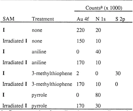
T able 1.1 .... ... 78 XPS analysis of the two regions in the polymer pattern.
CHAPTER FIVE
T able 5.1 ... ... 143 Summary of adhesive forces (nN) measured between functionalized tips and samples. These data were obtained by averaging the results from at least 300 force vs. displacement curves for each tip/sample combination.
I1
PART ONE
AN APPLICATION OF SELF-ASSEMBLED MONOLAYERS: SELECTIVE ELECTRODEPOSITION OF CONDUCTING POLYMERS
mo.
CHAPTER ONE
AN INTRODUCTION TO SELF-ASSEMBLED MONOLAYERS T "WY-i m om-ln
-Many critical materials properties originate in the main body-the bulk-of the material. When considering the stiffness of an I-beam, the optical properties of a lens, or the electrical conductivity of a copper wire, materials properties are largely the result of bulk physical and chemical structure. An increase in analytical sophistication over the past several decades has allowed the surfaces of materials and the interfaces between them to be studied. These experimental investigations have shown that the properties and performance of many materials are controlled at the boundary of materials in addition to the bulk: Metal fatigue occurs as cracks propagate along grain boundaries and coalesce into larger gaps, optical losses occur through reflection and scattering at interfaces, and as circuits become smaller, heat dissipation and local field effects play an increasingly important role in their electrical responses. In addition to materials, research in recent years has also shown that important biological processes occur at surfaces. Cell-cell, cell-protein, and protein-ligand interactions dominate most biological processes and all occur along some type of interface. Our understanding of surface behavior can be increased through chemical investigations of interfaces by relating microscopic chemical structure to macroscopic chemical, physical, or biological function, and by making specific and systematic chemical changes at interfaces to ultimately predict and control these properties at the molecular level.
This thesis describes the use of photosensitive self-assembled monolayers (SAMs) on gold substrates to produce patterns with different physical and chemical properties. Part One describes the use of monolayer patterns of different thickness on gold electrodes to direct the deposition of electropolymerized aniline, 3-methylthiophene, and pyrrole. Once deposited on the surface, these conducting polymers can be selectively removed and transferred to insulating supports. Adhesion to the polymer films is controlled via the terminal chemical groups of the SAM pattern. Part Two describes the development of chemical force microscopy, a new technique derived from conventional atomic force
microscopy. Microscopic probe tips and flat substrates coated with a thin layer of gold followed by SAMs terminating in different chemical groups allows adhesion and friction forces between specific chemical groups to me measured quantitatively on the molecular scale for the first time. The forces are directly related to the strength of the chemical interactions of molecular groups on the surface and tip, and are influenced by the surrounding medium, which is either air or a liquid. Comparisons between adhesion and friction forces on homogeneous monolayer surfaces show that in certain cases adhesion and friction are proportional at the nanometer scale. Images of functional group patterns on the substrate taken with tips ending in different functional groups show that this technique truly probes chemical interactions and may be used to generate "maps" of chemically heterogeneous surfaces.
This introductory chapter offers a brief review of self-assembled monolayers with particular emphasis on monolayers composed of alkanethiols or disulfides on gold surfaces. A wealth of research has been conducted on various monolayer systems.1 The
following review therefore is by no means comprehensive, but rather reflects several principles I feel are important and useful, and introduces some characterization techniques as a background for the remaining chapters. A brief description of the composition, formation, and utility of monolayers is first given, focusing on alkanethiols and disulfides on gold. Next, the techniques of contact angle measurements, ellipsometry, X-ray photoelectron spectroscopy, and cyclic voltammetry are described, along with what information about SAMs is gained from each of these methods. Several methods to generate SAM patterns-two-dimensional monolayer surfaces with different chemical or physical properties-are then briefly reviewed, followed by the fundamentals of our patterning method: alkyl disulfides terminating in aryl azide compose a photosensitive monolayer to which functionalized amines are selectively and covalently attached to generate patterns. The use of monolayers in "molecular architecture", the
surfac
X
SH Xfsmlyrfesr
Schme1.. A ilutrtion
of
thuiabe
self-ac
spnanosl ssmleo
Au surface
n
s, cesd
two-dimensionl molecular film.AI
[(H), heen
0-0, hc
tal
rup wih r eprsedc
on silanes oAn oxide surfaces and
anf-Silnane Monolayers and Langfaes,
alkyl) and R i(OR')3 (R = alkylcant
OHi groups and wih adsorbresed wa
with R groups projecting normal te
silanes including n-octadecyltrichlo
and mixed monolayers of two d
fabrication of multilayers, and the possibility using SAMs in several applications are finally discussed.
Self-Assembled Monolayers
A monolayer is a two-dimensional layer of molecules confined to an interface or surface. Self-assembly refers to the spontaneous formation of a monolayer upon introduction of a suitable interface to the appropriate molecules. For example, when a bare Au surface is immersed in a dilute (mM) organic solution of an alkanethiol (such as dodecanethiol in ethanol), a SAM spontaneously forms within minutes or hours, as
t X
Sillustrated in Scheme
SH SH XXXXXXXX 1. 1. Molecules that form monolayers on solid surfaces are composed of three parts: (1) head
ethtiolsfa laetil(uha
alen groups which covalently atac t te nShmustrate,
Au Au of three parts: (1) head
Scheme 1.1. An illustration of the self-assembly process. Alkanethiols
spontaneously assemble on Au surfaces, energetically driven by covalent groups which covalently
attachment of the head groups, S, to the Au substrate and by van der Waals
interactions between the alkyl chains, to form a continuous, ordered, attach to the substrate,
two-dimensional molecular film.
(2) long alkyl chains, [(CH2)n where n = 10-20], which pack tightly because of van der Waals forces, and (3)
tail groups which are expressed at the immediate surface. In addition to thiols and disulfides on Au, two other monolayer systems have been studied extensively: one based on silanes on oxide surfaces and another on Langmuir-Blodgett films.
Silane Monolayers and Langmuir-Blodgett Films.la,c Silanes based on RSiCl3 (R = alkyl) and RSi(OR')3 (R = alkyl, R' = methyl or ethyl) react at surfaces with exposed
OH groups and with adsorbed water to form an amorphous network of Si-O-Si bonds with R groups projecting normal to the surface. An early demonstration of long chain silanes including n-octadecyltrichlorosilane (OTS) discusses the formation of monolayers and mixed monolayers of two different silanes on glass substrates.2 Long-chain
alkylsilanes such as OTS form stable, uniform monolayers across the entire substrate (as opposed to islands) with a quasi-crystalline structure, as determined by ellipsometry and low-angle X-ray reflectivity.3 Short alkyl silanes also form monolayers;
3-aminopropyl-triethoxysilane (APS) is commercially available (Htils) and has been used extensively to fabricate "amine-terminated" surfaces on silicon, silica, and glass.4 Monolayers of APS
have also been formed on electrode surfaces such as platinum, tin oxide (SnO2), and
indium-tin oxide (In203/SnO2, ITO), and have been used in subsequent surface reactions
with various carboxylic acid derivatives.5 Because APS does not have a long chain of
methylene groups, it is structurally disordered compared to its long-chain counterparts. Recent attempts at forming long-chain silane monolayers with amines and other molecular groups have been developed to overcome this problem,6 and detailed structural
investigations of silane SAMs as a function of deposition temperature and alkyl chain length have been reported.7 Silane multilayers have also recently been studied at
attempts to form more elaborately designed surfaces.8
Silane monolayers are distinctly attractive because they readily form on two materials widely used in industry: silicon and glass. Formation of monolayers and multilayers on transparent substrates is especially attractive towards the development of optical devices.la,9 Also, silane surfaces are amenable to biological studies. Fluorescence microscopy has been used to detect the recognition of fluorescently labeled ligands to silanes selectively functionalized with DNA,10 peptides,11 and antibodies12 toward the
development of ligand binding assay techniques.
Monolayers of silanes have several disadvantages compared to those of thiols on Au, however. The formation of multilayer films is often unavoidable because silanes, especially trialkoxysilanes, are prone to polymerization. Also, because of the reactive nature of SiR3 and Si(OR)3 groups certain chemical functionalities (i.e., OH and COOH)
cannot be incorporated directly into the molecule and must be formed after monolayer assembly in a surface reaction. Methods at performing transformations at the surface to
alter the exposed functionality have been reported,4-6,13 but these systems have not yet
been extensively characterized. Limitations of silane monolayers are now being addressed14 and have not hindered efforts towards the development of optically or biologically active surfaces, but have probably contributed to the dearth of more detailed structure-property studies.
Langmuir-Blodgett films compose another monolayer system.1c In a very crude sense, LB films are similar to films of oil spread on water. These monolayers are composed of "amphiphilic" compounds-long chains similar to fatty acids that have a non-polar end that "likes" oil, and a polar end that "likes" water-and were first studied systematically by Irving Langmuir,15 who received the Nobel Prize in 1932 for his work
in surface chemistry. In this sense, the term "likes" can be interpreted to mean "is soluble in" which is itself a combination of several entropic and enthalpic terms involving the transfer of a solute particle into a solvent.16 A classic example of an amphiphilic
molecule is stearic acid, CH3(CH2)16CO2H. The carboxylic acid portion of the molecule
is soluble in water, whereas the nonpolar aliphatic chain is insoluble. Amphiphiles therefore aggregate at interfaces of water and air, or water and oil, with the polar portion of the molecule in the water phase. The degree of differential solubility is a critical parameter to the interfacial activity of amphiphiles. Consider stearic acid which forms LB monolayers: if the COOH group is changed to a larger, more polar S03" or N(CH3)4+
group, the molecule will become too water soluble for LB film formation. Likewise, monolayers will not form if the alkyl group is not sufficiently long. Propionic acid, with only two methylene groups, will never form well-ordered LB-type monolayers!
In the strictest sense, LB monolayers are not self-assembled monolayers. They lack the highest energy process of formation, the formation of a covalent bond to a solid surface via the head group, and therefore do not spontaneously form well-ordered assemblies. Structurally ordered monolayers may be formed, though, by compressing the amphiphiles at the interface with a moveable barrier. This technique uses what is now
known as a Langmuir trough, and was first demonstrated over 100 years ago.17 Indeed,
the most important indicator of LB monolayer properties is given by surface pressure/area isotherms-plots of the surface pressure as a function of the interface surface area. Films at low pressure are disordered. At high pressures they assume an almost crystalline structure until they collapse at even higher pressures. Well-ordered, compressed films can be transferred from the water/air interface (interfaces between water and organic liquids may also be used) to solid supports, as first demonstrated by Katherine Blodgett.18
Langmuir-Blodgett films transferred from a solution interface to a solid support have a monolayer structure similar to SAMs formed directly on solids, but like silane monolayers, suffer in several respects as models for interfacial behavior. Most importantly, the structure of LB films is not well defined, nor is it stable. Films may relax to a second, more stable structure after assuming an initial metastable form. Transferring LB films from the air/water interface to solid supports may also introduce perturbations on the microscopic structure and unlike silanes, transferred LB films have no strong association to the solid support and are not robust. The necessity for a polar end and a non-polar end on the molecule also limits the types of compounds that can be used, and thus limits the types of questions that can be addressed with LB films. Despite these drawbacks, LB films are actively used in fundamental surface science studies and in the development of molecular superstructures, thin-film devices, and optical devices.1 c,19
Self-Assembled Monolayers of Thiols and Disulfides on Gold
Long-chain alkanethiols (RSH) and dialkyl disulfides (RSSR') spontaneously assemble from solution to form monolayers on Au and compose the best understood monolayer system to date. Most of the thiols and disulfides used are commercially available or easily synthesized. The affinity of sulfur for Au is strong and accounts for the most energetic process in SAM formation (-30 kcal/mol).20 Though each covalent
cross-linking occurs between the alkanethiol groups that would compromise the overall SAM structure.
thiols: RS-H + Au -- RS--Au+ + H2(1.1)
disulfides: RS-SR + Au --- 2 RS--Au (1.2)
Like the other monolayer systems, van der Waals interactions between the alkyl chains also contribute to the enthalpy of SAM formation. Each additional CH2-CH 2
interaction contributes -1 kcal/mol.21 The combination of covalent attachment to the
a
XXXXXXX X X substrate, van der Waals interactions between the
alkyl chains, and the lack of additional structural requirements on the head groups results in the spontaneous formation of well-ordered, stable,
Au
Scheme 1.2. The alkyl chains of a rugged SAMs. For example, monolayers of SAM are close-packed and tilted by an
angle 0 with respect to the surface octadecanethiol, HS(CH2)1 7CH3, form SAMs
normal. Surface properties are determined
by the tail groups, X, which are exposed within seconds on clean Au substrates immersed in at the outer surface of the SAM.
dilute (-1-3 mM) ethanolic solutions. Though clean, freshly evaporated Au substrates give the best results, the adsorption of most
alkanethiols to Au is sufficiently strong to displace weakly adsorbed contaminants.
Spectroscopy and diffraction studies have been used to characterize the structure of SAMs on Au in detail. Several important structural features are now well understood:
1. Alkanethiols form ordered SAMs that are epitaxial with the supporting Au substrate. Early electron diffraction studies22 show that the symmetry of S atoms
on Au(111) is hexagonal with a spacing of 4.97A, which is very close to the spacing of next-nearest Au atoms, 4.99A. In this
F3
x 0 R300 structure, each S atom lies in a three-fold-hollow site, and each molecule occupies a surface area of 21.4 A2. Recently, X-ray diffraction, helium diffraction, and scanning probe microscopy have been used to investigate superlattices based on this structure and subtle factors such as rotation around the Au-S bond and chain torsion that definethe exact geometry of the unit cell.23 Additional structures, binding modes, and the migration of thiols on the surface are also being studied with several techniques.
2. The alkyl chains are predominantly trans-extended and tilted with respect to the surface normal (Scheme 1.2). Reflection-absorption (grazing-angle) IR spectroscopyla measures the magnitude and direction of transition dipoles of a thin films on a reflective sample. Used in conjunction with lattice models and molecular dynamics calculations, spectroscopic studies show that alkanethiols adsorb on polycrystalline Au(l 11) with an average tilt angle of 340 from the surface normal.24 The cant angle determines the distance between methylene
groups and is set to maximize the strength of the van der Waals interactions. Consequently, the cant angle changes according to the bonding and atom spacing of S atoms on the substrate. For instance, alkanethiols adsorb on Ag( 11) to form closely packed monolayers with tilt angles of only ~6-70.
3. Monolayers formed from either alkanethiols, RSH, or dialkyl disulfides, RSSR', have the same structure on Au(1 11) surfaces, RS--Au+/Au03, according to eqs 1.1 and 1.2 above.2 5,26 In other words, both thiols and disulfides form Au-thiolates, and in the absence of additional steric requirements, SAMs are formed from both compounds at the same rate.26 Dialkyl sulfides, RSR', also readily adsorb to Au surfaces to form robust SAMs. Due to the C-S-C bond angle however, both alkyl chains cannot extend from the surface to form a close-packed structure. Monolayers formed from dialkyl sulfides thus more closely resemble disordered organic liquids,27 not the crystalline arrays derived from alkanethiols and dialkyl disulfides.
4. Homogeneous mixed monolayers assemble from solution mixtures of two alkane-thiols that differ in their tail groups.28 If one alkanethiol, HS(CH2)nX, is used to
hydrophobic) and another alkanethiol is used to endow the surface with a different property (such as X = OH, which makes the surface hydrophilic), the two different thiols can be coadsorbed on the Au surface to form a well-ordered, mixed SAM with an intermediate surface property.
The stable, well-ordered structure of SAMs provides a reliable and flexible framework for surface chemistry studies. Alkanethiols and dialkyl disulfides with various tail groups (i.e., CH3, CF3, OH, COOH, C6H5) can be used to form SAMs with
specific chemical and physical properties. Photochemically and electrochemically active terminal groups can also be introduced, and surface reactions such as ester and amide bond formations can be studied and used to introduce additional functionalities in situ. The chemical composition and physical structure of these SAMs can be altered to study the molecular origins of specific surface properties.
Monolayer Patterning
Besides altering the structure and properties of homogeneous SAMs, patterning-controlling the lateral placement of two or more chemically different SAMs-provides an
additional method of synthetic control over surfaces. Patterns are useful in modeling biological interfaces such as cell surfaces, and are a prerequisite to the fabrication of magnetic, optical, or sensor devices.
A practical demonstration of patterning is shown in Figure 1.1, in which a SAM pattern is used to control the deposition of aniline, a conducting polymer. This system, described in Chapter Two, can be used to study mechanisms of polymer deposition and adhesion, and may have implications in device fabrication.
Monolayer patterns can be formed in one of several ways, as illustrated in Scheme 1.3. Perhaps the most basic way to localize monolayers to a specific region of the surface is to physically protect or "mask" the remaining regions with a protective layer. Patterns of photoresist are simply deposited on flat substrates to protect part of the surface from
Figure 1.1. A photograph of polyaniline selectively electrodeposited on a large Au-coated Si substrate. The polyaniline pattern (dark green) replicates the pattern initially generated on a photosensitive SAM.
-derivitization. A monolayer is then formed in the bare, unprotected regions of the surface. The original photoresist layer can then be removed. This method has been used with alkylsilanes to form surface patterns of CH3 groups (a hydrophobic surface) on
quartz (a hydrophilic surface) to control the behavior and orientation of cultured cells.29 Scheme 1.3. An illustration of several monolayer patterning methods. All methods
can be used to fabricate patterns with lateral dimensions on the micron scale. Monolayers are -30 A in thickness; substrate thickness ranges from 100-1000 A (Au supported on Si) to -1 mm (Si). photoresist pattern r M 7
Si
silane is depositedP_--photoresist is silane dissolved slane A- F i
a. Substrate Masking. Silanes are patterned on Si or glass using photoresist to mask the surface.
optical
mask ~ m
silane
orthiol [ SilAu
irradiated areas are a second / monolayer 1
rinsed to expose monolayer monolayer 2
the substrate M-" fills in ."
I SiIAu iA Si/Au
b. UV Photopatterning. Silane or thiol monolayers are destroyed by deep UV irradiation. They are rinsed away to expose regions of the substrate which can be coated with a second silane or thiol.
I
er stamp
Au Au is stamped to form SAM patterns a second SAM 1 thiol /SAM 2 fills in .Au
Au
c. Microcontact printing. A polymer stamp is inked with an alkanethiol solution and applied to Au substrates to form rugged, defect-free, micron-scale SAM patterns. A second thiol can be used to functionalize remaining areas of the Au substrate.
optical
mask
---photosensitive
SAM I Au
irradiation covalently attaches amine to change the apparent t
group at the SAM surface
ail tail group
ail /tail group
"
I
Au
I
d. Aryl Azide Photopatterning. A SAM with a photosensitive aryl azide tail group is irradiated
in the presence of a functionalized amine to introduce a second SAM tail group.
thiol
Though this method should be general for any type of surface (glass, Si, Au, etc.), it is not widely used, probably because of the incompatibility of the solvents used for photoresist stripping and monolayer formation, and fouling of the surface with photoresist before monolayer formation.
A more effective method of monolayer patterning is shown in Scheme 1.3b in which homogeneous SAMs are first formed and then selectively irradiated with UV light. In the case of silanes, the Si-C or C-C bond is broken and in an unknown mechanism, terminating the surface in OH;30 in the case of alkanethiols, the corresponding sulfonate
is formed.3 1 In both cases, irradiated portions of the monolayer are rinsed away and a
new, chemically different monolayer is formed in those regions. This approach is advantageous in that successive irradiations could conceivably be used to pattern multiple SAMs on the same surface, however complications are inherent. Assuming complete transformation of the irradiated region, incomplete desorption or dissolution of the irradiated monolayer, especially in areas of very small dimension, remains a distinct possibility. Complete formation of the second monolayer would then be questionable. Another practical limitation is the long irradiation time (-1 hr in some cases)27a,b
necessary for monolayer photoreaction, important when considering the fabrication of multiple samples.
Monolayer patterns can be formed without physically blocking the sample or selectively desorbing homogeneous monolayers. Whitesides and coworkers have used a pen and alkanethiol "ink" to write SAMs on Au surfaces.32 More recently, they have used stamps33 instead of pens, a more effective method to repeatedly and selectively transfer alkanethiols to large surfaces (Scheme 1.3c). Stamps several centimeters in size can easily be formed using lithographic techniques. Photoresist is used to create a master pattern on a flat substrate-a mold-onto which polydimethylsiloxane is deposited, cured, and peeled away to yield a flexible, free-standing, polymeric stamp. This stamp, which contains the negative of the original photoresist image, is "inked" with an