HAL Id: tel-01597904
https://tel.archives-ouvertes.fr/tel-01597904
Submitted on 29 Sep 2017HAL is a multi-disciplinary open access archive for the deposit and dissemination of sci-entific research documents, whether they are pub-lished or not. The documents may come from teaching and research institutions in France or abroad, or from public or private research centers.
L’archive ouverte pluridisciplinaire HAL, est destinée au dépôt et à la diffusion de documents scientifiques de niveau recherche, publiés ou non, émanant des établissements d’enseignement et de recherche français ou étrangers, des laboratoires publics ou privés.
mouse model
Maria Belen Lopez-Herdoiza
To cite this version:
Maria Belen Lopez-Herdoiza. Modeling C9ORF72 loss-of-function : a knockdown mouse model. Neu-rons and Cognition [q-bio.NC]. Université Pierre et Marie Curie - Paris VI, 2016. English. �NNT : 2016PA066239�. �tel-01597904�
This work is licensed under a Creative Commons Attribution-NonCommercial 4.0 International License.
Université Pierre et Marie Curie
Ecole Cerveau, Cognition, Comportement (ED-158)
Molecular Basis and Treatment of Neurodegenerative diseases
Modeling C9ORF72 loss-of-function:
a knockdown mouse model
By Maria-Belen LOPEZ HERDOIZA
Submitted and defended in partial satisfaction of the requirements for the degree of DOCTOR OF PHILOSOPHY
in NEUROSCIENCE on September 27th, 2016 In front of a jury composed of:
Pr. Alain TREMBLEAU President
Dr. Cedric RAOUL Referee
Dr. Adrian ISAACS Referee
Dr. Séverine BOILLEE Examiner
Dr. Valerie BUEE- SCHERRER Examiner
Dr. Luc DUPUIS Examiner
Pr. Alexis BRICE PhD Supervisor
“A good question is, of course, the key by which infinite answers can be educed”. Asimov (1982) “Let us keep looking, in spite of everything. Let us keep searching. It is indeed the best method of finding, and perhaps thanks to our eff orts, the verdict we will give such a patient tomorrow will not be the same we must give this man today.”
Charcot (1889)
iii
Acknowledgment
They usually say the acknowledgements are the easiest thing to write, especially after doing all the bibliographical work. Somehow, I find them particularly hard, especially since I communicate with all of you in different languages. I will try then with a classical approach and see how it goes…
First of all, I would like to thank Dr. Cedric Raoul and Dr.Adrian Isaacs for accepting to be referees for my PhD, they kindly tried to read me in English and for that I am really grateful. I look forward to keep discussing science with them in the future. I would like to thanks Pr. Luc Buée and Cedric again, who accompanied me during my thesis committees. Discussion with both of you was really enriching all along the project. These were my favorite moments and they made me remember why I like science so much. I would like to thanks Dr. Severine Boillée, Dr. Valérie Buée-Scherrer and Dr. Luc Dupuis for accepting to be members of my thesis defense.
A very special thanks to Dr. Morwena Latouche (“ma chef”, et je ne mets pas les autres surnoms :p) for everything. For trusting me with the project since the start and being available when I asked for help. For the scientific insight and the challenge you have given me over the years. And for all the other moments, inside and outside the lab. Thank you for being fun, joyful and full of energy. Working with you has taught me knowledge beyond science and I will cherish it along the way. What is left to say, you know it so let me finish with a “poulet poulet”.
I would also like to thank Pr. Alexis Brice for having me in his lab, and for being my PhD supervisor. Although always having a busy schedule, he always found some time to follow my project and assist to my presentations.
A special thanks also to Dr. Isabelle Le Ber, from whom I could always find time to ask questions and discuss ideas. Thank you very much for always being available and having an open door. I really liked belonging to this DFT team. I also thank Dr. Vincent Anquetil and Dr. Julien Branchu who helped me in the particular difficult part of writing the thesis manuscript. Your advices and corrections were, are and will be greatly appreciated. Thanks to all the other members of the DFT team, who always made discussion valuable and made me feel as part of the team, Paola, Agnes, Anne, Clemence, Kawtar and Matthieu.
Et là je change de langue, parce que c’est trop compliqué de vous remercier en anglais ^^ Merci beaucoup à tous les membres de l’équipe Brice, tous ceux que j’ai pu connaitre au cours de ces 3 années. Je pourrai faire une phrase pour chacun d’entre vous, mais j’aimerais vous accorder ce petit paragraphe. Les filles, Typhaine, Martina, Val, Marie merci infiniment pour ces moments passés à rigoler, à danser, à sourire. Votre amitié m’est très chère ! Merci aussi aux garçons, Max, Julien, Max encore :P, Raph… santé et bonheur pour vous tous.
iv Merci à tous les stagiaires, je pense que vous ne le savez pas, mais c’est vous l’âme même de l’équipe. J’ai adoré vous découvrir ces années Maeva, Marina, Julie, Baptiste, Marc, Laura, Marinne, Emeline, Jonas, Charlotte, Lyse, Celine, Remi… Merci aux autres aussi Agnès, Priscilla, Fiona, Livia, Claire avec qui j’ai pu partager des petits moments au labo…Un grand merci à Olga et Fred aussi, pour vos conseils, vos questions et vos discussions. Merci à Gio et Hamid et a tous les autres membres, vous êtes tellement nombreux !
Un merci spécial a Lydia qui a toujours été là pour m’aider. Sans toi, cela aura été beaucoup plus compliqué de rester ces 3 ans, surtout avec toutes les démarches administratives que j’ai dû faire.
Merci aussi à Annick Prigent et Brigitte Zeau avec qui j’ai pu passer des heures et des heures à couper, vous avez rendu cette tache beaucoup plus sympathique. Merci beaucoup à Magali Dumont pour tes conseils de tests comportementaux ainsi que pour ta disponibilité au moment de parler des ces tests. Merci à Aurélien Dauphin pour le cours d’imageJ et toute l’aide que tu m’as apporté. Merci à Delphine Boutellier, Carinne Dalle, Delphine Roussel et à tous les autres membres des plateformes avec qui j’ai pu interagir au cours de ce projet. Merci a Steph ;) Godard-Bauché et a Caro Le Duigou pour les magnifiques collaborations qu’on a eu. Vous m’avez gâté les filles…
Merci aux Ajites et co. (woooow !) pour tout. Vous etes l’ambiance de l’institut et sans vous, je pense que la recherche sera beaucoup beaucoup moins drôle. Merci Elise, Kevin, Melissa, Mark, Kristen, Aysegoul, Alizée, Sean, Martin, Ben, Tristan, Jean… je ne peux pas tous vous citer parce que cela prendra 2 pages. Sachez juste que je vous adore tous !! Une petite dédicace à Manale… c’est quand même grâce à toi que j’ai fait la thèse et le MBA. Tu as une force que j’admire, merci pour tous tes conseils mon amie !
Un grand merci à groupe escalade :p Vous êtes tellement importants pour cette thèse que sans vous, j’aurai décroché dès la deuxième année ^^ Merci Madlyne, Goki, Flo, Romain, Maud, Vasco, Celia, Lucie, Kevin, Jeanne, Charlie, Momo, Patricia et Patricia^^, Cyrielle, Pierre, Jerome, Remi, Vincent, Solenne…
Et aussi un grand merci pour mes BIPs… Betty, Camille, Charlotte, Bruneee, Popo, JB, Jeje, Amir, Hugo, Tom, Helene. Merci à Simon et Vero pour tout le soutien au cours des années…. Merci à mes copines du coeur Adelie, Manuela, Magda, Barbara, Arya y las que están en Ecuador Pao, Dani, Stefy, Sofy, Lore, Majo, Lu, Rosita, Bolo, Gordo, Tita, Juanse y Migue. A mi familia parisina Vicky, Fiti y Nico, que aunque estén por todo el mundo, siempre los siento cerca !
Y para mi familia, cada uno de ustedes que no logro poner en estas páginas… gracias especialmente Amira y Carlos por haberme dado la oportunidad de seguir mis sueños y la estabilidad para poder hacer los estudios que quise. Este trabajo es para ustedes porque sin ustedes, todo esto no hubiera sido posible. Los amo ! Y para ti peque, que eres y siempre serás el mejor regalo que me dieron mis padres.
v
French abstract – Résumé en français détaillé
Les démences frontotemporales (DFT) et la sclérose latérale amyotrophique (SLA) sont deux maladies neurodégénératives dévastatrices. La mutation du gène C9ORF72 a été identifiée comme la cause commune la plus fréquente de ces deux pathologies (c9FTD/ALS). Depuis la découverte de cette mutation, de nombreux efforts ont été faits pour identifier les mécanismes pathogéniques et ainsi développer de nouvelles thérapies. La mutation du gène C9ORF72 correspond à une expansion GGGGCC située dans le premier intron pour les transcrits V1 et V3 ou dans le promoteur du transcrit V2. Chez les patients, l’expansion hexanucléotidique est répétée plusieurs centaines de fois (~700 à 1,000) alors que les individus sains ne présentent pas plus de 30 répétitions. Plusieurs hypothèses pourraient expliquer les mécanismes conférant un effet toxique à la mutation et amenant à une neurodégénérescence. En premier lieu une toxicité de l’ARN mutant peut résulter de l’accumulation d’ARNs contenant les expansions dans des foci nucléaires séquestrant des protéines de liaison à l’ARN et des éléments d’épissage. Ces transcrits avec expansion peuvent aussi être traduits par un mécanisme non conventionnel indépendant de l’ATG (traduction RAN). Les dipeptides répétés résultants de cette traduction s’agrègent et pourraient s’avérer toxiques. Finalement, il a été montré que l’expansion cause la réduction de l’expression du gène C9ORF72 provoquant ainsi un phénomène d’haploinsuffisance qui pourrait lui aussi être délétère. Ces différentes hypothèses ne sont pas mutuellement exclusives et la neurodégénérescence peut être due à leur association. Il est donc indispensable de différentier les implications individuelles de la toxicité de l’ARN et des dipeptides ainsi que l’effet de l’haploinsuffisance.
Dans ce contexte, nous nous sommes intéressés à l’effet de la perte de fonction du gène C9ORF72. Pour étudier sa relation avec la DFT et la SLA, nous avons développé un modèle murin en utilisant un micro ARN interférence de synthèse pour diminuer les transcrits du gène C9orf72 murin et ainsi obtenir des souris avec un knockdown ubiquitaire de C9orf72, appelées souris miR-C9orf72. Cette méthode nous a permis d’obtenir des souris exprimant C9orf72 à un niveau semblable à celui des patients, tout en respectant la même variabilité (20 à 70% d’expression). Nous avons réalisé une caractérisation exhaustive de ces souris afin d’identifier des altérations au niveau comportemental, histologique et cellulaire se rapprochant des atteintes de type DFT et/ou SLA.
vi Nous avons montré que les souris miR-C9orf72 développent une atteinte de type DFT tout en présentant une motricité normale. A partir de l’âge de 5 mois, les souris miR-C9orf72 présentent une altération du comportement social ainsi qu’un comportement de type dépressif, anomalies typiques que l’on peut retrouver chez les modèles murins de DFT. Cependant, l’activité synaptique hippocampique, qui est l’un des supports majeurs des comportements sociaux chez la souris, semble conservée chez ces souris en vieillissant. Les souris miR-C9orf72 ne présentent pas de troubles majeurs de locomotion ou d’équilibre, ni de perte de motoneurones, ce qui exclut une atteinte de type SLA. Toutefois, nous avons observé un déficit de force musculaire à partir de l’âge de 12 mois malgré une morphologie du tissu musculaire intacte. Ce déficit de force pourrait s’expliquer par une atteinte de la jonction neuromusculaire (JNM). En effet, même si le contact entre les régions pre- et post-synaptiques de la JNM est maintenu, nous avons identifié une augmentation de signes de stress cellulaires tels que la présence de boursoufflures dans l’axone présynaptique ainsi qu’une fragmentation de la JNM chez les souris miR-C9orf72 âgées (15 et 23 mois).
Nous avons évalué si la perte de fonction du gène pouvait causer une neurodégénérescence ou une gliose. Le nombre de neurones est conservé au niveau du cortex frontal et du cortex moteur et nous n’avons pas identifié d’activation microgliale ou d’astrogliose dans ces mêmes régions.
Ensuite, nous nous sommes intéressés à l’état des protéines TDP-43 et p62 dans le cerveau de souris miR-C9orf72. En effet, dans le cerveau et la moelle épinière de patients FTD et ALS, nous observons une relocalisation cytoplasmique de la protéine TDP-43 phosphorylée ainsi que son agrégation alors que chez les individus sains, cette protéine à une localisation nucléaire. De même, des agrégats contenant la protéine p62 sont retrouvés dans le cerveau de patients DFT portant la mutation C9ORF72. Les souris miR-C9orf72 ne présentent pas d’agrégation cytoplasmique de protéine TDP43. Cependant, nous avons observé une augmentation du nombre de cellules corticales contenant une accumulation de p62 chez ces souris. Nous avons identifié que ces structures étaient aussi positives pour le marqueur de lysosomes Lamp1 dans les neurones et les microglies.
En conclusion, l’étude des souris miR-C9orf72, présentant un knockdown du gène C9orf72 a montré que la perte de fonction est impliquée dans l’apparition de symptômes propres à la DFT. Au niveau cellulaire, cette perte de fonction conduit à une altération de la voie autophagique dont le rôle délétère dans la DFT reste à déterminer. De plus, nous avons montré que la perte de fonction de C9orf72 n’est pas un élément déterminant pour causer la mort neuronale ou induire une agrégation de la protéine TDP-43, suggérant que les
vii mécanismes de toxicité de l’ARN et des agrégats de dipeptides pourraient jouer un rôle majeur dans ces aspects.
D’après les différents travaux publiés à ce jour, il apparait que des souris hétérozygotes ou avec un knock-out neuro-spécifique pour C9ORF72 ne développent pas d’atteinte motrice ou de diminution de la survie. De plus, des souris knock-out ubiquitaires pour C9orf72 présenteront une altération spécifique du comportement social à partir de l’âge de 6 mois. Contrairement aux souris miR-C9orf72 et aux souris hétérozygotes, les souris knock-out pour le gène C9orf72 présentent aussi une immunité périphérique anormale à partir de l’âge de 2 mois, avec le développement d’une splénomégalie et d’une lymphoadénopathie. Chez ces souris, les macrophages et les microglies présentent un défaut au niveau du trafic lysosomal tardif. Ces observations relient la fonction de la protéine C9ORF72 à la voie autophagique en tant qu’échangeur GDP/GTP, et une déficience dans son expression sera à l’origine d’une défaillance au niveau de la maturation lysosomes-endosomes, conduisant à une atteinte immunitaire en périphérie et dans le système nerveux central. Néanmoins, des études supplémentaires sont encore nécessaires pour identifier la fonction de la protéine C9ORF72 et les effets spécifiques à sa déficience partielle chez les patients.
L’étude de souris modélisant le gain-de-fonction toxique des ARN mutants et des dipeptides répétés a montré que l’expansion GGGGCC peut causer l’altération de comportements moteur et cognitif avec une perte neuronale et une délocalisation de la protéine TDP-43 lorsque celle-ci est surexprimée. Les mêmes observations n’ont pas été reproduites quand l’expansion est exprimée à un niveau physiologique, même si des foci d’ARN et des agrégats de dipeptides sont présents, suggérant qu’un facteur additionnel est requis pour induire la neurodégénérescence. De ce fait, nous émettons l’hypothèse que la perte de fonction de C9ORF72 est un élément requis pour mener au déséquilibre cellulaire causant une sensibilité accrue aux toxicités de l’ARN et des dipeptides produits par l’expansion GGGGCC. Les souris miR-C9orf72, présentant une déficience en C9orf72 semblable à celle des patients, pourraient ainsi être utilisées comme modèles pour tester les effets toxiques spécifiques de l’ARN mutant et des dipeptides répétés. Ainsi, ce nouveau modèle pourra donc servir comme outil pour déchiffrer les mécanismes moléculaires de la pathologie et identifier des cibles thérapeutiques spécifiques aux c9FTD/ALS.
viii
Index
FIGURES INDEX X TABLE INDEX XI ABBREVIATIONS XII INTRODUCTION 14 I. GENERAL INTRODUCTION 14II. NEURODEGENERATIVE DISEASES 16
1. PROTEIN AGGREGATION IN NEUROLOGICAL DISEASE 16
1.1. Inclusion bodies and aggresomes 19
1.1.1. Kinetics of aggregate formation 19
1.1.2. Inclusion bodies 21
1.2. Awry autophagy 23
1.3. p62, a link between autophagy and the ubiquitin proteasome system 26
1.4. Ubiquitin proteasome system and the unfolded protein response 27
1.5. Nucleocytoplasmic shuttling dysfunction 30
2. RNA METABOLISM ALTERATION IN NEUROLOGICAL DISEASE 32
2.1. Molecular mechanisms leading to repeat expansion formation 34
2.2. Molecular mechanisms leading to repeat-mediated toxicity in the case of non-coding
expansions 35
III. FRONTOTEMPORAL DEGENERATION AND AMYOTROPHIC LATERAL SCLEROSIS: TWO ENDS OF A SAME DISEASE
SPECTRUM 39
1. FEATURES OF FRONTOTEMPORAL DEMENTIA 39
1.1. Epidemiology and clinical features 39
1.2. Diagnosis 41
1.3. Neuropathology 42
1.4. Genetics 45
2. FEATURES OF AMYOTROPHIC LATERAL SCLEROSIS 48
2.1. Epidemiology, clinical features and genetics 48
2.2. Diagnosis 50
2.3. Neuropathology 52
3. HOW HAVE A DEMENTIA AND A MOTOR NEURON DISEASE BEEN LINKED TOGETHER? 55
4. THE C9ORF72 REPEAT EXPANSION, TYING UP FTD AND ALS 56
4.1. Transcripts and protein variation in C9orf72 carriers 57
4.2. Inclusions in C9orf72 patients 58
IV. UNDERLYING PHYSIOPATHOLOGICAL MECHANISMS IN FTD AND ALS 60 1. IMPLICATIONS OF TDP43 IN FTD AND ALS AND ITS ROLE AS AN MRNA BINDING PROTEIN 60
1.1. TARDBP gene and TDP43 protein 61
1.2. Pathological TDP43 65
1.2.1. TARDBP mutations 65
1.2.2. TDP43 proteinopathies and aggregation 66
1.2.3. TDP43 hyperphosphorylation 67
ix
1.2.5. TDP43 mislocalization 68
1.2.6. TDP43 pathological models 69
1.2.7. TDP43 mediated neurodegeneration 71
2. C9ORF72 IN ALS AND FTD PHYSIOPATHOLOGY 75
2.1. C9orf72 gene and protein 75
2.2. Pathological C9orf72 77
2.2.1. Repeat-expansion toxic gain-of-function contribution to disease pathogenesis 77
2.2.2. C9orf72 loss-of-function contributions to disease pathogenesis 84
2.3. Implications of the C9orf72 repeat expansion in cellular pathways: what do we know so far? 87
2.3.1. C9orf72 repeat expansion causes nucleolar stress 89
2.3.2. C9orf72 repeat expansion causes splicing defects 89
2.3.3. C9orf72 repeat expansion causes nucleocytoplasmic transport impairment 90
V. AIMS OF THE STUDY AND CHOICE OF MODEL 94
RESULTS: PARTIAL C9ORF72 LOSS OF FUNCTION IN MICE LEADS TO MILD BEHAVIOR ANOMALIES EVOKING FTD-LIKE SYMPTOMS WITHOUT MOTOR NEURON DISEASE (LOPEZ-HERDOIZA ET. AL) 96
DISCUSSION AND FUTURE PERSPECTIVES 140
I. ROLE OF THE C9ORF72 LOSS-OF-FUNCTION MECHANISM IN THE DISEASE COURSE: GAINING INSIGHT FROM THE
MIR-C9ORF72 MOUSE MODEL 140
II. BEHAVIOR EVALUATION OF MICE MODELS AND CORRELATIONS TO CLINICAL PATHOLOGY 146 III. HYPOTHESIS OF UNDERLYING MECHANISMS DRIVING NEURODEGENERATION IN C9FTD/ALS 150
1. PROTEIN HOMEOSTASIS, AGGREGATION AND TDP43 PATHOLOGY 150
2. MICROGLIA C9ORF72 IMPLICATIONS 151
3. ALS/FTD MECHANISMS NOT DIRECTLY RELATED TO THE C9ORF72 LOSS-OF-FUNCTION… OR ARE THEY? 153
3.1. Nucleocytoplasmic transport 153
3.2. RNA metabolism 154
3.3. Prion-like spreading and TDP43 155
CONCLUSION 156
REFERENCES 158
ANNEXES 186
x
Figures index
Figure 1 Formation of structured protein aggregates. 20
Figure 2 Model for aggresomes formation. 21
Figure 3 Autophagy pathway 25
Figure 4 ER stress-mediated apoptosis and the Unfolded Protein Response (UPR) 29
Figure 5 Schematic representation of nucleocytoplasmic shuttling 31
Figure 6 Coding and non-coding expansions associated to neurodegenerative diseases 34
Figure 7 Roles of RBPs in RNA metabolism 36
Figure 8 RNA interference and the RISC complex 37
Figure 9 Clinical symptoms of FTD 40
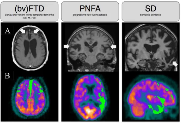
Figure 10 Overview of FTD alterations – Brain images 42
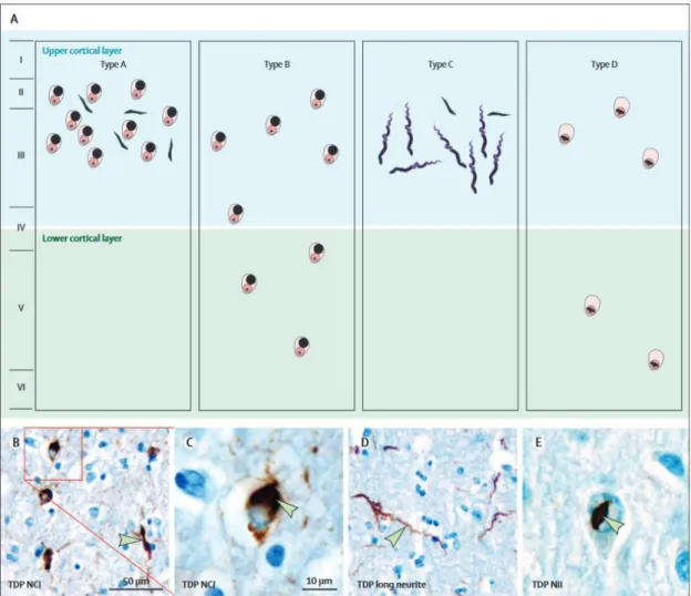
Figure 11 Pathological inclusions of TDP43 found in FTD and ALS 43
Figure 12 Schematic representation of TDP43, p62 and DPR inclusions type 44
Figure 13 Possible diseases mechanisms of the C9orf72 repeat expansion. 48
Figure 14 ALS common symptoms 51
Figure 15 Tools for ALS differential diagnosis 52
Figure 16 The “dying-forward” and “dying-back” motor neuron hypothesis. 54
Figure 17 Schematic representation of the FTD and ALS continuum 55
Figure 18 Physiological roles of TDP43 (and FUS) 63
Figure 19 TDP43 gene representation and localization of known mutations 65
Figure 20 TDP43 mutations and their functional effects 66
Figure 21 Stages of TDP43 spreading and deposition in FTD (left) and ALS (right) 72
Figure 22 TDP43 toxicity models 74
Figure 23 C9orf72 gene and protein isoforms 76
Figure 24 RBPs and the C9orf72 repeat expansion 78
Figure 25 RAN translation products of the C9orf72 repeat expansion 80
Figure 26 RAN products associated toxicity 81
Figure 27 Models of nucleocytoplasmic transport impairment mediated by GGGGCC toxicity 92
Figure 28 Behavioral characterization of miR-C9orf72 mice 95
Figure 29 Behavior alterations in the absence of C9orf72 142
Figure 30 Mouse models of FTD and associated behavioral assessments 147
xi
Table index
Table 1 Aggregating proteins in neurodegenerative diseases 18
Table 2 Identified genes causing familial FTD 46
Table 3 Identified genes causing familial ALS 50
Table 4 Pathological mechanism of the C9orf72 expansion Adapted from Haeusler et al. (2016) 88
xii
Abbreviations
3-MA 3-methyladenine
AD Alzheimer disease
ALS Amyotrophic Lateral Sclerosis
ALS-PDC Amyotrophic Lateral Sclerosis-Parkinsonims-Dementia Complex
ANG Angiogenine
APOA3 Apolipoprotein A3
ASOs Antisense oligonucleotids
ATF6 Autophagy factor 6
ATXN2 Ataxin 2
BAC Bacterial artificial chromosome
BMAA β-methylamino-L-alanin
BMI Body mass index
bvFTD Behavioral variant frontotemporal dementia C9-L C9orf72 long isoform
C9ORF72 Chromosome 9 open reading frame 72 C9-S C9orf72 short isoform
CELF CUG-BP and ETR-3-like factors
CFTR Cystic Fibrosis Transmembrane conductance Regulator CHMP2B Charged mutivesicular body protein 2B
CMT2B Charcot Marie Tooth type 2B
CTFs C terminal fragments
DENN Differentially expressed in normal and neoplastic cells DM1 - 2 Myotonic distrophy 1 -2
DN Dystrophic neurites
DPRs Dipeptide repeats
EMG Electromyogramme
ER Endoplasmic reticulum
ERAD Endoplasmic-reticulum-associated protein degradation ESCRT Endosomal sorting complex required for transport
FTD Frontotemporal dementia
FUS Fused in Sarcoma
FXTAS Fragile X–associated tremor/ataxia syndrome
GEF Guanine nucleotide exchange factor
HD Huntington disease
hNFL Human neurofilament
hnRNP Heterogeneous nuclear ribonucleoproteins
HRE Hexanucleotide repeat expansion
hsp heat shock protein
IPSC Induced pluripotent stem cell
LIR LC3-interacting region
MAPT Microtubule-associated protein tau
MBLN Muscleblind
miRNA microRNA
xiii
MRI Magnetic resonance imaging
mTOR Mammalian target of rapamicin
NCI Neuronal cytoplasmic inclusion
NCT Nucleocytoplasmic transport
NES Nuclear export signal
nfvPPA Non fluent variant primary progressive aphasia NIIS Neuronal intranuclear inclusions
NLS Nuclear localization signal
NPC Nuclear pore complex
Nups Nucleoporins
OPTN Optineurin
PD Parkinson disease
PET Positron emission tomography
PGRN Progranulin
PrPSC Prion proteins
Rab-GEFs Rab guanosine exchange factors
RAN Repeat associated non ATG translation
RBPs RNA binding proteins
RISC RNA-induced silencing complex
RNAi RNA interference
RNP RNA molecules linked to ribonucleoproteins
RRM RNA recognition motifs
SCA Spinocerebellar ataxia
SMN Survival of motorneuron
SOD1 Super oxide dismutase 1
SQSTM1 Sequestosome 1
STAMBP Signal-transducing adaptor molecule binding protein svPPA semantic variant primary progressive aphasia
TDP43 TAR DNA-binding protein 43
TNF Tumor necrosis factor
TRAF6 TNF associated factor 6
UBA Ubiquitin associated
UBQL2 Ubiquilin-2
UPR Unfolded protein response
UPS Ubiquitin proteasome system
UTR Untranslated region
VEN Von Economo neurons
14
Introduction
I. General introduction
Neurodegeneration is the generic term used to describe progressive loss-of-function or death of neurons. Neurodegenerative diseases occur as a result of progressive degeneration and/or cell death throughout different, non-fully characterized mechanisms. As world population lifespan increases, so does the prevalence of neurodegenerative diseases such as Alzheimer’s Disease (AD), Parkinson’s Disease (PD), Huntington’s Disease (HD), Amyotrophic Lateral Sclerosis (ALS), Fronto-Temporal Dementia (FTD), among others.
Causes for these pathologies are mostly unknown. Treatments proposed nowadays tend only to alleviate symptomatology, and not to treat the essential deleterious processes. The increased identification of loci and genes implicated in familial forms has provided answers on the underlying pathological mechanisms allowing for the development of new treatments targeting specific impaired pathways. The need to develop more accurate models to study identified pathways at a cellular and molecular level has never been more important, as they will also be key to test for therapies and to identify new, more efficient therapeutic targets.
As research advances, so does our knowledge on mechanisms leading to neurodegeneration, and surprisingly, similarities relating one disease to another on a sub-cellular level are often found. Most of these diseases present pathological hallmarks such as protein aggregation due to misfolding or atypical protein assembly, as is the case in AD with β-amyloid plaques accumulation, α-synuclein aggregates in PD, or TAR-DNA binding Protein 43 (TDP43) aggregates in FTD and ALS. Another common mechanism corresponds to alterations in RNA metabolism. RNA toxicity was firstly described in myotonic dystrophy 1 and myotonic dystrophy 2 (DM1 and DM2), a multisystemic disorder caused by the abnormal expansion of a non-coding trinucleotide sequence (Ranum and Day, 2002). In several cases, both protein aggregation and RNA toxicity seem to occur in parallel as is the case with hexanucleotide GGGGCC repeat expansion (HRE) in C9ORF72 a mutation responsible for both ALS and FTD (C9FTD/ALS). Little is known about the pathomechanisms of the HRE in C9FTD/ALS, and several hypotheses implicating production of a toxic RNA, peptide-aggregates toxicity or loss-of-protein function have been proposed. The C9ORF72 HRE is the most common cause of both ALS and FTD, and of the syndrome ALS/FTD. These two apparently different neurodegenerative diseases are related by numerous similarities including clinical presentation, mutated genes and identical neuropathological lesions with the presence of TDP43 inclusions. The course through which
15 the mutation of C9ORF72 drives pathogenicity remains elusive. However, the development of multiple and complementary animal models reproducing punctual aspects of the mutation has contributed to clarification of the underlying mechanisms leading to disease development or evolution. The study of familial history, clinical data and neuropathology of patients has allowed the establishment of relationships between different mechanisms, but also has given a broader picture of neurodegeneratives diseases showing that in some cases, one syndrome may overlap with another.
In this work, I will present my research in mechanisms involved in neurodegeneration based on the study of the C9ORF72 mutation. As previously mentioned, several mechanisms seem to co-exist in patients carrying the HRE, which make it an interesting model for studying pathways common to neurodegenerative diseases. To present this work, I will initially introduce background of mutual underlying mechanism in neurodegenerative diseases focusing on protein aggregation and RNA metabolism alterations and how their influence may lead to neurodegeneration (Chapter I). I will then follow to to a global presentation of FTD and ALS, two related but clinically different syndromes. I will pay a particular attention to the implication of the C9ORF72 mutation to both of these pathologies (Chapter II). Thirdly, I will merge the concepts presented in the two initial chapters to review molecular mechanisms and how they may lead to FTD and/or ALS. For this, I will analyze knowledge on TDP43, the main aggregating protein in FTD and ALS, which is also and RNA binding protein (RBP) directly implicated in RNA metabolism and then review the different mechanisms driven by the C9ORF72mutation (Chapter III). Finally, I will present the aims of my PhD project for which on the C9orf72 loss-of-function mechanism and its implications in ALS/FTD (Chapter IV).
16
II. Neurodegenerative diseases
1. Protein aggregation in neurological disease
Maintaining homeostasis is one of the major processes the cell has to carry out. The need for cellular adaptation emerges when fluctuations in the environment or of its intrinsic state occur. This will lead the cell to re-organize processes and functions accordingly. Mechanisms regulating cellular survival in aged or degenerative cells remain still poorly understood, but knowledge points toward aged-related differences between mechanisms. The neurodegenerative cell will yet have supplementary alterations to adapt adding to aging and exterior fluctuations, leading to a possible overflow in adaptive mechanisms. From this, the analysis of the brain of patients with different neurodegenerative disorders showed the presence of intracellular accumulation of protein aggregates, suggesting the cell is not able to compensate for variations. Presence of protein aggregates is a common feature of these diseases and their toxicity has been hypothesized as they may eventually lead to degeneration and death of cells (Miller and Kaplan, 2011). On the contrary, in normal conditions a rapid controlled apoptotic suicide is commonly observed when the cell is confronted to deprivation or stressful conditions (Nikoletopoulou et al., 2013). Increasing evidence indicates that, in the aged or degenerative nervous system, prolonged exposure to relatively minor insults might lead to cumulative neuronal oxidative stress and/or DNA damage, which in turn causes neurons to atrophy and degenerate.
Protein folding is essential for cellular protein homeostasis, allowing biological pathways to ensure cellular viability. Correctly folded proteins acquire a native three-dimensional functional structure that has to be maintained throughout the lifetime of each protein. Membrane and secretory proteins folding occurs mainly in the Endoplasmic Reticulum (ER) lumen, which allows for post-translational modifications due to its oxidizing conditions and numerous resident chaperons (Parakh and Atkin, 2016). Cytoplasmic proteins do not normally transit the ER and thus, rarely present disulphide bond formation, which are important for the structural stability of proteins and endorse for multi-protein complexes formation. Several E3 ligases ensure protein folding before the protein has left the ribosome, and chaperones from the hsp family will ensure correct protein trafficking and avoid aggregation (Amm et al., 2014). Protein misfolding is common to all proteins; mutations, heat, oxygen radicals, heavy metal ions and other stresses can enhance abnormal conformations and disturb proper folding and even alter already properly folded proteins. At
17 least, 30% of newly synthetized proteins are misfolded (Amm et al., 2014; Parakh and Atkin, 2016). To ensure proper protein folding, under normal conditions, several cellular protein quality control systems monitor newly synthetized proteins that maintain protein homeostasis, also known as proteostasis (Amm et al., 2014). When this system fails to correctly refold proteins, they will be directed to degradation by the proteasome, particularly the Ubiquitin Proteasome System (UPS) or autophagy system.
During protein misfolding, conformational changes occur in the hydrophobic core of the protein, leaving it exposed and prone to aggregation. “Conformational diseases” are a group of disorders characterized by the conversion of the tertiary structure of a protein to a different, newly stable form, resulting in cellular perturbation. If the misfolded protein is not shuttled towards degradation, it can oligomerize and aggregate. These latter processes could cause toxic effects by (1) a loss-of-function effect due to the lack of biological activity from the misfolded protein or (2) a toxic gain of function of the protein in its new form, both of these effects eventually leading to an altered cellular survival followed by cellular dysfunction and death in the long term. Intracellular and extracellular protein aggregation both are linked to several diseases and are commonly observed in neurodegenerative diseases (Table 1) (Hardy and Orr, 2006; Kopito, 2000; Ogen-Shtern et al., 2016; Parakh and Atkin, 2016).
18 Table 1 Aggregating proteins in neurodegenerative diseases
Accumulating proteins are pathological features of each disorder and proteins that misfold in each case are specific to a respective condition and a lesion site, although similarities in protein aggregation can occur in multiple diseases. Such is the case of TDP43 pathology in both ALS and FTD, or Tau aggregation in AD and FTD (Eftekharzadeh et al., 2016; Ogen-Shtern et al., 2016). Even though clinical manifestations are different, these “aggregating protein assemblies” eventually leading to neuronal dysfunction may share common pathways in neurodegenerative diseases. Yet, our knowledge of underlying mechanisms is still limited and it has become essential for us to increase our understanding of initial events leading to disease. From a molecular level, we need to characterize the triggers and processes of protein misfolding, aggregation and formation and the potential underlying cell toxicity mechanisms. Also, the transition between monomeric and aggregated – passing through partially unfolded – states has yet to be elucidated. From a physiological level it is important to understand whether these aggregates affect cell homeostasis and survival by altering cytoplasmic function due to abnormal interactions of misfolded or mutated proteins with common partners leading to toxicity, or transcriptional deregulations due to the
Disease Protein Characteristic pathology Pathologic species
Parkinson’s disease (PD) Alpha synuclein Intracellular
Membrane associated
Tetramers
Alzheimer’s disease (AD) Tau Intracellular Fibrils
Beta-sheets Extracellular Dimers Oligomers
Huntington’s disease (HD) Huntingtin (Htt) Intracellular Intranuclear Oligomer Aggregates Amyotrophic lateral sclerosis (ALS) TDP43 Intracellular Aggregates FUS
SOD1 Intracellular Dimers Aggregates
Frontotemporal dementia (FTD)
TDP43 Intracellular Aggregates
FUS Intracellular Aggregates
Tau Intracellular Tangles
19 aggregates themselves or to RNA modification, or altering nucleocytoplasmic shuttling, or causing nuclear function deregulation with the culmination in degeneration.
1.1. Inclusion bodies and aggresomes 1.1.1. Kinetics of aggregate formation
Protein aggregates are oligomeric complexes of non-native conformers that assemble from altered interactions among structured, kinetically trapped intermediates of the protein-folding pathway. Protein aggregation is caused by alterations in the protein primary structure produced by mutations, RNA modification, translation mis-incorporation or by partial unfolding during thermal or oxidative stress (Kopito, 2000). These factors will cause hydrophobic domains to be exposed allowing them to interact with other proteins and sequester them in an autocatalytic process, where misfolded oligomers can act as “seeds” to form larger misfolded structures (Parakh and Atkin, 2016). Structured protein aggregates are called amyloid assemblies and tend to be insoluble and metabolically stable under physiological conditions. Unstructured protein aggregates are called amourphous (Kopito, 2000). The suggested pathway leading to structural transition indicates that the natively folded protein monomers becomes partially unfolded and transit into an unstable disordered state from where it adopts a misfolded state which will enable assembly into energetically stable complexes (Eftekharzadeh et al., 2016).
Initially, it was believed that the native state of proteins was specified only by the amino acid sequence, and that folding was neither templated nor assisted by other factors. But, in 1972, it was shown that proteins have at least two states: a native state and an amyloid state, which is more stable (Anfinsen, 1972; Pauling et al., 1951). Amyloids are formed by nucleation, a templating process with strong thermodynamic effects that allows an energetic funnel to drive protein assembly (Bryngelson et al., 1995; Govindarajan et al., 2006). Once the nucleus or “seed” is formed, there is an exponential growth of aggregation or seeded polymerization, as proteins tend to stay in the lowest free energy state (Walker et al., 2013). Properly folded proteins have their hydrophobic regions buried within the structure, ensuring the lowest possible energy state, but misfolded proteins have exposed hydrophobic regions (Ogen-Shtern et al., 2016). Chaperons of the “quality control” (QC) machinery ensure that protein aggregates do not accumulate in unstressed cells by selectively degrading inappropriately folded proteins before they can aggregate. These molecular chaperons interact transitorily with protein intermediates concealing hydrophobic surfaces from forming intra- or
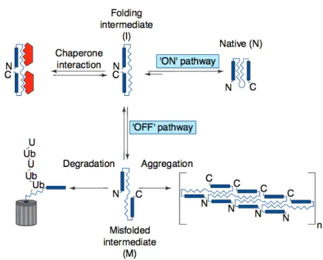
20 intermolecular interaction and block or and minimize aggregation (Kopito, 2000; Ogen-Shtern et al., 2016). By this, they ensure correct protein folding and the maintenance of protein homeostasis (known as proteostasis), which are fundamental for cell viability (Figure 1).
As a last resort, cellular pathways will cause the assembling of beta sheet-containing domains into amyloid fibrils in which the hydrophobic surfaces will be less exposed to damaging interactions (Ogen-Shtern et al., 2016). Accumulation of misfolded proteins can arise when these quality control pathways malfunction. Even if the formation of amyloid structures correlates with cell death, the relation between aggregates and neuronal loss has not been fully elucidated. Nevertheless, the question remains to whether aggregates are directly pathogenic or if it is the transition from natively folded proteins into amyloid that reflects or triggers toxicity.
Unfolded protein Intermediate(s) Native
Figure 1 Formation of structured protein aggregates.
The overall efficiency of protein folding is the proportion of intermediates that assume a native conformation. Intermediates can assume misfolded, off-pathway conformation, which are prone to oligomerize into non-native aggregates. The ubiquitin-proteasome pathway degrades protein intermediates before they can aggregate. Adapted from Kopito (2000)
21 1.1.2. Inclusion bodies
Inclusion bodies are intracellular foci, and in the case of neurodegenerative diseases, aggregated proteins mainly form them. Besides the major aggregated protein species, immunohistochemical analysis have revealed the presence of molecular chaperones, including heat shock proteins (hsp Hsc70, Hsp40), the proteasome subunits 19S and 26S, ubiquitin, intermediate filament proteins (Kopito, 2000; Ogen-Shtern et al., 2016). Whether the composition of these inclusion bodies reflects malfunction of the protein degradation and folding machinery or variable activities of cellular deubiquitinating enzymes has not yet been elucidated. Inclusion bodies are present in low copy number, usually one per cell and, in the case of neurodegenerative diseases they are accompanied by displaced and abnormally phosphorylated intermediate filaments (neurofilaments).
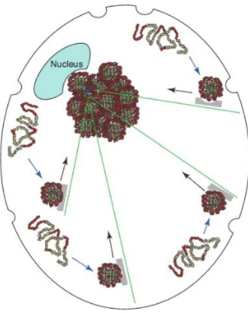
The mechanisms leading to formation of inclusion bodies in degenerative neurons represent an intense field of study. A common feature of trapped proteins is the presence of intrinsically disordered domains that could facilitate non-covalent hydrophobic boding or ionic interactions. It is now greatly supported that the formation of inclusion bodies in animal cells can take place by fusion of individual protein aggregates into a limited number of foci
Small aggregates formed at the cell periphery are derived along microtubules tracks (green lines) on retrograde motors (grey) to a juxtanuclear, pericentriolar localization. Adapted from Kopito (2000)
22 driven by active retrograde transport of the misfolded proteins on microtubules (Figure 2), contrary to the idea that they were formed by a diffusion process (García-Mata et al., 1999; Johnston et al., 1998; Kopito, 2000).
Inclusions bodies that are dependent on active microtubule transport are called “aggresomes”. Forms of misfolded or misassembled proteins that are prone to form inclusion bodies such as presenilin 1 (Johnston et al., 1998), peripheral myelin protein PMP22 (Notterpek et al., 1999) or ALS causing mutation in superoxide dismutase 1 (SOD1) enzyme (Johnston et al., 2000) are located at the pericentriolar region, suggesting a central role for this region in aggresomes formation. This region is known to contain proteins responsible for microtubule nucleation and anchoring. Like so, it is not surprising to see that protein aggregates accumulation in the pericentriolar region will result in expansion of the centrosome, disruption of Golgi’s normal architecture and disorganization of microtubules (Quintyne et al., 1999). The Golgi apparatus function depends on dynamic vesicular traffic (Lippincott-Schwartz, 1998), and Golgi cisternae displacement is a common feature in neurons of AD brains and spinal cords from ALS patients and mouse models of ALS (Gonatas et al., 1998), relating microtubule-based transport of aggregating proteins to cellular mechanisms that could be implicated in neurodegeneration (Kopito, 2000).
On the contrary, a protector effect of inclusion bodies has been also suggested, as they may have a role in protein homeostasis and act as cellular bins reducing levels of toxic intermediate forms, as the formation and accumulation of protein aggregates also takes place during normal aging (Cuanalo-Contreras et al., 2013). Protein turnover is essential for maintaining cellular homeostasis by removing defective proteins, by contributing to the pool of amino acids required for protein synthesis within limited nutriment availability and for allowing an optimal equilibrium of proteins themselves. Proteostasis, or protein homeostasis is responsible for the synthesis, folding and turnover of proteins (Balch et al., 2008), and pathways including the unfolded protein response (UPR), the UPS, autophagy and encapsulation of damaged proteins in aggresomes maintain the balance in the cell. Studies and genetic evidence showed that 30% of all newly synthetized cellular proteins are defective ribosomal products that will be degraded shortly after their synthesis and that normal cells must cope with a continuous flux of misfolded proteins that must be degraded (Nedelsky et al., 2008; Zhou and Schekman, 1999). For this, the proteasome active sites need to be able to directly interact with protein aggregates in order to degrade them. Chaperons will help the proteasome system by unfolding aggregates, but the system can be overwhelmed as the imbalance tilts towards aggregation. Whether protein aggregation will be able to over-drown
23 the cell will depend upon the competition between the effective degradation of misfolded substrates and the aggregation process itself (Kopito, 2000). With aging, degrading mechanisms do not perform equally and as so, protein aggregates are able to better stabilize themselves, thus tending to accumulate (Taylor and Dillin, 2011).
1.2. Awry autophagy
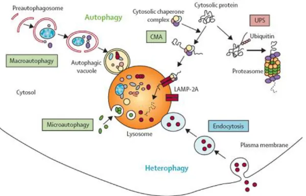
One of the main mechanisms of protein degradation is through autophagy. The autophagic route facilitate their disposal of aggregates by delivering them to a centralized region, as can be the pericentriolar region for example (Johnston et al., 1998). Evidence suggest that autophagy serves two major purposes in mammalian cells: (1) to provide an amino-acid supply under poor environmental conditions, which is known as “adaptive autophagy” and (2) to continually degrade proteins in the cytoplasm, which is known as “constitutive autophagy”. This process is the major process through which long-lived cytoplasmic components, such as excess or damaged organelles and aggregate-prone proteins, will be degraded by lysosomes (Komatsu et al., 2007). Three main types of autophagy have been characterized so far: (1) chaperon-mediated autophagy, which involves specific protein recognition and delivery to lysosomes, (2) microautophagy, which is poorly understood and (3) macroautophagy (referred here as autophagy) (Figure 3). Knowledge of chaperone-mediated autophagy and microautophagy implications in neurodegeneration remains poorly characterize, so I will not explore them in this work. On the other hand, autophagy implications in neurons have been studied deeper. Neurons are highly metabolically active post-mitotic cells. As they cannot dilute toxic waste burdens by cell division, they may try to get rid of aggregating proteins throughout autophagy. Constitutive autophagy activity in the brain is low when compared to other tissues, but it plays a crucial role as altering its function causes major neurodegenerative phenotypes (Mizushima, 2004). Mechanistically, large cytoplasmic regions, organelles and peripherally formed aggregates are engulfed within a double membrane vesicle, probably by fusion with an endosome, to create an autophagosome. In turn, the autophagosome will then fusion with lysosomes leading to turnover and recycling of cellular constituents (Komatsu et al., 2006; Wong and Cuervo, 2010). Formation of the autophagosome is regulated by the beclin VPS34 and the mTOR kinases complexes (Taylor and Dillin, 2011). When the pathway becomes overwhelmed, the continual delivery of protein aggregates might turn the deliver site into a cellular junkyard, as is the case in affected brains from patients of AD, PD, ALS, FTD and polyQ diseases in which neurons present an
24 abnormal amount of autophagosomes (Nedelsky et al., 2008; Wong and Cuervo, 2010). Accordingly, impairment of autophagy causes neurodegeneration in animal models. Autophagy deficient mice develop progressive deficits in motor function including an ataxic walking pattern, limb clasping reflexes and tremor. Modification of autophagy pathways also alters phenotype in animal models of neurodegeneration, either by reducing or accelerating degeneration (Hara et al., 2006; Komatsu et al., 2007, 2006; Nedelsky et al., 2008). Implications of autophagy have been further accepted when mutations or defects in autophagy-lysosomal function have been shown to have an impact on familial neurological diseases. For example mutation in a GTPase exchange factor that participates in trafficking autophagosomes and fusion with lysosomes called Rab7, result in dominantly inherited axonal neuropathy Charcot-Marie-Tooth type 2b (CMT2B) (Nedelsky et al., 2008) and autophagy related genes OPTN, SQSTM1 and CHMP2B will lead to ALS or FTD (Fecto et al., 2011; Le Ber et al., 2013; Maruyama et al., 2010; Skibinski et al., 2005).
Previous examples concern mutations in the autophagy pathway leading to neurodegeneration. A separate question implicates the role of autophagy in neurodegenerative diseases caused by mutations in genes unrelated to the autophagy function itself. Upregulation of autophagy can be induced by pharmacologically inhibiting the kinase mTOR (mammalian target of rapamycin). The mTOR pathway regulates a variety of cellular functions including cellular proliferation, motility, and survival, among others. Failure in this pathway has been associated with several pathologies including autism and AD (Rosner et al., 2008; Tang et al., 2014) . Thus, mutations that affect the mTOR pathway can alter autophagy and eventually, lead to neurodegeneration (Damme et al., 2014). Interestingly, the other autophagy pathway called microautophagy, which comprises the direct invagination of lysosomal membrane with cytoplasmic components, is also under the control of the mTOR pathway (Bitto et al., 2014), suggesting a interlace of the different types of autophagy pathways. Inducing macro autophagy by mTOR pharmacological inhibition has been shown to accelerate clearance of protein aggregates in vitro and protect against neurodegeneration in animal models, particularly delaying ALS progression in mouse models (Nedelsky et al., 2008; Rea et al., 2014; Wong and Cuervo, 2010). On the same line, TDP43 aggregation, the aggregating protein in both FTD and ALS, can be induced by inhibiting autophagy with 3-methyladenine (3-MA) or by affecting the endosomal sorting complex required for transport (ESCRT) machinery, which has a role in endosome formation and protein sorting (Cipolat Mis et al., 2016). As so, autophagy acts in a coordinated manner with other pathways such as endocytosis (Figure 3). Functional endosomes are necessary for clearance of
25 autophagosomes, and the disruption of endosomal proteins can lead to accumulation of polyubiquitinated TDP43 in FTD models (Wong and Cuervo, 2010).
The implications of autophagy seem to go beyond the local alteration of cellular homeostasis. A possible connection with aggregates spreading has been suggested as the kinetics of aggregate pathology progression suggests a non cellular-autonomous spreading: aggregates released by the cell through the autophagy pathway will be able to infect other cells. These aggregates will induce misfolding of proteins and aggregation in “healthy” cells in a prion-like manner. Furthermore, it has been shown that endocytosis is a major route of cellular entry for pathogenic forms of prion proteins (PrPSC), and the coupling of autophagy and endocytosis can act as a bridge for aggregates propagation. Evidence in this direction is increasing for diseases like AD, HD, FTD and others (Eftekharzadeh et al., 2016; Ogen-Shtern et al., 2016) but further investigation is still needed to determine whether or not this mechanism is effectively responsible for propagation of aggregating proteins in neurodegenerative diseases.
Autophagy is an intracellular degradation pathway that delivers cytoplasmic constituents to the lysosome. It is an independent system from the endosomal-lysosomal pathway and the proteasome degradation pathway, although the three of them interact. There are three types of autophagy—macroautophagy, microautophagy, and chaperone-mediated autophagy—and the term “autophagy” usually indicates macroautophagy Adapted from
amsbio.com
26 1.3. p62, a link between autophagy and the ubiquitin proteasome system
Autophagy is a key aspect of cellular homeostasis, and as previously seen, its failure causes various effects associated with neurodegeneration such as alterations in local axonal membrane trafficking and turnover (Komatsu et al., 2007). Its coordinated role with other protein degradation pathways allows for clearance of aggregating proteins that accumulate during disease. Several proteins link degradation pathway in a coordinated manner, of which, p62 has a particular role. p62, also known as sequestosome 1 (SQSTM1), is a multi-complex ubiquitin binding protein that recognizes toxic cellular waste and facilitates degradation through the UPS or through autophagosome-lysosome pathways (Bitto et al., 2014; Lin et al., 2013). The p62 protein is commonly found in inclusion bodies containing polyubiquitinated protein aggregates and expressed in stressful conditions such as oxygen radical stress and inhibition of proteasomal activity. It is also found in the brain of C9orf72 mutation carriers, under the form of TDP43 negative, ubiquitin positive inclusions. Of notice, mutations in the p62 gene can cause classical, adult onset Paget bone disease and FTD/ALS (Rea et al., 2014). It is a multifunctional protein capable of binding to ubiquitin at the surface of protein aggregates through an ubiquitin-associated (UBA) domain. Through this interaction, p62 brings autophagosome formation by linking with a LC3-interacting region (LIR) in a highly selective manner to ensure protein degradation (Birgisdottir et al., 2013; Pankiv et al., 2007);. The primary sequence of p62 has two nuclear localization signals (NLS) and one nuclear export signal (NES), allowing the protein to shuttle between nucleus and cytoplasm (Bitto et al., 2014). Another function of p62 has been described in amino acid sensing by its association with the mTORC1 complex subunit “Raptor”, and with several other Rab GTPases. The Rab protein family is implicated in regulation of multiple steps of membrane trafficking, including vesicle formation, vesicle movement along tubulin and actin networks, and membrane fusion. Through this interaction, p62 relays information from amino acid pool to the mTOR pathway, probably acting as a control mechanism to prevent excess autophagy and to avoid amino acid starvation (Bitto et al., 2014). Almost all inclusions of protein aggregates are p62 positive in autophagy deficient cells, suggesting that protein complexes aggregate in a p62 dependent manner (Komatsu et al., 2007). Furthermore, in several neurodegenerative diseases, protein aggregates co-localize with p62 (Bitto et al., 2014; Rea et al., 2014).
Contrarily to what was observed with modifications of the autophagy pathway, the ablation of p62 does not result in improvement or exacerbation of behaviors abnormalities,
27 such as tremor or abnormal limb clasping, in autophagy-deficient animals (Komatsu et al., 2007). Mutations of p62 that are associated with FTD/ALS locate close to or within the UBA domain and will lead to its defective recognition. These will directly impact upon protein-to-protein interactions, leading to an increase in NF-kB signaling pathways, through the interaction with TRAF6 (TNF associated factor 6) protein (Rea et al., 2014; Wooten et al., 2005). The NF-kB signaling pathway is important for neuronal differentiation and survival (Rea et al., 2014), and its role in ALS/FTD has been more broadly studied. Several genes causing ALS have been associated with altered NF-kB signaling in cellular and animal models, including OPTN, VCP and TDP43, and studies show upregulation of NF-kB expression in spinal motor neurons of ALS patients (Rea et al., 2014; Zhao et al., 2015). Interestingly, these genes have been shown to alter interaction with LIR regions, which are fundamental for autophagosome formation and growing evidence points towards the interplay between NF-kB signaling and the autophagy pathway.
1.4. Ubiquitin proteasome system and the unfolded protein response
A cross talk exists between autophagy and the UPS, as blockage of autophagy results in massive accumulation of polyubiquitinated aggregates (Wong and Cuervo, 2010). This cross talk is mediated by p62, which is a putative substrate of both systems. The UPS is another protein degradation pathway that can be clogged by the presence of protein aggregates (Schmidt and Finley, 2014). The presence of ubiquitin within these inclusions suggests ineffective protein degradation by the proteasome. This inefficacy can arise from direct proteasome malfunction or from the inefficient targeting of misfolded proteins to the degrading pathway. In vitro studies have shown a conserved proteasome activity in the presence of aggregating proteins (Dantuma and Bott, 2014), but reduced proteasome activity has been reported in the substantia nigra of PD patients, and SOD1 aggregates, present in familial forms of ALS, have been found to sequester proteasome subunits, making it complex to assert between one hypothesis rather than the other (Ogen-Shtern et al., 2016).
Correctly folded proteins are transported to their final cellular localization by carrier proteins across cellular membranes such as the plasma membrane, endoplasmic reticulum and nuclear envelope via the secretory pathway. Proteins are also trafficked between membrane-bound organelles inside membrane vesicles (Rapoport, 2007). On the other hand, misfolded proteins are recognized and targeted for retro-translocation in the cytoplasm, where the UPS will be able to degrade them through a process known as ER-associated degradation (ERAD).
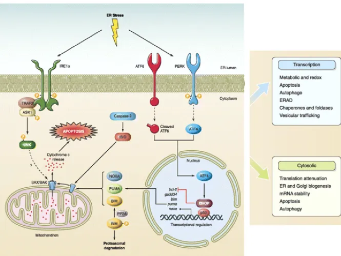
28 Inhibition of ERAD causes accumulation of misfolded proteins in the pericentriolar region of the cell. Accumulation of abnormally folded proteins at the ER engages an adaptive stress response (Figure 4). In the presence of this ER stress, the cell will activate the UPR mechanism to (1) diminish protein synthesis and to (2) up-regulate ER chaperons and (3) ERAD factors with the aim to reduce aggregate accumulation. If ER stress is not successfully diminished, the UPR will initiate an (4) apoptotic phase culminating in cellular death (Figure 4) (Hetz et al., 2011; Schmidt and Finley, 2014). Aggregating proteins in neurodegenerative diseases, such as tau or α-synuclein, are capable of inducing the UPR. In AD, Tau and Aßeta soluble oligomers are likely to inhibit ERAD leading to UPR activation and in PD, α-synuclein is capable of inhibiting autophagy factor 6 (ATF6) trafficking to the Golgi and thus causing UPR activation (Ogen-Shtern et al., 2016; Schmidt and Finley, 2014).
Yet, whether autophagy and proteasome degradation targets similar normal or misfolded proteins is a question that remains to be answered. Furthermore, as these two systems crosstalk between each other, it is often difficult to determine which of these processes, if any, is associated with the initial disease mechanisms. It is plausible that these two systems act in a simultaneous and complementary manner, particularly when aberrant proteins overwhelm the disposal capacity of the proteasome.
29
Under prolonged or chronic ER stress due to accumulation of abnormally folded proteins, activation of an apoptotic response will be engaged. There are three major ER-UPR stress sensors, IRE1α, PERK, and ATF6, which transduce information about the folding status of protein in the ER to the cytosol and the nucleus to restore folding capacity. Activation of IRE1α controls selective mRNA decay and also leads to the processing of the mRNA encoding XBP1, a transcription factor that upregulates many essential UPR. Active IRE1α also regulates stress responses mediated by JNK, ERK, and NFκB. Activation of PERK decreases the general protein synthesis rate through phosphorylation of the initiation factor eIF2α. eIF2α phosphorylation will increases the translation of the ATF4 mRNA, which will induces the expression of genes involved in amino acid metabolism, autophagy, antioxidant responses, and apoptosis. Activation of the pro-apoptotic BAX and BAK at the mitochondria is a key step in the induction of apoptosis, leading to the release of cytochrome c and activation of downstream caspases. The transcription factor CHOP activated by IRE1α, PERK, and ATF6 will downregulates the anti-apoptotic protein BCL-2. In addition, active IRE1α binding to TRAF2 will lead to the activation of the pro-apoptotic kinases JNK and ASK. Adapted from Hetz et al. (2011)
30 1.5. Nucleocytoplasmic shuttling dysfunction
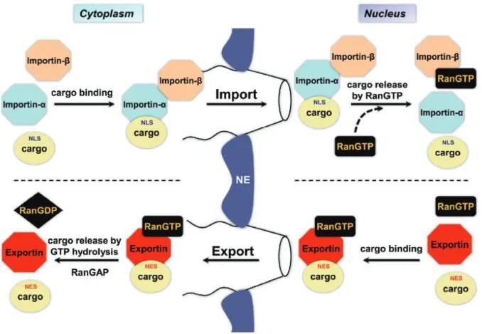
Several mechanisms could explain altered subcellular distribution of proteins, but growing evidence has placed disruption in nucleocytoplasmic transport (NCT) as central to the impairment of neuronal functions leading to neurodegeneration. Aggregation in the cytoplasm is able to interfere with nucleocytoplasmic shuttling and RNA transport (Woerner et al., 2015). Thus, the study of mechanisms altering NCT may play a significant role in understanding pathogenesis of neurodegenerative diseases.
Small proteins (<40kDa) can passively diffuse through the nuclear envelope but larger proteins will require active transport to be carried out between the nucleus and the cytoplasm with the assistance of importins for nuclear import and exportins for nuclear export (Figure 5). Molecules are able to enter and exit the nucleus through the nuclear pore complex (NPC). This large complex is composed of over 30-50 protein subunits called nucleoporins (Nups), each of them composed by several hundred individual protein-molecules (Patel and Chu, 2011; Rout et al., 2000). With aging, the composition of the nuclear pore can be altered and become “leakier” and in pathological conditions, the nuclear contour itself can be modified. In ALS and in PD models, staining of NPC nups-p62 protein has shown irregularities in the nuclear contour that present ruffled abnormal edges. In AD cases, Nrf2 protein, which is also implicated in NPC conformation, presents cytoplasmic mislocalization rather than the normal nuclear one (Patel and Chu, 2011). As neurons are polarized cells, molecule shuttle along the axon and the soma is a highly regulated process in order to maintain correct functional structure. Lesion studies have shown that neurons can activate a high response rate to damage, in order to avoid alterations. For example, in the case of nerve lesion, local translation of axonal mRNA increases importin β protein levels allowing for the formation of units that transport proteins backward and forward to ensure repairing (Patel and Chu, 2011). This shuttling will activate signals such as phosphorylation or acetylation that in turn, can alter the nuclear to cytoplasmic ratios of several proteins. Oxidative stress and protein aggregates are yet other stimuli that will lead to imbalance in nuclear to cytoplasmic ratio of proteins, but also in nucleoporins and other proteins of the nuclear import and export machinery (Patel and Chu, 2011; Woerner et al., 2015).
From this point, protein aggregates are of particular interest, as identical proteins with different targeting sequences, either to the nucleus or the cytoplasm can form distinct biochemical aggregates that differ in toxicity. Such is the case with expression of cytoplasmic-prone aggregating proteins that will cause altered distribution of the NPC
31 proteins, interfering with import and export proteins and mRNA transportation. Nucleus-prone aggregating proteins, on the other hand, will have no effect in NPC protein distribution or function, as was shown in in vitro models of protein aggregation (Woerner et al., 2015). Mechanisms through which cytoplasmic protein aggregates exert this toxicity in the NPC remain elusive. The toxic effect could pass through aberrant interactions with other proteins, resulting in their functional impairment and sequestration, especially since cytoplasmic aggregates bind splicing factors, RBPs and other proteins involved in the nuclear transport (Woerner et al., 2015). Nuclear aggregates, in the other hand, bind mainly RBPs, probably due to their intrinsic properties which make them more prone these association or to their
Figure 5 Schematic representation of nucleocytoplasmic shuttling
Import of NLS-containing cargo protein through the NPC occurs via Importin a/importin ß heterodimer. In the nucleus, RanGTP binds to importin ß to induce release of cargo protein. Importin ß will then be transported to the cytoplasm with the exportin CAS (cellular apoptosis susceptibility protein) to be recycled upon GTP hydrolysis. Export of NES-containing cargo protein occurs with the interaction of exportins. Protein release to the cytoplasm will occur upon GTP hydrolysis. NFT2 (nuclear transport factor 2) will recycle RanGDP back to the nucleus. Ran regulators, RanGAP in the cytoplasm and RanGEF in the nucleus, highly preserve RanGDP/RanGTP gradient, as it is fundamental for determining the sense of NCT. NE = Nuclear envelop Adapted from Patel and Chu (2011)
32 interaction with specific nuclear proteins such as nucleophosmin 1 rather than cytoplasmic proteins (Woerner et al., 2015).
Nuclear export is a less well-known process and it mainly concerns RNA molecules linked to ribonucleoproteins (RNP), such as tRNAs, ribosomal subunits and mRNAs. Emerging evidence has given a role to nuclear export in the regulation of microRNA (miRNAs) biogenesis process (Muqbil et al., 2013). miRNAs are short non-coding RNAs of 18 to 25 nucleotides in length that can regulate the expression of a myriad of genes and their implications will be explored in the following section.
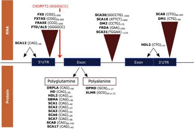
2. RNA metabolism alteration in neurological disease
Regulation of RNA metabolism in neurons is highly elaborated, involving compartmentalized expression through long distance mRNA trafficking, local translation and alternative splicing (Gao and Taylor, 2014). Interest in this pathway rose after the identification of altered RNA metabolism in abnormal microsatellite expansions cases. The identification, in 1991 that the CGG expansion in the 5’ non coding region of the FMR1 gene was responsible of Fragile-X syndrome (FXTAS) opened the door to the discovery of numerous neurological and neuromuscular diseases caused by repeat expansions (Fu et al. 1991, Oberle et al. 1991; Verkek et al. 1991). Over 40 neurological, neurodegenerative or neuromuscular diseases are explained by these type of mutations, and they include some of the most common inherited disorders such as HD, spinocerebellar ataxias (SCAs), and FTD/ALS caused by an expanded repeat in C9orf72 (c9FTD/ALS) (DeJesus-Hernandez et al., 2011; Loureiro et al., 2016; Maizels, 2015; Ranum and Cooper, 2006; Renton et al., 2011). Typically, the number of repeats in non-affected individuals is lower than 30, but it is higher that 40-50 in patients, and may exceed 1000 repeats in some cases (Sicot and Gomes-Pereira, 2013). Some of these disorders present genetic anticipation, as genetically unstable repeats can undergo further expansion during intergenerational transmission (Mirkin, 2007), whereas for some others, the type of inheritance has not been yet identified. More than 20 years later, our understanding of these “microsatellite-expansion diseases” has advanced, but has also risen questions concerning how these expansions are be able to result in disease. I will not refer to microsatellites present in the genome that are not disease-causing expansions. In this chapter, I will describe findings in the field of neurodegenerative diseases with a focus on the neuropathological mechanisms driven by RNA toxicity, pointing to aspects linked to the hexanucleotide repeat expansion in c9FTD/ALS.