HAL Id: lirmm-02304800
https://hal-lirmm.ccsd.cnrs.fr/lirmm-02304800
Submitted on 3 Oct 2019
HAL is a multi-disciplinary open access
archive for the deposit and dissemination of
sci-entific research documents, whether they are
pub-lished or not. The documents may come from
teaching and research institutions in France or
abroad, or from public or private research centers.
L’archive ouverte pluridisciplinaire HAL, est
destinée au dépôt et à la diffusion de documents
scientifiques de niveau recherche, publiés ou non,
émanant des établissements d’enseignement et de
recherche français ou étrangers, des laboratoires
publics ou privés.
Breathing detection from tracheal sounds in both
temporal and frequency domains in the context of
phrenic nerve stimulation
Xinyue Lu, David Guiraud, Serge Renaux, Thomas Similowski, Christine
Azevedo Coste
To cite this version:
Xinyue Lu, David Guiraud, Serge Renaux, Thomas Similowski, Christine Azevedo Coste. Breathing
detection from tracheal sounds in both temporal and frequency domains in the context of phrenic
nerve stimulation. EMBC 2019 - 41st International Engineering in Medicine and Biology Conference,
IEEE, Jul 2019, Berlin, Germany. �lirmm-02304800�
Breathing detection from tracheal sounds in both temporal and
frequency domains in the context of phrenic nerve stimulation
Xinyue LU
1,2, David GUIRAUD
1, Serge RENAUX
2, Thomas SIMILOWSKI
3, Christine AZEVEDO
1Abstract— Electrical stimulation of the phrenic nerves via implanted devices allows to counteract some disadvantages of mechanical ventilation in patients with high tetraplegia or Ondine’s syndrome. Existing devices do not allow to monitor breathing or to adapt the electroventilation to patients’ actual needs. A reliable breathing monitor with an inbuilt alarm function would improve patient safety. In our study, a real-time acoustic breathing detection method is proposed as a possible solution to improve implanted phrenic stimulation. A new algorithm to process tracheal sounds has been developed. It combines breathing detection in both temporal and frequency domains. The algorithm has been applied on recordings from 18 healthy participants. The obtained average sensitivity, speci-ficity and accuracy of the detection are: 99.31%, 96.84% and 98.02%, respectively. These preliminary results show a first positive proof of the interest of such an approach. Additional experiments are needed, including longer recordings from individuals with tetraplegia or Ondine Syndrome in various environments to go further in the validation.
I. INTRODUCTION
Individuals with a central respiratory paralysis, are es-sentially supplied by mechanical ventilation. In France, around 44 new high tetraplegia per year induce a ventilatory dependence. There are also about 60 congenital central alveolar hypoventilation in which 10%-15% patients have a ventilatory dependence [1].
However, severe drawbacks of mechanical ventilation are reported: (1) the induced positive pressure disturbs the ve-nous return [2], (2) ventilators are noisy and limit mobility which reduces the quality of life, (3) tracheotomy may cause respiratory infections, loss of olfaction and impossibility to speak [3]. In the case where phrenic nerves and diaphragm remain functional, the mechanical ventilation can be replaced by an implanted phrenic stimulation device which could cancel some of the disadvantages mentioned above. Further-more, phrenic stimulation induces a more natural respiration and reduces health care costs [2][3].
The main disadvantage of implanted phrenic stimulation is that it stimulates at a fixed frequency and intensity preset into the external controller unit. Therefore, the stimulation does not adapt to patients’ respiratory state. For example, high tetraplegia requires more respiratory volume while sitting than in lying position [4]; patients with Ondine syndrome do not need a permanent stimulation all day long. Indeed, they still have some remaining spontaneous respiratory cycles. Finally, the absence of a reliable respiratory monitoring
1INRIA, University of Montpellier, Montpellier, France 2NeuroResp, Les Aires, France
3Hospital Group Piti´e-Salpˆetri`ere Charles Foix, Paris, France
system makes most of patients switching to mechanical ventilator during nighttime.
The gold standard for apnea/hypoventilation evaluation is the polygraph, which includes an pulse oximeter and at least one respiratory flow sensor [5]. In a clinical use, flow sensors could be nasal cannula, pneumotachograph, thermistor or plethysmograph. But these sensors need to be placed over the face or are sensitive to patient’s movements. They are therefore not compatible with an implanted phrenic stimulation system which is portable and for a daily living use. With this in mind, we investigated an acoustic method. The proposed tracheal sounds recording requires only one tiny microphone fixed on the neck, which is the only physical contact with the patient. Another advantage of the absence of electrical contact with the user guarantees that the detection method will not influence the stimulation system.
For a better quality of signal, the best place for tracheal sounds recording is at suprasternal notch [6]. Many previous studies have shown some positive results on respiration analysis from tracheal sounds in sleep apnea, especially for obstructive sleep apnea. Their detection algorithms are either based on the envelope of sounds signals (in time domain) [6][7][8][9][10], or based on sounds spectral power (in frequency domain) [11][12][13][14][15][16]. Some groups also use statistical methods for respiratory phase detection and classification [17], and even for air flow estimation [18][19][20][21] associated with sounds entropy or the log of the sounds variance. But only few methods are developed for real-time applications (processing delay within seconds) with robustness requirements, indeed, all these studies have been carried out in quiet and controlled acoustic environments with stable sources of noises, and with limited movements of the subjects (during sleep).
The present study aims at a real-time and continuous breath detection (day and night), even during wakefulness in noisy environments. We proposed a new algorithm to detect respiration phases, by combining the signal processing both in the temporal and the frequency domains. We assessed the performances of the algorithm in emulated noisy environ-ments.
II. MATERIAL AND METHODS A. Equipment
Tracheal sounds are recorded by an omni-directional mi-crophone (pro-signal, ABM-705-RC), which is inserted into a 3D printed bell-shape support [6]. This support is fixed at the suprasternal notch by using a medical rubber bandage
(fig. 1). The microphone signal is amplified (x230) and fil-tered (100Hz-1200Hz, Band-pass second order Butterworth, custom made) in order to keep only the frequencies which are relevant for detecting airflow sounds [12]. The signal is then digitized at 4.6 kHz, 16 bits, using a NUCLEO-F429ZI card. Data are transferred to a PC and processed using MatlabTM software.
Fig. 1: position of microphone
B. Protocol
In this study, 18 healthy subjects (4 women and 14 men aged from 20 to 60 years old) participated. Signals were recorded in a sitting position. All recordings are performed in the same room and with a similar quiet environment.
Each recording lasted 30s. The procedure consisted in 3 succeeding phases: (1) breathing normally during the first 10s, (2) expiring and then holding respiration for 10s, (3) breathing normally again for the last 10s.
III. DETECTION ALGORITHM
For each processing cycle, 3 segments of 1024 samples (s(n)) are treated with an overlapping of one segment. The length of overlapping corresponds the processing delay, here is 1024/f s ≈ 0.22s. This delay is within the acceptable alarm delay for stimulation system, which is around 3s.
As the detection flow diagram in Fig. 2 shown, s(n) is first filtered by a 6thorder high-pass Butterworth filter at 300 Hz to remove cardiac noises. The filtered respiratory signal r(n) is processed both in temporal and frequency domains and the obtained results are then combined to get the final detection d(n). Three parameters (in red) ratio T , ratio F and ratio adapt are patient-dependent and are presented below.
A. Temporal domain
A 2nd order low pass Butterworth filter is used on the energy of the temporal signal, i.e. 2∗r(n)2, to get its envelope e(n). The cut-off frequency is around 1 Hz (corresponding to cut-off pulsation ωn = 0.01 rd.s−1). During holding breath,
information from environment noises are used to set patient-specific thresholds as it is considered as an apnea period. The minimum temporal threshold S T is obtained by multiplying the ratio T with the mean of the envelope amplitude during apnea (12s-19s). For each detected respiratory event (inspira-tion, expiration and pause/apnea), its’ begging (Ttempbeginning)
Fig. 2: Diagram of the detection algorithm
and ending points (Ttempending) are noted to calculate its center
time Ttempcenter. Furthermore, one detected respiratory event is
rejected if it lasts less than 0.4s. B. Frequency domain
In the frequency domain, the filtered signal is divided into 3 segments of 1024 samples (rseg(k)). FFT is applied on
each segment with Hanning window (Rseg(k)). Depending
on the recording device, the main respiratory frequency band could be different [22]. In this study, as shown in Fig. 3, normal respiration frequency contents are centered between 300 Hz and 900 Hz. The best discrimination was obtained with the power spectral density P SD(m) of each segment computed between 300 Hz and 600 Hz:
P SD = (
600
X
k=300
Rseg(k)2)/1024 (1)
The minimum threshold is obtained by multiplying the ratio F with the mean PSD during apnea (12s-19s). In addition, a moving threshold is also applied to adapt different density levels; for example, stronger breathing occurs after apnea. This adaptive threshold is obtained by multiplying the ratio adapt with the mean P SD(m) over 3 segments: the previous, the actual and the next segments. As in tem-poral domain, the beginning (Tf reqbeginning) and ending points (Tf reqending) for each detected event are noted to get centered time Tcenter
f req .
C. Combination
For the same respiratory event, detection result may have a little time lag in different domains. This time lag can’t exceed the minimum duration for one normal inspiration or expiration, which is around one second. So the respiratory event will be finally validated if the time lag |Ttempcenter −
Tcenter
f req | is less than 1 s. Then, the result d(n) depends
on Ttempbeginning and Ttempending because detection in temporal domain is more accurate if S T is well defined.
IV. RESULTS AND EVALUATION OF THE DETECTION ALGORITHM
An example of one breathing detection result is shown in Fig. 4. Three detection signals are illustrated in this figure: the temporal detection is in orange, the frequency detection is in green and the combined detection is in yellow. All detection signals have two states, indicating if there is a respiratory event (high) or not (low). In this example, all respiratory events are successfully detected.
Fig. 4: Detection result obtained with one recording of one subject.
The algorithm is evaluated on its specificity, sensitivity and accuracy, which are defined as follows:
Specif icity = T N T N + F P (2) Sensitivity = T P T P + F N (3) Accuracy = T P + T N T P + T N + F P + F N (4) where T N , F P , T P and F N are the number of true negative, false positive, true positive and false negative, respectively.
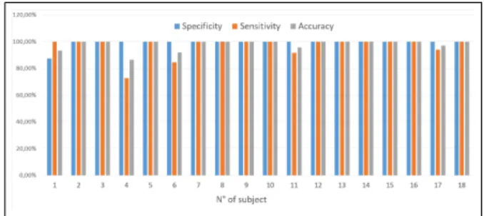
The scores for these 3 indexes have been computed for all 18 subjects and are plotted in figure 5. With a mean value of 99.31%, 96.84% and 98.02% for specificity, sensitivity and accuracy, respectively.
V. DISCUSSION/CONCLUSION
Temporal detection can help eliminating short intermittent noises by analyzing the duration of the detected event. An example is shown in figure 4, the short noise at 24thsecond
Fig. 5: Detection results evaluation
is eliminated by the temporal detection even though it is considered as a respiratory event in the frequency domain.
Frequency detection is useful for discriminating sounds without specific peaks or amplitudes in the time domain, such as breathing and snoring [16]. Some noises appear during apnea shown in Fig. 6. They are detected in temporal domain, but are rejected by the frequency detection because their main frequency bands are different to the one of respiration.
Fig. 6: Frequency detection helps in eliminating long noises
The reason why temporal detection has better event timing accuracy is that it reacts on each sample, whereas frequency detection can only detect on segments of 1024 samples. For the same reason, frequency detection may detect twice the same event, just like the detection at 27thsecond in Fig. 4. But thanks to the combination method by checking detected events distance, these repeated detected events are avoided.
In the case of phrenic stimulation, the first aim is to determine when the patient stops breathing. We should avoid false positive detection of respiration so a high specificity. Even if the timing of each respiratory event is not accurate, the result will not lead to a dangerous situation for patients. The definition of apnea is an absence of respiratory air flow for more than 10s [5]. It means that the lowest acceptable condition is when missing maximum 10s respiration per minute, which corresponds to specificity and accuracy of 82.86% and 90.48%, respectively. The proposed algorithm gives 99.31% of specificity and 98.02% of accuracy, this result is far more than the minimum values mentioned above. We have also tested the developed algorithm on a 60s recording for one subject, with 6 phases of 10s corresponding to: (1) normal respiration, (2) apnea, (3) normal respiration, (4) speech, (5) normal respiration, (6) normal respiration with
a played video at background. The corresponding detection result is showed in figure 7. The specificity, sensitivity and accuracy are 100%, 97.22%, 98.59%, respectively. This re-sult meets also the minimum requirements mentioned before.
Fig. 7: Detection result with speech and background noises The proposed algorithm has a better detection result com-pared to a similar continuous breathing detection study done by Kalkbrenner et al. [16] which showed a detection rate of 21%-80% for respiratory detection. But their study detects also heartbeats and movements which gives a more complete monitoring. A more recent study of the same group [9] advance in result with a sensitivity of 92.8% and a specificity of 99.7%. But this algorithm, based on temporal domain with 3 envelopes, needs to scan the entire recording, so it cannot be applied in real-time, and is not adapted to phrenic stimulation system monitoring.
The downside of our study is that the proposed algorithm is only applied on 18 recordings from 18 subjects, and that each recording lasts only 30s that is much shorter than those of other studies. More recordings are needed from target patients. This method depends on several subject individual parameters. With the proposed adaptive threshold detection in frequency domain, we obtained accurate results in these short recordings. The threshold decision method still needs to be improved to be used in real-time. The sensitivity of the detection performances to these parameters will also be precisely evaluated in the future. Furthermore, the presence of respiratory events is only verified by hearing and observ-ing recordobserv-ings which may cause a miss of precision. Future recordings synchronized and compared with other reference signal, such as pneumotachography, plethysmography, etc. are needed.
On the other hand, the analysis based on sounds is sensi-tive to noise, a noise reduction algorithm may be useful. Even respiratory features vary a little bit between each subject, but the recorded signal is still repetitive. In this case, a detection based on AI in tracheal sounds may be more efficient. To adapt to most of daytime environments and to have more robust detection, it could combine more various detection methods, such as seismograph (pulse oximeter), phonocardiogram (cardiac sounds), etc.
ACKNOWLEDGMENT
We would like to thank Dr. Gerhard Baer, founder of AtroStim, for reading our Manuscript.
REFERENCES
[1] HAS, “COMMISSION D’EVALUATION DES PRODUITS ET PRESTATIONS - ATROSTIM,” Tech. Rep., 2009. [Online]. Available: https://www.has-sante.fr/portail/upload/docs/application/ pdf/2009-05/cepp-2056 atrostim.pdf
[2] T. Similowski and J.-p. Derenne, “Stimulation phr´enique implant´ee,” M´edecine th´erapeutique, vol. 7, pp. 457–469, 2001.
[3] F. Le Pimpec-Barthes, J. Gonzalez-Bermejo, J. P. Hubsch, A. Duguet, C. Mor´elot-Panzini, M. Riquet, and T. Similowski, “Intrathoracic phrenic pacing: A 10-year experience in France,” Journal of Thoracic and Cardiovascular Surgery, vol. 142, no. 2, pp. 378–383, 2011. [4] A. F. DiMarco, “Diaphragm Pacing,” Clinics in Chest Medicine,
vol. 39, no. 2, pp. 459–471, 6 2018.
[5] M.Akkari, P.Franco, and F.Chalueau, “Enregistrements du sommeil chez l’enfant,” in Syndrome d’apn´ees-hypopn´ees obstructives du sommeil de l’enfant, 2016, pp. 50–60.
[6] P. Corbishley and E. Rodr´ıguez-Villegas, “Breathing detection: To-wards a miniaturized, wearable, battery-operated monitoring system,” IEEE Trans. Biomed. Eng., vol. 55, no. 1, pp. 196–204, 2008. [7] A. Kulkas, E. Huupponen, J. Virkkala, M. Tenhunen, A. Saastamoinen,
E. Rauhala, and S.-L. Himanen, “Intelligent methods for identifying respiratory cycle phases from tracheal sound signal during sleep,” Computers in Biology and Medicine, vol. 39, no. 11, pp. 1000–1005, 11 2009.
[8] Y. Nam, B. A. Reyes, and K. H. Chon, “Estimation of Respiratory Rates Using the Built-in Microphone of a Smartphone or Headset,” IEEE Journal of Biomedical and Health Informatics, vol. 20, no. 6, pp. 1493–1501, 2016.
[9] C. Kalkbrenner, M. Eichenlaub, S. R¨udiger, C. Kropf-Sanchen, W. Rottbauer, and R. Brucher, “Apnea and heart rate detection from tracheal body sounds for the diagnosis of sleep-related breathing dis-orders,” Medical and Biological Engineering and Computing, vol. 56, no. 4, pp. 671–681, 2017.
[10] A. Martin and J. Voix, “In-Ear Audio Wearable: Measurement of Heart and Breathing Rates for Health and Safety Monitoring,” IEEE Trans. Biomed. Eng., vol. 65, no. 6, pp. 1256–1263, 6 2018.
[11] J. S. Chuah and Z. K. Moussavi, “Automated Respiratory Phase Detection by Acoustical Means,” IEEE EMBC 1998, no. March, pp. 21–24, 1998.
[12] P. Hult, T. Fjallbrant, S. Dahle, P. Danielsson, and P. Ask, “A method for respiration monitoring by the use of a bioacoustic signal,” in First International Conference on Advances in Medical Signal and Information Processing, vol. 2000. Bristol, UK,: IET, 2000, pp. 22– 25.
[13] P. Hult, T. Fjallbrant, B. Wranne, O. Engdahl, and P. Ask, “An improved bioacoustic method for monitoring of respiration,” Technol Health Care, vol. 12, no. 4, pp. 323–332, 2004.
[14] A. Kulkas, E. Huupponen, J. Virkkala, M. Tenhunen, A. Saastamoinen, E. Rauhala, and S. L. Himanen, “New tracheal sound feature for apnoea analysis,” Medical and Biological Engineering and Computing, vol. 47, no. 4, pp. 405–412, 2009.
[15] A. Yadollahi and Z. Moussavi, “Automatic breath and snore sounds classification from tracheal and ambient sounds recordings,” Medical Engineering and Physics, vol. 32, no. 9, pp. 985–990, 2010. [16] C. Kalkbrenner, P. Stark, G. Kouemou, M. E. Algorri, and R. Brucher,
“Sleep monitoring using body sounds and motion tracking,” IEEE EMBC 2014, vol. 6000, pp. 6941–6944, 2014.
[17] S. Le Cam, C. Collet, and F. Salzenstein, “Acoustical respiratory signal analysis and phase detection,” in IEEE ICASSP 2008, vol. 1. IEEE, 3 2008, pp. 3629–3632.
[18] A. Yadollahi and Z. M. Moussavi, “A robust method for estimating respiratory flow using tracheal sounds entropy,” IEEE Trans. Biomed. Eng., vol. 53, no. 4, pp. 662–668, 2006.
[19] A. Yadollahi and Z. Moussavi, “Acoustic obstructive sleep apnea detection,” in IEEE EMBC 2009, vol. 4, no. 2. IEEE, 9 2009, pp. 7110–7113.
[20] A. Yadollahi, E. Giannouli, and Z. Moussavi, “Sleep apnea monitoring and diagnosis based on pulse oximetery and tracheal sound signals,” Medical and Biological Engineering and Computing, vol. 48, no. 11, pp. 1087–1097, 2010.
[21] S. Huq and Z. Moussavi, “Acoustic breath-phase detection using tracheal breath sounds,” Medical and Biological Engineering and Computing, vol. 50, no. 3, pp. 297–308, 2012.
[22] T. Penzel and A. K. Sabil, “The use of tracheal sounds for the diagnosis of sleep apnoea,” Breathe, vol. 13, no. 2, pp. e37–e45, 2017.