2016 ESC Guidelines for the diagnosis and
treatment of acute and chronic heart failure
The Task Force for the diagnosis and treatment of acute and chronic
heart failure of the European Society of Cardiology (ESC)
Developed with the special contribution of the Heart Failure
Association (HFA) of the ESC
Authors/Task Force Members: Piotr Ponikowski
*
(Chairperson) (Poland),
Adriaan A. Voors
*
(Co-Chairperson) (The Netherlands), Stefan D. Anker (Germany),
He´ctor Bueno (Spain), John G. F. Cleland (UK), Andrew J. S. Coats (UK),
Volkmar Falk (Germany), Jose´ Ramo´n Gonza´lez-Juanatey (Spain), Veli-Pekka Harjola
(Finland), Ewa A. Jankowska (Poland), Mariell Jessup (USA), Cecilia Linde (Sweden),
Petros Nihoyannopoulos (UK), John T. Parissis (Greece), Burkert Pieske (Germany),
Jillian P. Riley (UK), Giuseppe M. C. Rosano (UK/Italy), Luis M. Ruilope (Spain),
Frank Ruschitzka (Switzerland), Frans H. Rutten (The Netherlands),
Peter van der Meer (The Netherlands)
Document Reviewers: Gerasimos Filippatos (CPG Review Coordinator) (Greece), John J. V. McMurray (CPG Review
Coordinator) (UK), Victor Aboyans (France), Stephan Achenbach (Germany), Stefan Agewall (Norway),
Nawwar Al-Attar (UK), John James Atherton (Australia), Johann Bauersachs (Germany), A. John Camm (UK),
Scipione Carerj (Italy), Claudio Ceconi (Italy), Antonio Coca (Spain), Perry Elliott (UK), Çetin Erol (Turkey),
Justin Ezekowitz (Canada), Covadonga Ferna´ndez-Golfı´n (Spain), Donna Fitzsimons (UK), Marco Guazzi (Italy),
*Corresponding authors: Piotr Ponikowski, Department of Heart Diseases, Wroclaw Medical University, Centre for Heart Diseases, Military Hospital, ul. Weigla 5, 50-981 Wroclaw, Poland, Tel:+48 261 660 279, Tel/Fax: +48 261 660 237, E-mail:[email protected].
Adriaan Voors, Cardiology, University of Groningen, University Medical Center Groningen, Hanzeplein 1, PO Box 30.001, 9700 RB Groningen, The Netherlands, Tel:+31 50 3612355, Fax:+31 50 3614391, E-mail:[email protected].
ESC Committee for Practice Guidelines (CPG) and National Cardiac Societies document reviewers: listed in the Appendix. ESC entities having participated in the development of this document:
Associations: Acute Cardiovascular Care Association (ACCA), European Association for Cardiovascular Prevention and Rehabilitation (EACPR), European Association of Cardiovascular Imaging (EACVI), European Heart Rhythm Association (EHRA), Heart Failure Association (HFA).
Councils: Council on Cardiovascular Nursing and Allied Professions, Council for Cardiology Practice, Council on Cardiovascular Primary Care, Council on Hypertension. Working Groups: Cardiovascular Pharmacotherapy, Cardiovascular Surgery, Myocardial and Pericardial Diseases, Myocardial Function, Pulmonary Circulation and Right Ventricular Function, Valvular Heart Disease.
The content of these European Society of Cardiology (ESC) Guidelines has been published for personal and educational use only. No commercial use is authorized. No part of the ESC Guidelines may be translated or reproduced in any form without written permission from the ESC. Permission can be obtained upon submission of a written request to John Wiley & Sons, the publisher of the European Journal of Heart Failure and the party authorized to handle such permissions on behalf of the ESC ([email protected]). Disclaimer. The ESC Guidelines represent the views of the ESC and were produced after careful consideration of the scientific and medical knowledge and the evidence available at the time of their publication. The ESC is not responsible in the event of any contradiction, discrepancy and/or ambiguity between the ESC Guidelines and any other official recom-mendations or guidelines issued by the relevant public health authorities, in particular in relation to good use of healthcare or therapeutic strategies. Health professionals are encour-aged to take the ESC Guidelines fully into account when exercising their clinical judgment, as well as in the determination and the implementation of preventive, diagnostic or therapeutic medical strategies; however, the ESC Guidelines do not override, in any way whatsoever, the individual responsibility of health professionals to make appropriate and accurate decisions in consideration of each patient’s health condition and in consultation with that patient and, where appropriate and/or necessary, the patient’s caregiver. Nor do the ESC Guidelines exempt health professionals from taking into full and careful consideration the relevant official updated recommendations or guidelines issued by the competent public health authorities, in order to manage each patient’s case in light of the scientifically accepted data pursuant to their respective ethical and professional obligations. It is also the health professional’s responsibility to verify the applicable rules and regulations relating to drugs and medical devices at the time of prescription.
The article has been co-published with permission in European Heart Journal and European Journal of Heart Failure. All rights reserved in respect of European Heart Journal.
Maxime Guenoun (France), Gerd Hasenfuss (Germany), Gerhard Hindricks (Germany), Arno W. Hoes
(The Netherlands), Bernard Iung (France), Tiny Jaarsma (Sweden), Paulus Kirchhof (UK/Germany), Juhani Knuuti
(Finland), Philippe Kolh (Belgium), Stavros Konstantinides (Germany/Greece), Mitja Lainscak (Slovenia),
Patrizio Lancellotti (Belgium), Gregory Y. H. Lip (UK), Francesco Maisano (Switzerland), Christian Mueller
(Switzerland), Mark C. Petrie (UK), Massimo F. Piepoli (Italy), Silvia G. Priori (Italy), Adam Torbicki (Poland),
Hiroyuki Tsutsui (Japan), Dirk J. van Veldhuisen (The Netherlands), Stephan Windecker (Switzerland), Clyde Yancy
(USA), Jose Luis Zamorano (Spain)
The disclosure forms of all experts involved in the development of these guidelines are available on the ESC website
http://www.escardio.org/guidelines
.
-Keywords
Guidelines † Heart failure † Natriuretic peptides † Ejection fraction † Diagnosis † Pharmacotherapy †
Neuro-hormonal antagonists † Cardiac resynchronization therapy † Mechanical circulatory support †
Transplantation † Arrhythmias † Co-morbidities † Hospitalization † Multidisciplinary management
Table of Contents
Abbreviations and acronyms . . .
893
1. Preamble . . . .
896
2. Introduction . . . .
898
3. Definition, epidemiology and prognosis . . . .
898
3.1 Definition of heart failure . . . .
898
3.2 Terminology . . . .
899
3.2.1 Heart failure with preserved, mid-range and reduced
ejection fraction . . . 899
3.2.2 Terminology related to the time course of heart
failure . . . 899
3.2.3 Terminology related to the symptomatic severity
of heart failure . . . 900
3.4 Prognosis . . . .
900
4. Diagnosis . . . .
900
4.1 Symptoms and signs . . . .
900
4.2 Essential initial investigations: natriuretic peptides,
electrocardiogram, and echocardiography . . . .
901
4.3 Algorithm for the diagnosis of heart failure
4.3.1 Algorithm for the diagnosis of heart failure in the
non-acute setting
4.3.2 Diagnosis of heart failure with preserved ejection
fraction
5. Cardiac imaging and other diagnostic tests . . . .
904
5.1 Chest X-ray . . . .
904
5.2 Transthoracic echocardiography . . . .
904
5.2.1 Assessment of left ventricular systolic function . . . . 904
5.2.2 Assessment of left ventricular diastolic function . . . 905
5.2.3 Assessment of right ventricular function and
pulmonary arterial pressure . . . 905
5.3 Transoesophageal echocardiography . . . .
905
5.4 Stress echocardiography . . . .
905
5.5 Cardiac magnetic resonance . . . .
905
5.6 Single-photon emission computed tomography and
radionuclide ventriculography . . . .
905
5.7 Positron emission tomography . . . .
905
5.8 Coronary angiography . . . .
906
5.9 Cardiac computed tomography . . . .
906
5.10 Other diagnostic tests . . . .
907
5.10.1 Genetic testing in heart failure . . . 907
6. Delaying or preventing the development of overt heart
failure or preventing death before the onset of symptoms . . . . .
908
7. Pharmacological treatment of heart failure with reduced
ejection fraction . . . .
909
7.1 Objectives in the management of heart failure . . . .
909
7.2 Treatments recommended in all symptomatic patients
with heart failure with reduced ejection fraction . . . .
910
7.2.1 Angiotensin-converting enzyme inhibitors . . . 910
7.2.2 Beta-blockers . . . 910
7.2.3 Mineralocorticoid/aldosterone receptor antagonists . 910
7.3 Other treatments recommended in selected symptomatic
patients with heart failure with reduced ejection fraction . . .
910
7.3.1 Diuretics . . . 910
7.3.2 Angiotensin receptor neprilysin inhibitor . . . 913
7.3.3 I
f- channel inhibitor . . . 914
7.3.4 Angiotensin II type I receptor blockers . . . 914
7.3.5 Combination of hydralazine and isosorbide
dinitrate . . . 914
7.4 Other treatments with less certain benefits in
symptomatic patients with heart failure with reduced ejection
fraction . . . .
914
7.4.1 Digoxin and other digitalis glycosides . . . 914
7.4.2 n-3 polyunsaturated fatty acids . . . 915
7.5 Treatments not recommended (unproven benefit) in
symptomatic patients with heart failure with reduced ejection
fraction . . . .
915
7.5.1 3-Hydroxy-3-methylglutaryl-coenzyme A reductase
inhibitors (‘statins’) . . . 915
7.5.2 Oral anticoagulants and antiplatelet therapy . . . 915
7.5.3 Renin inhibitors . . . 915
7.6 Treatments not recommended (believed to cause harm)
in symptomatic patients with heart failure with reduced
ejection fraction . . . .
916
7.6.1 Calcium-channel blockers . . . 916
3.3 Epidemiology, aetiology and natural history of heart failure
900
. . . .
902
. . . 902
8. Non-surgical device treatment of heart failure with reduced
ejection fraction . . . .
916
8.1 Implantable cardioverter-defibrillator . . . .
916
8.1.1 Secondary prevention of sudden cardiac death . . . . 916
8.1.2 Primary prevention of sudden cardiac death . . . 917
9. Treatment of heart failure with preserved ejection fraction
9.1 Effect of treatment on symptoms in heart failure with
preserved ejection fraction . . . .
920
9.2 Effect of treatment on hospitalization for heart failure in
heart failure with preserved ejection fraction . . . .
920
9.3 Effect of treatment on mortality in heart failure with
preserved ejection fraction . . . .
920
9.4 Other considerations . . . .
920
10. Arrhythmias and conductance disturbances . . . .
920
10.1.1 Prevention of atrial fibrillation in patients with heart
failure . . . 921
10.1.2 Management of new-onset, rapid atrial fibrillation in
patients with heart failure . . . 921
10.1.3 Rate control . . . 921
10.1.4 Rhythm control . . . 922
10.1.5 Thromboembolism prophylaxis . . . 923
10.2 Ventricular arrhythmias . . . .
923
10.3 Symptomatic bradycardia, pauses and
atrio-ventricular block . . . .
924
11. Co-morbidities . . . .
925
11.1 Heart failure and co-morbidities . . . .
925
11.2 Angina and coronary artery disease . . . .
925
11.2.1 Pharmacological management . . . 925
11.2.2 Myocardial revascularization . . . 925
11.3 Cachexia and sarcopenia (for frailty, please refer to
Section 14) . . . .
926
11.4 Cancer . . . .
926
11.5 Central nervous system (including depression, stroke and
autonomic dysfunction) . . . .
927
11.6 Diabetes . . . .
927
11.7 Erectile dysfunction . . . .
928
11.8 Gout and arthritis . . . .
928
11.9 Hypokalaemia and hyperkalaemia . . . .
928
11.10 Hyperlipidaemia . . . .
928
11.12 Iron deficiency and anaemia . . . .
929
11.13 Kidney dysfunction (including chronic kidney disease,
acute kidney injury, cardio-renal syndrome, and prostatic
obstruction) . . . .
930
11.14 Lung disease (including asthma and chronic obstructive
11.16 Sleep disturbance and sleep-disordered
11.17 Valvular heart disease . . . .
932
11.17.1 Aortic stenosis . . . 932
11.17.2 Aortic regurgitation . . . 932
11.17.3 Mitral regurgitation . . . 932
11.17.4 Tricuspid regurgitation . . . 932
12. Acute heart failure . . . .
933
12.1 Definition and classification . . . .
933
12.2 Diagnosis and initial prognostic evaluation . . . .
934
12.3 Management . . . .
938
12.3.1 Identification of precipitants/causes leading to
decompensation that needs urgent management . . . 938
12.3.2 Criteria for hospitalization in ward vs intensive
care/coronary care unit . . . 939
12.3.3 Management of the early phase . . . 939
12.3.4 Management of patients with cardiogenic shock . . 944
12.4 Management of evidence-based oral therapies . . . .
944
12.5 Monitoring of clinical status of patients hospitalized due
to acute heart failure . . . .
945
12.6 Criteria for discharge from hospital and follow-up in
high-risk period . . . .
945
12.7 Goals of treatment during the different stages of
management of acute heart failure . . . .
945
13. Mechanical circulatory support and heart transplantation . . .
946
13.1 Mechanical circulatory support . . . .
946
13.1.1 Mechanical circulatory support in acute heart
failure . . . 946
13.1.2 Mechanical circulatory support in end-stage chronic
heart failure . . . 946
13.2 Heart transplantation . . . .
948
14. Multidisciplinary team management . . . .
949
14.1 Organization of care . . . .
949
14.2 Discharge planning . . . .
951
14.3 Lifestyle advice . . . .
951
14.4 Exercise training . . . .
951
14.5 Follow-up and monitoring . . . .
951
14.6 The older adult, frailty and cognitive impairment . . . . .
952
14.7 Palliative and end-of-life care . . . .
952
15. Gaps in evidence . . . .
953
16. To do and not to
messages from the Guidelines . . . .
954
17. Web Addenda . . . .
955
18. Appendix . . . .
956
19. References . . . .
956
Abbreviations and acronyms
ACC/AHA
American College of Cardiology/American
Heart Association
ACCF/AHA
American College of Cardiology Foundation/
American Heart Association
ACE
angiotensin-converting enzyme
ACEI
angiotensin-converting enzyme inhibitor
ACS
acute coronary syndrome
AF
atrial fibrillation
AHF
acute heart failure
AHI
apnoea/hypopnoea index
AIDS
acquired immunodeficiency syndrome
AKI
acute kidney injury
Aldo-DHF
aldosterone receptor blockade in diastolic
heart failure
AL
amyloid light chain
ALT
alanine aminotransferase
8.2 Cardiac resynchronization therapy . . . .
918
8.3 Other implantable electrical devices . . . .
919
. .
919
10.1 Atrial fibrillation . . . .
921
11.11 Hypertension . . . .
928
pulmonary disease) . . . .
931
11.15 Obesity . . . .
931\
breathing . . . .
931
do
AMI
acute myocardial infarction
AMICA
Atrial fibrillation Management In Congestive
heart failure with Ablation
ANP
A-type natriuretic peptide
ANS
autonomic nervous system
ARB
angiotensin receptor blocker
ARNI
angiotensin receptor neprilysin inhibitor
ARVC
arrhythmogenic right ventricular
cardiomyopathy
AST
aspartate aminotransferase
ASV
assisted servo-ventilation
ATLAS
Assessment of Treatment with Lisinopril And
Survival
ATTR
transthyretin-mediated amyloidosis
AV
atrio-ventricular
AVP
arginine vasopressin
b.i.d.
bis in die (twice daily)
BioPACE
Biventricular Pacing for Atrio-ventricular
Block to Prevent Cardiac Desynchronization
BiPAP
bilevel positive airway pressure
BiVAD
biventricular assist device
BLOCK-HF
Biventricular versus Right Ventricular Pacing in
Heart Failure Patients with Atrio-ventricular
Block
BMI
body mass index
BNP
B-type natriuretic peptide
BP
blood pressure
bpm
beats per minute
BSA
body surface area
BTB
bridge to bridge
BTC
bridge to candidacy
BTD
bridge to decision
BTR
bridge to recovery
BTT
bridge to transplantation
BUN
blood urea nitrogen
CABANA
Catheter ABlation versus ANtiarrhythmic
drug therapy for Atrial fibrillation
CABG
coronary artery bypass graft/grafting
CAD
coronary artery disease
CARE-HF
CArdiac REsynchronization in Heart Failure
CASTLE-AF
Catheter Ablation versus Standard
contional Treatment in patients with LEft
ven-tricular dysfunction and Atrial Fibrillation
CCB
calcium-channel blocker
CCM
cardiac contractility modulation
CCS
Canadian Cardiovascular Society
CCU
coronary care unit
CHA
2DS
2-VASc
Congestive heart failure or left ventricular
dys-function, Hypertension, Age
≥75 (doubled),
Diabetes, Stroke (doubled)-Vascular disease,
Age 65–74, Sex category (female)
CHARM-Alternative Candesartan in heart failure assessment of
reduction in mortality and morbidity
CHARM-Added
Candesartan Cilexetil in Heart Failure
Assess-ment of Reduction in Mortality and Morbidity
CHARM-Preserved
Candesartan Cilexetil in Heart Failure
Assess-ment of Reduction in Mortality and Morbidity
CI
cardiac index
CI-AKI
contrast-induced acute kidney injury
CIBIS II
Cardiac Insufficiency Bisoprolol Study II
CK
creatine kinase
CKD
chronic kidney disease
CK-MB
creatine kinase MB
CMP
cardiomyopathy
CMR
cardiac magnetic resonance
COMPANION
Comparison of Medical Therapy, Pacing, and
Defibrillation in Heart Failure
CONFIRM-HF
Ferric CarboxymaltOse evaluatioN on
per-Formance in patients with IRon deficiency
in coMbination with chronic Heart Failure
CONSENSUS
Cooperative North Scandinavian Enalapril
Survival Study
COPD
chronic obstructive pulmonary disease
COPERNICUS
Carvedilol Prospective Randomized
Cumula-tive Survival
COX-2 inhibitor
cyclooxygenase-2 inhibitor
CPAP
continuous positive airway pressure
CPG
Committee for Practice Guidelines
CRT
cardiac resynchronization therapy
CRT-D
defibrillator with cardiac resynchronization
therapy
CRT-P
pacemaker with cardiac resynchronization
therapy
CSA
central sleep apnoea
CSR
Cheyne-Stokes respiration
CT
computed tomography
CYP3A4
cytochrome P450 3A4
DCM
dilated cardiomyopathy
DES
desmin
DHA
docosahexaenoic acid
DIG-PEF
ancillary Digitalis Investigation Group trial
DNA
deoxyribonucleic acid
DOSE
Diuretic Optimization Strategies Evaluation
DPD
3,3-diphosphono-1,2-propanodicarboxylic
acid
DPP4i
dipeptidyl peptidase-4 inhibitor
DT
destination therapy
e
′early diastolic tissue velocity
ECG
electrocardiogram
Echo-CRT
Echocardiography Guided Cardiac
Resyn-chronization Therapy
ECLS
extracorporeal life support
ECMO
extracorporeal membrane oxygenation
ED
emergency department
EF
ejection fraction
eGFR
estimated glomerular filtration rate
EHRA
European Heart Rhythm Association
EMA
European Medicines Agency
EMB
endomyocardial biopsy
EMF
endomyocardial fibrosis
EMPHASIS-HF
Eplerenone in Mild Patients Hospitalization
and Survival Study in Heart Failure
EPA
eicosapentaenoic acid
EPHESUS
Eplerenone Post-Acute Myocardial Infarction
Heart Failure Efficacy and Survival Study
ESC
European Society of Cardiology
EU
European Union
EULAR
European League Against Rheumatism
Ex-DHF
Exercise training in Diastolic Heart Failure
FACIT-Pal
Functional Assessment of Chronic Illness
Therapy - Palliative Care
FAIR-HF
Ferinject Assessment in Patients with Iron
Deficiency and Chronic Heart Failure
FCM
ferric carboxymaltose
FiO
2fraction of inspired oxygen
GFR
glomerular filtration rate
GGTP
gamma-glutamyl transpeptidase
GH
growth hormone
GLS
global longitudinal strain
GLP-1
glucagon-like peptide 1
HAS-BLED
Hypertension, Abnormal renal/liver function
(1 point each), Stroke, Bleeding history or
predisposition, Labile international
normal-ized ratio, Elderly (.65 years), Drugs/alcohol
concomitantly (1 point each)
HbA1c
glycated haemoglobin
HCM
hypertrophic cardiomyopathy
HES
hypereosinophilic syndrome
HF
heart failure
HFA
Heart Failure Association
HFmrEF
heart failure with mid-range ejection fraction
HFpEF
heart failure with preserved ejection fraction
HFrEF
heart failure with reduced ejection fraction
H-ISDN
hydralazine and isosorbide dinitrate
HIV/AIDS
human immunodeficiency virus/acquired
immune deficiency syndrome
HR
heart rate
Hs troponin
high sensitivity troponin
IABP
intra-aortic balloon pump
IABP-SHOCK
IntraAortic Balloon Pump in Cardiogenic Shock
IABP-SHOCK II
IntraAortic Balloon Pump in Cardiogenic
Shock II
ICD
implantable cardioverter-defibrillator
ICU
intensive care unit
IHD
ischaemic heart disease
IL
interleukin
INH
Interdisciplinary Network for Heart Failure
INTERMACS
Interagency Registry for Mechanically
Assisted Circulatory Support
IN-TIME
Implant-based multiparameter
telemonitor-ing of patients with heart failure
IPD
individual patient data
I-PRESERVE
Irbesartan in Heart Failure with Preserved
Ejection Fraction Study
i.v.
intravenous
IVC
inferior vena cava
IVRT
isovolumetric relaxation time
KCCQ
Kansas City Cardiomyopathy Questionnaire
LA
left atrial/atrium
LAE
left atrial enlargement
LAVI
left atrial volume index
LBBB
left bundle branch block
LGE
late gadolinium enhancement
LMNA
lamin A/C
LMWH
low-molecular-weight heparin
LV
left ventricular/left ventricle
LVAD
left ventricular assist device
LVEDP
left ventricular end diastolic pressure
LVEDV
left ventricular end diastolic volume
LVEF
left ventricular ejection fraction
LVESV
left ventricular end systolic volume
LVID
left ventricular internal dimension
LVMI
left ventricular mass index
LVSD
left ventricular systolic dysfunction
MADIT-CRT
Multicenter Automatic Defibrillator
Implant-ation Trial with Cardiac ResynchronizImplant-ation
Therapy
MCS
mechanical circulatory support
MERIT-HF
Metoprolol CR/XL Randomised Intervention
Trial in Congestive Heart Failure
MR
mineralocorticoid receptor/magnetic
resonance
MRA
mineralocorticoid receptor antagonist
MR-proANP
mid-regional pro A-type natriuretic peptide
MV
mitral valve
MV A-Wave
mitral valve late diastolic flow
MV E-Wave
mitral valve early diastolic flow
MYBPC3
cardiac myosin binding protein C
MYH7
cardiac b-myosin heavy chain
n-3 PUFA
n-3 polyunsaturated fatty acid
NEP
neprilysin
NOAC
non-vitamin K antagonist oral anticoagulant
NP
natriuretic peptide
NPPV
non-invasive positive pressure ventilation
NSAID
non-steroidal anti-inflammatory drug
NSTE-ACS
non-ST elevation acute coronary syndrome
NT-proBNP
N-terminal pro-B type natriuretic peptide
NYHA
New York Heart Association
o.d.
omne in die (once daily)
OMT
optimal medical therapy
OSA
obstructive sleep apnoea
PaCO
2partial pressure of carbon dioxide in arterial
blood
PAH
pulmonary arterial hypertension
PaO
2partial pressure of oxygen in arterial blood
PARADIGM-HF
Prospective Comparison of ARNI with ACEI
to Determine Impact on Global Mortality and
Morbidity in Heart Failure Trial
PARAMOUNT
LCZ696 Compared to Valsartan in Patients
With Chronic Heart Failure and Preserved
Left-ventricular Ejection Fraction
PCWP
pulmonary capillary wedge pressure
PDE5I
phosphodiesterase 5 inhibitor
Peak VO
2peak oxygen uptake
PEP-CHF
Perindopril in Elderly People with Chronic
Heart Failure
PET
positron emission tomography
PLN
phospholamban
PPV
positive pressure ventilation
PRISMA 7
seven-item, self-completion questionnaire to
identify older adults with moderate to severe
disabilities
PROTECT II
Prospective,
Multi-center,
Randomized
Controlled Trial of the IMPELLA RECOVER
LP 2.5 System Versus Intra Aortic Balloon
Pump (IABP) in Patients Undergoing Non
Emergent High Risk PCI
PS-PEEP
pressure-support positive end-expiratory
pressure
PV
pulmonary vein
PVR
pulmonary vascular resistance
QALY
quality-adjusted life year
QRS
Q, R, and S waves (combination of three of
the graphical deflections)
RA
right atrium/atrial
RAAS
renin – angiotensin – aldosterone system
RAFT
Resynchronization-Defibrillation for
Ambu-latory Heart Failure Trial
RALES
Randomized Aldactone Evaluation Study
RCT
randomized controlled trial
RELAX
Phosphodiesterase-5 Inhibition to Improve
Clinical Status and Exercise Capacity in
Diastolic Heart Failure
REVERSE
REsynchronization reVErses Remodeling in
Systolic left vEntricular dysfunction
RV
right ventricular/ventricle
RVAD
right ventricular assist device
SADHART
Sertraline Antidepressant Heart Attack
Randomized Trial
SAVE
Survival After Veno-arterial ECMO
SBP
systolic blood pressure
SCD-HeFT
Sudden Cardiac Death in Heart Failure Trial
SDB
sleep-disordered breathing
SENIORS
Study of the Effects of Nebivolol Intervention
on Outcomes and Rehospitalisations in
Seniors with Heart Failure
SERVE-HF
Treatment of sleep-disordered breathing
with predominant central sleep apnoea with
adaptive Servo-ventilation in patients with
chronic heart failure
SHIFT
Systolic Heart failure treatment with the I
finhibitor ivabradine Trial
SIGNIFY
Study Assessing the Morbidity – Mortality
Benefits of the I
fInhibitor Ivabradine in
Patients with Coronary Artery Disease
SOLVD
Studies of Left Ventricular Dysfunction
SPECT
single-photon emission computed
tomography
SpO
2transcutaneous oxygen saturation
SPPB
Short Physical Performance Battery
SPRINT
Systolic Blood Pressure Intervention
Trial
STEMI
ST segment elevation myocardial
infarction
STICH
Surgical Treatment for Ischemic Heart
Failure
STS
structured telephone support
TAPSE
tricuspid annular plane systolic excursion
TAVI
transaortic valve implantation
TDI
tissue Doppler imaging
TECOS
Trial Evaluating Cardiovascular Outcomes
with Sitagliptin
TEHAF
Telemonitoring in Patients with Heart
Failure
Tele-HF
Telemonitoring to Improve Heart
Failure Outcomes
TIA
transient ischaemic attack
TIBC
total iron-binding capacity
t.i.d.
ter in die (three times a day)
TIM-HF
Telemedical Interventional Monitoring in
Heart Failure
TOE
transoesophageal echocardiography
TOPCAT
Treatment of Preserved Cardiac Function
Heart Failure with an Aldosterone
Antagonist
TR
tricuspid regurgitation
TRV
tricuspid regurgitation velocity
TSAT
transferrin saturation
TSH
thyroid-stimulating hormone
TTE
transthoracic echocardiography
TTN
titin
ULT
urate lowering therapy
VAD
ventricular assist device
Val-HeFT
Valsartan Heart Failure Trial
VE-VCO
2ventilatory equivalent ratio for carbon
dioxide
VT
ventricular tachycardia
VV interval
interventricular pacing interval
WBC
white blood cells
WISH
Weight Monitoring in Patients with Severe
Heart Failure
WRF
worsening renal function
1. Preamble
Guidelines summarize and evaluate all available evidence on a
par-ticular issue at the time of the writing process, with the aim of
assist-ing health professionals in selectassist-ing the best management strategies
for an individual patient with a given condition, taking into account
the impact on outcome, as well as the riskbenefit ratio of particular
diagnostic or therapeutic means. Guidelines and recommendations
should help health professionals to make decisions in their daily
practice. However, the final decisions concerning an individual
pa-tient must be made by the responsible health professional(s) in
con-sultation with the patient and caregiver as appropriate.
A great number of Guidelines have been issued in recent years by
the European Society of Cardiology (ESC) as well as by other
soci-eties and organisations. Because of the impact on clinical practice,
quality criteria for the development of guidelines have been
estab-lished in order to make all decisions transparent to the user. The
re-commendations for formulating and issuing ESC Guidelines can be
found on the ESC website (
http://www.escardio.org/Guidelines-&-Education/Clinical-Practice-Guidelines/Guidelines-development/
Writing-ESC-Guidelines
). ESC Guidelines represent the official
pos-ition of the ESC on a given topic and are regularly updated.
Members of this Task Force were selected by the ESC to
re-present professionals involved with the medical care of patients
with this pathology. Selected experts in the field undertook a
com-prehensive review of the published evidence for management
(in-cluding diagnosis, treatment, prevention and rehabilitation) of a
given condition according to ESC Committee for Practice
Guide-lines (CPG) policy. A critical evaluation of diagnostic and therapeutic
procedures was performed, including assessment of the risk-benefit
ratio. Estimates of expected health outcomes for larger populations
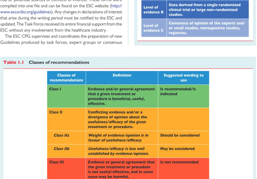
were included, where data exist. The level of evidence and the
strength of the recommendation of particular management options
were weighed and graded according to predefined scales, as
out-lined in Tables
1.1
and
1.2
.
The experts of the writing and reviewing panels provided
declara-tions of interest forms for all reladeclara-tionships that might be perceived as
real or potential sources of conflicts of interest. These forms were
compiled into one file and can be found on the ESC website (
http://
www.escardio.org/guidelines
). Any changes in declarations of interest
that arise during the writing period must be notified to the ESC and
updated. The Task Force received its entire financial support from the
ESC without any involvement from the healthcare industry.
The ESC CPG supervises and coordinates the preparation of new
Guidelines produced by task forces, expert groups or consensus
panels. The Committee is also responsible for the endorsement
pro-cess of these Guidelines. The ESC Guidelines undergo extensive
re-view by the CPG and external experts. After appropriate revisions
the Guidelines are approved by all the experts involved in the Task
Force. The finalized document is approved by the CPG for
publica-tion in the European Heart Journal. The Guidelines were developed
after careful consideration of the scientific and medical knowledge
and the evidence available at the time of their dating.
The task of developing ESC Guidelines covers not only integration
of the most recent research, but also the creation of educational tools
and implementation programmes for the recommendations. To
im-plement the guidelines, condensed pocket guidelines versions,
sum-mary slides, booklets with essential messages, sumsum-mary cards for
non-specialists, and an electronic version for digital applications
(smartphones, etc.) are produced. These versions are abridged and
thus, if needed, one should always refer to the full text version, which
is freely available on the ESC website. The National Cardiac Societies
of the ESC are encouraged to endorse, translate and implement all
ESC Guidelines. Implementation programmes are needed because
Table 1.1
Classes of recommendations
it has been shown that the outcome of disease may be favourably
in-fluenced by the thorough application of clinical recommendations.
Surveys and registries are needed to verify that real-life daily
prac-tice is in keeping with what is recommended in the guidelines, thus
completing the loop between clinical research, writing of guidelines,
disseminating them and implementing them into clinical practice.
Health professionals are encouraged to take the ESC Guidelines
fully into account when exercising their clinical judgment, as well as
in the determination and the implementation of preventive,
diagnos-tic or therapeudiagnos-tic medical strategies. However, the ESC Guidelines
do not override in any way whatsoever the individual responsibility
of health professionals to make appropriate and accurate decisions
in consideration of each patient’s health condition and in
consult-ation with that patient and the patient’s caregiver where appropriate
and/or necessary. It is also the health professional’s responsibility to
verify the rules and regulations applicable to drugs and devices at the
time of prescription.
2. Introduction
The aim of all the ESC Guidelines is to help health professionals to
make decisions in their everyday life based on the best available
evi-dence. We will soon be celebrating the 30th anniversary of clinical
trials that for the first time incontrovertibly demonstrated that the
miserable outcome of patients with heart failure (HF) can be
mark-edly improved.
2Since then, in the area of HF management we have
witnessed and celebrated numerous highs, which have definitely
outnumbered several lows, all of which have allowed us to unravel
the pathophysiology of this clinical syndrome, but more importantly
has led to better care of our patients.
3In the year 2016, no one
would any longer dispute that, by applying all evidence-based
dis-coveries, HF is now becoming a preventable and treatable disease.
The aim of this document is to provide practical, evidence-based
guidelines for the diagnosis and treatment of HF. The principal
changes from the 2012 guidelines relate to:
(i) a new term for patients with HF and a left ventricular ejection
fraction (LVEF) that ranges from 40 to 49% — ‘HF with
mid-range EF (HFmrEF)’; we believe that identifying HFmrEF as a
separate group will stimulate research into the underlying
char-acteristics, pathophysiology and treatment of this population;
(ii) clear recommendations on the diagnostic criteria for HF with
re-duced EF (HFrEF), HFmrEF and HF with preserved EF (HFpEF);
(iii) a new algorithm for the diagnosis of HF in the non-acute
set-ting based on the evaluation of HF probability;
(iv) recommendations aimed at prevention or delay of the
devel-opment of overt HF or the prevention of death before the
on-set of symptoms;
(v) indications for the use of the new compound sacubitril/
valsartan, the first in the class of angiotensin receptor
neprily-sin inhibitors (ARNIs);
(vi) modified indications for cardiac resynchronization therapy
(CRT);
(vii) the concept of an early initiation of appropriate therapy going
along with relevant investigations in acute HF that follows the
‘time to therapy’ approach already well established in acute
coronary syndrome (ACS);
(viii) a new algorithm for a combined diagnosis and treatment
ap-proach of acute HF based on the presence/absence of
conges-tion/hypoperfusion.
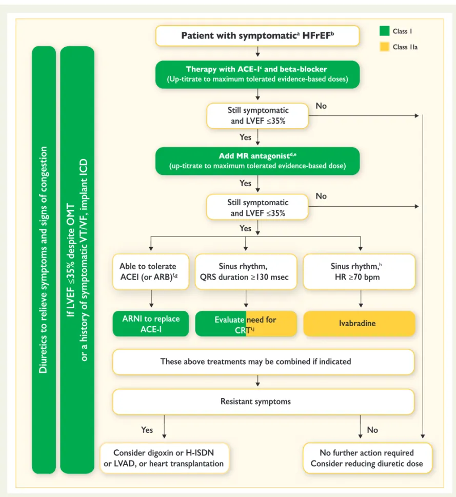
We followed the format of the previous ESC 2012 HF Guidelines.
Therapeutic recommendations state the treatment effect supported
by the class and level of recommendation in tabular format; in the
case of chronic HF due to left ventricular systolic dysfunction
(LVSD) the recommendations focus on mortality and morbidity
outcomes. Detailed summaries of the key evidence supporting
gen-erally recommended treatments have been provided. For diagnostic
recommendations a level of evidence C has been typically decided
upon, because for the majority of diagnostic tests there are no data
from randomized controlled trials (RCTs) showing that they will
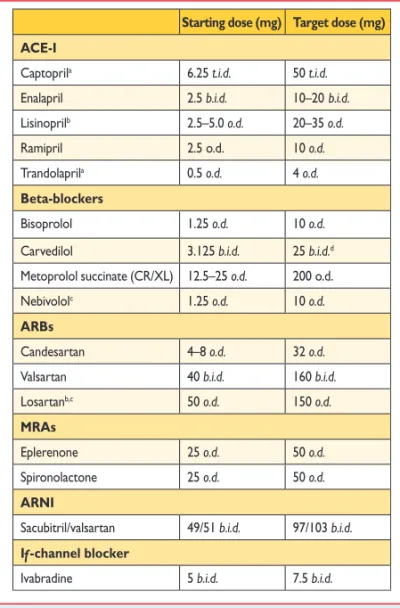
lead to reductions in morbidity and/or mortality. Practical guidance
is provided for the use of the important disease-modifying drugs and
diuretics. When possible, other relevant guidelines, consensus
statements and position papers have been cited to avoid unduly
lengthy text. All tables should be read in conjunction with their
ac-companying text and not read in isolation.
This document is the result of extensive interactions between the
Task Force, the review team and the ESC Committee for Practice
Guidelines. It represents a consensus of opinion of all of the experts
involved in its development. Concurrently to the development of
the 2016 ESC Guidelines on HF, the group writing the “2016
ACC/AHA/HFSA Focused Update on New Pharmacological
Ther-apy for Heart Failure” independently developed its
recommenda-tions on new pharmacotherapy for Heart Failure. Both working
groups/Task Force independently surveyed the evidence, arrived
at similar conclusions, and constructed similar, but not identical,
re-commendations. Given the concordance, the respective
organiza-tions simultaneously issued aligned recommendaorganiza-tions on the use
of these new treatments to minimize confusion and improve the
care of patients with HF.
3. Definition, epidemiology and
prognosis
3.1 Definition of heart failure
HF is a clinical syndrome characterized by typical symptoms
(e.g. breathlessness, ankle swelling and fatigue) that may be
accom-panied by signs (e.g. elevated jugular venous pressure, pulmonary
crackles and peripheral oedema) caused by a structural and/or
func-tional cardiac abnormality, resulting in a reduced cardiac output and/
or elevated intracardiac pressures at rest or during stress.
The current definition of HF restricts itself to stages at which clinical
symptoms are apparent. Before clinical symptoms become apparent,
patients can present with asymptomatic structural or functional cardiac
abnormalities [systolic or diastolic left ventricular (LV) dysfunction],
which are precursors of HF. Recognition of these precursors is
import-ant because they are related to poor outcomes, and starting treatment
at the precursor stage may reduce mortality in patients with
asymp-tomatic systolic LV dysfunction
4,5(for details see Section 6).
Demonstration of an underlying cardiac cause is central to the
diagnosis of HF. This is usually a myocardial abnormality causing
sys-tolic and/or diassys-tolic ventricular dysfunction. However,
abnormal-ities of the valves, pericardium, endocardium, heart rhythm and
conduction can also cause HF (and more than one abnormality is
of-ten present). Identification of the underlying cardiac problem is
cru-cial for therapeutic reasons, as the precise pathology determines the
specific treatment used (e.g. valve repair or replacement for valvular
disease, specific pharmacological therapy for HF with reduced EF,
reduction of heart rate in tachycardiomyopathy, etc).
3.2 Terminology
3.2.1 Heart failure with preserved, mid-range and reduced
ejection fraction
The main terminology used to describe HF is historical and is based
on measurement of the LVEF. HF comprises a wide range of
pa-tients, from those with normal LVEF [typically considered as
≥50%; HF with preserved EF (HFpEF)] to those with reduced
LVEF [typically considered as ,40%; HF with reduced EF (HFrEF)]
(Table
3.1
). Patients with an LVEF in the range of 40 – 49% represent
a ‘grey area’, which we now define as HFmrEF (Table
3.1
).
Differen-tiation of patients with HF based on LVEF is important due to
different underlying aetiologies, demographics, co-morbidities and
response to therapies.
6Most clinical trials published after 1990
se-lected patients based on LVEF [usually measured using
echocardiog-raphy, a radionuclide technique or cardiac magnetic resonance
(CMR)], and it is only in patients with HFrEF that therapies have
been shown to reduce both morbidity and mortality.
The diagnosis of HFpEF is more challenging than the diagnosis of
HFrEF. Patients with HFpEF generally do not have a dilated LV, but
instead often have an increase in LV wall thickness and/or increased
left atrial (LA) size as a sign of increased filling pressures. Most have
additional ‘evidence’ of impaired LV filling or suction capacity, also
classified as diastolic dysfunction, which is generally accepted as
the likely cause of HF in these patients (hence the term ‘diastolic
HF’). However, most patients with HFrEF (previously referred to
as ‘systolic HF’) also have diastolic dysfunction, and subtle
abnormal-ities of systolic function have been shown in patients with HFpEF.
Hence the preference for stating preserved or reduced LVEF over
preserved or reduced ‘systolic function’.
In previous guidelines it was acknowledged that a grey area exists
between HFrEF and HFpEF.
7These patients have an LVEF that
ranges from 40 to 49%, hence the term HFmrEF. Identifying HFmrEF
as a separate group will stimulate research into the underlying
characteristics, pathophysiology and treatment of this group of
pa-tients. Patients with HFmrEF most probably have primarily mild
sys-tolic dysfunction, but with features of diassys-tolic dysfunction
(Table
3.1
).
Patients without detectable LV myocardial disease may have
other cardiovascular causes for HF (e.g. pulmonary hypertension,
valvular heart disease, etc.). Patients with non-cardiovascular
path-ologies (e.g. anaemia, pulmonary, renal or hepatic disease) may have
symptoms similar or identical to those of HF and each may
compli-cate or exacerbate the HF syndrome.
3.2.2 Terminology related to the time course of heart
failure
In these guidelines, the term HF is used to describe the symptomatic
syndrome, graded according to the New York Heart Association
(NYHA) functional classification (see Section 3.2.3 and Web
Table 3.2), although a patient can be rendered asymptomatic by
treatment. In these guidelines, a patient who has never exhibited
the typical symptoms and/or signs of HF and with a reduced LVEF
is described as having asymptomatic LV systolic dysfunction. Patients
who have had HF for some time are often said to have ‘chronic HF’.
A treated patient with symptoms and signs that have remained
gen-erally unchanged for at least 1 month is said to be ‘stable’. If chronic
stable HF deteriorates, the patient may be described as
‘decompen-sated’ and this may happen suddenly or slowly, often leading to
hos-pital admission, an event of considerable prognostic importance.
New-onset (‘de novo’) HF may also present acutely, for example,
as a consequence of acute myocardial infarction (AMI), or in a
sub-acute (gradual) fashion, for example, in patients with a dilated
cardio-myopathy (DCM), who often have symptoms for weeks or months
before the diagnosis becomes clear. Although symptoms and signs
of HF may resolve, the underlying cardiac dysfunction may not, and
patients remain at the risk of recurrent ‘decompensation’.
Occasionally, however, a patient may have HF due to a problem
that resolves completely (e.g. acute viral myocarditis, takotsubo
car-diomyopathy or tachycarcar-diomyopathy). Other patients, particularly
those with ‘idiopathic’ DCM, may also show substantial or even
complete recovery of LV systolic function with modern
disease-modifying therapy [including angiotensin-converting enzyme
inhibi-tor (ACEI), beta-blocker, mineralocorticoid recepinhibi-tor antagonist
Table 3.1
Definition of heart failure with preserved (HFpEF), mid-range (HFmrEF) and reduced ejection fraction
(HFrEF)
BNP ¼ B-type natriuretic peptide; HF ¼ heart failure; HFmrEF ¼ heart failure with mid-range ejection fraction; HFpEF ¼ heart failure with preserved ejection fraction; HFrEF ¼ heart failure with reduced ejection fraction; LAE ¼ left atrial enlargement; LVEF ¼ left ventricular ejection fraction; LVH ¼ left ventricular hypertrophy; NT-proBNP ¼ N-terminal pro-B type natriuretic peptide.
a
Signs may not be present in the early stages of HF (especially in HFpEF) and in patients treated with diuretics. b
(MRA), ivabradine and/or CRT]. ‘Congestive HF’ is a term that is
sometimes used, and may describe acute or chronic HF with
evi-dence of volume overload. Many or all of these terms may be
accur-ately applied to the same patient at different times, depending upon
their stage of illness.
3.2.3 Terminology related to the symptomatic severity
of heart failure
The NYHA functional classification (Web Table 3.2) has been used
to describe the severity of symptoms and exercise intolerance.
However, symptom severity correlates poorly with many measures
of LV function; although there is a clear relationship between the
se-verity of symptoms and survival, patients with mild symptoms may
still have an increased risk of hospitalization and death.
8–10Sometimes the term ‘advanced HF’ is used to characterize
pa-tients with severe symptoms, recurrent decompensation and severe
cardiac dysfunction.
11The American College of Cardiology
Founda-tion/American Heart Association (ACCF/AHA) classification
de-scribes stages of HF development based on structural changes and
symptoms (Web Table 3.3).
12The Killip classification may be used to
describe the severity of the patient’s condition in the acute setting
after myocardial infarction (see Section 12).
133.3 Epidemiology, aetiology and natural
history of heart failure
The prevalence of HF depends on the definition applied, but is
ap-proximately 1 – 2% of the adult population in developed countries,
rising to
≥10% among people .70 years of age.
14–17Among
peo-ple .65 years of age presenting to primary care with breathlessness
on exertion, one in six will have unrecognized HF (mainly
HFpEF).
18,19The lifetime risk of HF at age 55 years is 33% for
men and 28% for women.
16The proportion of patients with HFpEF
ranges from 22 to 73%, depending on the definition applied, the
clin-ical setting (primary care, hospital clinic, hospital admission), age and
sex of the studied population, previous myocardial infarction and
the year of publication.
17,18,20–30Data on temporal trends based on hospitalized patients suggest
that the incidence of HF may be decreasing, more for HFrEF than
for HFpEF.
31,32HFpEF and HFrEF seem to have different
epidemio-logical and aetioepidemio-logical profiles. Compared with HFrEF, patients
with HFpEF are older, more often women and more commonly
have a history of hypertension and atrial fibrillation (AF), while a
his-tory of myocardial infarction is less common.
32,33The characteristics
of patients with HFmrEF are between those with HFrEF and HFpEF,
34but further studies are needed to better characterize this population.
The aetiology of HF is diverse within and among world regions.
There is no agreed single classification system for the causes of
HF, with much overlap between potential categories (Table
3.4
).
Many patients will have several different
pathologies—cardiovascu-lar and non-cardiovascupathologies—cardiovascu-lar—that conspire to cause HF.
Identifica-tion of these diverse pathologies should be part of the diagnostic
workup, as they may offer specific therapeutic opportunities.
Many patients with HF and ischaemic heart disease (IHD) have a
history of myocardial infarction or revascularization. However, a
normal coronary angiogram does not exclude myocardial scar
(e.g. by CMR imaging) or impaired coronary microcirculation as
al-ternative evidence for IHD.
In clinical practice, a clear distinction between acquired and
inher-ited cardiomyopathies remains challenging. In most patients with a
definite clinical diagnosis of HF, there is no confirmatory role for
routine genetic testing, but genetic counselling is recommended in
patients with hypertrophic cardiomyopathy (HCM), ‘idiopathic’
DCM or arrhythmogenic right ventricular cardiomyopathy
(ARVC) (see Section 5.10.1), since the outcomes of these tests
may have clinical implications.
Over the last 30 years, improvements in treatments and their
im-plementation have improved survival and reduced the hospitalization
rate in patients with HFrEF, although the outcome often remains
un-satisfactory. The most recent European data (ESC-HF pilot study)
demonstrate that 12-month all-cause mortality rates for hospitalized
and stable/ambulatory HF patients were 17% and 7%, respectively,
and the 12-month hospitalization rates were 44% and 32%,
respect-ively.
35In patients with HF (both hospitalized and ambulatory), most
deaths are due to cardiovascular causes, mainly sudden death and
worsening HF. All-cause mortality is generally higher in HFrEF than
HFpEF.
35,36Hospitalizations are often due to non-cardiovascular
causes, particularly in patients with HFpEF. Hospitalization for
cardio-vascular causes did not change from 2000 to 2010, whereas those
with non-cardiovascular causes increased.
313.4 Prognosis
Estimation of prognosis for morbidity, disability and death helps
pa-tients, their families and clinicians decide on the appropriate type
and timing of therapies (in particular, decisions about a rapid
transi-tion to advanced therapies) and assists with planning of health and
social services and resources.
Numerous prognostic markers of death and/or HF hospitalization
have been identified in patients with HF (Web Table 3.5). However,
their clinical applicability is limited and precise risk stratification in
HF remains challenging.
In recent decades, several multivariable prognostic risk scores
have been developed for different populations of patients with
HF,
36–41and some are available as interactive online applications.
Multivariable risk scores may help predict death in patients with
HF, but remain less useful for the prediction of subsequent HF
hos-pitalizations.
37,38A systematic review examining 64 prognostic
models
37along with a meta-analysis and meta-regression study of
117 prognostic models
38revealed only a moderate accuracy of
models predicting mortality, whereas models designed to predict
the combined endpoint of death or hospitalization, or only
hospital-ization, had an even poorer discriminative ability.
4. Diagnosis
4.1 Symptoms and signs
Symptoms are often non-specific and do not, therefore, help
discrim-inate between HF and other problems (Table
4.1
).
42–46Symptoms and
signs of HF due to fluid retention may resolve quickly with diuretic
therapy. Signs, such as elevated jugular venous pressure and
displace-ment of the apical impulse, may be more specific, but are harder to
detect and have poor reproducibility.
18,46,47Symptoms and signs
may be particularly difficult to identify and interpret in obese
indivi-duals, in the elderly and in patients with chronic lung disease.
48–50Younger patients with HF often have a different aetiology, clinical
pres-entation and outcome compared with older patients.
51,52A detailed history should always be obtained. HF is unusual in an
individual with no relevant medical history (e.g. a potential cause of
cardiac damage), whereas certain features, particularly previous
myocardial infarction, greatly increase the likelihood of HF in a
pa-tient with appropriate symptoms and signs.
42–45At each visit, symptoms and signs of HF need to be assessed, with
particular attention to evidence of congestion. Symptoms and signs
are important in monitoring a patient’s response to treatment and
stability over time. Persistence of symptoms despite treatment
usu-ally indicates the need for additional therapy, and worsening of
symptoms is a serious development (placing the patient at risk of
ur-gent hospital admission and death) and merits prompt medical
attention.
4.2 Essential initial investigations:
natriuretic peptides, electrocardiogram
and echocardiography
The plasma concentration of natriuretic peptides (NPs) can be used
as an initial diagnostic test, especially in the non-acute setting when
echocardiography is not immediately available. Elevated NPs help
establish an initial working diagnosis, identifying those who require
further cardiac investigation; patients with values below the
cut-point for the exclusion of important cardiac dysfunction do not
require echocardiography (see also Section 4.3 and Section 12).
Patients with normal plasma NP concentrations are unlikely to
have HF. The upper limit of normal in the non-acute setting for
Table 3.4
Aetiologies of heart failure
DISEASED MYOCARDIUM
Ischaemic heart disease
Myocardial scar
Myocardial stunning/hibernation Epicardial coronary artery disease Abnormal coronary microcirculation Endothelial dysfunction
Toxic damage Recreational substance abuse Alcohol, cocaine, amphetamine, anabolic steroids. Heavy metals Copper, iron, lead, cobalt.
Medications Cytostatic drugs (e.g. anthracyclines), immunomodulating drugs (e.g. interferons monoclonal antibodies such as trastuzumab, cetuximab), antidepressant drugs, antiarrhythmics, non-steroidal Radiation
Immune-mediated damage
Related to infection Bacteria, spirochaetes, fungi, protozoa, parasites (Chagas disease), rickettsiae, viruses (HIV/AIDS). Not related to infection Lymphocytic/giant cell myocarditis, autoimmune diseases (e.g. Graves’ disease, rheumatoid
arthritis, connective tissue disorders, mainly systemic lupus erythematosus), hypersensitivity and eosinophilic myocarditis (Churg–Strauss).
Related to malignancy
Not related to malignancy Amyloidosis, sarcoidosis, haemochromatosis (iron), glycogen storage diseases (e.g. Pompe disease), lysosomal storage diseases (e.g. Fabry disease).
Metabolic derangements
Hormonal
disease, Addison disease, diabetes, metabolic syndrome, phaeochromocytoma, pathologies related to pregnancy and peripartum.
Nutritional
(e.g. malignancy, AIDS, anorexia nervosa), obesity.
Genetic abnormalities Diverse forms HCM, DCM, LV non-compaction, ARVC, restrictive cardiomyopathy (for details see respective expert documents), muscular dystrophies and laminopathies.
ABNORMAL LOADING CONDITIONS
Hypertension Valve and myocardium structural defects
Acquired Mitral, aortic, tricuspid and pulmonary valve diseases.
Congenital Atrial and ventricular septum defects and others (for details see a respective expert document). Pericardial and
endomyocardial pathologies
Pericardial Constrictive pericarditis Pericardial effusion Endomyocardial
High output states Volume overload
ARRHYTHMIAS
Tachyarrhythmias Atrial, ventricular arrhythmias.
Bradyarrhythmias Sinus node dysfunctions, conduction disorders.
ARVC ¼ arrhythmogenic right ventricular cardiomyopathy; DCM ¼ dilated cardiomyopathy; EMF ¼ endomyocardial fibrosis; GH ¼ growth hormone; HCM ¼ hypertrophic cardiomyopathy; HES ¼ hypereosinophilic syndrome; HIV/AIDS ¼ human immunodeficiency virus/acquired immune deficiency syndrome; LV ¼ left ventricular.
B-type natriuretic peptide (BNP) is 35 pg/mL and for N-terminal
pro-BNP (NT-proBNP) it is 125 pg/mL; in the acute setting, higher
values should be used [BNP , 100 pg/mL, NT-proBNP , 300 pg/
mL and mid-regional pro A-type natriuretic peptide (MR-proANP)
, 120 pmol/L]. Diagnostic values apply similarly to HFrEF and
HFpEF; on average, values are lower for HFpEF than for HFrEF.
54,55At the mentioned exclusionary cut-points, the negative predictive
values are very similar and high (0.94 – 0.98) in both the non-acute
and acute setting, but the positive predictive values are lower
both in the non-acute setting (0.44 – 0.57) and in the acute setting
(0.66 – 0.67).
54,56–61Therefore, the use of NPs is recommended
for ruling-out HF, but not to establish the diagnosis.
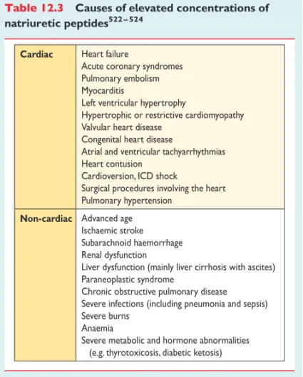
There are numerous cardiovascular and non-cardiovascular
causes of elevated NPs that may weaken their diagnostic utility in
HF. Among them, AF, age and renal failure are the most important
factors impeding the interpretation of NP measurements.
55On the
other hand, NP levels may be disproportionally low in obese
pa-tients
62(see also Section 12.2 and Table
12.3
).
An abnormal electrocardiogram (ECG) increases the likelihood
of the diagnosis of HF, but has low specificity.
18,46,63,64Some
abnor-malities on the ECG provide information on aetiology (e.g.
myocar-dial infarction), and findings on the ECG might provide indications
for therapy (e.g. anticoagulation for AF, pacing for bradycardia,
CRT if broadened QRS complex) (see Sections 8 and 10). HF is
un-likely in patients presenting with a completely normal ECG
(sensitiv-ity 89%).
43Therefore, the routine use of an ECG is mainly
recommended to rule out HF.
Echocardiography is the most useful, widely available test in
pa-tients with suspected HF to establish the diagnosis. It provides
im-mediate information on chamber volumes, ventricular systolic and
diastolic function, wall thickness, valve function and pulmonary
hypertension.
65–74This information is crucial in establishing the
diagnosis and in determining appropriate treatment (see Sections
5.2 – 5.4 for details on echocardiography).
The information provided by careful clinical evaluation and the
above mentioned tests will permit an initial working diagnosis and
treatment plan in most patients. Other tests are generally required
only if the diagnosis remains uncertain (e.g. if echocardiographic
images are suboptimal or an unusual cause of HF is suspected)
(for details see Sections 5.5 – 5.10).
4.3 Algorithm for the diagnosis of heart
failure
4.3.1 Algorithm for the diagnosis of heart failure in the
non-acute setting
An algorithm for the diagnosis of HF in the non-acute setting is
shown in Figure
4.1
. The diagnosis of HF in the acute setting is
discussed in Section 12.
For patients presenting with symptoms or signs for the first time,
non-urgently in primary care or in a hospital outpatient clinic
(Table
4.1
), the probability of HF should first be evaluated based
on the patient’s prior clinical history [e.g. coronary artery disease
(CAD), arterial hypertension, diuretic use], presenting symptoms
(e.g. orthopnoea), physical examination (e.g. bilateral oedema,
in-creased jugular venous pressure, displaced apical beat) and resting
ECG. If all elements are normal, HF is highly unlikely and other
diag-noses need to be considered. If at least one element is abnormal,
plasma NPs should be measured, if available, to identify those
who need echocardiography (an echocardiogram is indicated if
the NP level is above the exclusion threshold or if circulating NP
levels cannot be assessed).
55–60,75–784.3.2 Diagnosis of heart failure with preserved ejection
fraction
The diagnosis of HFpEF remains challenging. LVEF is normal and
signs and symptoms for HF (Table
4.1
) are often non-specific and
do not discriminate well between HF and other clinical conditions.
This section summarizes practical recommendations necessary for
proper diagnosis of this clinical entity in clinical practice.
The diagnosis of chronic HFpEF, especially in the typical elderly
patient with co-morbidities and no obvious signs of central fluid
overload, is cumbersome and a validated gold standard is missing.
To improve the specificity of diagnosing HFpEF, the clinical diagnosis
needs to be supported by objective measures of cardiac dysfunction
at rest or during exercise. The diagnosis of HFpEF requires the
fol-lowing conditions to be fulfilled (see Table
3.1
):
† The presence of symptoms and/or signs of HF (see Table
4.1
)
† A ‘preserved’ EF (defined as LVEF
≥50% or 40–49% for
HFmrEF)
† Elevated levels of NPs (BNP .35 pg/mL and/or NT-proBNP
.125 pg/mL)
† Objective evidence of other cardiac functional and structural
al-terations underlying HF (for details, see below)
Table 4.1
Symptoms and signs typical of heart failure
Symptoms
Signs
Typical
Breathlessness
Orthopnoea
Paroxysmal nocturnal dyspnoea
Reduced exercise tolerance
Fatigue, tiredness, increased time
to recover after exercise
Ankle swelling
Elevated jugular venous pressure
Third heart sound (gallop rhythm)
Laterally displaced apical impulse
Less typical
Nocturnal cough
Wheezing
Bloated feeling
Loss of appetite
Confusion (especially in the
elderly)
Depression
Palpitations
Dizziness
Syncope
Bendopnea
53Weight gain (>2 kg/week)
Weight loss (in advanced HF)
Tissue wasting (cachexia)
Cardiac murmur
Peripheral oedema (ankle, sacral,
scrotal)
Pulmonary crepitations
Reduced air entry and dullness to
percussion at lung bases (pleural
effusion)
Tachycardia
Irregular pulse
Tachypnoea
Cheyne Stokes respiration
Hepatomegaly
Ascites
Cold extremities
Oliguria
Narrow pulse pressure
Figure 4.1
Diagnostic algorithm for a diagnosis of heart failure of non-acute onset
BNP ¼ B-type natriuretic peptide; CAD ¼ coronary artery disease; HF ¼ heart failure; MI ¼ myocardial infarction; NT-proBNP ¼ N-terminal
pro-B type natriuretic peptide.
a