HAL Id: hal-02988643
https://hal.archives-ouvertes.fr/hal-02988643
Submitted on 4 Nov 2020
HAL is a multi-disciplinary open access
archive for the deposit and dissemination of
sci-entific research documents, whether they are
pub-lished or not. The documents may come from
teaching and research institutions in France or
abroad, or from public or private research centers.
L’archive ouverte pluridisciplinaire HAL, est
destinée au dépôt et à la diffusion de documents
scientifiques de niveau recherche, publiés ou non,
émanant des établissements d’enseignement et de
recherche français ou étrangers, des laboratoires
publics ou privés.
To cite this version:
Rémy Char, Philippe Pierre. The RUFYs, a Family of Effector Proteins Involved in Intracellular
Trafficking and Cytoskeleton Dynamics. Frontiers in Cell and Developmental Biology, Frontiers media,
2020, 8, �10.3389/fcell.2020.00779�. �hal-02988643�
doi: 10.3389/fcell.2020.00779
Edited by: Roberto Botelho, Ryerson University, Canada Reviewed by: Daniel G. S. Capelluto, Virginia Tech, United States Brian Paul Ceresa, University of Louisville, United States *Correspondence: Rémy Char char@ciml.univ-mrs.fr Philippe Pierre pierre@ciml.univ-mrs.fr Specialty section: This article was submitted to Membrane Traffic, a section of the journal Frontiers in Cell and Developmental Biology Received: 11 June 2020 Accepted: 24 July 2020 Published: 11 August 2020 Citation: Char R and Pierre P (2020) The RUFYs, a Family of Effector Proteins Involved in Intracellular Trafficking and Cytoskeleton Dynamics. Front. Cell Dev. Biol. 8:779. doi: 10.3389/fcell.2020.00779
The RUFYs, a Family of Effector
Proteins Involved in Intracellular
Trafficking and Cytoskeleton
Dynamics
Rémy Char
1* and Philippe Pierre
1,2,3*
1Aix Marseille Université, Centre National de la Recherche Scientifique, Institut National de la Santé et de la Recherche
Médicale, Centre d’Immunologie de Marseille-Luminy, Marseille, France,2Institute for Research in Biomedicine and Ilidio
Pinho Foundation, Department of Medical Sciences, University of Aveiro, Aveiro, Portugal,3Shanghai Institute
of Immunology, School of Medicine, Shanghai Jiao Tong University, Shanghai, China
Intracellular trafficking is essential for cell structure and function. In order to perform
key tasks such as phagocytosis, secretion or migration, cells must coordinate their
intracellular trafficking, and cytoskeleton dynamics. This relies on certain classes of
proteins endowed with specialized and conserved domains that bridge membranes
with effector proteins. Of particular interest are proteins capable of interacting with
membrane subdomains enriched in specific phosphatidylinositol lipids, tightly regulated
by various kinases and phosphatases. Here, we focus on the poorly studied RUFY family
of adaptor proteins, characterized by a RUN domain, which interacts with small
GTP-binding proteins, and a FYVE domain, involved in the recognition of phosphatidylinositol
3-phosphate. We report recent findings on this protein family that regulates endosomal
trafficking, cell migration and upon dysfunction, can lead to severe pathology at the
organismal level.
Keywords: RUFY, cancer, neurodegenerative diseases, immunity, RUN, FYVE, phosphatidylinositol 3-phosphate, cytoskeleton
INTRODUCTION
The organization of cells into multiple membranous compartments with specific biochemical
functions requires complex intracellular traffic and sorting of lipids and proteins, to transport them
from their sites of synthesis to their functional destination. Intracellular transport involves lipid
vesicles or tubules with the capacity to fuse with one another or to be secreted. They collectively
participate in the dynamic exchanges necessary for cell homeostasis (
Rothman, 2002
;
Søreng
et al., 2018
). Membrane traffic is tightly coordinated with protein synthesis, signal transduction of
environmental stimuli and cytoskeleton organization, allowing the implementation of key cellular
functions such as endocytosis, exocytosis, or migration (
McMahon and Gallop, 2005
;
Habtezion
et al., 2016
;
Vega-Cabrera and Pardo-López, 2017
;
MacGillavry and Hoogenraad, 2018
;
Margaria
et al., 2019
;
Tapia et al., 2019
;
Buratta et al., 2020
;
Stalder and Gershlick, 2020
).
Several families of molecular components required for orchestrating membrane vesicle exchange
and transport during this process are conserved. They include adaptor and coat proteins, small
GTP-binding proteins (GTPases), as well as Synaptosome Associated Protein (SNAP) Receptor
(SNARE) proteins and SNARE binding proteins (
Juliano, 2018
).
The vast superfamily of GTPases is involved in the establishment
or regulation of virtually every step of intracellular membrane
trafficking. They behave as molecular switches that can alternate
between active and inactive states, through GTP binding and
hydrolysis into GDP (
Takai et al., 2001
;
Stenmark, 2009
). The
largest group of GTPases involved in intracellular membrane
traffic is the Rab proteins family (
Lamb et al., 2016
). Rab GTPases
specifically localize to different intracellular compartments,
regulating vesicle formation and sorting, as well as transport
along the cytoskeletal network. Each Rab protein can be recruited
to specific membrane subdomains of a defined organelle and
is associated to multiple effectors controlling membrane fusion
and trafficking. Rab interaction with the membrane fusion
complexes and cytoskeleton regulators is therefore crucial for
cellular functions, including endocytosis and autophagy (
Chen
and Wandinger-Ness, 2001
;
Bruce et al., 2010
;
Geng et al., 2010
;
Thomas and Fromme, 2020
;
Yuan and Song, 2020
).
Here, we review the literature concerning a less-well known
family of proteins involved in the complex biochemical crosstalk
established between the cytoskeleton and intracellular vesicles.
This small group of proteins was named RUFY for “RUN and
FYVE domain-containing.” RUFYs share a common structural
domain organization, including an N-terminal RUN domain,
one or several coiled-coil (CC) repeats and a C-terminal FYVE
domain (Figure 1A). The molecular structures of the different
RUFY proteins has been described (
Dunkelberg and
Gutierrez-Hartmann, 2001
;
Mari et al., 2001
;
Kukimoto-Niino et al., 2006
;
Kitagishi and Matsuda, 2013
), but their function in endocytic
regulation and their physiological relevance at the organismal
level are still poorly characterized (
Kitagishi and Matsuda, 2013
;
Terawaki et al., 2016
). We revisit here how the
rufy gene
family was annotated, and propose the addition of a novel
member, the
fyco1 (FYVE and Coiled-coil containing domain
1) gene given its sequence and functional similarities with
the other
rufy genes (
Pankiv et al., 2010
;
Terawaki et al.,
2015
). We also highlight recent findings on the implication of
RUFY proteins in the regulation of cytoskeleton and endosome
dynamics and their contribution to immunity, cancer and
neurodegenerative diseases.
Endocytosis and Autophagy
Endocytosis and autophagy are membrane traffic pathways
required for degradation and recycling of extracellular
and intracellular components, respectively (
Birgisdottir and
Johansen, 2020
). These pathways have a common endpoint at the
lysosome, where their cargo is degraded. These both pathways
intersect at several stages throughout vesicle formation, transport
and fusion and share some of the components of their molecular
machineries (Figure 1B).
There are numerous co-existing endocytic pathways, which
initiate by the formation of nascent endocytic vesicles formed
from plasma membrane invaginations and scissions. These
endocytic vesicles undergo homotypic fusion and are rapidly
targeted to sorting endosomes (SE). Sorting events initiated in
SE determine the fate of internalized cargo molecules, such as
recycling to plasma membrane, degradation in lysosomes, or
other trafficking events (
Naslavsky and Caplan, 2018
; Figure 1B).
On their way to degradation, sorted cargo accumulate in
early endosomes (EE), that further mature into late endosomes
(LE) through multiple events of cargo and lipid sorting.
Late endosomes adopt a membrane organization termed
multivesicular bodies, that are enriched in lysobisphosphatidic
acid and contain intraluminal vesicles (
Gruenberg, 2020
). Next,
LE potentiate their hydrolytic competence by fusing with
lysosomes (
Pillay et al., 2002
) resulting in the degradation
of their contents, providing nutrients and key factors to
the cell (
Doherty and McMahon, 2009
;
Kaksonen and Roux,
2018
). Notably, endosomes play a role in signal transduction
by serving as signaling platforms either for surface activated
receptors like Toll-like receptors and epidermal growth factor
receptor or metabolic sensors such as mechanistic target
of rapamycin complex 1 (mTORC1;
Argüello et al., 2016
).
Often they promote the degradation of their targets, leading
to signal termination (
Chung et al., 2010
). The endocytic
pathway has also specialized functions in differentiated cells
such as neurotransmitter release and recycling in neurons, or
antigen processing and presentation in professional antigen
presenting cells, like B cells or dendritic cells (
Argüello
et al., 2016
;
Solé-Domènech et al., 2016
;
Hinze and Boucrot,
2018
). Endocytosis events and endosomes positioning is
highly dependent on the dynamic and spatial re-organization
of the different cytoskeleton networks that include actin,
intermediate filaments, or microtubules (
Fletcher and Mullins,
2010
;
Pegoraro et al., 2017
).
Complementary to endocytosis, autophagy is an intracellular
process by which cells degrade and recycle their own cytoplasmic
materials (
Mizushima and Komatsu, 2011
). Autophagy plays a
central role in many physiological processes including stress
management, development, immunity and aging (
Puleston and
Simon, 2014
;
Zhong et al., 2016
;
Fîlfan et al., 2017
;
Moretti
et al., 2017
;
Doherty and Baehrecke, 2018
). Autophagy is
partially controlled though mTORC1 activity and is responsible
for degradation and recycling of misfolded proteins, as well
as obsolete organelles (
Galluzzi et al., 2017
). The endpoint of
autophagy is to deliver cytoplasmic material to lysosomes, where
like for endocytosed cargo, it is degraded. Several autophagy
processes can be distinguished based on the entry mode of
the cytosolic components destined for degradation (Figure 1B).
Macroautophagy involves engulfment of cytoplasmic contents
into a double membrane vesicle termed the autophagosome.
The autophagosome fuses then with lysosomes, becoming
an autolysosome, in which its cargo is degraded (
Galluzzi
et al., 2017
). The presence of specific phosphoinositides
lipids, together with Rab GTPases, at a given membrane
compartment is often directly correlated with compartment
function. One of the common mechanism regulating endocytosis
and autophagy is an accumulation of phosphatidylinositol
3-phosphate (PtdIns(3)P) at surface of EE and on intraluminal
vesicles of multivesicular endosomes and on autophagosomes
(
Nascimbeni et al., 2017
; Figure 1B). PtdIns(3)P is also observed
at sites of LC3−associated phagocytosis another pathway of
internalization used by the cells to ingest large particulate
material or microbes. PtdIns(3)P is therefore a beacon used by the
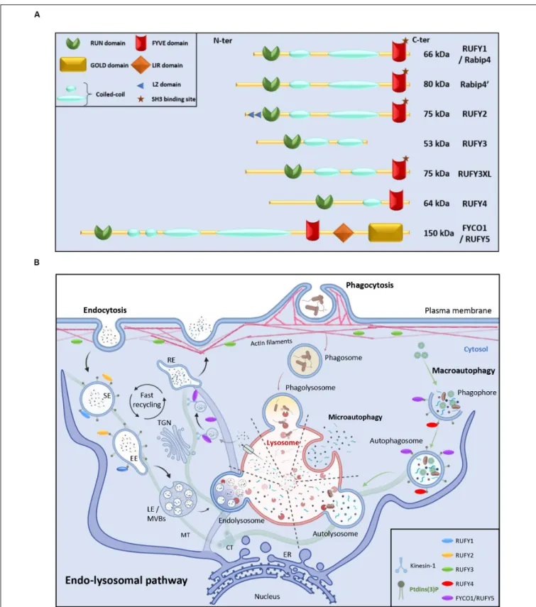
FIGURE 1 | RUN and FYVE domain containing-proteins in the endo-lysosomal pathway. (A) Schematic representation of the RUFY proteins family. (B) Description of the endo-lysosomal and autophagy pathways and presumed functional locations of RUFY proteins. Extracellular material is ingested by endocytosis or
phagocytosis. The action of different endosomes allows cargo to be sorted, recycled or degraded in a complex and regulated process involving fusion, maturation and transport along the cytoskeleton. Alternatively, during autophagy, obsolete components present in cytosol are captured in autophagosomes prior fusion with lysosomes and degradation (macroautophagy) or directly internalized through endosomal invagination (microautophagy). SE, sorting endosome; EE, early endosome; TGN, trans golgi network; LE, late endosome; MVBs, multi vesicular bodies; RE, recycling endosome, MT, microtubule; CT, centrioles; ER, endoplasmic reticulum. The location of PI3P and RUFY proteins known activity is shown. Created with BIoRender.com.
cellular machinery to regulate endosomal sorting and autophagy
(
Birgisdottir and Johansen, 2020
).
RUN Domains
The presence of a single copy of a RUN and a FYVE domain
at their extremities is the key characteristic defining the RUFY
family members. RUN domains were named after three proteins
bearing similar peptide motifs, RPIP8, UNC-14 and NESCA
(new molecule containing SH3 at the carboxy−terminus) (
Ogura
et al., 1997
;
Matsuda et al., 2000
). RUN domains are present in
multiple proteins (RUN proteins) in a large panel of organisms
(Figure 2) and principally allow direct interactions with small
GTPases of the Rap and Rab families (
Callebaut et al., 2001
;
Yoshida et al., 2011
). RUN domains adopt a hydrophobic
globular structure bearing six conserved blocks named A to F
(Figure 3A). These blocks correspond to eight
α-helices and
some 3
10-helices. The first helix is crucial to limit hydrophobic
exposure and maintain protein solubility of RUN-containing
proteins (
Callebaut et al., 2001
;
Kukimoto-Niino et al., 2006
).
In spite of strong conservation among the domains present
in RUN-containing proteins, the proteins they interact with,
their effectors, are highly variable (
Mari et al., 2001
) and the
structural features of the RUN domain alone are not sufficient
to define binding specificity for one or several members of the
GTPase superfamily (
Fukuda et al., 2011
). Most RUN
domain-bearing proteins bind small GTPases, but interactions with other
molecules like kinesin 1 have also been described (
Boucrot
et al., 2005
). A direct physical link between RUN proteins with
actin filaments and microtubules has been also demonstrated
(
Torti et al., 1999
), reinforcing the idea that these molecules are
also critical for cellular functions requiring actin remodeling,
such as migration or phagocytosis (
Price and Bos, 2004
;
Bos,
2005
;
Miertzschke et al., 2007
;
Xu et al., 2007
; Figure 4A).
Additional functions for RUN domains have been described,
for example for the RUN domain present in NESCA, which
blocks TRAF6-mediated polyubiquitination of the NF-kappa-B
essential modulator and consequently induces NF-kB activation.
This is just one of the ways in which RUN proteins can
act in signal transduction and the coordination of membrane
traffic with actin dynamics upon external stimulation (
Yoshida
et al., 2011
). As well as promoting endosomal fusion through
their binding to Rab or Rap GTPases (
Callebaut et al., 2001
;
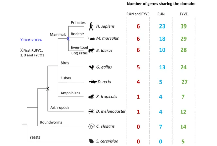
FIGURE 2 | Evolution of RUN and FYVE domain or rufy genes among living organisms. Diagram illustrating the evolution of the rufy genes. Species representative of various taxonomic groups are listed, data were extracted from the Differential Expression Atlas Genes database (EMBL-EBI). Next to each species studied, the number corresponds to the number of genes having in its sequence a FYVE (green), RUN (blue) or both (red) domain. The “X” corresponds to the appearance of a common rufy ancestor gene.
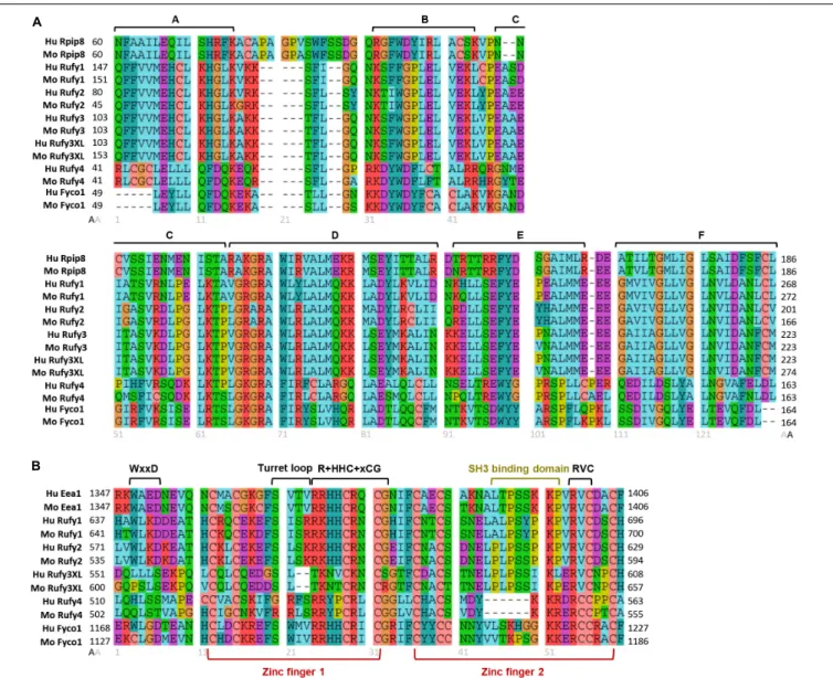
FIGURE 3 | Molecular organization of RUN and FYVE domains from the RUFY proteins family. Alignment of the protein sequences of the RUN (A) and FYVE (B) domains of the RUFY proteins family in human and mouse. (A) RUN consensus blocks are represented by segments (A–F). Rpip8 sequence is used as RUN domain reference, (B) FYVE conserved motives and zinc fingers are represented by segments. In the alignment, “x” is any amino acid and “+” represents positively charged amino acid. Eea1 sequence is used as FYVE domain reference. For all alignment, amino acids are colored according to their properties: Cyan for hydrophobic positions (A, V, I, L, M), turquoise for aromatic positions (F, Y, W, H), red for basic residues (K, R), purple for acidic residues (D, E), green for polar uncharged (N, Q, S, T), salmon for cysteine (C), orange for glycine (G) and yellow for proline (P). Gray numbers below alignment means the amino acids position after alignment. Black numbers surrounding the alignments represent the start (left) and end (right) positions of the domains in the peptide sequence of each protein. Alignment were realized with Seaviewer analyzer software (Gouy et al., 2010). Accession numbers for protein are following: human Rpip8 (NP_001138297.1), mouse Rpip8 (NP_058039.1), human Eea1 (NP_003557.3), mouse Eea1 (NP_001001932.1), human RUFY1 (NP_079434.3), mouse RUFY1 (NP_766145.1), human RUFY2 (NP_060457.4), mouse RUFY2 (NP_081701.2), human RUFY3 (NP_055776.1), mouse RUFY3 (NP_081806.1) human RUFY3XL (NP_001032519.1), mouse RUFY3XL (NP_001276703.1), human RUFY4 (NP_940885.2), mouse RUFY4 (NP_001164112.1), human FYCO1 (NP_078789.2), mouse FYCO1 (NP_001103723.2).
Yoshida et al., 2011
), their interaction with motor proteins, like
kinesin or myosin, suggests a role for RUN domains in regulating
vesicular and organelle transport (
Callebaut et al., 2001
;
Yoshida
et al., 2011
). Via these different mechanisms, RUN proteins have
been implicated in neuronal development (
Honda et al., 2017b
),
signaling (
Sun et al., 2012
), migration (
Yoshida et al., 2011
),
and regulation of various cellular function like endocytosis or
exocytosis (
Kitagishi and Matsuda, 2013
).
FYVE Domains
FYVE-domain-bearing proteins (for Fab1, YOTB/ZK632.12,
Vac1, and EEA1) are specifically found in association with
membranous organelles enriched in PtdIns(3)P and highly
conserved among eukaryotes, including yeast (
Hayakawa et al.,
2007
; Figure 2). FYVE domains adopt a zinc finger conformation
(
Misra and Hurley, 1999
;
Kutateladze and Overduin, 2001
). In
addition to FYVE, ten types of zinc finger folds have been
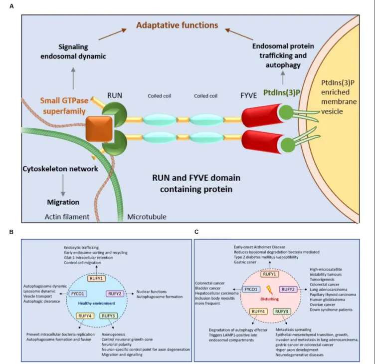
FIGURE 4 | RUFY proteins are important for intracellular trafficking, signaling and cytoskeleton dynamics. (A) Schematic representation of the RUN and FYVE domains activity of RUFY proteins. RUN domains act on signaling, endosomal protein trafficking and cytoskeletal network dynamics via small GTPase proteins. FYVE domains bind PtdIns(3)P and regulates autophagy and endosome trafficking. (B) Function of RUFY proteins in homeostatic conditions. (C) Consequences of alterations in RUFY proteins functions at the cellular and organismal level.
characterized, including conventional, Gal4, GATA-1, TFIIS,
MetRS, LIM, RING domain, PKC CRD, and PHD domains.
Zinc fingers are structural conformations adopted by peptide
chains upon coordination of two Zn
2+cations within a cysteine
rich region (
Schwabe and Klug, 1994
;
Stenmark et al., 1996
).
Unlike most molecules bearing zinc fingers, FYVE proteins
display only one copy of the domain located at any position
along the peptide chain, highlighting its autonomy as a structural
unit. FYVE zinc fingers can stabilize protein or
protein-DNA/RNA interactions (
Dunkelberg and Gutierrez-Hartmann,
2001
). A “classical” FYVE domain has eight potential zinc
coordinating tandem cysteine positions and is characterized by
having basic amino acids around the cysteines. Many members
of this family also include two histidine residues in a sequence
motif including WxxD, CxxC, R+HHC+xCG and RVC where
“x” means any amino acid and “+” a positively charged amino
acid (Figure 3B). Most deviations from this sequence can
reduce the domain affinity for zinc and destabilize it (
Stenmark
et al., 1996
;
Misra and Hurley, 1999
;
Stenmark and Aasland,
1999
;
Kutateladze and Overduin, 2001
). Within this structural
framework, specific modifications in the non-conserved residues
of the domain can radically affect FYVE protein subcellular
localization and function, by forming a “turret loop” and a
dimerization interface (
Hayakawa et al., 2004
).
With regard to their affinity for PtdIns(3)P, FYVE
domain-containing proteins are mostly found associated to EE or
phagosomes (
Stenmark et al., 1996
;
Gaullier et al., 1998
;
Stenmark and Aasland, 1999
; Figure 4A). The presence of
FYVE domains is therefore correlated to the regulation of
membrane traffic, through specific recognition of PtdIns(3)P
domains by “R+HHC+xCG” motifs (
Gaullier et al., 1998
),
and modulation by associated phosphatidylinositol kinases.
PtdIns(3)P is generated from phosphatidylinositol by Class III
PtdIns 3-kinases (PI3K), like Vps34, on target membranes
such as nascent autophagosome (omegasomes) (
Melia et al.,
2020
), or EE (
Di Paolo and De Camilli, 2006
;
Raiborg et al.,
2013
;
Scott et al., 2014
; Figure 1B). In turn, accumulation of
PtdIns(3)P recruits and activates effector proteins containing
FYVE domains, favoring transport or fusion of target organelles
(
Stenmark and Gillooly, 2001
;
Axe et al., 2008
;
Burman and
Ktistakis, 2010
;
Schink et al., 2013
). Affinity for PtdIns(3)P is
determined by the pair of histidine residues present in the
“R+HHC+XCG” motif of the FYVE domain (
Stahelin et al.,
2002
;
Diraviyam et al., 2003
;
Lee et al., 2005
;
He et al., 2009
).
This affinity can also be harnessed by FYVE proteins to link
endosomes with mRNA, ribonucleoprotein particles (mRNP)
and associated ribosomes, playing a role in their long-distance
transport in the cell (
Pohlmann et al., 2015
). Importantly,
many FYVE proteins homodimerize. Dimerization multiplies the
conserved residues displayed by the different signature motifs
present in the FYVE domain and contributes to a network of
hydrogen bonding and electrostatic interactions that provides
positive selection for binding several PtdIns(3)P head groups.
PH-dependent insertion of FYVE domain into cell membranes
(
He et al., 2009
;
Pankiv et al., 2010
) is reinforced by additional
hydrophobic membrane interactions with the turret loop and/or
tandem lysine residues. These non-specific interactions promote
FYVE domain access to phosphate head groups, that are
hindered by the close packing of lipid molecules. This bivalent
mechanism increases therefore greatly FYVE domains specificity
for PtdIns(3)P-enriched domains and discrimination against
other mono- or polyphosphorylated PtdIns species (
Misra and
Hurley, 1999
;
Stenmark and Aasland, 1999
;
Dumas et al., 2001
;
Kutateladze and Overduin, 2001
).
FYVE proteins are therefore key players in endocytosis and
autophagy and mutations in FYVE domains can alter profoundly
these functions, as well as cellular homeostasis (
Kamentseva et al.,
2020
). For example, EEA1 protein (early endosome antigen 1) is
known to be crucial for endosome dynamics and any mutation in
its conserved residues or the oligomerization site can drastically
reduce the affinity between its FYVE domain and PtdIns(3)P
(
Stenmark et al., 1996
;
Gaullier et al., 2000
). In this context,
RUFYs proteins, by bearing a N-terminal RUN domain, one
or several copies of a coiled-coil domain next to a C-terminal
FYVE domain (Figure 1A) have all the features required to
carry-out specific adaptor functions to regulate endocytosis or
autophagy by impacting on organelle fusion and mobility along
the cytoskeleton.
The RUFY Proteins Family
The RUFY family encompass four genes named
rufy1 to 4,
sharing homologies and displaying specific tissue expression
and alternative splicing.
Rufy genes are relatively conserved
genes, absent from prokaryotes and fungi. Upon evolution, the
emergence of the common ancestor appeared in vertebrates and
arthropods, which possess one ortholog (CG31064) (Figure 2).
No RUFY protein could be detected In
Caenorhabditis elegans
and only a FYVE-bearing protein (T10G3.5) considered as an
ortholog of human EEA1 shows some sequence similarities
with the RUFY family.
T10G3.5 exhibits PtdIns(3)P binding
activity and is involved in endocytosis, being mostly expressed
in epidermis and intestine of
C. elegans (
Hayakawa et al.,
2007
). In chordates, Rubicon (RUN domain and cysteine-rich
domain containing, Beclin 1-interacting protein) and FYVE And
Coiled-Coil Domain Autophagy Adaptor 1 (FYCO1), display
structural and functional features, potentially categorizing them
as RUFY proteins. Rubicon was identified as a component
of the Class III PI3K complex and a negative regulator
of autophagy and endosomal trafficking (
Matsunaga et al.,
2009
;
Zhong et al., 2009
). Like RUFYs, Rubicon contains
multiple functional domains that interact with other proteins,
including a RUN, a CC and a FYVE-like domains (
Wong
et al., 2018
). However, despite these similarities, the poor
degree of sequence homology and the lack of conservation
of its FYVE-like domain, which was found not to bind to
PI(3)P (
Burman and Ktistakis, 2010
), prevented Rubicon’s
integration within the RUFY proteins family, conversely to
FYCO1, which we propose here to name RUFY5 and detail the
characteristics below.
RUFY1
RUFY1, previously named Rabip4 is an 80 kDa protein,
mainly expressed in the brain, kidney, lung, placenta and
testis. There are two RUFY1 isoforms Rabip4, and Rabip4’
that has an additional 108 amino acid upstream of the
N-terminal RUN domain (Figure 1A). They were both shown
to interact with the small endosomal GTPases Rab4, Rab5,
and Rab14 (
Fouraux et al., 2004
;
Vukmirica et al., 2006
;
Table 1
). RUFY1 inactivation inhibits efficient recycling of
endocytosed transferrin, implicating RUFY1 in the regulation
of EE functions through cooperative interactions with Rab4
and Rab14 (
Cormont et al., 2001
;
Yamamoto et al., 2010
;
Nag
et al., 2018
). This was further demonstrated by the alteration of
epidermal growth factor receptor endocytic trafficking kinetics
in cells depleted of RUFY1 (
Gosney et al., 2018
) and the
hijacking of RUFY1 by the bacteria
P. gingivalis to escape
lysosomal degradation (
Takeuchi et al., 2016
). In melanocytes,
RUFY1 was found to form a complex with rabenosyn-5,
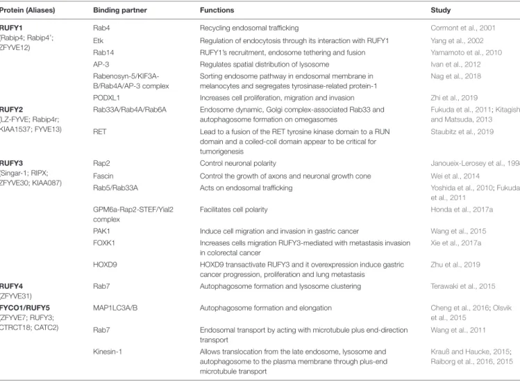
TABLE 1 | Summary of RUFY proteins functional interactions.
Protein (Aliases) Binding partner Functions Study
RUFY1 (Rabip4; Rabip4’; ZFYVE12)
Rab4 Recycling endosomal trafficking Cormont et al., 2001
Etk Regulation of endocytosis through its interaction with RUFY1 Yang et al., 2002
Rab14 RUFY1’s recruitment, endosome tethering and fusion Yamamoto et al., 2010
AP-3 Regulates spatial distribution of lysosome Ivan et al., 2012
Rabenosyn-5/KIF3A-B/Rab4A/AP-3 complex
Sorting endosome pathway in endosomal membrane in melanocytes and segregates tyrosinase-related protein-1
Nag et al., 2018
PODXL1 Increases cell proliferation, migration and invasion Zhi et al., 2019
RUFY2
(LZ-FYVE; Rabip4r; KIAA1537; FYVE13)
Rab33A/Rab4A/Rab6A Endosome dynamic, Golgi complex-associated Rab33 and autophagosome formation on omegasomes
Fukuda et al., 2011;Kitagishi and Matsuda, 2013
RET Lead to a fusion of the RET tyrosine kinase domain to a RUN domain and a coiled-coil domain appear to be critical for tumorigenesis
Staubitz et al., 2019
RUFY3 (Singar-1; RIPX; ZFYVE30; KIAA087)
Rap2 Control neuronal polarity Janoueix-Lerosey et al., 1998
Fascin Control the growth of axons and neuronal growth cone Wei et al., 2014
Rab5/Rab33A Acts on endosomal trafficking Yoshida et al., 2010;Fukuda
et al., 2011
GPM6a-Rap2-STEF/Yial2 complex
Facilitates cell polarity Honda et al., 2017a
PAK1 Induce cell migration and invasion in gastric cancer Wang et al., 2015
FOXK1 Increases cells migration RUFY3-mediated with metastasis invasion in colorectal cancer
Xie et al., 2017a
HOXD9 HOXD9 transactivate RUFY3 and it overexpression induce gastric cancer progression, proliferation and lung metastasis
Zhu et al., 2019
RUFY4 (ZFYVE31)
Rab7 Autophagosome formation and lysosome clustering Terawaki et al., 2015
FYCO1/RUFY5 (ZFYVE7; RUFY3; CTRCT18; CATC2)
MAP1LC3A/B Autophagosome formation and elongation Cheng et al., 2016;Olsvik
et al., 2015
Rab7 Endosomal transport by acting with microtubule plus end-direction transport
Wang et al., 2011
Kinesin-1 Allows translocation from the late endosome, lysosome and autophagosome to the plasma membrane through plus-end microtubule transport
Krauß and Haucke, 2015;
Raiborg et al., 2016, 2015
KIF3A-B, Rab4A and adaptor protein-3 (AP-3) to differentially
regulate tyrosinase-related protein-1 and tyrosinase sorting in
endosomes, contributing to melanosome maturation (
Nag et al.,
2018
; Table 1). Moreover, silencing the Rabip4’ isoform of
RUFY1 was shown to promote outgrowth of plasma membrane
protrusions, and to regulate the spatial distribution of lysosomes
at their tips, through an interaction with AP-3 (
Ivan et al.,
2012
; Figures 1B, 4B). RUFY1 is also capable of controlling
cell migration by regulating integrin trafficking (
Vukmirica
et al., 2006
), presumably via endocytosis. In full agreement
with a role of RUFY1 in regulating endosomal dynamics, a
single nucleotide polymorphism (S705A) in the
rufy1 gene was
associated with high blood glucose levels and type 2 diabetes
mellitus susceptibility in an exome-wide association study
(EWAS;
Yamada et al., 2017
). This result is consistent with the
early finding that Rabip4 expression leads to Glucose
transporter-1 (Glut-transporter-1) intracellular retention (
Cormont et al., 2001
).
Interestingly RUFY1 display a SH3-binding motif “PxxPxP”
embedded in the FYVE domain and is able to interacting
with the epithelial and endothelial tyrosine kinase (ETK),
and possibly regulates endocytosis through this interaction
(
Yang et al., 2002
). Another EWAS, aiming to find early-onset
Alzheimer’s Disease (AD) susceptibility genes, identified RUFY1
among genes involved in endo-lysosomal transport and known
to be important for the development of AD (
Kunkle et al.,
2017
; Figure 4C).
RUFY2
RUFY2 (or Leucine zipper FYVE-finger protein, LZ-FYVE) is a
75 kDa protein originally identified as an activating transcription
factor-2 interactor embryogenesis (
Dunkelberg and
Gutierrez-Hartmann, 2001
), preferentially located in the nucleus and
expressed during. After development, RUFY2 expression remains
high in the brain, lung, liver and the gastrointestinal tract
(
Yang et al., 2002
). RUFY2 displays two N-terminal leucine
zipper domains as well as a C-terminal FYVE-finger domain.
Although it is likely to have a nuclear function at early stages
of embryonic development, the presence of a FYVE domain
suggests a cytoplasmic role for RUFY2 in regulating membrane
traffic in fully differentiated cells. Importantly, the RUN domain
of RUFY2 was shown to associate specifically with the Golgi
complex-associated Rab33A (
Fukuda et al., 2011
; Table 1). Given
the reported interaction of Rab33A and Rab33B with Atg16L
and its putative role in regulating autophagy (
Fukuda and Itoh,
2008
), RUFY2 could contribute to autophagosome formation
through a dual interaction with Rab33A and PtdIns(3)P on
omegasomes (Figures 1B, 4B). Irrespective of its function,
rufy2
expression is subject to modulation by the micro RNA miR-155
(
Bofill-De Ros et al., 2015
), which is an important regulator of
immune cells development and inflammatory responses (
Ceppi
et al., 2009
). The
rufy2 gene is also frequently found mutated
in cancer cells, with the most frequent mutations converting
it into a strong target for nonsense mediated mRNA decay,
thereby decreasing considerably its expression (
Shin et al.,
2011
; Figure 4C).
RUFY3
RUFY3, also known as Rap2-interacting protein X (RIPX)
(
Kukimoto-Niino et al., 2006
) or Single Axon-Related 1 (Singar1)
(
Mori et al., 2007
), is the best characterized member of the
RUFY family. RUFY3, the smallest of the RUFY proteins with
a molecular weight of 53 kDa (Figure 1A), is mostly expressed
in neurons (
Kitagishi and Matsuda, 2013
). Neuronal RUFY3 is
atypical, since it lacks a FYVE domain and is considered as part
of the RUFY family based on strong sequence similarities with
the other members, notably in the RUN and coiled-coil domains
(Figure 2A). RUFY3 is distributed between the cytosol and at the
plasma membrane, but not in intracellular vesicles, presumably
because it lacks a FYVE domain. In artificial conditions, like
following expression of the dominant gain of function mutant
form of Rab5 (Q79L) in U937 cells, RUFY3 was found associated
in large vesicle structures and to co-immunoprecipitate with
Rab5, via an interaction with its carboxyl terminal domain and
surprisingly not its RUN domain (
Yoshida et al., 2010
). Like
RUFY2, RUFY3 was also shown in a 2-hybrid screen and by
co-immunoprecipitation to bind Rab33, through its coiled-coil
domain 1 (CC1;
Fukuda et al., 2011
). In 293T and 3Y1 cell
lines however, RUFY3 was shown not to interact with several
small GTPases, including Rab2, Rab5, Rab7, Rho, and Ras.
This suggests that either RUFY3 requires cell specific partner
proteins or post-translation modifications to be able to bind to
small GTPases. RUFY3 was first described as interacting with
Rap2, a small Ras-like GTPase, via a 173 residue fragment (83–
255) located in the RUN domain (
Janoueix-Lerosey et al., 1998
;
Kukimoto-Niino et al., 2006
; Table 1). Together with Rap1, Rap2
interacts with Ras effectors, such as Raf, PI3K, and Ral guanine
nucleotide dissociation stimulator, inhibiting activation of their
downstream targets, and thus suppressing Ras oncogenic activity
(
Kukimoto-Niino et al., 2006
;
Nussinov et al., 2020
). In the adult
nervous system, Rap1 and Rap2 also regulate the maturation
and plasticity of dendritic spine and synapses. By forming
a complex together with Rap2 and Fascin, RUFY3 interacts
with the filamentous actin network and controls the growth of
axons and neuronal growth cone (
Wei et al., 2014
; Table 1).
Recent mechanistic studies indicate that RUFY3 accumulates in
lipid rafts by forming a Glycoprotein M6A
(GPM6a)-RUFY3-Rap2-STEF/Yial2 complex (
Honda et al., 2017a
; Table 1).
This complex activates the Rac guanine nucleotide exchange
factor (
Honda et al., 2017b
), impacting actin organization and
promoting neuronal polarity and growth (Figure 4B). RUFY3
seems therefore to have different axogenic functions in brain
(
Mori et al., 2007
;
Honda et al., 2017b
) and not surprisingly, roles
for RUFY3 in amyotrophic lateral sclerosis (
Arosio et al., 2016
),
major depressive disorder (
Aberg et al., 2018
) and AD (
Zelaya
et al., 2015
) have been reported. Olfactory dysfunction occurs in
90% of AD cases and is correlated with elevated
rufy3 expression
in glomerular and mitral layers of the olfactory bulb (
Zelaya et al.,
2015
). RUFY3 is cleaved by caspase 3 and critically required for
caspase-mediated degeneration of tropomyosin receptor kinase
A positive sensory axons
in vitro and in vivo (
Hertz et al., 2019
;
Figure 4C
). Removal of neuronally enriched RUFY3 is able to
block caspase 3-dependent apoptosis, while dephosphorylation of
RUFY3 at residue S34 appears required for its degradation (
Hertz
et al., 2019
). Analysis of
rufy3-deficient mice supports a second
distinct function for RUFY3 in neuronal growth and polarity,
since mutant embryos show defects in axonal projection patterns.
These occur in addition to the prevention of CASP3-dependent
apoptosis in dorsal root ganglions. RUFY3 appears therefore to be
key for nervous system development, remodeling and function,
explaining the embryonic lethality displayed upon
rufy3 genetic
inactivation in mouse (
Hertz et al., 2019
).
With the current advance in genomics and single cell RNA
sequencing, specific gene expression patterns can be revised and
more accurately defined. Analysis of several genomic databases
(BioGPS, NCBI, Human Atlas Protein, ImmGen, Ensembl) reveal
that, in addition to neurons, RUFY3 expression can be detected
in other tissues and cell types. Moreover, the
rufy3 gene appears
to have many transcriptional variants, leading to the expression
of different protein isoforms. Two of these isoforms display a
C-terminal region extended by 150 amino acids, compared to the
previously identified neuronal isoform of RUFY3. Importantly,
these previously uncharacterized longer isoforms (RUFY3XL)
possess the same RUN domain and a putative FYVE domain
in their C-terminus (Figure 1A), indicating that RUFY3 is a
legitimate member of the RUFY family. In contrast to classic
FYVE zinc fingers, genomic databases reveal this putative FYVE
domain appears to lack the tandem histidine residue cluster
that defines affinity for PtdIns(3)P (Figure 3B). Interestingly, the
SH3 binding site embedded in the RUFY1 and RUFY2 FYVE
domains is also present in RUFY3XL, suggesting a potential
signal transduction activity for this uncharacterized isoform.
The translation of
rufy3xl mRNA into a functional protein and
its capacity to bind PtdIns(3)P remain to be demonstrated.
If true, a role for RUFY3 in the coordination of endosome
dynamics or organelle transport could be hypothesized. This
idea is supported by the observation that RUFY3 is present in
Staufen2-containing messenger ribonucleoprotein particles, that
are used to transport mRNAs along neuronal dendrites to their
site of translation (
Maher-Laporte et al., 2010
). FYVE proteins
have already been implicated in endosome-mediated transport of
mRNP (
Pohlmann et al., 2015
) and RUFY3XL could therefore
also perform this function. The existence of FYVE domain
bearing isoforms, might extend and diversify its function in other
specialized cells.
RUFY4
RUFY4 is a 64 kDa that is atypical among the RUFY family
members, since it bears several non-conserved residues in
its RUN domain and it lacks the tandem histidine cluster
and the SH3 binding domain normally present in the FYVE
domain (Figures 1A, 3A,B). RUFY4 was shown to interact
with PtdIns(3)P enriched membranes (
Terawaki et al., 2015,
2016
). Interestingly, EMBL-EBI and SMART genomic databases
show that
rufy4 is present only in mammals, suggesting that
rufy4 is the most recently evolved gene in the RUFY family
(Figure 2). RUFY4 levels are extremely low in most cells and
tissues with the exception of lungs and lymphoid organs. RUFY4
was found to be strongly induced
in vitro in dendritic cells
differentiated from bone marrow progenitors in presence of
GM-CSF and IL-4.
In vivo, its expression was confirmed in
alveolar macrophages and in lung dendritic cells isolated from
asthmatic mice (
Terawaki et al., 2015
). RUFY4 interacts with
Rab7 through its RUN domain and promotes the generation
of large autophagosomes (
Terawaki et al., 2015
; Figure 4B and
Table 1
). RUFY4 over-expression induces the degradation of the
autophagy effector LC3/ATG8 and triggers clustering of
LAMP1-positive late endosomal compartments. These compartments
are distinct from large abnormal autophagosome-like structures
positive for RUFY4 and Syntaxin-17, a Qa SNARE involved
in autophagosome formation and fusion (Figure 4C). RUFY4
was also proposed to interact with PLEKHM1 and the HOPS
complex, which are implicated in LE and lysosome dynamics
and positioning (
Terawaki et al., 2016
). RUFY4 seems therefore
able to harness the classical autophagy machinery to facilitate
autophagosome formation and increase autophagy flux by
acting at different biochemical steps (
Terawaki et al., 2015
). By
optimizing effector protein activity and organelle distribution,
RUFY4 expression facilitates the elimination of both damaged
mitochondria and intracellular bacteria in phagocytes. RUFY4
expression in HeLa cells can prevent replication of
Brucella
abortus (
Terawaki et al., 2015
) and
Salmonella typhimurium
(
Lassen et al., 2016
) suggesting that RUFY4 has a key role in
anti-bacterial responses in the lung. It also potentially acts to drive
immunity though the regulation of endocytosis and autophagy,
necessary for the presentation at the cell surface of antigens from
intracellular pathogens (
Terawaki et al., 2015
).
FYCO1
FYCO1 is a 150 kDa protein bearing a RUN and a FYVE
domains. In several databases,
fyco1 was misidentified as rufy3,
although these two genes are present on completely distinct
chromosomes, in human chromosome 3 and 4, respectively.
At the sequence level, although it is larger, FYCO1 appears
to be a RUFY4 ortholog gene (Figures 3A,B), suggesting that
FYCO1 belongs to the RUFY family. We therefore propose that
it could be annotated as RUFY5 to fit the family nomenclature.
Separating its N-terminal RUN domain from the FYVE zinc
finger, FYCO1 has several CC domains, as well as a LC3/ATG8
Interacting Region (LIR) and a Golgi Dynamic (GOLD) domain
in its C-terminus (Figure 1A). FYCO1 preferentially interacts
with MAP1LC3A/B of the Atg8-familly proteins through its LIR
(
Olsvik et al., 2015
;
Cheng et al., 2016
). Coiled-coil domains
promote FYCO1 dimerization and have been shown to mediate
the formation of a complex with Rab7, via a part of the CC
located upstream of the FYVE domain (
Pankiv et al., 2010
;
Wang
et al., 2011
; Table 1). Overexpression of FYCO1 was shown
to redistribute LC3- and Rab7-positive structures to the cell
periphery in a microtubule-dependent manner (
Pankiv et al.,
2010
). This effect is mediated by the central part of the CC region
and suggests a role for FYCO1 in the transport of autophagic
vesicles (Figure 4B). The capacity of FYCO1 to interact with
Rab7 and LC3A/B on the external surface of autophagosomes,
and PtdIns3P enriched membranes through its FYVE domain, is
likely to be key to its function as an adaptor protein. Indeed, these
interactions allow microtubule plus end-directed transport and
protrusion of endocytic organelles, including autophagosomes
(
Pankiv and Johansen, 2010
), LE (
Raiborg et al., 2015, 2016
),
lysosomes (
Mrakovic et al., 2012
;
Hong et al., 2017
;
Lie and
Nixon, 2019
), and phagosomes (
Ma et al., 2014
). Endoplasmic
reticulum (ER) and endosomes are connected through contact
sites, the numbers of which increase as endosomes mature.
The functions of such sites include to control the association
of endosomes with the minus-end-directed microtubule motor
dynein and to mediate endosome fission. Repeated LE–ER
contacts promote microtubule-dependent translocation of LEs
to the cell periphery and subsequent fusion with the plasma
membrane (
Raiborg et al., 2016
). Such fusion induces outgrowth
of protrusions and neurites in the neuroendocrine cell line PC12,
which require the ER-associated protein protrudin on the ER
and FYCO1 to interact with LEs and kinesin 1 (
Krauß and
Haucke, 2015
; Table 1). FYCO1 has been described as a novel
mediators of invalopodia formation and function of
Protrudin-mediated ER–endosome contact sites (
Pedersen et al., 2020
).
Multiple studies highlight the critical function of FYCO1 in
autophagy and autophagosome/endosome trafficking (
Dionne
et al., 2017
) with pathological consequences arising when FYCO1
function is altered (Figures 4B,C). Mutations in the
fyco1
gene affect autophagy and cause autosomal-recessive congenital
cataracts by altering lens development and transparency in
patients (
Chen et al., 2011, 2017
;
Brennan et al., 2012
;
Costello
et al., 2013
;
Chauss et al., 2014
;
Frost et al., 2014
;
Khan et al.,
2015
;
Gunda et al., 2018
;
Li et al., 2018
). Sequencing studies of
candidate genes potentially involved in several neuromuscular
or neurodegenerative diseases have identified rare variants of
autophagy related proteins like VCP and SQSTM1. Among these
genes, a missense
fyco1 variant was identified to cause sporadic
inclusion body myositis (
Güttsches et al., 2017
;
Rothwell et al.,
2017
;
Britson et al., 2018
; Figure 4C). Finally FYCO1 has been
implicated in the autophagic clearance of specialized particles or
aggregates, like male germ cell-specific RNP ribonucleoprotein
granules (
Da Ros et al., 2017
), post-mitotic bodies (
Dionne et al.,
2017
) or
α-synuclein aggregates (
Saridaki et al., 2018
).
RUFY Proteins and Cancer
As describe above, RUFY proteins play a central role in cellular
functions by regulating vesicular trafficking and its interactions
with the cytoskeleton. Neuronal deficit and neurodegeneration
are the most obvious manifestations of RUFY proteins alteration.
Not surprisingly, however, given their relatively broad adaptors
functions, RUFY proteins have taken center stage in the
oncology field.
The ETK tyrosine kinase has been shown to play a pivotal
role in a variety of cellular processes including proliferation,
differentiation, motility, and apoptosis (
Yang et al., 2002
;
Kung, 2011
;
Zhuang et al., 2014
;
Wang et al., 2018
). Tyrosine
phosphorylation of RUFY1 by ETK appears to be important
for its endosomal localization and could play an important
role promoting tumoral transformation by affecting downstream
effectors of PI3-kinase. RUFY1 was also shown to interact
with podocalyxin-like protein (PODXL), a transmembrane
glycoprotein with anti-adhesive properties associated with poor
prognosis of several cancers (
Taniuchi et al., 2018
;
He et al.,
2020
; Table 1). Gastric cancer progression is significantly
increased upon PODXL expression, a phenotype reduced by
concomitant RUFY1 silencing. Depletion of RUFY1 inactivates
the PI3K/AKT, NF-
κB and MAPK/ERK signaling pathways
and reduces drastically migration and invasion of cancer cells
in vitro (
Zhi et al., 2019
). Given the positive correlation
between
podxl and rufy1 expression in tissues and serum, rufy1
was proposed as a potential biomarker for gastric cancers
stratification (
Zhi et al., 2019
; Figure 3C). Like RUFY1, a role
for RUFY2 in various cancer has been reported (
Shin et al.,
2011
;
Zheng et al., 2014
;
Staubitz et al., 2019
).
Rufy2 is one
of the most frequently mutated genes in high-microsatellite
instability tumors and colorectal cancer (
Shin et al., 2011
).
Gene rearrangement of the proto-oncogene
ret with rufy2 have
been shown to drive tumorigenesis in lung adenocarcinoma
(
Zheng et al., 2014
) and papillary thyroid carcinoma (
Staubitz
et al., 2019
). The gene rearrangement leads to a fusion of the
RET tyrosine kinase domain with RUFY2 RUN domain and
coiled-coil domain; this appears to be critical for tumorigenesis
(
Staubitz et al., 2019
; Table 1).
Rufy2 mRNA is the target
of several microRNAs, including miR-146a, miR-196a-5p and
miR-155 (
Bofill-De Ros et al., 2015
). Dysregulated microRNA
targeting of RUFY2 expression was found important for
the development of human glioblastoma and ovarian cancer,
suggesting a tumor suppression role for RUFY2 (
Lukács et al.,
2019
;
Zheng et al., 2020
; Figure 4C). Given the key role of
RUFY3 in cell migration, membrane transport, and cellular
signaling, through its interaction with rap2, it is not surprising
that RUFY3 dysregulation has been implicated in several
cancer processes and metastatic tumor spread. The abnormal
expression of RUFY3 is linked to poor prognosis. It can promote
growth, invasion and metastasis in lung adenocarcinoma,
gastric cancer cells or colorectal cancer (
Xie et al., 2017a,b
;
Men et al., 2019
;
Zhu et al., 2019
). RUFY3 overexpression
and its interaction with P21-activated kinase-1 (PAK1) leads
to the formation of F-actin-enriched protrusive structures,
increased epithelial-mesenchymal transition and gastric cancer
cell migration (
Kumar and Vadlamudi, 2002
;
Vadlamudi and
Kumar, 2003
). Several transcription factors, including Forkhead
box k1 (FOXK1) and Homebox D9 (HOXD9) involved in
cancer progression (
Moens and Selleri, 2006
;
Tabuse et al., 2011
;
Lv et al., 2015
;
Peng et al., 2016
;
Wu et al., 2016
;
Liu et al.,
2018
;
Zhu et al., 2019
), have been shown to regulate RUFY3
expression and activity (
Xie et al., 2017a
;
Zhu et al., 2019
;
Table 1
). So far, no correlation has been found between
RUFY4 and any type of cancer. FYCO1 has also been
implicated in colorectal cancer progression (
Sillars-Hardebol
et al., 2010
) and recent studies have concluded that FYCO1
may serve as a biomarker in bladder cancer (
Eissa et al., 2017
)
or hepatocellular carcinoma (
Vongchan and Linhardt, 2017
;
Figure 4C
). Plus, FYCO1 can indirectly associated with cell
invasion (
Pedersen et al., 2020
).
CONCLUSION
Although they have been poorly characterized to date, RUFY
proteins play a central role in cellular homeostasis by regulating
endocytosis, autophagy and coordinating organelle transport
with signal transduction cascades. It is important to note
that RUFY proteins also provide a regulatory link between
cytoskeletal dynamics and membrane trafficking. Consequently,
these proteins have adaptive functions by acting on localized
actions (through PtdIns(3)P) and signaling (through small
GTPases), which can affect key biological functions in specialized
cells, such as migration, tissue repair or targeted secretion.
The dysregulated expression of RUFY proteins has therefore
severe consequences on cell differentiation and polarization,
causing cancers or neurodegenerative diseases. However, further
molecular and physiological analyses will be required to
understand how these proteins exert their functions in specialized
cell types like immune cells or neurons. Immunocytes require
endocytosis and migration to perform their functions within
primary and secondary lymphoid organs or at sites of infection.
The restricted expression of RUFY4, as well as the existence of
splicing variants of RUFY3 in alveolar macrophages and dendritic
cells, suggest a role for these molecules in phagocytes. Of
importance will be the characterization of the different molecules
interacting either with their RUN or FYVE domains in a cell
specific manner. Identification of these RUFY’s interactors will
be crucial to establish the functionality of the domains and their
importance for signaling on one end and subcellular targeting at
the other end. The coiled-coil structural domains found in the
central part of the RUFY proteins should also be scrutinized.
CC domains, in addition to support homodimerization and
increase affinity for PtdIns(3)P, could also be determinant in
promoting RUFY proteins interactions with effector molecules,
like Rab7, as observed for FYCO1. The nature and pattern of
expression of these effector molecules will allow to sort the
different activities displayed by the RUFYs in individual cell
types and thereby shed light on their physiological importance
in health and diseases.
AUTHOR CONTRIBUTIONS
Both
authors
contributed
equally
to
the
design
and
implementation of the research and writing of the manuscript.
FUNDING
The PP laboratory is “Equipe de la Fondation de la
Recherche
Médicale”
(FRM)
sponsored
by
the
grant
DEQ20180339212. The project was supported by grants from
l’Agence Nationale de la Recherche (ANR) «DCBIOL Labex
0043», «INFORM Labex
ANR-11-LABEX-0054» funded by the “Investissements d’Avenir” French
government program. The research was also supported by
the Ilídio Pinho foundation and FCT – Fundaç˘ao para
a Ciência e a Tecnologia – and Programa Operacional
Competitividade
e
Internacionalizaç˘ao
–
Compete2020
(FEDER) – references POCI-01-0145-FEDER-016768 and
POCI-01-0145-FEDER-030882.
ACKNOWLEDGMENTS
We acknowledge Dr. Jonathan Ewbank for his constructive
comments.
REFERENCES
Aberg, K. A., Dean, B., Shabalin, A. A., Chan, R. F., Han, L. K. M., Zhao, M., et al. (2018). Methylome-wide association findings for major depressive disorder overlap in blood and brain and replicate in independent brain samples.Mol. Psychiatry 25, 1344–1354. doi: 10.1038/s41380-018-0247-6
Argüello, R. J., Reverendo, M., Gatti, E., and Pierre, P. (2016). Regulation of protein synthesis and autophagy in activated dendritic cells: implications for antigen processing and presentation.Immunol. Rev. 272, 28–38. doi: 10.1111/imr.12427 Arosio, A., Sala, G., Rodriguez-Menendez, V., Grana, D., Gerardi, F., Lunetta, C., et al. (2016). MEF2D and MEF2C pathways disruption in sporadic and familial ALS patients.Mol. Cell. Neurosci. 74, 10–17. doi: 10.1016/j.mcn.2016.02.002 Axe, E. L., Walker, S. A., Manifava, M., Chandra, P., Roderick, H. L., Habermann,
A., et al. (2008). Autophagosome formation from membrane compartments enriched in phosphatidylinositol 3-phosphate and dynamically connected to the endoplasmic reticulum.J. Cell Biol. 182, 685–701. doi: 10.1083/jcb.200803137 Birgisdottir, ÅB., and Johansen, T. (2020). Autophagy and endocytosis –
interconnections and interdependencies.J. Cell Sci. 133:jcs228114. doi: 10.1242/ jcs.228114
Bofill-De Ros, X., Santos, M., Vila-Casadesús, M., Villanueva, E., Andreu, N., Dierssen, M., et al. (2015). Genome-wide miR-155 and miR-802 target gene identification in the hippocampus of Ts65Dn Down syndrome mouse model by miRNA sponges.BMC Genomics 16:907. doi: 10.1186/s12864-015-2160-6 Bos, J. L. (2005). Linking rap to cell adhesion.Curr. Opin. Cell Biol. 17, 123–128.
doi: 10.1016/j.ceb.2005.02.009
Boucrot, E., Henry, T., Borg, J.-P., Gorvel, J.-P., and Méresse, S. (2005). The intracellular fate ofSalmonella depends on the recruitment of kinesin. Science 308, 1174–1178. doi: 10.1126/science.1110225
Brennan, L. A., Kantorow, W. L., Chauss, D., McGreal, R., He, S., Mattucci, L., et al. (2012). Spatial expression patterns of autophagy genes in the eye lens and induction of autophagy in lens cells.Mol. Vis. 18, 1773–1786.
Britson, K. A., Yang, S. Y., and Lloyd, T. E. (2018). New developments in the genetics of inclusion body myositis.Curr. Rheumatol. Rep. 20:26. doi: 10.1007/ s11926-018-0738-0
Bruce, E. A., Digard, P., and Stuart, A. D. (2010). The Rab11 pathway is required for influenza A virus budding and filament formation.J. Virol. 84, 5848–5859. doi: 10.1128/JVI.00307-10
Buratta, S., Tancini, B., Sagini, K., Delo, F., Chiaradia, E., Urbanelli, L., et al. (2020). Lysosomal exocytosis, exosome release and secretory autophagy: the autophagic- and endo-lysosomal systems go extracellular. Int. J. Mol. Sci. 21:2576. doi: 10.3390/ijms21072576
Burman, C., and Ktistakis, N. T. (2010). Regulation of autophagy by phosphatidylinositol 3-phosphate.FEBS Lett. 584, 1302–1312. doi: 10.1016/j. febslet.2010.01.011
Callebaut, I., de Gunzburg, J., Goud, B., and Mornon, J.-P. (2001). RUN domains: a new family of domains involved in Ras-like GTPase signaling.Trends Biochem. Sci. 26, 79–83. doi: 10.1016/S0968-0004(00)01730-8
Ceppi, M., Schmidt, E., and Pierre, P. (2009). Genetic modification of murine dendritic cells by RNA transfection. Methods Mol. Biol. 531, 145–156. doi: 10.1007/978-1-59745-396-7_10
Chauss, D., Basu, S., Rajakaruna, S., Ma, Z., Gau, V., Anastas, S., et al. (2014). Differentiation state-specific mitochondrial dynamic regulatory networks are revealed by global transcriptional analysis of the developing chicken lens.G3 4, 1515–1527. doi: 10.1534/g3.114.012120
Chen, J., Ma, Z., Jiao, X., Fariss, R., Kantorow, W. L., Kantorow, M., et al. (2011). Mutations in FYCO1 cause autosomal-recessive congenital cataracts.Am. J. Hum. Genet. 88, 827–838. doi: 10.1016/j.ajhg.2011.05.008
Chen, J., Wang, Q., Cabrera, P. E., Zhong, Z., Sun, W., Jiao, X., et al. (2017). Molecular genetic analysis of Pakistani families with autosomal recessive congenital cataracts by homozygosity screening.Invest. Ophthalmol. Vis. Sci. 58, 2207–2217. doi: 10.1167/iovs.17-21469
Chen, W., and Wandinger-Ness, A. (2001). Expression and functional analyses of Rab8 and Rab11a in exocytic transport from trans-Golgi network.Methods Enzymol. 329, 165–175. doi: 10.1016/s0076-6879(01)29077-6
Cheng, X., Wang, Y., Gong, Y., Li, F., Guo, Y., Hu, S., et al. (2016). Structural basis of FYCO1 and MAP1LC3A interaction reveals a novel binding mode for Atg8-family proteins.Autophagy 12, 1330–1339. doi: 10.1080/15548627.2016. 1185590
Chung, I., Akita, R., Vandlen, R., Toomre, D., Schlessinger, J., and Mellman, I. (2010). Spatial control of EGF receptor activation by reversible dimerization on living cells.Nature 464, 783–787. doi: 10.1038/nature08827
Cormont, M., Mari, M., Galmiche, A., Hofman, P., and Le Marchand-Brustel, Y. (2001). A FYVE-finger-containing protein, Rabip4, is a Rab4 effector involved in early endosomal traffic.Proc. Natl. Acad. Sci. U.S.A. 98, 1637–1642. doi: 10.1073/pnas.031586998
Costello, M. J., Brennan, L. A., Basu, S., Chauss, D., Mohamed, A., Gilliland, K. O., et al. (2013). Autophagy and mitophagy participate in ocular lens organelle degradation.Exp. Eye Res. 116:141–150. doi: 10.1016/j.exer.2013. 08.017
Da Ros, M., Lehtiniemi, T., Olotu, O., Fischer, D., Zhang, F.-P., Vihinen, H., et al. (2017). FYCO1 and autophagy control the integrity of the haploid male germ cell-specific RNP granules. Autophagy 13, 302–321. doi: 10.1080/15548627. 2016.1261319
Di Paolo, G., and De Camilli, P. (2006). Phosphoinositides in cell regulation and membrane dynamics.Nature 443, 651–657. doi: 10.1038/nature05185 Dionne, L. K., Peterman, E., Schiel, J., Gibieža, P., Skeberdis, V. A., Jimeno, A.,
et al. (2017). FYCO1 regulates accumulation of post-mitotic midbodies by mediating LC3-dependent midbody degradation.J. Cell Sci. 130, 4051–4062. doi: 10.1242/jcs.208983
Diraviyam, K., Stahelin, R. V., Cho, W., and Murray, D. (2003). Computer modeling of the membrane interaction of FYVE domains.J. Mol. Biol. 328, 721–736. doi: 10.1016/S0022-2836(03)00325-5
Doherty, G. J., and McMahon, H. T. (2009). Mechanisms of endocytosis.Annu. Rev. Biochem. 78, 857–902. doi: 10.1146/annurev.biochem.78.081307.110540 Doherty, J., and Baehrecke, E. H. (2018). Life, death and autophagy.Nat. Cell Biol.
20, 1110–1117. doi: 10.1038/s41556-018-0201-5
Dumas, J. J., Merithew, E., Sudharshan, E., Rajamani, D., Hayes, S., Lawe, D., et al. (2001). Multivalent endosome targeting by homodimeric EEA1.Mol. Cell 8, 947–958. doi: 10.1016/S1097-2765(01)00385-9
Dunkelberg, J. C., and Gutierrez-Hartmann, A. (2001). LZ-FYVE: a novel developmental stage-specific leucine zipper, FYVE-finger protein.DNA Cell Biol. 20, 403–412. doi: 10.1089/104454901750361460
Eissa, S., Matboli, M., Awad, N., and Kotb, Y. (2017). Identification and validation of a novel autophagy gene expression signature for human bladder cancer patients.Tumour Biol. 39:1010428317698360. doi: 10.1177/1010428317698360 Fîlfan, M., Sandu, R. E., Z˘av˘aleanu, A. D., Gre¸si ¸T˘a, A., Gl˘avan, D. G., Olaru, D. G., et al. (2017). Autophagy in aging and disease.Rom. J. Morphol. Embryol. 58, 27–31.