Collagen Scaffolds and Injectable Biopolymer Gels for Cardiac Tissue
Engineering
by
Karen Kailin Ng
B.S. Materials Science and Engineering Northwestern University, 2006
SUBMITTED TO THE DEPARTMENT OF MATERIALS SCIENCE AND ENGINEERING IN PARTIAL FULFILLMENT OF THE REQUIREMENTS FOR THE DEGREE OF
DOCTOR OF PHILOSOPHY IN MATERIALS SCIENCE AND ENGINEERING AT THE
MASSACHUSETTS INSTITUTE OF TECHNOLOGY
FEBRUARY 2012
© 2011 Massachusetts Institute of Technology. All rights reserved.
Signature of Author: ____________________________________________________________________ Department of Materials Science and Engineering October 21, 2011 Certified by: __________________________________________________________________________
Myron Spector Senior Lecturer, Mechanical Engineering Professor of Orthopaedic Surgery (Biomaterials), Harvard Medical School Thesis Co-Advisor Certified by: __________________________________________________________________________
Ioannis Yannas Professor, Polymer Science & Engineering Thesis Co-Advisor Accepted by: _________________________________________________________________________
Gerbrand Ceder Professor, Materials Science and Engineering Chair, Departmental Committee on Graduate Students
3
Collagen Scaffolds and Injectable Biopolymer Gels for Cardiac Tissue Engineering by
Karen Kailin Ng
Submitted to the Department of Materials Science and Engineering On October 21, 2011 in partial fulfillment of the requirements for the degree of
Doctor of Philosophy in Materials Science and Engineering
Abstract
Three-dimensional biomaterial scaffolds have begun to shown promise for cell delivery for cardiac tissue engineering. Although various polymers and material forms have been explored, there is a need for: injectable gels that meet certain design specifications; a more in-depth characterization of the scaffold properties; and a deeper understanding of the relation of select properties to cellular behavior, to provide a rational basis for future in vivo studies.
The first objective of this thesis was to develop and characterize novel injectable biopolymer hydrogels capable of safely undergoing covalent cross-linking in vivo to provide a mechanically tunable nanofibrillar scaffold. Soluble type I collagen gels with genipin and transglutaminase cross-linkers, and gelatin-hydroxyphenylpropionic acid (Gtn-HPA) gels, the cross-linking of which are modulated by horse radish peroxidase and hydrogen peroxide, were investigated. The gels were characterized on the basis of rheological properties, resistance to degradation, and effects on stem cell behavior. Another objective was to evaluate the simultaneous differentiation of embryonic carcinoma cells (ECCs) incorporated in the gels into the three cell types in cardiac tissue—cardiomyocytes, neural cells, and vascular endothelial cells—and to determine the effects of certain properties of the gels on the differentiation profile, using mesenchymal stem cells as a comparative control.
The injectable collagen-genipin and Gtn-HPA gels were found to be mechanically tunable hydrogel systems that supported cell encapsulation and proliferation at safe concentrations of the respective cross-linking agents. ECCs cultured as embryoid bodies (EBs) incorporated in the collagen-genipin and Gtn-HPA gels differentiated into cardiac, neural, and endothelial cells and combinations thereof, demonstrating the capability of EBs to express multiple cell lineages within the same EB. EBs cultured in collagen gels without cross-linkers and collagen gels with 0.25 mM genipin exhibited the highest differentiation efficiency compared to those cultured in monolayer, sponge-like scaffolds, and Gtn-HPA gels. The differentiation medium and culture time also had significant effects on differentiation efficiency. Notable findings included: the increased expression of neural and endothelial markers in EBs cultured in in mixed medium conditions compared to those cultured in neural or endothelial differentiation medium alone, and the correlation between angiogenic and neurogenic differentiation in the EBs in the non-cross-linked collagen gels for all media. Collectively, these findings show promise in using collagen gels cross-linked with 0.25 mM genipin, incorporated with EBs, for cellular therapy in cardiac tissue engineering applications.
Thesis Advisor: Myron Spector
Title: Senior Lecturer, Mechanical Engineering; Professor of Orthopaedic Surgery (Biomaterials), Harvard Medical School
5
Acknowledgments
The work presented herein could not have been accomplished without the help and guidance of numerous individuals, to whom I am deeply indebted.
First and foremost, I would like to express my sincere gratitude and thanks to my thesis advisor, Dr. Myron Spector, for the invaluable advice, guidance, and support over the years, not just on research and career paths, but also on life and relationships. Your enthusiasm, optimism, organization, wisdom, and creativity have been truly inspiring to me. I feel very fortunate that I was able to do learn from you and work in your research group.
To my thesis committee, Dr. Ioannis Yannas, Dr. Hemant S. Thatte, Dr. Krystyn Van Vliet, and Dr. Darrell Irvine – thank you for your guidance, ideas and support. It’s been a great experience learning from and interacting with all of you.
To the many people (past and present) in the VA Healthcare System Tissue Engineering group that have supported and helped me both in and outside of lab: Dr. Hu-Ping Hsu, Alix Weaver, Paul Elias, Cathal Kearney, Rahmat Cholas, Lily Jeng, Teck Chuan Lim, Daniel Macaya, Toh Wei Seong, Thomas Cheriyan, Hsi-Chin Wu, and Tzu-Wei Wang – thank you for your companionship, perspective, and encouragement. I enjoyed working with all of you and wish you the best in all you do.
To Dr. Simona Socrate at MIT, thank you for your assistance and insight with mechanical testing. To Dr. Xiu-Gui Lu at the VA Healthcare System at West Roxbury, thank you for your assistance with cell culture and western blot. To Dr. Gareth McKinley at MIT, thank you for your help with the rheological testing. To Patrick Boisvert at the Center for Materials Science and Engineering, thank you for your assistance with the ESEM.
To all my friends that have been an enormous source of encouragement, support, and joy in my life: Esther Cho, Doris Lin, Meredith Silberstein, Alisha Schor, Eugene Wu, Michelle Chiang, John Joo, Chi-Fong Wang, Friends07, sMITe, Hatch, Wuwu, and Mixed Nuts – you have made my graduate school experience a memorable and fulfilling one. Thanks for your kindness, laughter, generosity, and distractions. I couldn’t have done it without you.
To my family, especially my mother Suenee, my brother Gordon, and my grandma Chia Shia – for accepting, supporting, and loving me unconditionally.
To my husband, Bryan Ng – thank you for your never-ending unconditional love, support, and encouragement. Thank you for believing in me and never letting me give up.
This thesis is dedicated to my father, Dr. Vincent Shu.
Financial support for this work provided by the Department of Veterans Affairs is gratefully acknowledged. I am grateful to Ajinomoto Food Ingredients LLC for providing the microbial transglutaminase and the Institute of Bioengineering and Nanotechnology (Singapore) for providing the gelatin-hydroxyphenylpropionic acid.
7
Table of Contents
Chapter 1 Introduction ... 21
Chapter 2 Background and motivation ... 25
2.1 Tissue response to myocardial infarct ... 25
2.2 Cell types in cardiac tissue ... 26
2.3 Extracellular matrix molecules in cardiac tissue ... 26
2.4 Cardiac tissue engineering – An overview ... 27
2.5 Scaffolding materials ... 27
2.5.1 Porous sponge-like collagen scaffolds ... 27
2.5.2 Injectable hydrogels ... 28
2.6 Cell therapy ... 31
2.6.1 Mesenchymal stem cells ... 31
2.6.2 Embryonic carcinoma cells ... 35
Chapter 3 Differentiation of MSCs and ECCs into cardiac, neural, and endothelial cells in monolayer and in 3-D collagen scaffolds ... 37
3.1 Introduction ... 37
3.2 Objective, hypothesis, and rationale ... 38
3.3 Materials and methods ... 39
3.3.1 Isolation and culture of MSCs ... 39
3.3.2 MSC differentiation assays for chondrogenesis, osteogenesis, and adipogenesis .... 40
3.3.3 Culture of ECCs ... 41
3.3.4 Monolayer culture ... 41
3.3.5 Cardiac differentiation ... 41
3.3.6 Neural differentiation ... 41
3.3.7 Endothelial differentiation ... 42
3.3.8 Scaffold fabrication and cross-linking ... 42
3.3.9 Environmental scanning electron microscopy ... 43
3.3.10 Collagen scaffold culture ... 43
3.3.11 Analysis of DNA and glycosaminoglycan content ... 43
8
3.3.13 Histology and immunohistochemistry ... 44
3.3.14 Immunofluorescence ... 45
3.3.15 Western blot ... 45
3.3.16 Statistics ... 46
3.4 Results ... 46
3.4.1 MSC differentiation assays for chondrogenesis, osteogenesis, and adipogenesis .... 46
3.4.2 MSC and ECC pellet culture for chondrogenesis ... 47
3.4.3 MSC and ECC scaffold culture for chondrogenesis ... 49
3.4.4 α-SMA expression in monolayer culture ... 51
3.4.5 Monolayer cardiac differentiation ... 52
3.4.6 Monolayer neural differentiation ... 53
3.4.7 Monolayer endothelial differentiation ... 55
3.4.8 Scaffold culture ... 56
3.5 Discussion ... 61
3.5.1 Comparison of MSCs and ECCs for chondrogenesis ... 61
3.5.2 Comparison of MSCs and ECCs for cardiac, neural, and endothelial differentiation in monolayer and 3-D collagen scaffolds ... 63
3.6 Summary ... 68
Chapter 4 Injectable biopolymer gels for cardiac tissue engineering ... 71
4.1 Introduction ... 71
4.1.1 Injectable gels for cardiac tissue engineering ... 71
4.1.2 Biopolymers to be investigated ... 71
4.2 Objective, hypothesis, and rationale ... 74
4.3 Materials and methods ... 75
4.3.1 Collagen-genipin gels ... 75
4.3.2 Collagen-transglutaminase gels ... 76
4.3.3 Gelatin-hydroxyphenylpropionic acid hydrogels ... 76
4.3.4 Rheology testing ... 76
4.3.5 Unconfined compression testing ... 77
4.3.6 Absorbance and fluorescence measurements of collagen-genipin gels ... 77
9
4.3.8 Swelling ratio ... 78
4.3.9 Environmental scanning electron microscopy ... 78
4.3.10 Isolation and culture of MSCs ... 79
4.3.11 Culture of ECCs ... 79
4.3.12 Cell-incorporated gels ... 79
4.3.13 Rheological behavior of cell-incorporated gels ... 80
4.3.14 Cell viability ... 81 4.3.15 Cell proliferation ... 81 4.3.16 Statistics ... 81 4.4 Results ... 81 4.4.1 Rheological Behavior ... 81 4.4.2 Compression testing ... 83
4.4.3 Environmental scanning electron microscopy ... 87
4.4.4 Absorbance and fluorescence of collagen-genipin gels ... 89
4.4.5 Enzymatic degradation ... 90
4.4.6 Swelling ratio ... 93
4.4.7 Cell-incorporated gels ... 94
4.4.8 Rheological behavior of cell-incorporated gels ... 96
4.4.9 Cell viability ... 97
4.4.10 Cell proliferation ... 99
4.5 Discussion ... 101
4.5.1 Collagen-genipin gels ... 101
4.5.2 Collagen-transglutaminase gels ... 106
4.5.3 Gelatin-hydroxyphenylpropionic acid gels ... 106
4.6 Overview of hydrogels ... 109
4.7 Summary ... 110
Chapter 5 Differentiation of MSCs and ECCs into cardiac, neural and endothelial cells separately and simultaneously in biopolymer gels ... 113
5.1 Introduction ... 113
5.2 Objective, hypothesis, and rationale ... 114
10
5.3.1 Overview of experimental set-up ... 116
5.3.2 Culture of P19 embryonic carcinoma cells ... 117
5.3.3 Differentiation medium ... 117
5.3.4 Cell incorporation in collagen-genipin gels ... 118
5.3.5 Cell incorporation in gelatin-hydroxyphenylpropionic acid gels ... 118
5.3.6 Immunohistochemistry and immunofluorescence ... 119
5.3.7 Statistics ... 122
5.4 Results ... 122
5.4.1 Cardiac staining ... 123
5.4.2 Neural staining ... 128
5.4.3 Endothelial staining ... 133
5.4.4 Correlation of angiogenesis and neurogenesis ... 138
5.5 Discussion ... 140
5.6 Summary ... 151
Chapter 6 In vivo feasibility of biopolymer gels ... 153
6.1 Introduction ... 153
6.2 Objective, hypothesis, and rationale ... 154
6.3 Methods and materials ... 154
6.3.1 Gel injections into ex vivo cardiac tissue ... 154
6.3.2 Histological analysis ... 155
6.3.3 Pilot animal study ... 155
6.4 Results ... 156
6.4.1 Gel injections into ex vivo cardiac tissue ... 156
6.4.2 Animal study ... 157
6.5 Discussion ... 158
6.5.1 Gel injections into ex vivo cardiac tissue ... 158
6.5.2 Methods of infarct creation ... 159
6.5.3 Animal study ... 159
6.6 Summary ... 161
Chapter 7 Limitations and future work ... 163
11
Chapter 9 Appendix ... 169
9.1 Cell culture protocols ... 169
9.1.1 Freezing cells ... 169
9.1.2 Thawing cells ... 169
9.1.3 Changing media ... 170
9.1.4 Passaging cells ... 171
9.1.5 Counting cells ... 172
9.2 Scaffold fabrication protocols ... 174
9.2.1 Collagen slurry preparation ... 174
9.2.2 Virteous freeze-drying “ramp” protocol ... 175
9.2.3 Dehydrothermal treatment ... 175
9.2.4 Cross-linking collagen scaffolds by carbodiimide treatment ... 175
9.3 Cell-seeded collagen scaffold protocols ... 179
9.3.1 Agarose coating of well-plates ... 179
9.3.2 Seeding collagen scaffolds ... 180
9.4 Cell differentiation protocols ... 181
9.4.1 Osteogenic and adipogenic differentiation ... 181
9.4.2 Chondrogenic differentiation ... 182
9.4.3 Pellet assay for chondrogenic differentiation ... 184
9.4.4 Cardiac, neural, and endothelial differentiation ... 184
9.5 Hydrogel preparation protocols ... 189
9.5.1 Collagen-genipin gels ... 189
9.5.2 Collagen-transglutaminase gels ... 189
9.5.3 Gelatin-hydroxyphenylpropionic acid gels ... 190
9.6 Sample preparation for staining protocols ... 191
9.6.1 Tissue processing and paraffin-embedding ... 191
9.6.2 OCT embedding and cryosectioning ... 191
9.7 Staining protocols ... 194
9.7.1 Hematoxylin and eosin (H&E) staining ... 194
9.7.2 Safranin O staining ... 195
12
9.7.4 Immunohistochemical monolayer staining ... 198
9.7.5 Immunohistochemical section staining ... 204
9.8 Hydrogel characterization protocols ... 206
9.8.1 Critical point drying ... 206
9.8.2 Rheological testing ... 207
9.8.3 Compression testing ... 209
9.8.4 Enzymatic degradation studies ... 209
9.9 Biochemical assay protocols ... 211
9.9.1 Proteinase K digestion ... 211
9.9.2 Dimethylmethylene blue assay for sulfated glycosaminoglycans ... 211
9.9.3 PicoGreen dsDNA quantitation assay ... 213
9.9.4 Live/Dead staining for cell viability ... 216
9.10 Supplemental results ... 217
13
List of Figures
Figure 2-1 Histological stages of myocardial infarction [20]. ... 26
Figure 2-2 Collagen cross-linked with genipin [39] ... 29
Figure 2-3 Cross-linking structures genipin-fixed collagen [40] ... 30
Figure 2-4 Mesenchymal stem cell lineage [53] ... 32
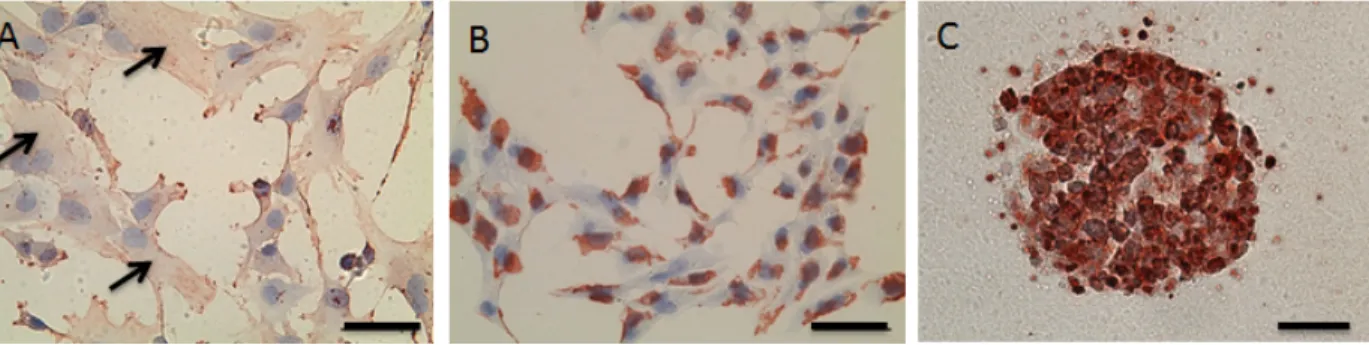
Figure 3-1 MSC differentiation assays (A,B) SafO stain and (C,D) type II collagen for chondrogenesis, (E) Von Kossa stain for osteogenesis [118], (F) Oil red stain for adipogenesis [118]. ... 47
Figure 3-2 Pig MSCs (A-D) and mouse ECCs (E-H) stained with H&E (A, E), Safranin-O (B,F), α-SMA (C,G) and type II collagen (D,H) at day 14 (n = 4). Scale bars, 100 µm (Inset scale bars, 20 µm). ... 48
Figure 3-3 DNA content, GAG content, and GAG to DNA ratio of the cell pellets at days 1 and 14 (n = 6, mean ± S.E.). ... 49
Figure 3-4 Scaffold contraction over 14 days for MSCs and ECCs seeded on (A) type I and (B) type II collagen scaffolds (n = 8, mean ± S.E.). ... 49
Figure 3-5 Pig MSCs (A,B) and mouse ECCs (C,D) stained with type II collagen (A,C) and Safranin-O (B,D) at day 14 (n = 6). Scale bars, 2 mm. ... 50
Figure 3-6 DNA and GAG contents, GAG:DNA ratio of scaffolds at day 1 and 14 (n = 4, mean ± S.E.)... 51
Figure 3-7 Immunohistochemistry of α-SMA in (A) pig MSCs, (B) dissociated mouse ECCs, and (C) EBs (n = 6). Scale bars, 50 µm. ... 52
Figure 3-8 MSCs cultured in cardiac medium (A-E) and stained immunohistochemically for cardiac Tn-I at (A) 7 days and (B) 14 days, (C,D) MHC at 14 days, (E) α-SMA at 14 days, and (F) MSCs cultured in complete medium and stained for MHC at 14 days (n = 3). ... 52
Figure 3-9 EBs cultured in cardiac medium (A-F) and stained immunohistochemically at 14 days for (A) α-actinin, (B) MHC, (C) Tn-I, (D) α-SMA, (E) Tn-I at 7 days, (F) Tn-I at 14 days, and (G) EBs cultured in complete medium and stained for Tn-I at 14 days (n = 3). Scale bars, 100 µm. ... 53
Figure 3-10 Number of EB outgrowths with beating cells (%) over time (n = 6, mean ± S.D.) .. 53
Figure 3-11 MSCs cultured in neural medium and stained immunohistochemically for (A) TUJ1 and (B) α-SMA at 1 day (n = 4). Scale bars, 50 µm. ... 54
Figure 3-12 EBs cultured in in neural medium and stained immunohistochemically for TUJ1 at (A) 2 days, (B) 7 days, (C) 14 days, and (D) EBs cultured in complete medium did not stain for TUJ1 for time points up until 14 days. Scale bar, 100 µm. ... 55
Figure 3-13 Western blot analysis confirms expression of N-Nos for EBs exposed to 10-7 M RA compared to those cultured in complete medium. ... 55
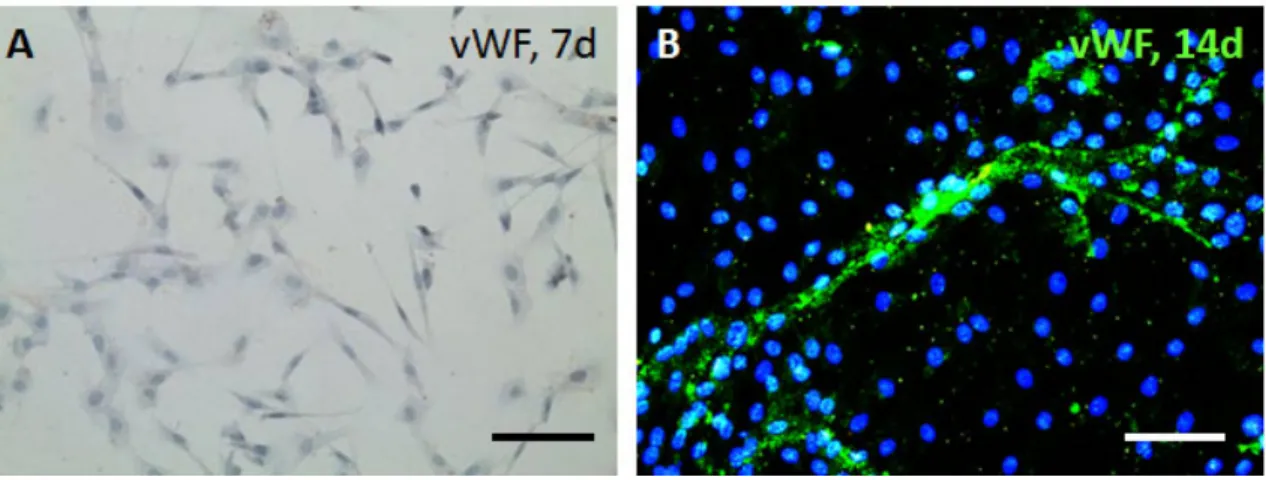
Figure 3-14 MSCs cultured in endothelial differentiation medium and stained immunohistochemically for vWF at (A) 7 days and (B) 14 days. Scale bar, 100 µm. ... 56
14
Figure 3-15 EBs cultured in endothelial differentiation medium and stained
immunohistochemically for vWF at (A) 7 days and (B) 14 days, (C) EBs cultured in complete medium and stained for vWF. Scale bar, 200 µm. ... 56 Figure 3-16 ESEM image of collagen scaffolds ... 57 Figure 3-17 Percent original diameter of scaffolds seeded with MSCs cultured in control,
cardiac, neural, and endothelial media for up to 14 days (n = 10, mean ± S.D.) ... 57 Figure 3-18 Gross appearance of (A) MSCs and (B) ECCs seeded in collagen scaffolds cultured
in cardiac media in 12-well plates for 7 days. H&E staining of (C) MSCs and (D) ECCs in cardiac medium at 7 days. ... 58 Figure 3-19 MSC and ECC differentiation in collagen scaffolds in (A,D) cardiac media, stained
for MHC at 14 days, (B,E) neural media, stained for TUJ1 at 1 and 7 days, (C,F)
endothelial media, stained for vWF at 14 days (n = 2). ... 60 Figure 3-20 Immunohistochemical staining for α-SMA for (A-D) MSCs and (E-H) ECCs
cultured in (A,E) control medium, (B,F) cardiac medium, (C,G) neural medium, and (D,H) endothelial medium. Scale bars, 50 µm. ... 60 Figure 4-1 Cryotube molds for cell-seeded Gtn-HPA gels ... 80 Figure 4-2 Rheological behavior of collagen-genipin gels: (A) shear storage modulus G’; and (B) gel time (n = 3, mean ± S.D.) ... 82 Figure 4-3 Rheological behavior of Gtn-HPA gels: (A) shear storage modulus G’; and (B) gel
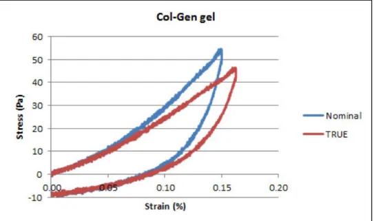
time (n = 3, mean ± S.D.)... 83 Figure 4-4 Typical nominal and true stress/strain curves for collagen-based gels ... 84 Figure 4-5 Typical effect of strain rate (10%/s vs. 1%/s) on collagen-0.25 mM genipin gels (n =
3) ... 84 Figure 4-6 Collagen-genipin compression results: (A) Typical loading and unloading curve
(collagen with 0.25 mM genipin), and (B) compression modulus for various
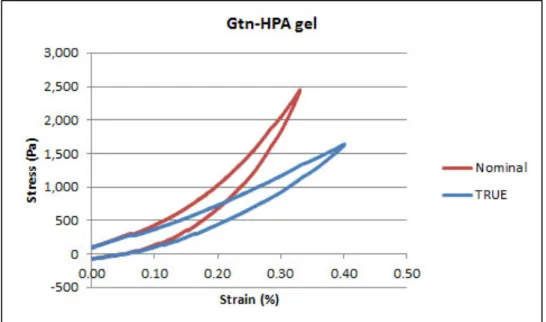
concentrations of genipin for 10%/s and 1%/s strain rates (n = 3, mean ± S.D.) ... 85 Figure 4-7 Typical nominal and true stress/strain curves for Gtn-HPA gels ... 86 Figure 4-8 Gtn-HPA compression testing: (A) typical true stress-strain curve, and (B)
compression moduli for 0.85 – 3.0 mM H2O2 (n = 3, mean ± S.D.) ... 87 Figure 4-9 Mooney-Rivlin characterization of Gtn-HPA gels: (A) Mooney plot; (B) shear
modulus, G (n = 3, mean ± S.D.) ... 87 Figure 4-10 ESEM images of collagen-genipin gels ... 88 Figure 4-11 Representative ESEM image of Gtn-HPA gel ... 89 Figure 4-12 Color change in collagen gels with: (A) no genipin; (B) 0.25 mM genipin; (C) 0.5
mM genipin; (D) 1.0 mM genipin ... 89 Figure 4-13 Weight remaining (%) of Collagen-genipin gels at various degradation times for
different concentrations of genipin (mM) (n = 3, mean ± S.D.) ... 90 Figure 4-14 Weight remaining (%) of collagen-genipin gels at various degradation times for
different concentrations of genipin (n = 3, mean ± S.D.) ... 91 Figure 4-15 Half-life of collagen-genipin gels matured for 1 and 24 hours (n=3, mean ± S.D.) . 92
15
Figure 4-16 Enzymatic degradation of Gtn-HPA gels for various concentrations of H2O2: (A) Weight remaining (%); and (B) Half-life (n = 3, mean ± S.D.) ... 93 Figure 4-17 Swelling ratio of (A) collagen-genipin and (B) Gtn-HPA gels (n = 3, mean ± S.D.)94 Figure 4-18 Contraction data for MSCs incorporated in (A) collagen-genipin gels (0-2mM
genipin); (B) collagen-TGase (unpurified) gels (0-50 mg/ml TGase); (C) collagen-TGase (purified) gels (0-100 µg/ml TGase); and (D) Gtn-HPA gels (1.0 and 1.7 mM H2O2) (n = 3, mean ± S.D.) ... 95 Figure 4-19 MSCs in collagen-genipin gels at day 7: (A) collagen-only; (B) collagen-0.25mM
genipin; (C) collagen-0.5mM genipin (n = 3) ... 95 Figure 4-20 MSCs in collagen-TGase (purified) gels at day 7: (A) collagen-only; (B) collagen-5
µg/ml TGase; (C) collagen-50 µg/ml TGase; (D) collagen-100 µg/ml TGase (n = 3) ... 96 Figure 4-21 MSCs in Gtn-HPA gels at day 7: (A) 1.0 mM H2O2 and (B) 1.7 mM H2O2 (n = 3) 96 Figure 4-22 Storage modulus for MSCs and ECCs at for various concentrations of genipin at 1
and 6 days (n = 4, mean ± S.D.) ... 97 Figure 4-23 Cell viability for MSCs incorporated collagen-genipin gels (0-2 mM genipin) (A)
before and (B) after excess genipin was removed (n = 3, mean ± S.D.) ... 98 Figure 4-24 Cell viability for MSCs incorporated in (A) collagen-TGase (unpurified) gels (0-50
mg/ml TGase); (B) collgen-TGase (purified) gels (0-100 µg/ml TGase); and (C) Gtn-HPA gels (1.0 and 1.7 mM H2O2) (n = 3, mean ± S.D.) ... 99 Figure 4-25 DNA content for MSCs incorporated in (A) collagen-genipin gels (0-2 mM genipin); (B) collagen-TGase (unpurified) gels (0-50 mg/ml TGase); (C) collagen-TGase (purified) gels (0-100 µg/ml TGase); and (D) Gtn-HPA gels (1.0 and 1.7 mM H2O2) (n = 4-6, mean ± S.D.) ... 100 Figure 4-26 Correlation of G (from Mooney-Rivlin analysis) and G’ (from rheological testing)
(n=3) ... 108 Figure 4-27 Materials characterization chart comparing (1) collagen scaffolds cross-linked with
DHT and EDAC; (2) collagen-genipin gels; (3) collagen-TGase gels; (4) Gtn-HPA gels ... 110 Figure 5-1 Sample images of EBs stained with MHC that would (A) not be counted as a positive
stain; and (B, C) would be counted as a positive stain. ... 121 Figure 5-2 Sample images of EBs stained with TUJ1 that would have typical % Area stained
values of (A) 5%, (B) 20%, and (C) 40%. ... 122 Figure 5-3 EB diameter for EBs incorporated in (A) Col-only gels, (B) Col-Gen gels, (C)
Gtn-HPA-comp gels, and (D) Gtn-HPA-stiff gels (n = 9, mean ± S.D.) ... 123 Figure 5-4 MHC expression of EBs cultured in C medium at 14 days in various gel types: (A)
Col-only, (B) Col-Gen, (C), Gtn-HPA-comp, and (D) Gtn-HPA-stiff gels. ... 124 Figure 5-5 MHC expression (% EBs stained) for EBs incorporated in various gel types cultured
in (A) N, (B) C/N, (C) C/N/E, and (D) Cntl media for 7 and 14 days (n = 3, mean ± S.D.). ... 125
16
Figure 5-6 MHC expression (% Area) stained for EBs cultured in (A) C, (B) C/N, (C) C/N/E, and (D) Cntl media in various gel types for 7 and 14 days (n = 15-20, mean ± S.D.). .. 126 Figure 5-7 MHC expression of EBs incorporated in Col-only gels at 14 days cultured in (A) C,
(B) N, (C) E, (D) C/N, (E) C/N/E, and (F) Cntl media ... 127 Figure 5-8 MHC expression of EBs incorporated in Col-only gels, cultured in C medium at (A) 7 and (B) 14 days ... 128 Figure 5-9 TUJ1 expression of EBs cultured in N medium at 14 days in various gel types: (A)
Col-only, (B) Col-Gen, (C), Gtn-HPA-comp, and (D) Gtn-HPA-stiff gels. ... 129 Figure 5-10 TUJ1 expression (% EBs stained) for EBs incorporated in various gel types cultured
in (A) N, (B) C/N, (C) C/N/E, and (D) Cntl media for 7 and 14 days (n=3, mean ± S.D.). ... 131 Figure 5-11 TUJ1 expression (% Area stained) for EBs incorporated in various gel types cultured
in (A) N, (B) C/N, (C) C/N/E, and (D) Cntl media for 7 and 14 days (n = 15-20, mean ± S.D.). ... 131 Figure 5-12 TUJ1 expression of EBs incorporated in Col-only gels at 14 days cultured in (A) C,
(B) N, (C) E, (D) C/N, (E) C/N/E, and (F) Cntl media. ... 132 Figure 5-13 TUJ1 expression of EBs incorporated in Col-only gels, cultured in N medium at (A)
7 and (B) 14 days ... 133 Figure 5-14 CD31 expression of EBs cultured in C/N/E media at 14 days in various gel types:
(A) Col-only, (B) Col-Gen, (C), Gtn-HPA-comp, and (D) Gtn-HPA-stiff gels. ... 134 Figure 5-15 CD31 expression (% EBs stained) while incorporated in various gel types cultured in
(A) N, (B) C/N, (C) C/N/E, and (D) Cntl media for 7 and 14 days (n = 3, mean ± S.D.). ... 135 Figure 5-16 CD31 expression (% Area stained) for EBs incorporated in various gel types
cultured in (A) N, (B) C/N, (C) C/N/E, and (D) Cntl media for 7 and 14 days (n = 15-20, mean ± S.D.). ... 136 Figure 5-17 CD31 expression of EBs incorporated in Col-only gels at 14 days cultured in (A) C,
(B) N, (C) E, (D) C/N, (E) C/N/E, and (F) Cntl media. ... 137 Figure 5-18 CD31 expression of EBs incorporated in Col-only gels, cultured in C/N/E media at
(A) 7 and (B) 14 days ... 138 Figure 5-19 TUJ1 (red) and CD31 (green) expression in EBs incorporated in Col-only gels
cultured in C/N/E medium at (A) 7 and (B) 14 days. ... 139 Figure 5-20 Linear regression plots for the expression (% Area) of (A) cardiac vs. neural; (B)
cardiac vs. endothelial; and (C) neural vs. endothelial markers for EBs incorporated in all gel types cultured in all differentiation media for 7 and 14 days. ... 139 Figure 5-21 Linear regression plot for the expression (% Area) of TUJ1 and CD31 for EBs
incorporated in Col-only gels cultured in all media for 14 days. ... 139 Figure 6-1 Col-Gen gels injected into ex vivo cardiac tissue: (A) Col-Gen gels dyed with trypan
blue; (B) Gross view of cardiac muscle; (C) Gross view of fixed cardiac tissue; (D) H&E stained paraffin-embedded section; (C) H&E stained cryosection. ... 157
17
Figure 6-2 Induction of myocardial infarct in swine through ligation of the LAD coronary artery: (A) the third left rib was marked for left anteromedial thoracotomy; (B) cauterizers were used to cut through muscle; (C) clamps were used to expose heart. ... 158 Figure 6-3 Induction of myocardial infarct in swine through occlusion of LAD coronary artery
via Gelfoam delivery through catheter: (A) femoral vein and artery were cannulated; (B) animal was positioned under a C-arm to identify the LAD coronary artery; (C) a delivery of Gelfoam, saline, and contrast was used to occlude the vessel ... 158 Figure 9-1 Hemacytometer ... 173 Figure 9-2 MHC expression (% EBs stained) for EBs incorporated in (A) collagen-only, (B)
collagen-genipin, (C) Gtn-HPA-comp, and (D) Gtn-HPA-stiff gels cultured in various media for 7 and 14 days (n = 3, mean ± S.D.). ... 217 Figure 9-3 MHC expression (% Area stained) for EBs incorporated in (A) collagen-only, (B)
collagen-genipin, (C) Gtn-HPA-comp, and (D) Gtn-HPA-stiff gels cultured in various media for 7 and 14 days (n = 15-20, mean ± S.D.). ... 218 Figure 9-4 TUJ1 expression (% EBs stained) for EBs incorporated in (A) collagen-only, (B)
collagen-genipin, (C) Gtn-HPA-comp, and (D) Gtn-HPA-stiff gels cultured in various media for 7 and 14 days (n = 3, mean ± S.D.). ... 219 Figure 9-5 TUJ1 expression (% Area stained) for EBs incorporated in (A) collagen-only, (B)
collagen-genipin, (C) Gtn-HPA-comp, and (D) Gtn-HPA-stiff gels cultured in various media for 7 and 14 days (n = 15=20, mean ± S.D.). ... 220 Figure 9-6 CD31 expression (% EB stained) for EBs incorporated in (A) collagen-only, (B)
collagen-genipin, (C) Gtn-HPA-comp, and (D) Gtn-HPA-stiff gels cultured in various media for 7 and 14 days (n = 3, mean ± S.D.). ... 221 Figure 9-7 CD31 expression (% Area stained) for EBs incorporated in (A) collagen-only, (B)
collagen-genipin, (C) Gtn-HPA-comp, and (D) Gtn-HPA-stiff gels cultured in various media for 7 and 14 days (n = 3, mean ± S.D.). ... 222 Figure 9-8 Linear regression plots for the expression (% Area stained) of (A) cardiac vs. neural;
(B) cardiac vs. endothelial; (C) neural vs. endothelial markers of EBs cultured in
collagen-only gels in all differentiation media for all culture times ... 223 Figure 9-9 Linear regression plots for the expression (% Area stained) of (A) cardiac vs. neural;
(B) cardiac vs. endothelial; (C) neural vs. endothelial markers of EBs cultured in
collagen-only gels in all differentiation media for 14 days ... 223 Figure 9-10 Linear regression plots for the expression (% Area stained) of (A) cardiac vs. neural;
(B) cardiac vs. endothelial; (C) neural vs. endothelial markers of EBs cultured in
collagen-genipin gels in all differentiation media for all culture times ... 224 Figure 9-11 Linear regression plots for the expression (% Area stained) of (A) cardiac vs. neural;
(B) cardiac vs. endothelial; (C) neural vs. endothelial markers of EBs cultured in
18
Figure 9-12 Linear regression plots for the expression (% Area stained) of (A) cardiac vs. neural; (B) cardiac vs. endothelial; (C) neural vs. endothelial markers of EBs cultured in Gtn-HPA-comp gels in all differentiation media for all culture times ... 225 Figure 9-13 Linear regression plots for the expression (% Area stained) of (A) cardiac vs. neural;
(B) cardiac vs. endothelial; (C) neural vs. endothelial markers of EBs cultured in Gtn-HPA-comp gels in all differentiation media for 14 d ... 225 Figure 9-14 Linear regression plots for the expression (% Area stained) of (A) cardiac vs. neural;
(B) cardiac vs. endothelial; (C) neural vs. endothelial markers of EBs cultured in Gtn-HPA-stiff gels in all differentiation media for all culture times ... 226 Figure 9-15 Linear regression plots for the expression (% Area stained) of (A) cardiac vs. neural;
(B) cardiac vs. endothelial; (C) neural vs. endothelial markers of EBs cultured in Gtn-HPA-stiff gels in all differentiation media for 14 d ... 226 Figure 9-16 Linear regression plots for the expression of cardiac and neural markers (% Area
stained) of EBs incorporated in all gels, cultured in (A) C; (B) N; (C) E; (D) C/N; (E) C/N/E, and (F) Cntl media, for 14 days. ... 227 Figure 9-17 Linear regression plots for the expression of cardiac and endothelial markers (%
Area stained) of EBs incorporated in all gels, cultured in (A) C; (B) N; (C) E; (D) C/N; (E) C/N/E, and (F) Cntl media, for 14 days. ... 228 Figure 9-18 Linear regression plots for the expression of neural and endothelial markers (% Area
stained) of EBs incorporated in all gels, cultured in (A) C; (B) N; (C) E; (D) C/N; (E) C/N/E, and (F) Cntl media, for 14 days. ... 229
19
List of Tables
Table 2-1 Cross-linking of collagen [37] ... 29 Table 2-2 Differentiation of MSCs into cardiomyocyte-like cells [20] ... 32 Table 3-1 Differentiation efficiency of MSCs and ECCs in cardiac, neural, and endothelial
differentiation media (% stained cells) (n = 3, mean ± S.D.) ... 59 Table 4-1 Design characteristics for gels for cardiac tissue engineering ... 72 Table 4-2 Weight remaining (%) and half-life times for collagen-genipin gels for 1 and 24 hours
maturation times. (n = 3, mean ± S.D.) ... 92 Table 4-3 Weight remaining (%) and half-life times for Gtn-HPA gels. (n = 3, mean ± S.D.) ... 93 Table 5-1: Independent variables and metrics for evaluation ... 116 Table 5-2: Mixed medium conditions ... 118
21
Chapter 1 Introduction
Cardiovascular disease is the leading cause of death in the Western world. Nearly 8 million people in the United States alone have suffered a myocardial infarction (MI), with 1.2 million new and recurrent cases each year (American Heart Association, 2011). In any given year, the survival rate from an MI is only 60%. Since myocardial tissue lacks intrinsic regenerative capability, MIs result in the substantial death of cardiomyocytes in the infarct zone followed by pathological remodeling of the heart. The remodeling process involves cardiac dilation, wall thinning, and severe deterioration of contractile function leading to congestive heart failure in 500,000 patients in the U.S. each year. Medical management for heart failure may improve symptoms and slow the progression to failure but does not restore a functioning myocardium. Once an MI has occurred, there is no treatment to recover the myocardial function from that portion of the heart. The current treatment of end-stage heart failure is heart transplantation, which is problematic due to the lack of organ donors and the complications associated with immunosuppressants. New solutions are needed to regenerate MI-damaged hearts. In particular, research in cardiac tissue engineering aims to restore contractility of the myocardium and prevent the adverse remodeling of the heart in patients who survive MIs.
Although many advances have been made in this field, regeneration is not complete and researchers face many problems including an appropriate cell source and a method of delivery to bring cells and therapeutics to the infarcted heart. Researchers combine cells, scaffolds, growth factors, and external stimuli including mechanical [1, 2] or electrical [3] exercise to differentiate stem cells into cardiomyocyte-like cells. Many different cell types are currently being investigated for cell-based therapies. Among these are neonatal cardiomyocytes [4], embryonic stem cells (ESCs) [5], ESC-derived cardiomyocytes [6], skeletal muscle cells [7], bone marrow cells [8], and mesenchymal stem cells (MSCs) [9, 10]. In particular, ESCs and MSCs have been studied extensively as promising cell candidates for cardiac tissue engineering. Totipotent ESCs can differentiate into beating cardiomyocyte clusters known as embryoid bodies (EBs) in vitro and have been integrated to pace an infarcted heart [6]. One particular type of embryonic stem cell line is the pluripotent P19 embryonic carcinoma cell (ECC) derived from an embryonal carcinoma in a C3H/He mouse. These ECCs are well characterized and have been used as a model embryonic cell type for various developmental and differentiation assays. Pluripotent bone marrow-derived MSCs can also differentiate into cardiomyocyte-like cells in vitro and improve cardiac function in vivo in a variety of different ways including stimulating neovascularization. Furthermore the implantation of autologous MSCs may reduce the chances of an adverse immune response.
These cells can be delivered to the myocardium in a variety of ways: injected directly into the diseased myocardium, delivered through an injectable gel, or seeded on a three-dimensional scaffold and then engrafted onto the infarct site. Although the actual mechanism of cardiac renewal is unknown and many theories have been proposed [11-13], researchers agree
22
that the ultimate functional goal of cardiac tissue engineering is the complete remodeling and restoration of cardiac function.
Collagen scaffolds and injectable gels have been investigated as promising methods of cell delivery to the myocardium. Porous collagen scaffolds with high surface areas allow the encapsulation of a high density of cells that can be engrafted onto a beating heart. Minimally invasive delivery vehicles such as injectable hydrogels provide a unique way to deliver cells and therapeutics without major surgery. Although many natural and synthetic polymers have been investigated, very few gel materials and linkers have the capability to form covalent cross-links in situ, thus allowing the mechanical, rheological, and degradative properties of the gels to be tuned and optimized. An ideal material may be one that mimics the natural environment during embryonic cardiac development and differentiation. The extracellular matrix in which cardiogenesis takes place is referred to as “cardiac jelly.”
Cardiac jelly is a homogenous network of type I collagen fibrils and fine filaments present between the endothelium and myocardium of the embryonic heart, which transforms into the connective tissue of the endocardium [14, 15]. The structural stability of cardiac jelly is ensured by the presence of elastin and collagen, whereas its gel-like appearance is controlled by the glycosaminoglycan content, which determines the degree of hydration of the jelly. A few researchers have investigated cardiac jelly-like, collagen-based gels to incorporate EBs to facilitate in vitro cardiac differentiation [16, 17].
In addition to the functional contractile unit cardiomyocyte, myocardium contains other cell types including endothelial and smooth muscle cells that comprise the vasculature to support a highly metabolic organ, and cardiac neurons known as Purkinje cells that innervate cardiac tissue to regulate the intrinsic electrical conduction system of the heart. The closely intertwined neural and vasculature networks function together to allow efficient contraction of all four chambers of the heart, thereby allowing selective blood perfusion through both the lungs and systemic circulation. Although researchers have considered incorporating vasculature into cardiac tissue engineering constructs, no methods have incorporated neural networks in cardiac tissue.
This overall goal of this thesis was to investigate two types of stem cells (MSCs and ECCs) cultured in various 3-D environments including sponge-like type I collagen scaffolds and novel injectable hydrogels with covalent cross-linkers, that support cell proliferation and differentiation. Specifically, the three hydrogel materials characterized included type I collagen cross-linked with a small molecule known as genipin, type I collagen cross-linked with a large enzyme known as transglutaminase, and gelatin-hydroxyphenylpropionic acid, whose cross-linking is modulated by horse radish peroxidase and H2O2. Furthermore, it investigates the novel concept of simultaneous differentiation in which a single cell culture is exposed to mixed medium conditions and differentiated into cardiac, neural, and endothelial lineages in a spatially relevant distribution.
23
The hypothesis is that the tunable properties of the injectable gels can be optimized to match that of cardiac jelly, which would provide the environment to produce the most mature cardiomyocytes, neural cells, and endothelial cells that are differentiated from ECCs. Furthermore, ECCs seeded in collagen gels with a covalent cross-linker can differentiate into cardiac, neural and endothelial cells in the same culture when exposed to appropriate media. The key parameters in the injectable gels incorporated with cells include the cell type, the scaffold form (sponge-like scaffold or injectable gel), the gel material and cross-linker, and the differentiation medium. The results are summarized in four manuscripts, whose abstracts are included as Chapters 3-6.
Chapter 2 describes the background and motivation underlying this thesis, including the basics of cardiac muscle structure and function and an overview of tissue culture methods for cardiac tissue engineering up to date.
Chapter 3 compares the differentiation efficiency of two stem cell types (adult porcine and caprine bone marrow-derived MSCs and P19 ECCs) in two in vitro culture environments (2-D monolayer and 3-(2-D collagen scaffolds) into the main cellular components of cardiac tissue, including cardiomyocytes, neural cells and vascular endothelial cells. MSCs and ECCs were also cultured in chondrogenic medium and investigated for associations among contraction, α-smooth muscle actin expression, and chondrogenesis in vitro.
Chapter 4 characterizes three mechanically tunable injectable gel systems that have the capability to form covalent cross-links in situ. These gel systems (collagen cross-linked with genipin, collagen cross-linked with transglutaminase, and gelatin-hydroxyphenylpropionic acid whose cross-linking is modulated with HRP and H2O2) were characterized in terms of mechanical and rheological behavior, resistance to enzymatic degradation, and cell behavior when incorporated in these gels. Furthermore, this chapter provides a thorough literature review about cardiac jelly as an optimal material for in vitro cardiac differentiation.
Chapter 5 describes the differentiation of MSCs and ECCs into cardiac, neural and endothelial cells separately and simultaneously in select gel types that were characterized in the previous chapter.
Chapter 6 investigates the in vivo feasibility of injectable collagen gels cross-linked with genipin and describes a pilot large animal study in which different methods of inducing MIs were investigated. Furthermore, this chapter provides an experimental set-up to investigate functional improvements of implanting a cell-incorporated gel into an infarcted heart.
Chapter 7 discusses the limitations of this thesis and identifies areas that need to be addressed in future work.
25
Chapter 2 Background and motivation
2.1 Tissue response to myocardial infarct
A myocardial infarction occurs when the blood supply to part of the heart muscle is blocked, usually by the sudden formation of a blood clot within a coronary artery, leading to death of part of the heart muscle. Large-scale tissue necrosis and massive cells loss occur in which more than a billion cells are lost. Heart failure ensues when contractile reserve is depleted below a critical threshold.
Because of its high metabolic rate and oxygen consumption, myocardium (Figure 2-1A) begins to undergo irreversible injury within 20 minutes of ischemia. Cell death spreads from the inner layers to the outer layers of the myocardium over 3-6 hours resulting in myocardial necrosis (Figure 2-1B) and eliciting an inflammatory response. Leukocytes, including neutrophils and macrophages, enter the infarct to phagocytose necrotic cell debris. Subsequently, granulation tissue consisting of fibroblasts and endothelial cells invade the necrotic area to initiate wound repair (Figure 2-1C). Over time, the granulation tissue remodels to form fibrotic scar tissue (Figure 2-1D). In humans, this myocardial repair process is completed in two months. MI results in thinning of the ventricular wall and dilation of the ventricular cavity (Figure 2-1E), termed “ventricular remodeling,” which increases the mechanical stress on the ventricular wall and debilitates contractile function. Healthy cardiac tissue in humans has a stiffness of 8 kPa in diastole and 40 kPa in systole [18]. After fibrotic tissue has formed, the stiffness of the left ventricle in both diastole and systole increased to 10 kPa and 60 kPa, respectively [18, 19]. Once an MI has occurred, there is no treatment to recover the myocardial function from that portion of the heart.
26
Figure 2-1 Histological stages of myocardial infarction [20].
2.2 Cell types in cardiac tissue
Myocardium contains many cell types including cardiomyocytes, fibroblasts, and vasculature composed of smooth muscle cells and endothelial cells lining the lumen. The functional contractile unit is the cardiomyocyte which comprises 25% of the cells in the myocardium and occupies up to 90% of the myocardial volume. A basement membrane composed of type IV collagen, laminin and fibronectin interfaces the cardiomyocyte and the surrounding collagen extracellular matrix. Cardiac fibroblasts comprise of 65% of cells in myocardium and function as mechanoelectric transducers in the heart by responding to mechanical deformation to polarize and depolarize during contraction of the cardiac muscle and help maintain electrical stability.
Myocardium contains nerves that not only support the blood vessels but also conduct electrical impulses from the SA (sino-atrial) and AV (atrial-ventricular) nodes to synchronize the beating of the heart. The heart rate is regulated by two nervous systems: sympathetic and parasympathetic. The parasympathetic nervous system is controlled by cholinergic neurotransmitters, which decrease blood pressure, heart rate, constrict coronary arteries. Adrenergic neurotransmitters regulate the sympathetic nervous system and increase blood pressure, heart rate, dilate coronary arteries. Incorporating neural networks in cardiac tissue is essential in regulating electrical impulses that originate from the atrioventricular (AV) and sinoatrial (SA) nodes.
2.3 Extracellular matrix molecules in cardiac tissue
Adult cardiac tissue is primarily composed of type I/III collagen, which comprises of 25% of the myocardial volume. Furthermore, cardiac tissue also contains vimentin, laminin, and
27
fibronectin [21]. Vimentin, the intermediate filament of mesenchymal cells, is present in fibroblasts, fibrocytes, and endothelial cells. Laminin is observed in the basal membrane of myocytes, smooth muscle and endothelial cells. Fibronectin is localized homogeneously throughout the extracellular space as matrix material in which the cellular elements and the various other proteins such as collagens are embedded.
Fetal cardiac development occurs in a gelatinous substance known as “cardiac jelly.” Cardiac jelly is a homogenous network of type I collagen fibrils and fine filaments present between the endothelium and myocardium of the embryonic heart, which transforms into the connective tissue of the endocardium [14]. The structural stability of cardiac jelly is ensured by the presence of elastin and collagen, whereas its gel-like appearance is controlled by the glycosaminoglycan content, which determines the degree of hydration of the jelly. The modulus of cardiac jelly has found to be 1-30 Pa, measured by various method s including microindentation [22] and mesomechanical testing [23]. A few researchers have investigated cardiac jelly-like gels to incorporate EBs to facilitate in vitro cardiac differentiation [16, 17, 24, 25].
2.4 Cardiac tissue engineering – An overview
Tissue engineering methods are employed to grow new myocardium in diseased hearts and restore cardiac function. Various strategies have been investigated, including stem cell injections [9, 26, 27], reconstruction of beating tissues ex vivo to be implanted in vivo [4], and cell-seeded scaffolds that are injected or implanted [28, 29]. Cell-seeded scaffolds can serve three purposes: to accommodate ingrowth and differentiation of endogenous progenitor cells [26, 30], to deliver therapeutic agents [31], and to host exogenous cells [32]. Furthermore, these cells, scaffolds, and growth factors can be combined with external stimuli including mechanical [1, 2] or electrical [3] exercise to differentiate stem cells into cardiomyocyte-like cells. In this section, various scaffold materials, including porous scaffolds and injectable hydrogels, and cell types, including mesenchymal stem cells and embryonic carcinoma cells, are explored.
2.5 Scaffolding materials
Scaffolding materials in the form of porous sponge-like scaffolds and injectable hydrogels have the capability to deliver large number of cells and growth factors to a defect site. 2.5.1 Porous sponge-like collagen scaffolds
The use of 3-D scaffolds that mimic the natural in vivo environment of cells has been shown to facilitate the reparative process and result in the successful repair and neovascularization of various functioning tissues. The 3-D environment of these porous sponge-like scaffolds serves as a desirable structural support for seeded or migrating endogenous cells and allows for a much greater surface to volume ratio for increased cell attachment as compared to a 2-D surface.
28
There are several design requirements for a scaffold to be used as an implant for cardiac regeneration. The scaffold needs to be biodegradable, non-toxic, able to be sutured to the defect site, facilitate cell attachment, regulate cell expression, and possess sufficient mechanical strength. Natural polymers such as collagen provide a native surface to cells, since it is a major component of the natural extracellular matrix (ECM) in cardiac tissue, and possess ligands that favor cellular attachment.
Type I collagen is a major constituent of cardiac ECM and is commended by its successful use as a regeneration template for dermis [33] and peripheral nerve [34] and its versatility in being able to be cross-linked using a variety of treatments, thus providing design control over its degradation rate and mechanical properties. Furthermore, beating cardiomyocytes can be seeded onto scaffolds, which can then be transplanted onto a beating heart [35, 36].
Various methods have been investigated to modulate the mechanical properties of porous collagen scaffolds. For example the cross-link density and thus stiffness of the scaffold can be modified by dehydrothermal or EDC treatment, modulating the degradation rate. Furthermore, properties such as pore size and pore connectivity can be tailored for various applications.
2.5.2 Injectable hydrogels
Injectable hydrogels have been explored as a minimally invasive method for the delivery of cells and growth factors. Cross-linking is an important characteristic that allows tenability of a polymer’s stiffness and resistance to degradation. And in particular to injectable gels, the cross-linker must be a covalent cross-linker able to form cross-links in situ.
Collagen gels
Collagen-based matrices are widely used in tissue regenerative applications due to its ubiquitous presence in the human body, antigenic behavior, and biodegradability. Thus it is critical to be able to manipulate the mechanical properties of collagen gels by cross-linking mechanisms.
Collagens comprise the majority of proteins in connective tissue such as skin, bone, cartilage, and tendons, and are therefore popular candidates for injectable biomaterials. Furthermore, collagen’s superior biocompatibility and nearly ubiquitous bioactivity have made it one of the most extensively investigated biomaterial scaffolds for tissue engineering. However, the effectiveness of collagen-based tissue engineered materials has been severely limited by their lack of mechanical strength.
A variety of methods exist to cross-link collagen gels [37]. Physical treatments such as ultraviolet (UV) and γ-irradiation and dehydrothermal treatments are not practical in because of their limited use in cellular tissues. Chemical treatments with aldehydes are often used to
29
preserve and stiffen tissues but these treatments are highly cytotoxic. In vivo, tissues are naturally cross-linked by enzymes such as lysyl oxidase and transglutaminase. However, use of these enzymes for bulk changes in mechanical properties in cultured tissue mimics is cost prohibitive. Chemical aldehydes are used as a fixative to preserve tissues but are highly toxic.
Table 2-1 Cross-linking of collagen [37]
Physical treatment UV radiation (254 nm) γ-‐irradiation Dehydrothermal treatment Chemical treatment Bifunctional agent
Aldehyde ex: glutaraldehyde (GTA)
Diisocyanate ex: hexamethylene diisocyanate (HMDC) Polyepoxy ex: polyglycerol polyglycidyl ether Activation of carboxylic acid groups
Carbodiimide ex: cyanamide 1-‐ethyl-‐3-‐(3-‐dimethyl aminopropyl) carboxdiimide HCl (EDC)
Acyl azide method ex: hydrazine diphenylphosphorylazide (DPPA)
Genipin has been investigated to modulate mechanical stiffness of collagen and gelatin. Genipin is the active compound found in Gardenia Jasminoides fruit extract and it crosslinks collagen through nucleophilic attack by primary amine groups on lysine and arginine residues on the C3 atom of genipin resulting in a gel strength comparable to glutaraldehyde, but it is 10,000-fold less cytotoxic (Figure 2-1) [38]. In addition to an increase in mechanical strength of collagen, genipin crosslinking is associated with a color change in which opaque collagen turns blue, and the crosslinks emit fluorescence at 630 nm when excited at 590 nm.
Figure 2-2 Collagen cross-linked with genipin [39]
Genipin may cross-link collagen in a variety of different mechanisms. Genipin molecules may react with amino groups within a tropocollagen molecule or between adjacent tropocollagen molecules to form intrahelical and interhelical crosslinks in the genipin-fixed tissue. In addition, intermicrofibrillar cross-links may be formed between collagen microfibrils via polymerization of genipin molecules before cross-linking (oligomeric cross-link).
30
Figure 2-3 Cross-linking structures genipin-fixed collagen [40]
Furthermore, the degradation rate of genipin-cross-linked gelatin was found to be significantly slower than glutaraldehyde-cross-linked counterparts [41]. The mechanical and rheological behavior of genipin-cross-linked gelatin has been investigated, revealing that with an increase in genipin concentration and temperature, the gelatin network shifts from being dominated by hydrogen bonds (physical links) to covalent linking (chemical cross-links) [42].
Although genipin is an attractive cross-linker for collagen, its cytotoxicity at high concentrations (5 mM) limits its usage to small concentrations [39]. Transglutaminase (TGase), a ubiquitous enzyme with a role in blood clotting, has also been investigated for cross-linking collagen gels [43]. TGases are able to covalently attach primary amine containing compounds to peptide bound glutamine, facilitating modification of the physical, chemical and biological properties of proteins [44]. Specifically, TGase catalyzes the formation of a covalent bond between the ε-amino group of a lysine residue and a γ-carboxamide group of glutamine, creating intramolecular covalent links within and between polymers. It has been mainly used in the food industry in processing meats and fish but has recently been used in the biomedical field for cross-linking natural polymers. TGase has been used to cross-link gelatin matrices to increase their strength [45] and, also, to incorporate cell adhesion factors within the gel matrix, enhancing cell proliferation [46].
Gelatin-hydroxyphenylpropionic (Gtn-HPA) gels
In the last few years, there has been a class of polymers that are enzymatically cross-linkable via pendant phenolic groups, which is catalyzed by H2O2 and HRP. The development of this cross-linking method 1has spurred the creation of many gels including hyaluronic acid-tyramine gels [47], carbomethyl-cellulose-acid-tyramine gels [48], gelatin-acid-tyramine gels [49], and dextra-tyramine/heparin-tyramine [50]. The H2O2 and HRP determined the hydrogel stiffness and the gelation rate of the hydrogel, respectively. Gtn-HPA gels have recently been investigated as an injectable material for cell delivery. Gelatin is a derivative of collagen containing single strands of collagen instead of the tropohelix of collagen, and has many of the same properties including cell adhesion. Although the mechanical and rheological properties of Gtn-HPA gels have been characterized, few studies have investigated cell behavior and differentiation while incorporated in the gels.
31 2.6 Cell therapy
Many different cell types are currently investigated for cell-based therapies. Among these are neonatal cardiomyocytes [4], ESCs [5], ESC-derived cardiomyocytes [6], skeletal muscle cells [7], bone marrow cells [8], and MSCs [9, 10]. These cells are injected into the diseased myocardium or seeded on a three-dimensional scaffold and then engrafted onto the infarct site. The actual mechanism of cardiac renewal is unknown but many theories have been proposed: 1) the exogenous cells stimulate endogenous progenitor cells to revascularize a myocardial infarct [11]; 2) the cells fuse with the existing cardiac myocytes, giving rise to hybrid cells expressing cardiac markers [12]; 3) the cells and subsequent new tissue formation attenuates pathological ventricular remodeling [13]. Although the actual mechanism of cardiac renewal is still under debate and research, researchers agree that the ultimate functional goal of cardiac tissue engineering is the complete remodeling and restoration of cardiac function.
Three main methods have been explored to deliver cells into the infracted myocardium. The first method is direct injection of cells into the myocardium [51]. However, direct injection has several drawbacks, including very low efficacy of cell engraftment and substantial cell death soon after the injection of cells. Nevertheless, there are currently ongoing clinical trials in humans investigating the safety and feasibility of direct injection of cells for cardiac therapy. The second method involves an intravenous injection of a cell suspension. Even though there have been positive results with this relatively painless method of delivery [52], a very small percentage of cells actually reach the myocardium. The final method of cell delivery is the implantation of a three-dimensional cell-seeded scaffold or injectable hydrogel. These constructs allow maintenance of cellular viability and differentiation, favor cell migration and promote angiogenesis [28].
2.6.1 Mesenchymal stem cells
Because of their pluripotency, accessibility and renewability, MSCs are a promising cell type for modalities implemented for treating heart failure. An MSC is a bone marrow-derived stromal cell that can differentiate into many tissue lineages, including chondrocytes, adipocytes, and osteocytes (Figure 2-4). Furthermore, evidence suggests MSCs can express phenotypic characteristics of endothelial, neural, smooth muscle, skeletal myoblasts, and cardiomyocytes [10].
32
Figure 2-4 Mesenchymal stem cell lineage [53]
It has been proposed that autologous MSCs cells may improve cardiac function by: transdifferentiation to cardiomyocytes [54]; fusion with cells present in the heart [12]; or release of regulatory cytokines that could induce the mitosis of cardiomyocytes or rescue injured cells [ref]. Furthermore, MSCs have the ability to home to sites of tissue damage or inflammation, participate in angiogenesis, and express cardiomyocyte-specific proteins [55].
MSCs exposed to a variety of physiologic or non-physiologic stimuli can differentiate into cells displaying several features of cardiomyocyte-like cells [56-59]. Most groups culture MSCs with 5-azacytidine, a DNA-demethylating agent which has been reported to enhance myogenic differentiation of pluripotent cells [60-62]. Other researchers have demonstrated the transdifferentiation of MSCs when cultured with fetal or neonatal cardiomyocytes [54, 63]. Under certain culture conditions, differentiated MSCs exhibit cardiac markers including cardiac alpha-myosin heavy chain (alpha-MHC), beta-MHC, cardiac troponin-I, -C, –T, and connexin-43, a protein responsible for intracellular connection and electrical coupling between cells. Furthermore, these cells display a myotube-like structure and beat synchronously.
Table 2-2 Differentiation of MSCs into cardiomyocyte-like cells [20]
MSC Sourceb/type of
Study Stimuli Differentiation features References
Rat/in vitro 5-aza, amphotericin b Multinucleated myotubes, spontaneous beating Wakitani et al., 1995 [61] Murine/in vitro 5-aza Myotube-like, spontaneous beating, desmin,
myosin, actinin, natriuretic peptides Makino et al., 1999 [60] Rat/in vivo Intramyocardial Tx 5-aza–
treated MSC
Myotube-like, troponin I-C Bittira et al., 2002 [57] Human/in vivo (mice) Intramyocardial Tx with fetal
cardiomyocytes
Cardiac alpha-MHC, Tn-I Min et al., 2002 [63] Human/in vivo (mice) Intramyocardial Tx Desmin, beta-MHC, alpha-actinin, Tn-T,
phospholamban, sarcomeric organization
Toma et al., 2002 [56] Human, cord blood/in
vitro
5-aza Cardiomyocyte-like, Tn-T Cheng et al., 2003 [64] Human, cord blood/in Intravenous Tx Human DNA (PCR human beta-globin) in Erices et al., 2003 [65]
33 vitro (mice) cardiac muscle
Murine/in vitro 5-aza Cardiomyocyte-like, isoproterenol-dependent beating, action potentials, alpha1A, 1B, 1D; β1 and β2 adrenergic and M1 and M2 muscarinic receptors
Fukuda et al., 2003 [66]
Murine/in vitro Co-culture with neonatal cardiomyocytes
Beta-MHC, Tn-I, atrial natriuretic peptide, connexin 43
Fukuhara et al., 2003 [54] Rat/in vivo Intramyocardial Tx, MSC
encoding prosurvival protein
Stopped remodeling, reduced intramyocardial inflammation, collagen deposition and hypertrophy; normalized myocardial volume and cardiac function
Mangi et al., 2003 [67]
Human/in vitro Co-culture with human cardiomyocytes
Beta MHC, beta-actin, troponin Rangappa et al., 2003 [68] Swine/in vivo (pig) Intramyocardial Tx MSC
inside fibrin matrix patches X-gal
+–myocyte-like, vWF+
angioblasts/capillaries Liu et al., 2004 [69] Rat/in vivo Intravenous Tx Desmin, Tn-T, connexin 43; cells in vascular
structures are vWF+; increase in capillary density,
cardiac function; decrease infarct size
Nagaya et al., 2004 [52] Human/in vivo (dog) Subepicardial Tx MSC
transfected with pacemaker gene
Spontaneous rhythms left-sided origin; MSCs form gap junctions with adjacent myocytes
Potapova et al., 2004 [70] Human/in vitro Insulin, dexamethasone,
ascorbic acid
Tn-I, sarcomeric tropomyosin, titin, α-act-positive Z bands
Shim et al., 2004 [62] Human, cord blood/in
vitro
Fibronectin, laminin, ECM-like peptides
Titin, myosin H/L chains, transcriptional activators (Nkx 2.5, GATA-4, E-HAND, D-HAND, MEF-2A)
Vanelli et al., 2004 [71] Human/in vitro 5-aza Beta-MHC, desmin, alpha cardiac actin,
myofilament-like, Tn-T
Hirata et al, 2005 [72]
Abbreviations: 5-aza, 5-azacytidine; Tx, transplantation; PCR, polymerase chain reaction for human beta-globin; MHC, myosin heavy chain; Tn, troponin, α-act, alpha-actinin.
a Data show a selective, but not exhaustive, list of findings ordered by year of publication. Tx, transplantation; PCR, polymerase chain reaction for human beta-globin.
b Tissue source is bone marrow unless indicated.
Electrical stimulation could play an important role in cardiomyogenic differentiation because functional myocardium is subjected to constant electrical stimulation in situ [73]. In particular rhythmic action potentials generated by the sinoatrial node are traversed to the myocardium via conducting myofibers (Purkinje fibers). There have been many studies on electrical exercise of both adult and neonatal primary cardiomyocytes [3, 74, 75], as well as embryonic stem cells [76]. However, very little studies have studied MSC response to electrical stimulation under cardiomyogenic conditions. Similarly, mechanical stimulation in the form of passive mechanical stretch loading or active contractile stretch stimulation, has been investigated for adult [77] and neonatal [78] cardiomyocytes, but very few studies have been conducted for adult MSCs. Although my project will not incorporate mechanical or electrical stimulation of cells, this is an important and interesting area of research for in vitro differentiation of stem cells. Even without complete differentiation into cardiomyocytes within infracted tissue, MSC cardiomyoplasty has been associated many significant functional improvements in the postinfarcted heart. Shake et al [13] implanted autologous MSCs by direct injection into infracted swine hearts. Piezoelectric crystals were placed in the ischemic region to measure regional wall thickness and contractile function. He found that the engrafted MSCs prevent pathologic left ventricular wall thinning and improve postinfarction hemodynamics. Lower
end-34
diastolic pressure in MSC-treated animals suggests improved diastolic relaxation and decreased wall stress that are likely attributable to favorable ventricular remodeling [10].
Liu et al [69] investigated autologous MSC-seeded fibrin gels for cardiac regeneration. The MSCs were exposed to 5-azacytidine before implantation and showed an increase in muscle-specific protein genes, including phospholamban, troponin-T, and α-muscle-muscle-specific actin. Three weeks after implantation, magnetic resonance imaging data indicated a significant increase of LV systolic wall thickening fraction in the infarct zone of transplanted hearts compared with controls. Light microscopy demonstrated that transplanted cells had differentiated into cells with myocyte-like characteristics and a robust increase of neovascularization as evidenced by von Willebrand factor-positive angioblasts and capillaries in transplanted hearts.
Many challenges remain in the field of cell-based cardiac repair [20]. First, less than 10% of injected cells, either intravenously or intramyocardially, are actually delivered to the heart [79]. Worse still, retention is extremely variable from study to study, making graft size unpredictable. A second problem is the extensive death of transplanted cells. Multiple studies indicated that ~90% of cells successfully delivered to the heart die within the first week [80]. The massive cell death is probably due to ischemia and mechanical stress during injection. Thus the problems of cell retention and survival can be resolved by engrafting a cell-seeded construct which allows for localization and retention of cells at the site of implantation. Furthermore, the scaffold can be designed to provide some mechanical reinforcement of the weakened cardiac wall.
A final problem in cell-based cardiac repair is scar tissue. Fibrotic scar in a postinfarcted heart prevents grafted cells from forming electromechanical junctions with the host myocardium required for synchronous contraction [29]. Part of this problem can be ameliorated by neovascularization, which will allow macrophages to migrate to the infarct area and phagocytose fibrotic tissue. Furthermore, it has been found that porous scaffolds elicit a tissue response that promotes angiogenesis at the infarct site [28, 81].
In the last few years, several clinical trials have been initiated to assess the effect of transplantation of autologous cells in myocardial regeneration after acute MI [9]. In most of these studies however, the source of “repairing” cells has been the heterogeneous fraction of bone marrow cells, named bone marrow–derived mononuclear cells (BM-MNCs).
In one ongoing study sponsored by Osiris Therapeutics, patients received an intravenous dose (0.5, 1.6 or 5.0 million) of a mixture of human stem cells following an acute MI [82]. Other studies are investigating the safety of intravenous or intramyocardial injections of autologous MSCs [83, 84]. MSC-seeded scaffolds have not yet been investigated in clinical trials. The development of a cardiac patch in a large animal model provides a basis for translating this treatment to clinical trials.
35 2.6.2 Embryonic carcinoma cells
ESCs are an attractive source of stem cells for tissue engineering due to their unlimited renewability and pluripotency [85]. They have been shown to differentiate into cardiac [5], neural [86-88], and endothelial lineages [89]. One particular type of embryonic stem cell line is the pluripotent P19 ECC derived from an embryonal carcinoma in a C3H/He mouse. These P19 cells are well-characterized [90] and readily differentiate into neural cells in retinoic acid (RA) [91] and cardiac-like cells in DMSO [92], 5-azacytidine [93], or oxytocin [94]. Furthermore, ECCs have been differentiated into endothelial cells in the presence of vascular endothelial growth factor (VEGF) and display prominent capillary-like networks including platelet endothelial cell adhesion molecule-1 (PECAM-1)-positive cells that upregulate the expression of endothelial markers Tie-1, Tie-2, and Flk-1 [95].
In vitro culture of ESCs typically requires a feeder layer, which is a monolayer of cells (usually embryonic fibroblasts) to support the growth of and supply important metabolites for ESCs [96]. The P19 ECC is a unique pluripotent embryonic cell that does not require a feeder layer to be cultured in vitro, making it relatively less complicated to be used in culture. Many researchers have used it as a model system for studying development [97] or differentiation [16, 90]. P19 ECCs are used in these studies with the understanding that they face the same challenges as ESCs, and that the cells would have to be differentiated in vitro prior to implantation in order to obviate the formation of teratomas.
![Figure 3-1 MSC differentiation assays (A,B) SafO stain and (C,D) type II collagen for chondrogenesis, (E) Von Kossa stain for osteogenesis [118], (F) Oil red stain for adipogenesis [118]](https://thumb-eu.123doks.com/thumbv2/123doknet/14365650.503488/47.918.249.677.110.637/figure-differentiation-assays-collagen-chondrogenesis-kossa-osteogenesis-adipogenesis.webp)