HAL Id: hal-01257793
https://hal.sorbonne-universite.fr/hal-01257793
Submitted on 18 Jan 2016
HAL is a multi-disciplinary open access
archive for the deposit and dissemination of
sci-entific research documents, whether they are
pub-lished or not. The documents may come from
teaching and research institutions in France or
abroad, or from public or private research centers.
L’archive ouverte pluridisciplinaire HAL, est
destinée au dépôt et à la diffusion de documents
scientifiques de niveau recherche, publiés ou non,
émanant des établissements d’enseignement et de
recherche français ou étrangers, des laboratoires
publics ou privés.
Mice Are Protected from Hydronephrosis and Kidney
Fibrosis after Unilateral Ureteral Obstruction
Maria K. Tveitarås, Trude Skogstrand, Sabine Leh, Frank Helle, Bjarne M.
Iversen, Christos Chatziantoniou, Rolf K. Reed, Michael Hultström
To cite this version:
Maria K. Tveitarås, Trude Skogstrand, Sabine Leh, Frank Helle, Bjarne M. Iversen, et al.. Matrix
Metalloproteinase-2 Knockout and Heterozygote Mice Are Protected from Hydronephrosis and Kidney
Fibrosis after Unilateral Ureteral Obstruction. PLoS ONE, Public Library of Science, 2015, 10 (12),
pp.e0143390. �10.1371/journal.pone.0143390.t002�. �hal-01257793�
Matrix Metalloproteinase-2 Knockout and
Heterozygote Mice Are Protected from
Hydronephrosis and Kidney Fibrosis after
Unilateral Ureteral Obstruction
Maria K. Tveitar
ås
1,2, Trude Skogstrand
1,2,3, Sabine Leh
2,4, Frank Helle
2, Bjarne
M. Iversen
2†, Christos Chatziantoniou
5, Rolf K. Reed
1,6, Michael Hultström
1,2,7,8*
1 Department of Biomedicine, University of Bergen, Bergen, Norway, 2 Department of Clinical Medicine, University of Bergen, Bergen, Norway, 3 Department of Medicine, Haukeland University Hospital, Bergen, Norway, 4 Department of Pathology, Haukeland University Hospital, Bergen, Norway, 5 Inserm UMR 702, Université Pierre et Marie Curie, Paris VI, Paris, France, 6 Center for Cancer Biomarkers, CCBIO, University of Bergen, Bergen, Norway, 7 Department of Medical Cellbiology, Uppsala University, Uppsala, Sweden, 8 Anaesthesiology and Intensive Care Medicine, Department of Surgical Sciences, Uppsala University, Uppsala, Sweden
† Deceased.
*michael.hultstrom@mcb.uu.se
Abstract
Matrix Metalloproteinase-2 (Mmp2) is a collagenase known to be important in the
develop-ment of renal fibrosis. In unilateral ureteral obstruction (UUO) the obstructed kidney (OK)
develops fibrosis, while the contralateral (CL) does not. In this study we investigated the
effect of UUO on gene expression, fibrosis and pelvic remodeling in the kidneys of Mmp2
deficient mice (Mmp2-/-), heterozygous animals (Mmp2+/-) and wild-type mice (Mmp2+/+).
Sham operated animals served as controls (Cntrl). UUO was prepared under isoflurane
anaesthesia, and the animals were sacrificed after one week. UUO caused hydronephrosis,
dilation of renal tubules, loss of parenchymal thickness, and fibrosis. Damage was most
severe in Mmp2+/+ mice, while both Mmp2-/- and Mmp2+/- groups showed considerably
milder hydronephrosis, no tubular necrosis, and less tubular dilation. Picrosirius red
quantifi-cation of fibrous collagen showed 1.63
±0.25% positivity in OK and 0.29±0.11% in CL
(p
<0.05) of Mmp2+/+, Mmp2-/- OK and Mmp2-/- CL exhibited only 0.49±0.09% and 0.23
±0.04% (p<0.05) positivity, respectively. Mmp2+/- OK and Mmp2+/- CL showed 0.43
±0.09% and 0.22±0.06% (p<0.05) positivity, respectively. Transcriptomic analysis showed
that 26 genes (out of 48 examined) were differentially expressed by ANOVA (p
<0.05). 25
genes were upregulated in Mmp2+/+ OK compared to Mmp2+/+ CL: Adamts1, -2, Col1a1,
-2, -3a1, -4a1, -5a1, -5a2, Dcn, Fbln1, -5, Fmod, Fn1, Itga2, Loxl1, Mgp, Mmp2, -3, Nid1,
Pdgfb, Spp1, Tgfb1, Timp2, Trf, Vim. In Mmp2-/- and Mmp2+/- 18 and 12 genes were
expressed differentially between OK and CL, respectively. Only Mmp2 was differentially
regulated when comparing Mmp2-/- OK and Mmp2+/- OK. Under stress, it appears that
Mmp2+/- OK responds with less Mmp2 upregulation than Mmp2+/+ OK, suggesting that
there is a threshold level of Mmp2 necessary for damage and fibrosis to occur. In
a11111
OPEN ACCESS
Citation: Tveitarås MK, Skogstrand T, Leh S, Helle F, Iversen BM, Chatziantoniou C, et al. (2015) Matrix Metalloproteinase-2 Knockout and Heterozygote Mice Are Protected from Hydronephrosis and Kidney Fibrosis after Unilateral Ureteral Obstruction. PLoS ONE 10(12): e0143390. doi:10.1371/journal. pone.0143390
Editor: Nikos K Karamanos, University of Patras, GREECE
Received: April 13, 2014 Accepted: November 4, 2015 Published: December 16, 2015
Copyright: © 2015 Tveitarås et al. This is an open access article distributed under the terms of the
Creative Commons Attribution License, which permits unrestricted use, distribution, and reproduction in any medium, provided the original author and source are credited.
Data Availability Statement: All relevant data are within the paper.
Funding: The study was supported by the Western Health Authority of Norway (Helse Vest) (project no: 911685)www.helsevest.no, and the Swedish Society For Medical Research (SSMF)www.ssmf.se. The funders had no role in study design, data collection and analysis, decision to publish, or preparation of the manuscript.
conclusion, reduced Mmp2 expression during UUO protects mice against hydronephrosis
and renal fibrosis.
Introduction
Obstructive nephropathy is a common cause of kidney damage and renal insufficiency, both in
congenital obstructive nephropathy in children [
1
], and acquired obstruction caused by kidney
stones, malignancies and benign prostate hyperplasia [
2
]. In rodents unilateral ureteral
obstruction (UUO) is a well-studied model that leads to hydronephrosis with tubular dilation,
cortical atrophy and fibrosis. UUO is interesting both as a model of ureteral obstruction, and
for studying the fibrotic process as such [
3
]. The development and degree of fibrosis is
consid-ered to be one of the most reliable prognostic markers for loss of kidney function and
progres-sion towards end stage renal disease (ESRD) [
4
].
Matrix Metalloproteinase-2 (Mmp2), also known as gelatinase-A, is a 72 kDa collagenase
that is important in extracellular matrix metabolism. Mmp2 cleaves type IV collagen, and
degrades already denatured collagens [
5
]. In the kidney, Mmp2 is upregulated in several
patho-logical states [
6
–
9
]. Inhibition of Mmp2 activity results in disparate outcomes depending on
the phase of kidney disease studied and on the underlying cause [
10
,
11
]. For example, it has
been shown that Mmp2 facilitates fibrosis by participating in epithelial to mesenchymal
transi-tion [
12
]. Mmp2 has also been found to be involved in tubular repair after acute kidney injury
(AKI) [
13
], and Mmp2 deficiency protects against ischemia-reperfusion AKI [
14
].
Mmp2 knockout mice (Mmp2-/-) do not show major anatomical abnormalities, but are
born smaller and grow more slowly than the wild type (Mmp2+/+), suggesting that Mmp2 is
important for fetal development and growth [
15
]. Knockout of the Mmp2 gene occurs in exon
1, resulting in no Mmp2 expression neither at the RNA nor the protein level [
15
]. The
Mmp2-/- mice show reduced angiogenic response in oxygen-induced retinopathy [
16
], and are
more susceptible to diabetic nephropathy [
17
]. However, they are protected against
haemor-rhagic transformation during the early stages of cerebral ischemia and reperfusion [
18
].
Since UUO damage is closely connected to the remodeling of the renal pelvis and the
defor-mation of the kidney parenchyma, we hypothesized that Mmp2-deficiency would protect the
obstructed kidney (OK). However, a recent study of pharmacological inhibition in the UUO
model showed increased fibrosis, while cellular infiltration was decreased [
19
]. The aim of the
present study was to investigate the effect of homozygous and heterozygous genetic
inactiva-tion of Mmp2 on gene expression, fibrosis and pelvic remodeling in the kidneys of mice after
one week of UUO.
The fibrotic process was investigated in knockout animals (Mmp2-/-), heterozygotes
(Mmp2+/-) and wild-type C57Black6J (Mmp2+/+) mice. In addition, sham operated
individu-als from each group served as controls (Cntrl-/-, Cntrl+/-, Cntrl+/+). The genes selected for
investigation in this study were chosen due to their involvement in fibrosis and renal damage.
Materials and Methods
Animals
Mmp2 deficient C57BL/6J mice were generously provided by Dr. Werb [
15
] at Inserm UMR
702, Université Pierre et Marie Curie, Paris, and later transferred to Bergen for use in the
pres-ent project. The animals were kept and bred at the animal facility at the Departmpres-ent of
Bio-medicine in Bergen. The study consisted of 6 groups; Mmp2+/+ Control (Cntrl+/+) (n = 10)
Competing Interests: The authors would like to confirm that Christos Chatziantoniou is a PLOS ONE Editorial Board member, however this does not alter the authors' adherence to PLOS ONE Editorial policies and criteria.
and UUO (n = 10), Mmp2+/- Control (Cntrl+/-) (n = 10) and UUO (n = 11), and
Mmp2-/-Control (Cntrl-/-) (n = 10) and UUO (n = 9) (
Table 1
). The control groups were sham
operated.
Ethics Statement
The experiments were conducted in accordance with the guidelines of, and with approval
obtained from the Norwegian State Board for Biological Experiments with Living Animals
(Approval No: 2009
–1899). All surgery was carried out under isoflurane anaesthesia.
Ureteral obstruction
UUO was prepared under isoflurane anaesthesia. The left ureter was identified through a
sub-costal incision, and obstructed using a silk ligature at the level of the lower pole of the kidney.
The animals were sacrificed under isoflurane anaesthesia one week after obstruction. The
abdominal aorta was dissected and cannulated in order to perfuse the animal with ice-cold PBS
before the kidneys were removed. The kidneys were cut in transverse slices that were either
sta-bilised in RNA-later, or fixed in 4% formaldehyde, processed and embedded in paraffin.
Histology
Morphological damage was investigated by light microscopy using 3
μm sections, stained with
Periodic Acid-Schiff (PAS). In order to monitor the amount of collagen in the different
experi-mental groups, 7
μm sections were stained with Picrosirius Red and examined according to our
previously published protocol [
9
]. Briefly, digital images were captured randomly under
con-stant polarized light in a Leica DMLB microscope connected to a CCD ColorView IIIu camera.
Image acquisition and analysis was performed using CellD version 2.4. Intensity was separated
as a gray-scale image from the HSI colour-space. The detection threshold was the same for all
images. Collagen content was expressed as percent positive pixels of total pixels.
Quantitative RTPCR
RNA was extracted from the kidneys using an RNeasy mini kit (Qiagen, West Sussex, UK) as
described in the protocol provided by the manufacturer, and cDNA was synthesised from
RNA using Reverse Transcriptase Core Kit obtained from Eurogentec (Seraing, Belgium). A
custom-made Low Density Array (LDA) from Applied Biosystems was used to determine the
mRNA expression levels of a selection of genes. The method is based on the well-established
quantitative RT-PCR (QRT-PCR) technique, however, LDA has the benefit of enabling
quanti-fication of several genes simultaneously and at the same time maintaining the sensitivity of
QRT-PCR [
20
]. 18s ribosomal RNA was used as a standard. In addition Gapdh, Tbp, Pgk1 and
Ppia were used as housekeeping genes.
Statistical analysis
Data is presented as means ± standard error of the mean (SEM), except for comparison of the
expression of individual genes where the fold-change is used. The probability of chance
differ-ence was tested using ANOVA, with Fisher
’s test and à priori contrasts to test individual
com-parisons. Comparisons between CL and OK were paired. The Bonferroni correction was used
for gene-expression data, separately across samples and across genes for each comparison
made. P
< 0.05 was accepted as statistically significant.
Results
Kidney damage in UUO
A total of 60 mice were used in the UUO experiment (
Table 1
). UUO caused severe
hydrone-phrosis in Mmp2+/+, which was accompanied by tubular dilation, necrosis and atrophy, while
Mmp2-/- and Mmp2+/- showed considerably milder hydronephrosis, no tubular necrosis, and
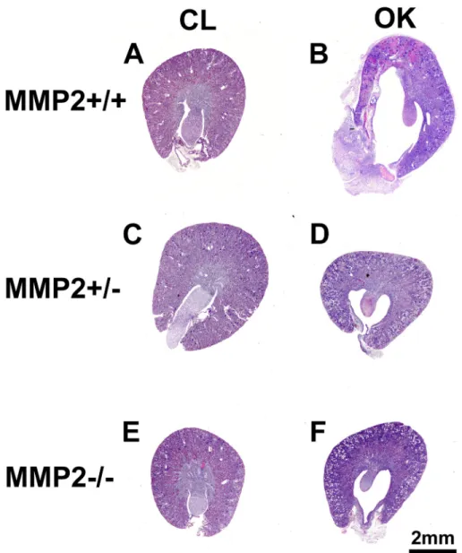
less tubular dilation (Figs
1
and
2
). Automatic image analysis of collagen content in Picrosirius
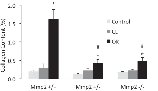
Red stained sections showed 1.63±0.25% positivity in OK and 0.29±0.11% in CL (p<<0.05) of
Mmp2+/+, whereas Mmp2-/- OK and Mmp2-/- CL only showed 0.49±0.09% and 0.23±0.04%
(p
<0.05) positivity, respectively, and Mmp2+/- OK and Mmp2+/- CL exhibited 0.43±0.09%
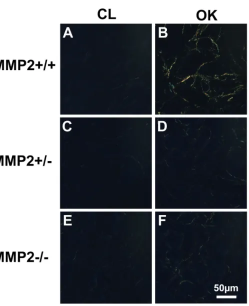
and 0.22±0.06% (p<0.05) positivity, respectively (Figs
3
and
4
). There were no significant
dis-crepancies between the levels of fibrosis in the control kidneys and the CL kidneys in any of the
groups.
Gene expression
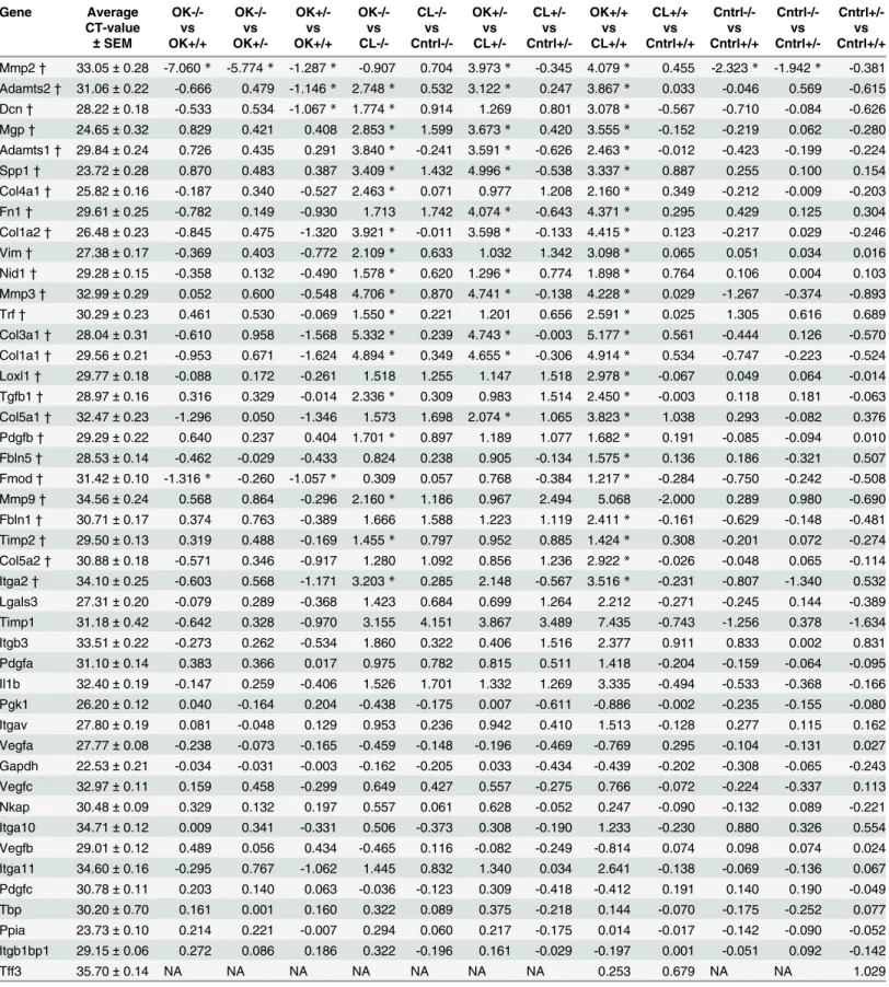
Using RTPCR, 26 out of 48 genes examined showed differential expression when ANOVA was
applied across groups (
Table 2
). None of the housekeeping genes showed significant changes,
and the results were comparable when using the different genes for normalisation. Data are
presented using 18S as standard. There was no difference in gene expression between CL and
control kidneys in any of the groups.
The greatest difference in gene expressions was seen when comparing the OK and CL in the
Mmp2+/+ mice, where 25 genes were significantly altered (
Table 2
). A comparison of
Mmp2-/- OK vs Mmp2-/- CL identified 18 significantly altered genes. Mmp2+/- OK displayed
12 significantly altered genes when compared to Mmp2+/- CL. Cntrl+/- did not differ from
Cntrl+/+ in any of the 48 tested genes. After UUO, Mmp2 expression was 7-fold and 5.5-fold
induced in Mmp2+/+ OK and Mmp2+/- OK, respectively, when compared to Mmp2-/- OK.
Comparisons between Mmp2-/- OK vs Mmp2-/- CL, and Mmp2+/- OK vs Mmp2+/- CL
showed that five genes in common did not respond in the same manner as in the Mmp2
+/+ OK vs Mmp2+/+ CL comparison. These genes are Loxl1, Fbln5, Fmod, Fbln1 and Col5a2.
None of these genes were significantly altered after UUO in Mmp2 -/- and Mmp2+/- mice. In
contrast, all five genes were upregulated in Mmp2+/+ mice. Fmod is downregulated in both
Mmp2-/- and Mmp2+/- OK groups compared to Mmp2+/+ OK. Mmp9 showed a 2.2-fold
upregulation in Mmp2-/- OK when compared to Mmp2-/- CL mice. Only Mmp2 was
differen-tially regulated when comparing Mmp2-/- OK and Mmp2+/- OK. In addition to the
house-keeping genes, 17 genes were not differentially expressed between any of the groups.
Discussion
The present study demonstrates that Mmp2-/- and Mmp2+/- mice display less
pelvic-remodel-ling and fibrosis compared to Mmp2+/+ mice after UUO. Both Mmp2-/- and Mmp2+/- mice
Table 1. Bodyweight and age of the mice at the start of the experiment.
Mmp2+/+ Mmp2+/-
Mmp2-/-Control UUO Control UUO Control UUO
n = 10 10 10 11 10 9
Bodyweight (g) 31±0.4 23±0.3 35±1 38±2 29±2 29±2
Age (weeks) 14±0 9±0 19±1 37±2 20±2 41±6
are protected, which may indicate an important effect of gene-dose. Furthermore, Mmp2
+/+ mice displayed more histological damage and showed the highest response in gene
expres-sion following UUO, with 25 genes significantly altered when comparing OK to CL. Both
Mmp2-/- and Mmp2+/- animals gave a milder response to UUO, showing 18 and 12
signifi-cantly altered genes after UUO, respectively. The main findings in this study are similar to
those of Du et al., described in a comprehensive paper in which they also reported reduced
infiltration of immune cells, and improved immunohistochemistry for a number of collagens
and renal injury markers [
21
]. Our RT-PCR results correlate to those of Du et al with similar
expression patterns for Mmp2, Mmp9, Timp1 and Timp2. However, our study presents a
wider range of molecular markers, and also data from heterozygotes beyond what has been
pre-viously reported.
Fig 1. Representative images of Period Acid-Schiff (PAS) stained transversal sections showing increased morphological damage in the Mmp2+/+ obstructed kidney (OK, 1B) compared to the other groups (1 D, F). The Mmp2+/+ OK (1B) shows severe hydronephrosis and inflammation of the renal pelvis, while the Mmp2-/- OK (1F) and Mmp2+/- OK (1D) only display slight dilatation and minimal inflammation. The contralateral kidneys (CL, 1 A, C, E) do not differ from each other and show normal morphology.
When comparing the control kidneys between the groups, the lack of Mmp2 does not seem
to affect the amount of collagen when the kidney is not under stress (
Fig 4
). As expected, the
level of Mmp2 in both Cntrl+/- and Cntrl+/+ animals is higher relative to the Cntrl-/- group.
There is no significant difference in Mmp2 expression between wild type and heterozygotes
under control conditions. Mmp2 gene expression in Mmp2+/- mice is not upregulated in OK
as much as in the Mmp2+/+ OK. This could suggest that there is a threshold level of Mmp2
necessary for damage and fibrosis to occur and this level might not be reached in the
Mmp2+/-mice, thus resulting in protection of the kidneys in Mmp2+/- mice as well. Taken together with
the fact that Mmp2 upregulation is required for regulation of several of the genes studied here,
this could point towards a cascade effect where a threshold level of Mmp2 is important for
ini-tiation of fibrogenesis.
Fig 2. Morphological damage in the obstructed kidney (OK, 2B, D, F) compared to the contralateral kidney (CL, 2 A, C, E). All OK kidneys show tubular dilatation, flattened tubular epithelium with loss of brush border and reactive nuclear enlargement. Only Mmp2+/+ OK (2B) shows necrotic tubules (asterix) and many apoptotic cells (arrows). The morphology of the CL kidney is normal. (PAS stain).
Some of the genes utilised in our study have been investigated previously in similar settings,
and could help elucidate the role of Mmp2 after UUO. The effect of pharmacological inhibition
of Mmp2 seems to be dependent on the time of administration in UUO-induced renal fibrosis.
Late administration of an inhibitor of Mmp2, TISAM, decreased the level of macrophage
infil-tration, while early administration did not, and contrary to our findings both early and late
inhibition of Mmp2 resulted in accelerated fibrosis in the mouse kidney [
19
]. Inhibition by
He4, an Mmp2/Mmp9 inhibitor and a pan-serine protease which has been shown to directly
interact with and inhibit Mmp2 and Mmp9, improved the outcome after UUO [
22
].
He4-inhi-bition, and thus an increase in Mmp2 and Mmp9 mediated collagen I digestion, resulted in less
fibrosis compared to the control group. Taken together, this suggests that genetic deficiency of
Mmp2 affects the development of fibrosis by a different mechanism than pharmacological
inhibition of Mmp2. This time dependency has also been described in a mouse model of Alport
Fig 3. Representative images of Picrosirius Red stained sections under polarised light from the obstructed kidney (OK, 3 B, D, F) compared to the contralateral kidney (CL, 3 A, C, E). There is a higher level of collagen in Mmp2+/+ OK (3 B) compared to OK in both Mmp2+/- and Mmp2-/- (3 D, F). Only minimal levels of collagen are detected in CL kidneys across all groups (3 A, C, E).
syndrome [
10
]. Early combination therapy with inhibitors of Mmp2, Mmp3 and Mmp9
signif-icantly delayed the onset of proteinuria, while treatment after onset of proteinuria accelerated
renal disease.
In addition, Mmp2 has been shown to affect vascular reactivity. For example endothelin-1
requires cleavage to produce the active hormone and Mmp2 contributes to this process [
23
].
Mmp2 has also been found to cleave adrenomedullin, resulting in production of a
vasocon-strictor peptide [
24
], and this may be an underlying mechanism that leads to kidney damage
[
25
]. One may suggest that Mmp2 deficiency decreases vasoconstriction and thus reduces
kid-ney damage in UUO.
Mmp9 deficiency seems to have a positive effect during UUO [
26
], while pharmacological
inhibition of Mmp9 has a time-dependent effect, with reduced epithelial to mesenchymal
tran-sition and fibrosis at the early or chronic stage, but not during the establishment of fibrosis
[
11
]. The present data show upregulation of Mmp9 in the obstructed kidney in Mmp2-/- mice
compared to CL, which may be a compensatory increase due to the lack of Mmp2 [
10
].
How-ever, Mmp9 was not upregulated after UUO in Mmp2+/- animals, and since both strains
showed less damage, it is unlikely that Mmp9 alone is responsible for the decreased fibrosis
after UUO.
Decorin (Dcn) deficient mice have been reported to show greater damage than WT after
UUO [
27
]. This could indicate that an upregulation of Dcn is a protective mechanism.
How-ever, while the expression of Dcn mRNA was increased after UUO in wild type and knockout
mice, there was no effect in heterozygous animals. These results, therefore, do not allow one to
draw firm conclusions regarding the potential protective role of Dcn. Timp2 is not
differen-tially expressed between the groups, yet there is an upregulation of Timp2 in the OK kidney
compared to the CL kidney in both Mmp2+/+ and Mmp2-/- mice, but not in the
Mmp2+/-group. However, we do not have any basis to explain why Mmp2+/- mice regulate Timp2
dif-ferently under the stress of UUO. Earlier data showed that Timp2 promotes injury through
activating Mmp2 [
28
]. In both Mmp2-/- and Mmp2+/- animals Timp2 has less Mmp2 to
acti-vate, which could be a mechanism behind the reduced rate of fibrosis and remodelling. Thus,
Fig 4. Image analysis of collagen in Picrosirius Red stained sections under polarised light. Collagen content is expressed as the percent of positive pixels to all pixels.* denotes P<0.05 compared to the contralateral kidney (CL). # denotes P<0.05 compared to OK in Mmp2+/+.
Table 2. Differential gene expression between strains (Mmp2 +/+, +/-, and -/-), and as paired comparisons between the obstructed (OK) and contra-lateral kidneys (CL) of the same animal.
Gene Average OK-/- OK-/- OK+/- OK-/- CL-/- OK+/- CL+/- OK+/+ CL+/+ Cntrl-/- Cntrl-/-
Cntrl+/-CT-value vs vs vs vs vs vs vs vs vs vs vs vs
± SEM OK+/+ OK+/- OK+/+ CL-/- Cntrl-/- CL+/- Cntrl+/- CL+/+ Cntrl+/+ Cntrl+/+ Cntrl+/- Cntrl+/+ Mmp2† 33.05± 0.28 -7.060 * -5.774 * -1.287 * -0.907 0.704 3.973* -0.345 4.079* 0.455 -2.323* -1.942 * -0.381 Adamts2† 31.06 ± 0.22 -0.666 0.479 -1.146* 2.748 * 0.532 3.122* 0.247 3.867* 0.033 -0.046 0.569 -0.615 Dcn† 28.22± 0.18 -0.533 0.534 -1.067* 1.774 * 0.914 1.269 0.801 3.078* -0.567 -0.710 -0.084 -0.626 Mgp† 24.65± 0.32 0.829 0.421 0.408 2.853* 1.599 3.673* 0.420 3.555* -0.152 -0.219 0.062 -0.280 Adamts1† 29.84 ± 0.24 0.726 0.435 0.291 3.840* -0.241 3.591* -0.626 2.463* -0.012 -0.423 -0.199 -0.224 Spp1† 23.72± 0.28 0.870 0.483 0.387 3.409* 1.432 4.996* -0.538 3.337* 0.887 0.255 0.100 0.154 Col4a1† 25.82± 0.16 -0.187 0.340 -0.527 2.463* 0.071 0.977 1.208 2.160* 0.349 -0.212 -0.009 -0.203 Fn1† 29.61± 0.25 -0.782 0.149 -0.930 1.713 1.742 4.074* -0.643 4.371* 0.295 0.429 0.125 0.304 Col1a2† 26.48± 0.23 -0.845 0.475 -1.320 3.921* -0.011 3.598* -0.133 4.415* 0.123 -0.217 0.029 -0.246 Vim† 27.38± 0.17 -0.369 0.403 -0.772 2.109* 0.633 1.032 1.342 3.098* 0.065 0.051 0.034 0.016 Nid1† 29.28± 0.15 -0.358 0.132 -0.490 1.578* 0.620 1.296* 0.774 1.898* 0.764 0.106 0.004 0.103 Mmp3† 32.99± 0.29 0.052 0.600 -0.548 4.706* 0.870 4.741* -0.138 4.228* 0.029 -1.267 -0.374 -0.893 Trf† 30.29± 0.23 0.461 0.530 -0.069 1.550* 0.221 1.201 0.656 2.591* 0.025 1.305 0.616 0.689 Col3a1† 28.04± 0.31 -0.610 0.958 -1.568 5.332* 0.239 4.743* -0.003 5.177* 0.561 -0.444 0.126 -0.570 Col1a1† 29.56± 0.21 -0.953 0.671 -1.624 4.894* 0.349 4.655* -0.306 4.914* 0.534 -0.747 -0.223 -0.524 Loxl1† 29.77± 0.18 -0.088 0.172 -0.261 1.518 1.255 1.147 1.518 2.978* -0.067 0.049 0.064 -0.014 Tgfb1† 28.97± 0.16 0.316 0.329 -0.014 2.336* 0.309 0.983 1.514 2.450* -0.003 0.118 0.181 -0.063 Col5a1† 32.47± 0.23 -1.296 0.050 -1.346 1.573 1.698 2.074* 1.065 3.823* 1.038 0.293 -0.082 0.376 Pdgfb† 29.29± 0.22 0.640 0.237 0.404 1.701* 0.897 1.189 1.077 1.682* 0.191 -0.085 -0.094 0.010 Fbln5† 28.53± 0.14 -0.462 -0.029 -0.433 0.824 0.238 0.905 -0.134 1.575* 0.136 0.186 -0.321 0.507 Fmod† 31.42± 0.10 -1.316 * -0.260 -1.057* 0.309 0.057 0.768 -0.384 1.217* -0.284 -0.750 -0.242 -0.508 Mmp9† 34.56± 0.24 0.568 0.864 -0.296 2.160* 1.186 0.967 2.494 5.068 -2.000 0.289 0.980 -0.690 Fbln1† 30.71± 0.17 0.374 0.763 -0.389 1.666 1.588 1.223 1.119 2.411* -0.161 -0.629 -0.148 -0.481 Timp2† 29.50± 0.13 0.319 0.488 -0.169 1.455* 0.797 0.952 0.885 1.424* 0.308 -0.201 0.072 -0.274 Col5a2† 30.88± 0.18 -0.571 0.346 -0.917 1.280 1.092 0.856 1.236 2.922* -0.026 -0.048 0.065 -0.114 Itga2† 34.10± 0.25 -0.603 0.568 -1.171 3.203* 0.285 2.148 -0.567 3.516* -0.231 -0.807 -1.340 0.532 Lgals3 27.31± 0.20 -0.079 0.289 -0.368 1.423 0.684 0.699 1.264 2.212 -0.271 -0.245 0.144 -0.389 Timp1 31.18± 0.42 -0.642 0.328 -0.970 3.155 4.151 3.867 3.489 7.435 -0.743 -1.256 0.378 -1.634 Itgb3 33.51± 0.22 -0.273 0.262 -0.534 1.860 0.322 0.406 1.516 2.377 0.911 0.833 0.002 0.831 Pdgfa 31.10± 0.14 0.383 0.366 0.017 0.975 0.782 0.815 0.511 1.418 -0.204 -0.159 -0.064 -0.095 Il1b 32.40± 0.19 -0.147 0.259 -0.406 1.526 1.701 1.332 1.269 3.335 -0.494 -0.533 -0.368 -0.166 Pgk1 26.20± 0.12 0.040 -0.164 0.204 -0.438 -0.175 0.007 -0.611 -0.886 -0.002 -0.235 -0.155 -0.080 Itgav 27.80± 0.19 0.081 -0.048 0.129 0.953 0.236 0.942 0.410 1.513 -0.128 0.277 0.115 0.162 Vegfa 27.77± 0.08 -0.238 -0.073 -0.165 -0.459 -0.148 -0.196 -0.469 -0.769 0.295 -0.104 -0.131 0.027 Gapdh 22.53± 0.21 -0.034 -0.031 -0.003 -0.162 -0.205 0.033 -0.434 -0.439 -0.202 -0.308 -0.065 -0.243 Vegfc 32.97± 0.11 0.159 0.458 -0.299 0.649 0.427 0.557 -0.275 0.766 -0.072 -0.224 -0.337 0.113 Nkap 30.48± 0.09 0.329 0.132 0.197 0.557 0.061 0.628 -0.052 0.247 -0.090 -0.132 0.089 -0.221 Itga10 34.71± 0.12 0.009 0.341 -0.331 0.506 -0.373 0.308 -0.190 1.233 -0.230 0.880 0.326 0.554 Vegfb 29.01± 0.12 0.489 0.056 0.434 -0.465 0.116 -0.082 -0.249 -0.814 0.074 0.098 0.074 0.024 Itga11 34.60± 0.16 -0.295 0.767 -1.062 1.445 0.832 1.340 0.034 2.641 -0.138 -0.069 -0.136 0.067 Pdgfc 30.78± 0.11 0.203 0.140 0.063 -0.036 -0.123 0.309 -0.418 -0.412 0.191 0.140 0.190 -0.049 Tbp 30.20± 0.70 0.161 0.001 0.160 0.322 0.089 0.375 -0.218 0.144 -0.070 -0.175 -0.252 0.077 Ppia 23.73± 0.10 0.214 0.221 -0.007 0.294 0.060 0.217 -0.175 0.014 -0.017 -0.142 -0.090 -0.052 Itgb1bp1 29.15± 0.06 0.272 0.086 0.186 0.322 -0.196 0.161 -0.029 -0.197 0.001 -0.051 0.092 -0.142 Tff3 35.70± 0.14 NA NA NA NA NA NA NA 0.253 0.679 NA NA 1.029 (Continued)
even though Timp2 expression in OK and CL in Mmp2-/- and Mmp2+/- mice differ, the
lim-ited possibility of an increase in Mmp2 at the level of expression would probably reduce this
effect. This may be the reason why they are also protected. In our study, Timp1 was not
affected by UUO, and this is supported by evidence that elimination of Timp1 alone was not
found to be sufficient in order to alter the severity of fibrosis during UUO [
29
].
The present results did not show consistent findings in the Pdgf system. This is not entirely
surprising since the Pdgf system is important in kidney fibrosis [
30
] and in UUO [
31
,
32
], but it
is not necessary for the development of fibrosis in the kidney [
33
]. There was no differential
expression of Pdgfa or Pdgfc. On the other hand Pdgfb was differentially expressed after UUO
in Mmp2-/- and Mmp2+/+ mice but not in Mmp2+/- animals. This is difficult to explain,
espe-cially since antagonists have a positive effect in several models of renal disease. Pdgfb, however,
also plays a role in the repair of cellular damage [
34
].
Tff3, Vgfa, Vgfb, Vgfc, Il1b and Lgals3 were not differentially expressed in the present study
even though they have been implicated in kidney disease. Specifically, Tff3 has been suggested
as a diagnostic marker in CKD since it increases in both serum and urine during CKD
progres-sion [
35
]. Vegfs have been demonstrated to slow down the progression of renal injury in
exper-imental models and can be administered in renoprotective therapy [
36
], but overstimulation
can induce glomerular pathology. Il1b is known to stimulate proliferation of human renal
fibroblasts as well as production of matrix proteins, while Lhals3 has been shown to protect
against fibrosis [
37
,
38
].
The clinical treatment of ureteral obstruction is primarily surgical, but as a strategy for
lim-iting the destruction of kidney tissue in progressive or recurring disease, some intervention
against Mmp2 may be useful. It is further unlikely that reduced Mmp2 expression provides
complete protection from the damage in UUO. There is no doubt that a longer period of UUO
would lead to more severe damage, even in mice with decreased Mmp2 expression.
In conclusion, both homozygous and heterozygous genetic inactivation of Mmp2 protect
mice against hydronephrosis and kidney fibrosis after UUO, as indicated by both histology and
gene expression. The genetic mechanism seems to be a reduced ability to respond with Mmp2
upregulation under stress, and this may suggest that there is a threshold level of Mmp2
neces-sary for pelvic remodelling and genetic events leading to activation of fibrosis.
Acknowledgments
The study was supported by the Western Health Authority of Norway (Helse Vest), and the
Swedish Society for Medical Research (SSMF).
Parts of this study were presented at Experimental Biology 2012 and published as a meeting
abstract [
39
].
Table 2. (Continued)
Gene Average OK-/- OK-/- OK+/- OK-/- CL-/- OK+/- CL+/- OK+/+ CL+/+ Cntrl-/- Cntrl-/-
Cntrl+/-CT-value vs vs vs vs vs vs vs vs vs vs vs vs
± SEM OK+/+ OK+/- OK+/+ CL-/- Cntrl-/- CL+/- Cntrl+/- CL+/+ Cntrl+/+ Cntrl+/+ Cntrl+/- Cntrl+/+ Il1a 35.93± 0.31 -0.429 1.094 -1.523 1.657 -1.016 -0.533 0.624 1.242 -0.540 -0.368 0.544 -0.912
Matn1 33.92± 1.25 NA NA NA NA NA NA NA NA NA NA NA NA
Cntrl denotes kidneys from control animals without obstruction.
†denotes significant variations between all groups, P<0.05 by ANOVA.
* denotes P<0.05 for the individual comparison. doi:10.1371/journal.pone.0143390.t002
Bjarne M. Iversen passed away before the submission of the final version of this manuscript.
Michael Hultström accepts responsibility for the integrity and validity of the data collected and
analysed.
Author Contributions
Conceived and designed the experiments: MKT TS FH SL BMI CC RKR MH. Performed the
experiments: MKT TS SL MH. Analyzed the data: MKT TS SL MH. Contributed reagents/
materials/analysis tools: MKT TS FH SL CC MH. Wrote the paper: MKT TS FH SL CC RKR
MH.
References
1. Heikkila J, Holmberg C, Kyllonen L, Rintala R, Taskinen S (2011) Long-term risk of end stage renal dis-ease in patients with posterior urethral valves. J Urol 186: 2392–2396. doi:10.1016/j.juro.2011.07.109
PMID:22014822
2. Hamdi A, Hajage D, Van Glabeke E, Belenfant X, Vincent F, Gonzalez F, et al. (2012) Severe post-renal acute kidney injury, post-obstructive diuresis and post-renal recovery. BJU Int 110: E1027–1034. doi:
10.1111/j.1464-410X.2012.11193.xPMID:22583774
3. Chevalier RL, Forbes MS, Thornhill BA (2009) Ureteral obstruction as a model of renal interstitial fibro-sis and obstructive nephropathy. Kidney Int 75: 1145–1152. doi:10.1038/ki.2009.86PMID:19340094
4. Nangaku M (2004) Mechanisms of tubulointerstitial injury in the kidney: final common pathways to end-stage renal failure. Intern Med 43: 9–17. PMID:14964574
5. Aimes RT, Quigley JP (1995) Matrix metalloproteinase-2 is an interstitial collagenase. Inhibitor-free enzyme catalyzes the cleavage of collagen fibrils and soluble native type I collagen generating the spe-cific 3/4- and 1/4-length fragments. J Biol Chem 270: 5872–5876. PMID:7890717
6. Hultstrom M, Leh S, Skogstrand T, Iversen BM (2008) Upregulation of tissue inhibitor of metallopro-teases-1 (TIMP-1) and procollagen-N-peptidase in hypertension-induced renal damage. Nephrol Dial Transplant 23: 896–903. PMID:17977875
7. Flamant M, Placier S, Dubroca C, Esposito B, Lopes I, Chatziantoniou C, et al. (2007) Role of matrix metalloproteinases in early hypertensive vascular remodeling. Hypertension 50: 212–218. PMID:
17515450
8. Catania JM, Chen G, Parrish AR (2007) Role of matrix metalloproteinases in renal pathophysiologies. Am J Physiol Renal Physiol 292: F905–911. PMID:17190907
9. Skogstrand T, Leh S, Paliege A, Reed RK, Vikse BE, Bachmann S, et al. (2013) Arterial damage pre-cedes the development of interstitial damage in the nonclipped kidney of two-kidney, one-clip hyperten-sive rats. J Hypertens 31: 152–159. PMID:23079683
10. Zeisberg M, Khurana M, Rao VH, Cosgrove D, Rougier JP, Werner MC, et al. (2006) Stage-specific action of matrix metalloproteinases influences progressive hereditary kidney disease. PLoS Med 3: e100. PMID:16509766
11. Tan TK, Zheng G, Hsu TT, Lee SR, Zhang J, Zhao Y, et al. (2013) Matrix metalloproteinase-9 of tubular and macrophage origin contributes to the pathogenesis of renal fibrosis via macrophage recruitment through osteopontin cleavage. Lab Invest 93: 434–449. doi:10.1038/labinvest.2013.3PMID:
23358111
12. Kalluri R, Neilson EG (2003) Epithelial-mesenchymal transition and its implications for fibrosis. J Clin Invest 112: 1776–1784. PMID:14679171
13. Kaneko T, Shimizu A, Mii A, Fujita E, Fujino T, Kunugi S, et al. (2012) Role of matrix metalloproteinase-2 in recovery after tubular damage in acute kidney injury in mice. Nephron Exp Nephrol 1metalloproteinase-2metalloproteinase-2: metalloproteinase-23–35. doi:10.1159/000346569PMID:23548779
14. Kunugi S, Shimizu A, Kuwahara N, Du X, Takahashi M, Terasaki Y, et al. (2011) Inhibition of matrix metalloproteinases reduces ischemia-reperfusion acute kidney injury. Lab Invest 91: 170–180. doi:10. 1038/labinvest.2010.174PMID:20956976
15. Itoh T, Ikeda T, Gomi H, Nakao S, Suzuki T, Itohara S (1997) Unaltered secretion of beta-amyloid pre-cursor protein in gelatinase A (matrix metalloproteinase 2)-deficient mice. J Biol Chem 272: 22389– 22392. PMID:9278386
16. Ohno-Matsui K, Uetama T, Yoshida T, Hayano M, Itoh T, Morita I, et al. (2003) Reduced retinal angio-genesis in MMP-2-deficient mice. Invest Ophthalmol Vis Sci 44: 5370–5375. PMID:14638740
17. Takamiya Y, Fukami K, Yamagishi S, Kaida Y, Nakayama Y, Obara N, et al. (2013) Experimental dia-betic nephropathy is accelerated in matrix metalloproteinase-2 knockout mice. Nephrol Dial Transplant 28: 55–62. doi:10.1093/ndt/gfs387PMID:23028104
18. Suofu Y, Clark JF, Broderick JP, Kurosawa Y, Wagner KR, Lu A (2012) Matrix metalloproteinase-2 or -9 deletions protect against hemorrhagic transformation during early stage of cerebral ischemia and reperfusion. Neuroscience 212: 180–189. doi:10.1016/j.neuroscience.2012.03.036PMID:22521821
19. Nishida M, Okumura Y, Ozawa S, Shiraishi I, Itoi T, Hamaoka K (2007) MMP-2 inhibition reduces renal macrophage infiltration with increased fibrosis in UUO. Biochem Biophys Res Commun 354: 133–139. PMID:17210124
20. Goulter AB, Harmer DW, Clark KL (2006) Evaluation of low density array technology for quantitative parallel measurement of multiple genes in human tissue. BMC Genomics 7: 34. PMID:16504128
21. Du X, Shimizu A, Masuda Y, Kuwahara N, Arai T, Kataoka M, et al. (2012) Involvement of matrix metal-loproteinase-2 in the development of renal interstitial fibrosis in mouse obstructive nephropathy. Lab Invest 92: 1149–1160. doi:10.1038/labinvest.2012.68PMID:22614125
22. LeBleu VS, Teng Y, O'Connell JT, Charytan D, Muller GA, Muller CA, et al. (2013) Identification of human epididymis protein-4 as a fibroblast-derived mediator of fibrosis. Nat Med 19: 227–231. doi:10. 1038/nm.2989PMID:23353556
23. Fernandez-Patron C, Radomski MW, Davidge ST (1999) Vascular matrix metalloproteinase-2 cleaves big endothelin-1 yielding a novel vasoconstrictor. Circ Res 85: 906–911. PMID:10559137
24. Martinez A, Oh HR, Unsworth EJ, Bregonzio C, Saavedra JM, Stetler-Stevenson WG, et al. (2004) Matrix metalloproteinase-2 cleavage of adrenomedullin produces a vasoconstrictor out of a vasodilator. Biochem J 383: 413–418. PMID:15307819
25. Ledbetter S, Kurtzberg L, Doyle S, Pratt BM (2000) Renal fibrosis in mice treated with human recombi-nant transforming growth factor-beta2. Kidney Int 58: 2367–2376. PMID:11115070
26. Wang X, Zhou Y, Tan R, Xiong M, He W, Fang L, et al. (2010) Mice lacking the matrix metalloprotei-nase-9 gene reduce renal interstitial fibrosis in obstructive nephropathy. Am J Physiol Renal Physiol 299: F973–982. doi:10.1152/ajprenal.00216.2010PMID:20844022
27. Schaefer L, Macakova K, Raslik I, Micegova M, Grone HJ, Schonherr E, et al. (2002) Absence of dec-orin adversely influences tubulointerstitial fibrosis of the obstructed kidney by enhanced apoptosis and increased inflammatory reaction. Am J Pathol 160: 1181–1191. PMID:11891213
28. Wang Z, Famulski K, Lee J, Das SK, Wang X, Halloran P, et al. (2014) TIMP2 and TIMP3 have diver-gent roles in early renal tubulointerstitial injury. Kidney Int 85: 82–93. doi:10.1038/ki.2013.225PMID:
23760282
29. Kim H, Oda T, Lopez-Guisa J, Wing D, Edwards DR, Soloway PD, et al. (2001) TIMP-1 deficiency does not attenuate interstitial fibrosis in obstructive nephropathy J Am Soc Nephrol 12: 736–748. PMID:
11274235
30. Kishioka H, Fukuda N, Wen-Yang H, Nakayama M, Watanabe Y, Kanmatsuse K (2001) Effects of PDGF A-chain antisense oligodeoxynucleotides on growth of cardiovascular organs in stroke-prone spontaneously hypertensive rats. Am J Hypertens 14: 439–445. PMID:11368465
31. Eitner F, Buecher E, van Roeyen C, Kunter U, Rong S, Seikrit C, et al. (2008) PDGF-C is a proinflam-matory cytokine that mediates renal interstitial fibrosis. Journal of the American Society of Nephrology 19: 281–289. doi:10.1681/ASN.2007030290PMID:18184860
32. Martin IV, Borkham-Kamphorst E, Zok S, van Roeyen CRC, Eriksson U, Boor P, et al. (2013) Platelet-Derived Growth Factor (PDGF)-C Neutralization Reveals Differential Roles of PDGF Receptors in Liver and Kidney Fibrosis. American Journal of Pathology 182: 107–117. doi:10.1016/j.ajpath.2012.09.006
PMID:23141925
33. Tang WW, Ulich TR, Lacey DL, Hill DC, Qi M, Kaufman SA, et al. (1996) Platelet-derived growth factor-BB induces renal tubulointerstitial myofibroblast formation and tubulointerstitial fibrosis. Am J Pathol 148: 1169–1180. PMID:8644858
34. Ostendorf T, Boor P, Van Roeyen CRC, Floege J (2014) Platelet-derived growth factors (PDGFs) in glomerular and tubulointerstitial fibrosis. Kidney International Supplements 4: 65–69. PMID:26312152
35. Du TY, Luo HM, Qin HC, Wang F, Wang Q, Xiang Y, et al. (2013) Circulating Serum Trefoil Factor 3 (TFF3) Is Dramatically Increased in Chronic Kidney Disease. Plos One 8.
36. Chade AR (2012) VEGF: Potential therapy for renal regeneration. F1000 Med Rep 4: 1. doi:10.3410/ M4-1PMID:22238513
37. Vesey DA, Cheung C, Cuttle L, Endre Z, Gobe G, Johnson DW (2002) Interleukin-1beta stimulates human renal fibroblast proliferation and matrix protein production by means of a transforming growth factor-beta-dependent mechanism. J Lab Clin Med 140: 342–350. PMID:12434136
38. Henderson NC, Mackinnon AC, Farnworth SL, Kipari T, Haslett C, Iredale JP, et al. (2008) Galectin-3 expression and secretion links macrophages to the promotion of renal fibrosis. Am J Pathol 172: 288– 298. doi:10.2353/ajpath.2008.070726PMID:18202187
39. Tveitarås MK S T, Helle F, Leh S, Reed RK, et al (2012) MMP2 deficient mice are protected from hydro-nephrosis after unilateral ureteral obstruction. FASEB Journal 26: 868.812.


