HAL Id: tel-01424170
https://tel.archives-ouvertes.fr/tel-01424170
Submitted on 2 Jan 2017HAL is a multi-disciplinary open access archive for the deposit and dissemination of sci-entific research documents, whether they are pub-lished or not. The documents may come from teaching and research institutions in France or abroad, or from public or private research centers.
L’archive ouverte pluridisciplinaire HAL, est destinée au dépôt et à la diffusion de documents scientifiques de niveau recherche, publiés ou non, émanant des établissements d’enseignement et de recherche français ou étrangers, des laboratoires publics ou privés.
Regulatory RNAs in Staphylococcus aureus : Function
and Mechanism
Thao Nguyen Le Lam
To cite this version:
Thao Nguyen Le Lam. Regulatory RNAs in Staphylococcus aureus : Function and Mechanism. Agri-cultural sciences. Université Paris Sud - Paris XI, 2015. English. �NNT : 2015PA112216�. �tel-01424170�
UNIVERSITÉ PARIS SACLAY - UNIVERSITÉ PARIS SUD
UFR SCIENTIFIQUE D’ORSAY - ÉCOLE DOCTORALE
STRUCTURE ET DYNAMIQUE DES SYSTÈMES VIVANTS
Thèse
Présentée pour obtenir le grade de
DOCTEUR EN SCIENCES DE L’UNIVERSITÉ PARIS SUD
Le 24 septembre 2015
Thao Nguyen LE LAM
Directeur de thèse : Dr. Philippe BOULOC
Laboratoire d’accueil :Signalisation et Réseaux de Régulations Bactériens Institute for Integrative Biology of the Cell (I2BC)
CEA, CNRS, Université Paris-Sud, Université Paris-Saclay, Orsay, France
Composition du jury :
Président du jury Pr. Nicolas BAYAN
Rapporteur Dr. Maude GUILLIER
Rapporteur Dr. Francis REPOILA
Examinateur Pr. Brice FELDEN
Examinateur Dr. Nara FIGUEROA-BOSSI
Directeur de thèse Dr. Philippe BOULOC
Characterization of regulatory RNAs
in Staphylococcus aureus
TABLE OF CONTENTS
FIGURES AND TABLES ... 5
ABBREVIATIONS ... 7 INTRODUCTION ... 9 I. Staphylococcus aureus ... 11 1. General features ... 11 2. Role in disease ... 12 3. Antibiotic resistance ... 14
II. Overview of adaptation and virulence in S. aureus ... 15
1. Some examples in adaptation ... 15
2. Host-pathogen interaction ... 25
III. Overview of bacterial competitive fitness ... 27
IV. Overview of regulatory RNAs in S. aureus ... 31
1. Identification of regulatory RNAs by various approaches ... 31
2. Classification ... 36
2.1 Regulatory small RNAs targeting mRNAs ... 36
2.2 Cis-regulatory elements ... 39
2.3 Protein-targeting RNAs ... 42
3. Role of regulatory RNAs in S. aureus ... 43
4. Proteins involved in S. aureus sRNA functions ... 51
4.1 RNA chaperone Hfq: a controversial factor ... 51
4.2 Main RNases in S. aureus ... 53
V. Selected samples of regulatory RNAs in some Firmicutes species ... 61
1. sRNA in the pathogenesis of Streptococcus ... 61
2. sRNA in the pathogenesis of Clostridium ... 62
3. sRNA in the pathogenesis of Listeria monocytogenes ... 63
4. sRNA in the pathogenesis of Bacillus subtilis ... 67
5. sRNAs in the pathogenesis of Enterococcus faecalis... 68
AIM OF THE THESIS ... 71
CHAPTER A1 ... 73
CHAPTER A2 ... 125
CHAPTER B ... 149
CHAPTER C ... 217
DISCUSSION & PERSPECTIVES ... 249
FIGURES AND TABLES
Figure 1: Staphylococcus aureus also known “the golden Staph”. ... 11
Figure 2: Sites of infection and diseases caused by S. aureus. ... 13
Figure 3: Model of SrrAB system. ... 17
Figure 4: Model for the NreBC sensor and regulation system in staphylococci. ... 18
Figure 5: The CtsR, HrcA regulon of S. aureus... 21
Figure 6: Mechanism of acid resistance of Gram-positive bacteria ... 23
Figure 7: Human lines of defense against pathogens ... 26
Figure 8: Mechanisms by which S. aureus subverts host innate immune defense ... 27
Figure 9: Scheme of a competitive fitness assay. ... 28
Figure 10: Signature-tagged mutagenesis in Salmonella. ... 30
Figure 11: Example of cis-encoded antisense RNAs (asRNAs). ... 36
Figure 12: Antisense regulation of plasmid pT181 replication. ... 37
Figure 13: Gene arrangement and regulatory functions of sRNAs. ... 38
Figure 14: C-rich box conserved motif in S. aureus sRNAs and an example of example sRNA/mRNA base pairing. ... 39
Figure 15: Diversity of riboswitches and mechanisms of gene control in bacteria. ... 40
Figure 16: Model of a regulatory cascade for methionine biosynthesis operon control ... 41
Figure 17: RNA polymerase interacts with 6S sRNA in E.coli ... 43
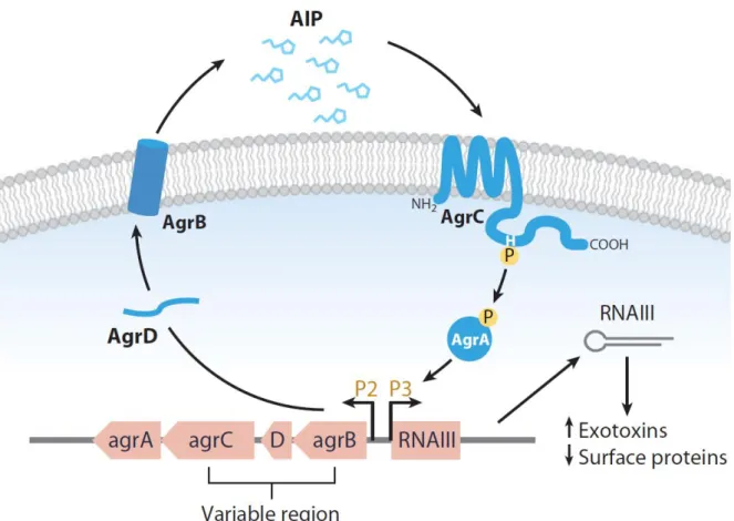
Figure 18: The accessory gene regulator (agr) quorum sensing system of S. aureus ... 44
Figure 19: Regulatory circuits involved in virulence gene expression ... 46
Figure 20: RsaE controls metabolic pathways. ... 48
Figure 21: RsaA and its regulatory circuits. ... 49
Figure 22: Base-pair association of SprD with sbi mRNA. ... 51
Figure 23: Effect of hfq deletion on pigmentation in different S. aureus strains. ... 51
Figure 24: Examples of regulatory RNAs and their mechanism of action in S. aureus. ... 53
Figure 25: Examples of RNase III functions. ... 57
Figure 26: Schematic representation of the saePQRS operon ... 58
Figure 27: S. aureus hemolysin production via RNA stability control by RNase Y and PNPase.. ... 60
Figure 28: Regulatory mechanism of FasX with the ska mRNA. ... 62
Figure 29: Examples of regulatory RNAs in Clostridium. ... 63
Figure 30: Regulatory mechanism of LhrA and chiA mRNA. ... 64
Figure 31: Interplay between a metabolite-sensing riboswitch and a temperature-sensing RNA thermometer. ... 65
Figure 32: SR1, a B. subtilis trans-encoded sRNA with dual-function. ... 68
Figure 33: Riboswitch-based regulation. ... 70
ABBREVIATIONS
B Sigma B
ADI Arginine deiminase
agr Accessory gene regulator
ahpCF Alkyl hydroperoxide reductase
AIP Autoinducing peptide
asRNAs antisense RNAs
BCAA Branched-chain amino acid
bp Base pairs
CA-MRSA Community-acquired MRSA
CcpA Catabolite control protein A
CcpN Control catabolite protein of gluconeogenic
ChiA Chitinase
CHIPS Chemotaxis inhibitory protein of Staphylococcus aureus
chp Chemotaxis-inhibiting protein
clfB Clumping factor B
coa Coagulase
colA Kappa-toxin or collagenase
cpd 2’,3’-cyclic nucleotide phosphodi-esterase
CS Cutting site
csp Cold shock protein
Dcp D-alanyl carrier protein
ds Double-strand
EA Ethanolamine
efb Fibrinogen-binding protein
eut Ethanolamine utilization
fbp Fibronectin binding protein
FMN Flavin mononucleotide
FNR Fumarate and nitrate reductase regulatory
ftn Ferritin
Fur Ferric uptake repressor
Glc-6P Glucosamine-6-phosphate
HA-MRSA Hospital-acquired MRSA
Hfq Host factor-I
hla -hemolysin
LTAs Lipoteichoic acids
MRSA Methicillin-resistant Staphylococcus aureus
narGHJI Nitrate reductase
narT Nitrate transporter
nir-operon Nitrite reductase
NO Nitrosative
PBP2 Penicillin binding protein 2
PC Pyrimidine compound
PG Plasminogen
PIA Polysaccharide intercellular adhesin
PNAG Poly-N-acetylglucosamine
PNPase Polynucleotide phosphorylase
PSM Phenol-soluble modulin
ptp Protein tyrosine phosphatase
PVL Panton-Valentine leukocidin
RBS Ribosome-binding site
rot Repressor of toxin
roxS Related to oxidative stress
sak Staphylokinase
SAM S-adenosyl methionine
SCIN Staphylococcal complement inhibitor
SCV Small colony variant
SD Shine-Dalgarno
sdhCAB Succinate dehydrogenase
sec Staphylococcal enterotoxin type C
sodA Superoxide dismutase
Spr Small pathogenicity island RNAs
SrhSR Staphylococcal resDE homologues
sRNAs Trans-encoded RNAs
SrrAB Staphylococcal respiration response
SSRs Small stable RNAs
TA Toxin-antitoxin
TCA Tricarboxylic acid
TCS Two-component system
TPP Thiamine pyrophosphate
TSST Toxic shock syndrome toxin
UTR Untranslated regions
VR-RNA VirR-regulated RNA
ACKNOWLEDGEMENT
“The mediocre teacher tells. The good teacher explains. The superior teacher demonstrates. The great teacher inspires.” ― William Arthur Ward.
First and foremost, I want to express my deeply gratitude to my greatest supervisor Philippe Bouloc with all his kindness, encouragement, conscientiousness and enthusiasm. It has be an honor to become his PhD student. He not only teaches me in term of science, but also inspires me in term of life. Word cannot express how grateful I am to him. Beside my supervisor, I would like to thank my PhD tutors Jean-Luc Pernodet and Lionello Bossi for their insightful discussion, suggestion and orientation for my project.
During my PhD time in France, all the members of my lab are the most important part of me. I would like to give special thanks to Annick Jacq, Chantal Bohn for all their kindly help, monitor, and discussion. My life is also fulfilled with joy with all labmates Tatiana Rochat, Frédérique Lartigue, Elena Disconzi, Rémy Bonnin, Yiqin Deng, Audrey Vingadassalon, Wang Ji, Ngoc An Nguyen, Florence Lorieux andWenfeng Liu. I would
like to thank also the staff members of the institute for providing me with all the necessary facilities for the research.
In addition, I would like to give my sincere thanks for the important corporation with Claire Poyart, Constantin Hays (Cochin Institute, Paris); Erwrin van Dick, Yan Jaszczyszyn, Claude Thermes (High throughput sequencing platform, Institute of Integrative Biology of the Cell (I2BC), Gif-sur-Yvette); and Claire Toffano-Nioche, Thuong Van Du Tran (Bioinformatic team, I2BC, Gif-sur-Yvette)
I would also like to thank my committee members: Nicolas Bayan, Maude Guillier, Francis Repoila, Brice Felden and Nara Figueroa-Bossi for their time, interest, brilliant comments and suggestion.
I grateful acknowledge the funding sourced for my thesis work. I was funded by the doctoral school scholarship (Gene Genome and Cell) for my first three years and was honored to receive the scholarship from Medical Research Foundation for fourth year. In addition, I was also supported by 3-month
Vietnamese-France scholarship. In particular, I am grateful to Michel Jacquet for his continuous encouragement and his help for Vietnamese student.
Last but not the least, I want to express my deeply thankful for the support, love and encouragement of my family and my lover. For my mother who raises me with full of love and her endless support and care. For my partner, Damien Esteve and his family who give me their tremendous supporting during my life. Thank you with all my heart and soul. Through their love, support and belief, I have been able to complete this entire journey.
I. Staphylococcus aureus
1. General features
In the second half of the nineteenth century, the Scotland surgeon Sir Alexander Ogston identified for the first time the bacterium Staphylococcus from a patient knee abscess joint (Ogston 1984). In 1884, based on pigmented colony types, the German physician and microbiologist Friedrich Julius Rosenbach renamed this bacterium Staphylococcus aureus to differentiate it from Staphylococcus albus, which is now called Staphylococcus epidermidis.
Staphylococcus aureus (in Greek Staphylo “bunch of grapes” and in Latin aureus “golden”) is a Gram-positive coccal (spherical) bacterium member of the Staphyloccaceae family (Figure 1).
Figure 1: Staphylococcus aureus also known “the golden Staph”.
This bacterium is non-motile, non-spore-forming, facultative anaerobic and grows in oxygenated conditions or ferments glucose to produce mainly lactic acid. Its colonies are fairly large, round, golden-yellow on rich medium and have a -hemolytic activity on blood agar plates. S. aureus is positive for catalase (decomposes hydrogen peroxide to water and oxygen), reduces nitrate to nitrite and ferments mannitol (in contrast to S. epidermidis).
Scientific classification Domain: Bacteria Kingdom: Eubacteria Phylum: Firmicutes Class: Bacilli Order: Bacillales Family: Staphylococcaceae Genus: Staphylococcus Species: aureus Binomial name Staphylococcus aureus Rosenbach 1884
S. aureus produces a membrane-associated coagulase, with reacts with prothrombin in the blood to form staphylothrombin. This complex triggers blood clotting by converting soluble fibrinogen to insoluble fibrin and may protect the bacteria from phagocytosis (Tortora et al. 2013). S. aureus is negative for urease.
S. aureus reproduces by binary fission. After division, the daughter cells often remain attached, generating bacterial clusters. S. aureus has a circular chromosome of 2.8 M base pairs (bp) with a low GC composition (32.8%). The genome has about 2700 coding sequences of which approximately 38% have unknown function.
2. Role in disease
S. aureus is one of the most common causes of hospital-acquired (nosocomial) and community-acquired infection. It causes disease by three main mechanisms: (1) invasion of tissues and inflammation, (2) toxin production and (3) biofilm formation. S. aureus expresses a large number of virulence factors that include:
Surface proteins, invasion factors (e.g., leukocidin, kinases, hyaluronidase).
Structures for evading phagocytes such as surface factors (e.g., capsule, protein A), biochemical compounds and enzymes (e.g., carotenoids, catalase, lipase, -lactamase) or immunological disguises (e.g., coagulase, protein A).
Membrane-damaging toxins, exfoliatin toxins and superantigens.
S. aureus is an important cause of death and morbidity in humans. Carriage rates of S. aureus may vary between human populations and different studies but can be divided in three types of population: non-carriers (approximately 20% of the population); persistent carriers (20-25%) and intermittent carriers (55-60%) (Lindsay 2008). In human, S. aureus primarily colonizes the nasal passage and axillae and can occasionally be found as part of the flora of the digestive and vaginal tracts (Williams 1963). When the protective layer of the human epithelium is breached and the mechanisms of host immunity fail, the bacterium is able to colonize a wide range of different organs. Most common infections caused by S. aureus are skin and soft tissue lesions such as boils, styles, and furuncles. However, when this bacterium enters the bloodstream, it can cause more serious and life-threatening infections such as pneumonia (lung infection), endocarditis (inflammation of the heart inner
layer, the endocardium), osteomyelitis (infection and inflammation of bone or bone marrow) or thrombophlebitis (inflammation of veins caused by blood clots), urinary tract infections, bacteremia (presence of bacteria in the blood), sepsis (whole body inflammation cause by an infection) (Figure 2).
Figure 2: Sites of infection and diseases caused by S. aureus.
(Fromhttp://www.tjclarkinc.com/bacterial_diseases/hold/staphylococcus.htm).
S. aureus produces enzymes and toxins that are also responsible for food poisoning, toxic shock syndrome and scalded skin syndrome (Figure 2). Food poisoning is caused by eating food contaminated with enterotoxins released by the bacteria rather than by its infection. Toxic shock syndrome is caused by the release of a toxic shock syndrome toxin (TSST, 22 kDa superantigens) into the bloodstream leading to high fever, vomiting, diarrhea, low blood pressure and potentially to death. Scalded skin syndrome occurs mainly in children under 5-year-old, especially newborn babies; it is due to exotoxins (exfoliatin A and B) that cause skin damages. S. aureus is also a substantial pathogen for animals, transmitted between species, and consequently, a worrying zoonotic agent (McEvoy et al. 2013). S. aureus can overwhelmingly colonize a variety of animals leading to infections in about three
in cow (inflammation of udder) in the dairy industry with an infection rate up to 10 to 12 percent (Tenhagen et al. 2009).
3. Antibiotic resistance
S. aureus has the potential to become resistant to multiple antibiotics, complicating significantly its treatment (Uhlemann et al. 2014). In the 1940s, the first treatment against S. aureus infections was penicillin. Unfortunately, S. aureus developed quickly resistances to this antibiotic due to the presence of penicillinase (a form of -lactamase) that degrades penicillin. Two decades later, more than 80% of hospital- and community-acquired S. aureus isolates were penicillin-resistant.
In 1959, methicillin, the first semi-synthetic penicillinase-resistant penicillin, was used in clinical treatments. Two years later, the first case of methicillin-resistant S. aureus (MRSA) was reported. Other antibiotics from the same group such as oxacillin, cloxacillin, dicloxacillin, flucloxacillin and nafcillin were developed to replace methicillin, but S. aureus strains became extensively resistant to all -lactam antibiotics due to the emergence of a penicillin binding protein 2 (PBP2) having a decrease binding affinity for penicillin. Glycopeptide antibiotics, including vancomycin, are the most efficient weapons against Gram-positive infections, including the problematic MRSA strains. The existence of vancomycin-resistant S. aureus (VRSA) that are already MRSA is a serious threat in human health since it could lead to a therapeutic dead-end (Zetola et al. 2005). In industrialized nations, 20-60% of all hospital S. aureus strains are methicillin-resistant (Hospital-acquired MRSA, HA-MRSA), and newly emerging community-acquired MRSA (CA-MRSA) combine antibiotic resistance with hyper-virulence. In 1999, it was reported that about 500,000 patients of United States hospitals were infected every year by S. aureus (Bowersox 1999).
The genome of MRSA strains carry SSCmec mobile genetic elements containing the mecA gene that confer resistance to methicillin and all other β-lactam antibiotics (Katayama et al. 2000). In addition, CA-MRSA strains not only have a short SCCmec but also a Panton-Valentine leukocidin locus (Vandenesch et al. 2003).
S. aureus can acquired resistance against virtually all antimicrobial agents available in hospitals and communities (Deleo et al. 2010). However, a new cell wall inhibitor named
teixobactin was recently reported as an efficient antibiotic for Gram-positive pathogens including drug-resistant strains (Ling et al. 2015). Teixobactin binds to motifs of lipid II and lipid III, which are precursor of peptidoglycan and cell wall teichoic acid, respectively and hence inhibits cell wall synthesis. So far, no resistant S. aureus strains with teixobactin were observed.
II. Overview of adaptation and virulence in S. aureus
1. Some examples in adaptation
Adaptation to environmental changes is a crucial step for survival and development. Most bacteria adapt to new conditions by changing genes expression including those encoding structural proteins, transporters and metabolic enzymes. The success of S. aureus as a virulent pathogen is due to its ability to respond to change in different environments. It tolerates dry conditions, nutrient deprivation and survives on different external surfaces (Oie & Kamiya 1996; O’Connell & Humphreys 2000). It can also grow in a variety of media within a broad range of temperatures (from 15oC to 45oC, optimal 37oC), pHs (acidic to alkaline) and high salt conditions (concentration up to 15 percent) (Bore et al. 2007).
Dissecting the transcriptional adaptation of S. aureus is central for understanding how this pathogen interacts with its various hosts and is able to cause life-threatening diseases. This chapter will present a few examples of S. aureus adaptation, which are reminiscent of conditions found during host infection.
1.1. Oxygen limitation
S. aureus transiting between hosts or when living on the skin is in an aerobic environment but when growing in an abscess, the oxygen concentration is limited. S. aureus which is a facultative anaerobe that respires with or without oxygen has to sense the oxygen level and adapts its response accordingly. Transcriptomes and proteomes were performed in anaerobic conditions in strain COL to investigate the influence of oxygen on S. aureus global gene expression (Fuchs et al. 2007). In limited oxygen conditions, 130 genes were upregulated and 77 genes were downregulated. As expected, many genes belonging to the glycolysis, fermentation and anaerobic pathways showed an increase of expression.
Moreover, some virulence factor genes were also upregulated such as pls, hlY, splCD, epiG, isaB. On the other way, many genes encoding ribosomal proteins, tRNA synthesis, elongation factor G and enzymes involved in the tricarboxylic acid (TCA) cycle, decrease their transcription.
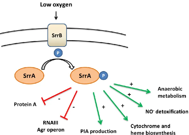
SrrAB (Staphylococcal respiration response) [or SrhSR (Staphylococcal resDE homologues)] was the first oxygen sensor system discovered in S. aureus. It is a two-component system (TCS) in which SrrB is a membrane sensor and SrrA is a cytoplasmic response regulator (Throup et al. 2001; Yarwood et al. 2001). It is homologous to the ResDE aerobic/anaerobic regulation system from Bacillus subtilis. The srrAB mutant had a growth defect in anaerobiosis while no phenotype was observed in aerobic condition. The transcriptome analysis of wild-type and srrAB strains grown in aerobic and micro-aerobic condition indicate that the expression of regulatory RNA RNAIII and spa (encoding the protein A) genes (see chapter IV.3 for more details) increase in srrAB mutant under micro-aerobic conditions. In addition, the SrrAB system is involved in nitrosative (NO) and hypoxia stress response (Kinkel et al. 2013). Various srrAB-required genes that were found to vary during two stress conditions are involved in cytochrome and heme biosynthesis, anaerobic metabolism and NO-detoxification.
SrrAB depletion affects the expression of genes involved in the TCA cycle, in fermentation and energy, arginine catabolism, xanthine catabolism and cell morphology (Throup et al. 2001). It also impacts biofilm formation and increases cell death. It has been proposed that SrrAB links the oxygen response to the regulation of virulence factors (Yarwood et al. 2001). Indeed, the srrAB mutant has an attenuated virulence in a murine model for hematogenous pyelonephritis infection as compared to a wild-type strain (Throup et al. 2001).
Under anaerobic condition, the response regulator SrrA-P binds to a 100 bp DNA sequence located in the upstream region of the ica gene to activate the production of polysaccharide intercellular adhesin (PIA). PIA is an important cell surface factor that protects S. aureus against human neutrophils (Ulrich et al. 2007). Therefore, the SrrAB TCS plays a positive role in PIA production helping bacterial survival against human defense mechanisms. A model of SrrAB activity is presented in figure 3.
Figure 3: Model of SrrAB system. PIA: Polysaccharide intercellular adhesin production.
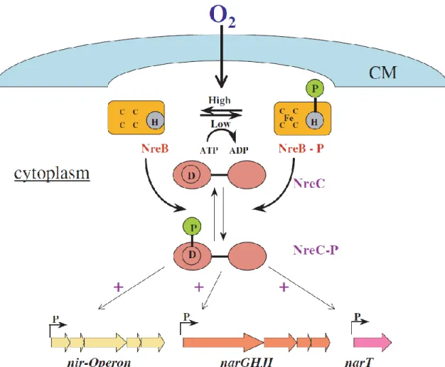
The second oxygen sensor system discovered was the TCS NreBC (Nitrogen regulation) present in some staphylococci such as S. aureus, S. epiderminis, S. carnosus (Kamps et al. 2004). NreB is a cytoplasmic oxygen sensor containing an O2-labile iron-sulfur
cluster considered as equivalent to the FNR (Fumarate and Nitrate reductase Regulatory) sensor, while NreC is a response regulator that controls gene expression involving in nitrogen regulation. In the presence of iron and low oxygen level, iron-sulfur cluster is formed and NreB autophosphorylates itself. Then, the active form NreB-P transfers its phosphoryl group to the response regulator NreC and activates it. NreC-P then positively controls the expression of the nitrite reductase (nir) operon , nitrate reductase (narGHJI) operon and narT (nitrate transporter) gene. On the contrary, under aerobic condition, NreB is dephosphorylated and its iron-sulfur cluster is destroyed (Figure 4).
Figure 4: Model for the NreBC sensor and regulation system in staphylococci.
[From (Kamps et al. 2004)].
1.2. Iron restriction
Metal ions are crucial elements for life processes including virulence. In bacteria, some metal ions like iron, magnesium and manganese are essential elements required for growth and play important roles in metalloproteins.
Iron (Fe) is mainly found in two common oxidation state, ferrous (Fe2+) and ferric (Fe3+). Ferrous irons are solube while ferric irons are insoluble and also the most stable form of iron. The difference in solubility of Fe2+ and Fe3+ leads to difficulties for bacteria to acquire iron. Therefore, bacteria not only develop mechanisms to uptake iron from the environment by solubilizing and assimilating it; but also have to compete for iron with other microorganisms or with the host. On the other hand, the quantity of intracellular iron has to be strictly controlled since an excess is toxic.
In S. aureus, there are three Ferric uptake repressors (Fur) homologues called Fur, PerR and Zur. When iron is present in bacteria, it binds to Fur. The Fe-Fur complex then binds to inverted repeat motifs called Fur boxes located in promoter regions of Fur-regulated genes. A Fur depletion in S. aureus generates a growth defect due to an excessive acquisition of iron and a decrease of the katA expression (Horsburgh, Ingham, and Foster 2001). There is a coupling between iron and oxidative regulations via the Fenton reaction. When peroxide or superoxide reacts with iron, they form harmful hydroxyl radicals which are reduced by catalases or peroxidases. The murine abscess model was used to test the role of Fur in virulence; fur mutant had a reduced virulence compared to the wild-type strain. A proteomic study revealed that in iron-limiting conditions or after fur depletion, genes involved in glycolysis, iron acquisition and transport were upregulated while genes in the TCA cycle and rsbU were downregulated (Friedman et al. 2006).
PerR is a second Fur homologue in S. aureus. PerR was shown to sense peroxide level inside the cell and control iron storage proteins (Horsburgh et al. 2001). The PerR-dependent regulon comprises not only oxidative stress resistance genes such as catalase (katA), alkyl hydroperoxide reductase (ahpCF) but also iron storage related genes such as ferritin (ftn), mgrA and fur. Like fur, the perR mutant had a reduced survival in murine abscess model infection (Horsburgh et al. 2001). PerR is also involved in the pathogenesis of Streptococcus pyogenes, Listeria monocytogenes and Enterococcus faecalis.
The third Fur-like protein in S. aureus is Zur. Its gene is within an operon encoding two putative membrane proteins with homology to zinc and other metal transporters (Lindsay & Foster 2001). In S. aureus, the depletion of Zur did not affect Zn2+ uptake but its overexpression was shown to affect the whole operon in a Zn2+-dependent manner. zur homologues were shown to regulate zinc uptake and ribosomal protein paralogs in B. subilis (Panina et al. 2003) and were involved in virulence in Salmonella enterica (Campoy et al. 2002) and Xanthomonas campestris (Tang et al. 2005). In contrast, S. aureus zur did not play a role in pathogenicity in a mouse skin infection model (Lindsay & Foster 2001).
1.3. Temperature
In E. coli, CspA is a major cold shock protein (Goldstein et al. 1990). In S. aureus, cspA was found to positively regulate the yellow pigment 4,4’-diaponeurosporene (Katzif et al. 2005). Transcriptome and mRNA turnover rates in response to cold shock were studied by Affymetrix GeneChips (Anderson et al. 2006). The cspA gene was moderately induced (2-fold) by a cold shock, while for two other cold shock genes, cspB was upregulated 9-fold and no change was observed for cspC expression. In addition, 46 genes upregulated and 416 genes downregulated by cold shock were identified. Many virulence factor genes [i.e. seo (enterotoxin), lip (lipase), srtA (sortase)], and regulatory genes [i.e. lexA (a SOS repressor) and SACOL0958 (general stress protein)] were also induced in cold shock condition. Surprisingly, cspC was strongly induced in oxidative stress (with hydrogen peroxide), salt condition by arsenate and various antibiotics such as ciprofloxacin, rifampicin, ampicillin, and cephalothin (Chanda et al. 2009).
1.3.2. Heat shock
CtsR and HrcA, SarA and Sigma B are regulators involved in heat shock adaptation in S. aureus (Clements & Foster 1999).
HcrA is a repressor of class I heat shock genes encoding dnaK and the groESL operons. CtsR has a dual function: it is a repressor of class III heat shock genes (encoding Clp ATP-dependent proteases) but also works together with HcrA to repress chaperon protein genes (Chastanet et al. 2003) (Figure 5). Sigma B (B) is a transcriptional factor that associates with RNA polymerase to initiate transcription. In S. aureus, B is an alternative “stress” sigma factor that modulates many stress response genes.
Figure 5: The CtsR, HrcA regulon of S. aureus
[Adapted from (Chastanet et al. 2003)].
Studies of the transcriptomic profile in response to heat shock revealed that 98 genes increased and 42 genes decreased (Anderson et al. 2006). Like for a cold shock, the viability of bacteria was not affected. The three heat shock genes, ctsR, clpB and clpC were significantly increased. In addition, many putative virulence genes [i.e. hla (-hemolysin), pathogenicity island genes, urea-ureG (urease system)] were upregulated. Interestingly, 11 genes were induced in both heat and cold shock conditions; they may belong to the same family of temperature-mediated response genes.
1.4. pH
Electron transport chain is the last process of aerobic respiration to generate the energy. The main function of this chain is transferring electrons from donors to acceptors and associated with proton pumps that transfer protons (hydrogen ions) across a membrane to create an electrochemical proton gradient that powers ATP production. pH or the hydrogen ion concentration is the main factor affecting the cytoplasmic pH homeostasis of bacteria. Therefore, to maintain the growth, bacteria need to develop pH sensing and mechanisms to keep balance their cytoplasmic pH homeostasis.
1.4.1. Acid shock
Many bacteria, including pathogens, need to adapt to acidic conditions, i.e. in dairy food like yogurts, fermented milk or inside the gastrointestinal system. For example, the
the phagosome. The ability to tolerate acidic pH is considered as a virulence factor for bacteria.
There are 8 known mechanisms for acid resistance in Gram-positive bacteria (Figure 6). They can maintain their intracellular pH either by proton pumps (GAD system, F1F0ATPase) that increase the uptake of hydrogen ion or by generating alkaline products such as NH3/ NH4+ (via an arginine deiminase (ADI) pathway) and an urease system that
counteracts acid pH. Other mechanisms are protein and DNA repair systems. Some genes involved are dnaK, groEL, htr, clp ATPases and lo18 for protein repair systems and recA, uvr and smn for DNA repair systems.
During an acidic challenge, bacteria alter their general energy and metabolism, and cell envelope, a switch that is needed for the adaptation. The role of the cell membrane is demonstrated by changes in membrane fatty acid profiles (Cotter & Hill 2003). A Streptococcus mutants strain with a dltC (encoding the D-alanyl carrier protein, Dcp) deletion was more sensitive to acid with a longer doubling-time and reduced growth yield than the parental strain (Boyd et al. 2000). The inactivation of dltC prevents the D-alanylation of lipoteichoic acids (LTAs), a main cell wall compound in Gram-positive bacteria.
Bacteria also resist to media acidification via global regulators such as TCSs and sigma factors. For example, in L. monocytogenes the depletion of lisRK encoding a TCS generates an altered acid shock response. The lisRK mutant was more sensitive to long time exposure to acid in stationary phase while it was more resistant to short time exposure during pre-stationary phase compared to the wild-type strain (Cotter et al. 1999).
Figure 6: Mechanism of acid resistance of Gram-positive bacteria
[Adapted from (Cotter & Hill 2003)].
During its life cycle, S. aureus can undergo various pH conditions within or outside the host. It can survive in human body, colonize and cause infection in different places which have acidic to alkaline pH such as external labia (pH 3.8 - 4.5), lysosomal compartments (pH 4.5 - 5.5) and wound sites (pH 8.9).
The B mutant is more sensitive to acid stress with a rapid loss of viability. S. aureus can be killed by a pH 2 but its resistance to acid increases if it is first pre-incubated at pH4, a non-lethal condition (Chan et al. 1998). In addition, the sodA (encoding a major superoxide dismutase) mutant has a reduced viability at low pH as compared to the wild-type strain (Clements et al. 1999). These results demonstrated the role of B and SodA to adapt to acid stress. However, the mechanism of sodA involved in acid resistance is still not clear.
Several studies were performed to determine the gene expression under different acidic conditions, such as growth in mild acidic condition (pH5.5) (Weinrick et al. 2004);
(Anderson et al. 2010). The results from Anderson et al not only confirmed many genes and overlapped with previous studies but also extended the network of genes related to acid stress adaptation. Interestingly, a total of 15 virulence genes were found downregulated in acidic condition including 7 known genes involved in acidic adaptation from (Bore et al. 2007). Five new additional virulence factors i.e. spa, chemotaxis-inhibiting protein CHIPS (chp), clumping factor B (clfB), fibrinogen-binding protein (efb) and staphylokinase precursor (sak) were revealed in this study. Moreover, 4 TCS such as SaeRS, LytSR, ArlSR and GdpS were also observed to be downregulated in response in acidic condition.
S. aureus is also a major cause of food poisoning. During food conservation, it undegoes stress associated with organic acids like lactic and acetic acids. S. aureus responses to medium containing these acids was explored by microarrays (Rode et al. 2010). First, a large variation in growth patterns was observed: bacterial growth was inhibited in medium containing acetic acid whereas bacteria exposed to lactic acid had a longer lag phase than when growing in medium containing HCl. Interestingly, only the pH of the culture containing lactic acid increased up to pH 7.5 during growth. Thus, compared with HCl induction, the response to lactic acid stress induced a specific mechanism to increase pH by accumulating ammonium and removing acid groups with production of diacetyl (2,3-butane dione) and pyrazines .
1.4.2. Alkaline shock
The first study on alkaline effect in S. aureus was reported in 1992 (Regassa & Betley 1992). The expression of the agr (accessory gene regulator) quorum sensing system (see chapter IV) examined in alkaline condition (pH from 6.5 to 8) revealed that the expression of RNAIII, one of the transcripts of agr locus, was higher at pH 7 but mostly vanished at pH 8. The expression of sec (staphylococcal enterotoxin type C), a target of the agr system, was also reduced in alkaline stress. Alkaline stress was found to strongly induce B transcription and 122 B-dependent genes (involved in capsule biosynthesis, Na+/H+ antiporter system and autolysin) were upregulated during this stress (Pané-Farré et al. 2006).
A microarray assay was also performed in alkaline condition (pH10 during 30 min) (Anderson et al. 2010). Cell viability in alkaline stress was not affected, but 128 transcripts
increased while 773 transcripts decreased. Downregulated genes were involved in nucleotide biosynthesis, amino acid metabolism and translation while upregulated were involved in amino acid biosynthesis (lysine, valine, isoleucine, histidine and threonine) and virulence (capsule biosynthesis). Interestingly, the alkaline shock stimulon induces the expression of (p)ppGpp, the activator of the stringent response.
2. Host-pathogen interaction
S. aureus is a versatile human pathogen, hence this bacterium needs to develop efficient mechanisms to survive in host defense and resist to the host immune system.
Briefly, the human immune system comprises a non-specific (innate) and specific (adaptive or acquired) immunity systems (Figure 7). The non-specific system includes two lines of defense. The first line consists of physical and chemical barriers, natural flora and mechanical barriers. If bacteria can pass the first line, they will face the second line of defense that includes defensive molecules, phagocytosis, complement and protective mechanisms. The specific immune system involves lymphocytes and antibodies which will recognize and eliminate pathogens, their toxins products, and also confers long-term protection by developing immunological memories.
Figure 7: Human lines of defense against pathogens
http://www2.bakersfieldcollege.edu/bio16/15_innate_immune.htm
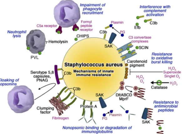
S. aureus developed different mechanisms to evade the innate immune system by inhibiting phagocyte functions, blocking complement activation, resisting antimicrobial peptides, lysing neutrophils (Nizet 2007) (Figure 8).
Figure 8: Mechanisms by which S. aureus subverts host innate immune defense. Phagocyte
recruitment is limited by binding of CHIPS (Chemotaxis inhibitory protein of Staphylococcus aureus) to chemokine receptors. Complement activation is blocked by protein Efb binding of soluble C3 and inhibition of the both the classic/lectin and alternative C3 convertases by SCIN (Staphylococcal complement inhibitor). Golden carotenoid pigment provides an antioxidant shield whereas catalase detoxifies hydrogen peroxide. Resistance to cationic antimicrobial peptides is afforded by positive charge modifications of the cell wall, aureolysin-mediated proteolysis, and binding/inactivation by staphylokinase. Protein A binds Fc domains of Igs in a nonopsonic manner, whereas fibrinogen binding clumping factor and the surface polysaccharide capsule and poly-N-acetylglucosamine (PNAG) act to cloak surface bound opsonins from phagocyte recognition. The heptameric pore-forming toxins -hemolysin and Panton-Valentine leukocidin (PVL) preferentially target leukocyte membranes. The plasminogen (PG) binding protein staphylokinase (SAK) activates the zymogen to the active protease plasmin, which can degrade complement opsonin C3b and the immunoglobulin Fc domain [From (Nizet 2007)].
III. Overview of bacterial competitive fitness
The fitness of a bacterial strain could be explained from a Darwinian point of view. From his theory, “survival of the fittest” is the main concept of natural selection that is mechanism by which species adapt and evolve. It could be defined as an increase of
frequency or its probability of survival in competition with others. The individuals having variants that best fit to the environment (fittest) have better potential for survival, reproduction and passing their desirable variations to their offsprings.
Based on this concept, one way to define the bacterial fitness in the laboratory conditions is a competitive fitness assay by growing two or more strains together that allow them to compete and evaluate their ratio at different times (Figure 9). In addition, growth in the presence of other strains reflects a more “real” situation, as the fitness of one strain may be affected by the genetically different surrounding strains. Therefore, these experiments could reveal patterns of interaction or epistasis among different strains and possibly whether particular combinations of strains interact synergistically or antagonistically (Zhan & McDonald 2013). Interestingly, variants leading to improved fitness in one growth condition can lead to altered fitness in another condition, as the result is often a compromise (Mariam et al. 2004) (MacLean & Vogwill 2014).
Figure 9: Scheme of a competitive fitness assay. The strain to be assayed is mixed with a reference
The classical way to carry out fitness assays is to label the strains with different antibiotic resistance or fluorescent markers. However, one impediment is the limited number of available markers; hence fitness assays are difficult to perform on a large scale. As a result, one possibility to easily follow many strains in the same culture is to introduce specific DNA sequences for each constructed strains. These sequences, called DNA barcodes, can then be quantitatively detected within a mix culture. A DNA barcode acts as a specific “marker” that represents the relative presence of a strain in the population.
The first application of DNA barcodes was in Salmonella typhimurium to identify genes involved in pathogenesis using a murine model of typhoid fever (Hensel et al. 1995). Briefly, the DNA barcodes contained 40 random nucleotides flanked by common priming regions on each side. The DNA barcodes were ligated with transposons and used to mutagenize S. typhimurium genome. A bank of 1152 transposon-tagged mutants was obtained and arrayed in twelve 96-well microtiter plates. DNA colony blots were made from microtiter dishes by replica plating them on a membrane. The mutants of each microtiter plate were pooled together and used to infect mice. After 3 days of infection, spleens were recovered, homogenized and plated to recover the infecting mutants. Approximately 10,000 obtained colonies were pooled together. Chromosomal DNAs were extracted and DNA barcodes were amplified (using conserved priming regions for all tags), radiolabeled and hybridized with DNA colony blots (Figure 4). Virulence genes were found by identifying DNA barcodes that were present in the control samples but not in the infected ones. These DNA barcodes corresponded to insertions leading to attenuated virulence (Figure 10).
Figure 10: Signature-tagged mutagenesis in Salmonella. A) Design of a signature tag. Each tag has a
unique central sequence of 40 bp ([NK] 20; N = A, C, G, or T; K = G or T), flanked by invariable arms of 20 bp, which are common to all the tags. B) Signature-tagged mutagenesis screening in mice. A complex pool of tags (shown as colored rectangles) is ligated to transposons. The tagged transposons are then used to mutagenize bacteria, which are subsequently assembled into a library. Only bacteria with tags that are efficiently amplified by PCR and are not cross reactive with other tags in hybridization experiments are selected for inclusion in the pool that is used to infect the mice. [From (Mazurkiewicz et al. 2006)].
This technique was subsequently developed in yeast (Shoemaker et al. 1996; Pierce et al. 2007; Smith et al. 2009; Han et al. 2010; Chen et al. 2012), and used with other bacteria (Rooney et al. 2008; Hobbs et al. 2010).
In many published fitness protocols, tags were analyzed by hybridizing labeled PCR products on dedicated DNA arrays (Hensel et al. 1995; Shoemaker et al. 1996; Pierce et al. 2007; Rooney et al. 2008; Hobbs et al. 2010). These experiments are rather heavy and expensive as each tested condition required at least one array. The protocol was adapted to deep sequencing technology to improve its sensitivity and its ease-of-use (Smith et al. 2009; Han et al. 2010) (chapter A).
IV. Overview of regulatory RNAs in S. aureus
Besides the central roles of mRNA, rRNA and tRNA in translation, the regulatory role of RNAs in prokaryote gene expression is nowadays well established. They can act either in trans, targeting RNAs and proteins, or in cis by affecting adjacent or associated sequences. Through sophisticated mechanisms, these regulatory RNAs fine-tune genetic expression to allow bacterial fitness and adaptation to varied environments including those within their dedicated hosts. They usually exert their functions at the levels of transcription and/or translation of their mRNA targets (Storz et al. 2011; Guillet et al. 2013).
1. Identification of regulatory RNAs by various approaches
The first regulatory RNAs in S. aureus was discovered in 1993 and named RNAIII (see chapter IV.3 for more details). Later, several studies contributed to identify numerous S. aureus regulatory RNAs based on computational prediction (Geissmann et al. 2009; Marchais et al. 2009; Pichon & Felden 2005), Affymetrix microarrays (Anderson et al. 2010; Roberts et al. 2006; Anderson et al. 2006), sequence cDNA libraries (Abu-Qatouseh et al. 2010) and high throughput sequencing (Bohn et al. 2010; Beaume et al. 2010; Lasa et al. 2011; Lioliou et al. 2012; Howden et al. 2013). Several targets from these identified sRNAs were experimentally validated (see Table 1). So far, the transcription of approximately 250 staphylococcal regulatory RNAs was experimentally confirmed [review from (Felden et al. 2011)].
Table 1: Summary of experimentally validated regulatory RNAs in S. aureus a (Tomasini et al. 2014) Study Strain used sRNA discovery methodology (number of in silico predicted sRNAs)
Experimentally validated sRNAs e Experimental validation method
Target and mechanism Comment
(Roberts et al. 2006)
UAMS-1 Gene chip analysis SSR42 (srn_4470, RsaX28, Teg27, sRNA363) NB, RT-PCR Spa, hla, hglC, lukF Unknown
(Pichon & Felden 2005) and (Sayed et al. 2011) Mu50 (clonal Complex 5) Bioinformatic predictions SprA
SprA2 (srn_4550, WAN014FZW, IGR2049,
IGR8bis, Teg26as, sRNA371), SprA3 (IGR2125), SprB (srn_3600, Teg9, IGR18),
SprC (srn_3610, Teg10), SprD (srn_3800,
Teg14_sRNA300), SprE, SprF, SprG, SprFG2,
SprFG3
4.5S RNA (ffs, Teg42, IGRLF1, WAN01CBPQ,
sRNA98), 6S RNA (Teg97, ssrS, ssr80, IGR2, WAN01CC8T, sRNA256), RNAIII (srn_3910, sRNA317), tmRNA (tmR, WAN014GIY, Teg150, ssrA, sRNA166), RNase P (RseP, rnp, Teg65, IGR1215, sRNA240)
NB b NB b NB b
ABC transporter (SA2216), possible antisense sRNA Unknown, SprA2 encodes a cytolytic peptide Housekeeping ncRNAs (Geissmann et al. 2009) RN6390, COL, Newman, HG001 Bioinformatic predictions and experimental validation
RsaE (srn_2130, RsaON, Sau20, Teg92, IGR6,
sRNA183),
RsaA (srn_1510, rsaOJ, Teg88, sau64, IGR1,
IGR14, sRNA132), RsaB (srn_3410), RsaC (srn_1590, Teg90, sRNA135), RsaD (srn_1640, Teg91, sRNA138), RsaF, RsaG (srn_0510, Teg93, sRNA31), RsaH (srn_1910, rsaOK, Teg94, sau6059, IGR7, sRNA162), RsaI (srn_4390, rsaOG, Teg24, sRNA356), RsaJ (srn_4530, sprAs2prime, Teg96, sau5837, sRNA369), RsaK (srn_0440, Teg38, sRNA27)
NB, PE, RACE NB, PE, RACE
Masking of ribosomal binding site for oppB, sucD, SA0873
Unknown
Genetic manipulation demonstrated a role for RsaE in controlling metabolic pathways
Study Strain used sRNA discovery methodology (number of in silico predicted sRNAs)
Experimentally validated sRNAs e Experimental validation method
Target and mechanism Comment
(Marchais et al. 2009) N315 (clonal Complex 5) Bioinformatic (NAPP) (189) and Northern analysis
RsaOA, RsaOB (srn_0860,Teg40, sRNA61), RsaOC (srn_1770, RsaX08, Teg50), RsaOD
(srn_3160, Teg67_sRNA250), RsaOE (srn_3490, Teg73_sRNA276), RsaOF
(srn_1930.3, Teg12), RsaOGb (srn_4390, RsaI, Teg24, sRNA356) NB Unknown (Jesper S Nielsen et al. 2011) N315 (clonal complex 5) Bioinformatic search for intergenic B
consensus sites and experimental validation
SbrA (srn_2290, RsaOO, Teg54, sbrA, sRZN), SbrB (srn_2830, Teg111), SbrC (srn_1580)
NB B regulated. SbrC interacts with SA0587 (mntC) sbrA and sbrB potential CDS (Abu-Qatouseh et al. 2010) - Cloning and sequencing of cDNAs Ssr-72, Ssr-80 (6S, Teg97, ssrS, IGR2,
WAN01CC8T, sRNA256), Ssr-87, Sau-02 (Teg19a, Teg102, sRZN, sRNA190), Sau-13 (srn_5000), Sau-19 (srn_4680, Teg131, RsaX21, sRNA382), Sau-24 (srn_2610, Teg81),
Sau-27 (srn_2690), Sau-30 (srn_4260,
SSR154, sRZI), Sau-31 (srn_4250), Sau-41 ( srn_1070), Sau-50 (srn_3040), Sau-53 (srn_0430), Sau-59 (srn_2340), Sau-63 (srn_0950, Teg146, sRNA83), Sau-64, Sau-66 (srn_1780), Sau-5949 (srn_3460, Teg120, sRNA272), Sau-5971 (srn_0880), Sau-6053 ( srn_2200, sRNA189, Teg78), Sau-6072
NB c NB
Unknown
Unknown. Sau-66, putative posttranslational control of antisense gene SA0671
142 candidate sRNA identified
Study Strain used sRNA discovery methodology (number of in silico predicted sRNAs)
Experimentally validated sRNAs e Experimental validation method
Target and mechanism Comment
(Bohn et al. 2010) N315 (clonal complex 5) 454 pyrosequencing followed by experimental validation
RsaON (srn_2130, rsaE, Sau20, Teg92, IGR6,
sRNA183)d
RsaOH, RsaOI (srn_1490, SSR156, Teg47,
Sau6477, sRZR, sRNA131), RsaOL (srn_1960, Teg100, Sau07, IGR14, sRNA168), RsaOM (srn_2030, Teg52_IGR20_sRNA172), RsaOO (srn_2290, Teg54, sbrA, sRZN), RsaOP (srn_2350), RsaOQ (srn_2880, Teg82, IGR23, sRNA230), RsaOR (srn_3820.1, SprX2, ssr6, teg15, IGR12), RsaOT (srn_4670, ssr43, RsaON, Teg29, sRNA381), RsaOU (srn_4800, sRZG), RsaOV (srn_4840, Sau40, sRZV)
RsaOW, RsaOX
NB NB
Binds opp3A mRNA ribosome binding site. Overexpression of RsaE reduces central metabolic pathways and increases amino acid pool 30 sRNAs identified, 14 new (Beaume et al. 2010) N315 (clonal complex 5)
Illumina sequencing Teg1 (srn_0050, sRNA3), Teg4, Teg17
(srn_0360, sRNA21), Teg18 (srn_0770, sRNA53), Teg19b, Teg21 (srn_4130),
Teg24 (srn_4390, rsaOG, RsaI, sRNA356), Teg26 (srn_4460, sRNA362), Teg28
(srn_4590, sRNA375), Teg35 (srn_0030, sRNA2), Teg38 (srn_0440, rsaK, sRNA27),
Teg42 (4.5S, ffs, IGRLF1, WAN01CBPQ,
sRNA98), Teg45 (srn_1350, sRNA120), Teg47 (srn_1490, RsaOI, SSR156, Sau6477, sRZR, sRNA131), Teg55 (srn_2370, sRNA198),
Teg56 (srn_2440, sRNA203), Teg57
(srn_2450, Sau6229, sRNA204), Teg60 (srn_2520, sRNA208), Teg61 (srn_2620, sRNA216), Teg69 (srn_3260, sRNA257),
Teg70 (srn_3300, sRNA262), Teg72
RT-PCR Unknown 195 sRNAs predicted by HTS
a
Validated by Northern blot, RNA extremity mapping, or RT-qPCR.
b
Also experimentally validated using Northern blot in the study by (Abu-Qatouseh et al. 2010).
c
Originally described by (Anderson et al. 2006).
d Previously described and validated. NB, Northern blot; PE, primer extension; RACE, random amplification of cDNA ends; RT-PCR, real-time PCR. Several long and
stable
RNAs (SSR) expressed under particular conditions of growth have been assigned by microarrays (Roberts et al. 2006).
e
A number of different names have been assigned to many of these sRNAs. In this table, the initial name from the relevant study has been used. A full list of alternate names can be found in the supplementary table from (Felden et al. 2011). Recently, a uniform nomenclature for S. aureus regulatory RNAs was proposed (Sassi et al. 2015).
(srn_3340), Teg73 (srn_3490, RsaOE, sRNA276), Teg76 (srn_0930, sRNA74), Teg91 (srn_1640, rsaD, sRNA138), Teg2pl
2. Classification
Regulatory RNAs exert usually their activity by base-pairing with target mRNAs or by binding to proteins. Some regulatory RNAs have a cis-regulatory activity on associated RNA sequences. Most regulatory RNAs are active under specific conditions and influence gene expression in response to environmental changes. They can bind mRNA ribosome-binding site (RBS), inhibit and stimulate translation or RNA degradation. According to their predicted mode of action, regulatory RNAs can be categorized as follows.
2.1 Regulatory small RNAs targeting mRNAs 2.1.1 Cis-encoded antisense RNAs
Cis-encoded antisense RNAs (asRNAs) are expressed from DNA strands opposite to genes. The predicted putative target of asRNAs is the mRNA expressed from the opposite strand. asRNAs share extended regions of complete complementarity with their target (often 75 nucleotides or more) with which they fully or partially overlap to activate or inhibit mRNA functions (Waters & Storz 2009; Georg & Hess 2011) (Figure 11). Most studied asRNAs are from bacteriophages, plasmids, and transposons and control phage development, plasmid replication and copy-number control of mobile elements, respectively.
The first asRNA reported in S. aureus was RNAI from plasmid pT181 (Novick et al. 1989). RNAI associates by base-pairing with repC mRNA and consequently inhibits the expression of RepC, a replication initiation protein. A second asRNA, RNAII, transcribed from the same promoter as RNAI, but longer than RNAI is also involved in pT181 replication as show on figure 12.
Figure 12: Antisense regulation of plasmid pT181 replication. (A) Genetic organization of pT181
plasmid and its control region. RNAI and RNAII are asRNAs. (B) Schematic secondary structure model of repC mRNA leader region as proposed by (Novick et al. 1989). The formation of a large helical domain formed by helices I and III favors the formation of an anti-terminator hairpin that stimulates the transcription of the complete mRNA. In this structure, the Shine and Dalgarno sequence (SD) and the initiation codon are available for translation. (C) The antisense RNAI traps a transient hairpin structure of repC mRNA during transcription, and the formation of the RNAI-mRNA duplex stabilizes a Rho-independent terminator to arrest transcription. RepC synthesis is thus prevented. [From (Romilly et al. 2012)].
RsaOW is an example of an asRNA expressed from mobile elements, which pairs with the 5’UTR of the transposase gene of IS1181. RsaOW has eight copies from rsaOW1 to rsaOW8 in N315 genome and is expressed constitutively likely to block expression of the
asRNAs were also reported from S. aureus core genome. The first high-throughput sequencing of N315 transcriptome revealed that at least 1.3% of mRNAs were covered by asRNAs (Beaume et al. 2010). However, this percentage is likely much higher. In this recent study, long (>75 nt) and short (<50 nt) transcripts of strain NCTC8325 were analyzed by RNA deep sequencing. The authors concluded that 49% of the ORFs are covered on at least 50% of their length by long asRNAs and up to 75% of ORF in case of short transcripts (Lasa et al. 2011).
2.1.2 Trans-encoded RNAs (sRNAs)
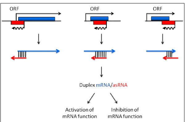
In contrast to cis-encoded asRNAs, trans-encoded RNAs (often referred to as sRNAs), are expressed from DNA regions with no RNA expressed on their opposite strand. sRNAs are usually small, typically between 50 and 500 nucleotides in length, and non-coding (Gottesman and Storz 2011). Their RNA targets are usually located at different positions on the chromosome. Hence, the base pairing length between a sRNA and its target is often limited, typically about 10-25 nucleotides (Waters & Storz 2009). Their mode of action is quite depicted in figure 13.
Figure 13: Gene arrangement and regulatory functions of sRNAs. Genes encoding trans-encoded sRNAs (red) are located separately from the genes encoding their target RNAs (blue) sRNAs have a limited base-pair complementarity with their targets. (Left panel) sRNAs can base pairing to 5’UTRs and block ribosome-binding site (RBS) and/or (Middle panel) target the mRNAs to degradation by ribonuclease. (Right
panel) sRNAs can also prevent the formation of an inhibitory structure which sequester RBSs. [From (Waters & Storz 2009)].
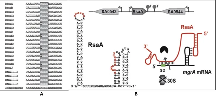
In S. aureus, the sRNA/RNA interaction seems to require a longer pairing than in many other organisms, possibly because S. aureus has a low GC content genome therefore generating weaker RNA-RNA interactions. Most of sRNAs have a conserved “seed” motif UCCC to initiate the pairing with RNA targets (Figure 14) (Geissmann et al. 2009).
A B
Figure 14: C-rich box conserved motif in S. aureus sRNAs and an example of example sRNA/mRNA base
pairing. (A) Alignment of the C-rich sequence motifs of S. aureus sRNAs [From (Geissmann et al. 2009)]. (B) Repression of mgrA mRNA translation by the sRNA RsaA. Two regions of RsaA base pair i) with the mgrA mRNA RBS and ii) with the coding region via a loop-loop interaction. The two regions are essential to repress translation and to enhance the mgrA mRNA degradation [From (Fechter et al. 2014)].
2.2 Cis-regulatory elements
In bacteria, cis-regulatory RNAs may be located in the 5’ or 3’ untranslated regions (UTR) of mRNA and hence act in cis. Under specific condition, cis-regulatory RNAs can sense environmental signals such as metabolites (riboswitches), uncharged tRNAs (T-boxes), metal ions, pH and temperature (thermoswitches) and then adopt different conformations thus regulating the expression of downstream genes.
The most widespread examples of cis-regulatory sRNAs in bacteria are riboswitches that play a major role in regulating genes involved in metabolic pathways. Riboswitches
sense metabolites such as cofactors, vitamins, amino acids, nucleotides, the second messenger cyclic di-GMP, metal ions. Generally, riboswitches are composed of two parts: i) the aptamer region which binds ligands (metabolites) and ii) the expression platform which changes its structure in response to the ligand binding. mRNA modifications usually involve alternative hairpin structures creating transcription terminators and antiterminators or structures occluding and exposing RBSs, thus controlling translation. In most cases, the presence of ligands inhibit transcription or translation (Figure 15).
Figure 15: Diversity of riboswitches and mechanisms of gene control in bacteria. Mechanisms of
modulation of gene expression are highly divergent in prokaryotes and involve control of transcription, translation and mRNA stability. SD (Shine-Dalgarno), Pol (RNA Polymerase), ORF (Open Reading Frame) [From (Serganov & Nudler 2013)].
Many S. aureus riboswitches were discovered by biocomputing analysis of genome sequences to identify conserved metabolite-binding domains (Barrick & Breaker 2007) (Yao et al. 2007); among them riboswitches sensing S-adenosylmethionine (SAM), thiamine pyrophosphate (TPP), flavin mononucleotide (FMN), lysine, glycine, guanine, 7-aminomethyl-7-deazaguanine (preQ1) and glucosamine-6-phosphate (Glc-6P) (Geissmann et al. 2009; Marchais et al. 2009; Abu-Qatouseh et al. 2010; Beaume et al. 2010; Bohn et al. 2010; Ten Broeke-Smits et al. 2010).
Antibiotic binding to riboswitches can be used to develop new treatments for multiple drug resistant strains. Some riboswitches are known as antimicrobial targets such as TPP, lysine, FMN and guanine riboswitches [review in (Mulhbacher, St-Pierre, et al. 2010)]. Recently, novel putative ligands that can bind to guanine riboswitches were selected based
on a model of the crystal structures. Two pyrimidine-based molecules named pyrimidine compound 1 (PC1) and pyrimidine compound 2 (PC2) were identified. PC1 showed antibacterial activity in vitro against S. aureus and Clostridium difficile. PC1 was also tested in a mouse model and shown to decrease S. aureus growth in the mammary gland (Mulhbacher, Brouillette, et al. 2010).
Another example of riboswitch is associated with the tightly control of the methionine biosynthesis operon in S. aureus. This regulation is a complex combination of the action of stringent-mediated repressor CodY, T-box riboswitch and methionine metlCFE-mdh operon as explained in figure 16 (Schoenfelder et al. 2013).
Figure 16: Model of a regulatory cascade for methionine biosynthesis operon control. (i) At high amino
acid concentration, branched-chain amino acids (BCAA) and GTP are bound to the CodY repressor, increasing its affinity for target DNA binding; downstream genes are repressed (small picture, bottom left). (ii) Low amino acid levels will trigger the stringent response due to stalled ribosomes, which leads to an increase in RelA-mediated ppGpp alarmone synthesis resulting in less GTP. Subsequently, CodY dissociates from the DNA activating downstream transcription of the T-box leader RNA. The T-box acts as the crucial check-point sensing uncharged tRNAifMet levels and determines transcription of the met biosynthesis genes in a highly
methionine-dependent manner. Rapid degradation of the met mRNA by the RNA degradosome is an additional mechanism to limit unnecessary translation of methionine biosynthesis mRNA. [From (Schoenfelder et al. 2013)].
2.3 Protein-targeting RNAs
Protein-targeting RNAs are regulatory RNAs that directly interact with proteins to modulate their activity. Some of these proteins belong to ribonucleoprotein complexes and contribute to the function of housekeeping complexes such as M1 [the RNA component of Ribonuclease P, RNase P (Esakova & Krasilnikov 2010)], 4.5S RNA [component of the signal recognition particles (Ribes et al. 1990)] and tmRNA [tRNA-like mRNA-like dual functions (Keiler et al. 1996)]. Protein-targeting sRNAs can regulate protein activity by mimicking their substrate. In E. coli, some well-known examples are 6S sRNA that interacts with 70 RNA polymerase (Wassarman 2007), CrsB sRNA that interacts with CsrA protein (carbon storage regulator) (Babitzke & Romeo 2007) and GlmY sRNA that interacts with YhbJ protein (Görke & Vogel 2008). In S. aureus, tmRNA, RNase P RNA, 4.5S and 6S were detected by comparative genomics and experimentally validated (Pichon & Felden 2005). However, this bacterium does not have the CsrB/CsrA and GlmY/YhbJ systems.
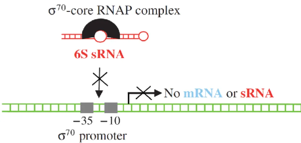
6S sRNA is a global regulator conserved in bacteria (Barrick et al. 2005; Trotochaud & Wassarman 2005). In E.coli, 6S sRNA sequestrates 70-core RNA polymerase by mimicking a promoter sequence (Figure 17). This interaction represses the expression of many 70 -dependent transcription in stationary phase due to the abundance of 6S in this phase (Wassarman & Storz 2000). Moreover, 6S sRNA also upregulates the general stress σS -dependent transcription in vivo (Trotochaud & Wassarman 2005; Cavanagh & Wassarman 2014). 6S sRNA was found to be highly expressed in stationary phase of four S. aureus pathogenic strains (Pichon & Felden 2005). However, there is no homolog of σS in S. aureus. Instead, σB is the general stress sigma factor involved in environmental response and virulence factor expression (Gertz et al. 2000). Therefore, staphylococcal 6S RNA is possibly associated with different regulations than in E. coli.
Figure 17: RNA polymerase interacts with 6S sRNA in E.coli. RNA synthesis by the “70–core RNA polymerase” complex (black). 6S sRNA (red) sequesters the polymerase complex during nutrient limitation (stationary phase of growth), and restrain gene expression to the ones controlled by an alternative factor [From (Pichon & Felden 2007)].
3. Role of regulatory RNAs in S. aureus
Regulatory RNAs play multiple roles in S. aureus. Their expression was observed in several stress conditions including starvation, antibiotic treatment and host infection (Anderson et al. 2006; Geissmann et al. 2009; Beaume et al. 2010; Bohn et al. 2010; Anderson et al. 2010; Howden et al. 2013). Several examples of S. aureus regulatory RNAs involved in metabolism, stress response, environmental adaptation and virulence will be presented below.
RNAIII is the most well-known Staphylococcal sRNA. It is the intracellular effector of the agr system (Novick et al. 1993) that controls the expression of many virulence genes by a
two-component signaling module. The agr locus encodes a quorum sensing system
(agrBDCA) and RNAIII driven by the P2 and P3 promoters, respectively (Figure 18). AgrD encodes an autoinducing peptide (AIP) while AgrB is a transmembrane protein that processes and secretes this peptide. At the high cell densities, the secreted AIP molecules accumulate and reach a threshold level that can bind and activate the receptor histidine kinase AgrC. Then, the phosphorylated AgrC can activate the sensor regulator AgrA by transferring its phosphate group. As a consequence, the phosphorylated AgrA binds to the P2 and P3 promoters to induce RNAII and RNAIII transcripts. Therefore, RNAIII accumulates and its maximal expression is in late-exponential and stationary growth phases.
![Figure 6: Mechanism of acid resistance of Gram-positive bacteria [Adapted from (Cotter & Hill 2003)]](https://thumb-eu.123doks.com/thumbv2/123doknet/12888239.370473/26.892.129.747.142.618/figure-mechanism-resistance-gram-positive-bacteria-adapted-cotter.webp)