HAL Id: tel-02144589
https://tel.archives-ouvertes.fr/tel-02144589v2
Submitted on 4 Jun 2019
HAL is a multi-disciplinary open access archive for the deposit and dissemination of sci-entific research documents, whether they are pub-lished or not. The documents may come from teaching and research institutions in France or abroad, or from public or private research centers.
L’archive ouverte pluridisciplinaire HAL, est destinée au dépôt et à la diffusion de documents scientifiques de niveau recherche, publiés ou non, émanant des établissements d’enseignement et de recherche français ou étrangers, des laboratoires publics ou privés.
Characterisation of semaphorin 3A-chondroitin sulphate
interaction in the central nervous system
Lynda Djerbal
To cite this version:
Lynda Djerbal. Characterisation of semaphorin 3A-chondroitin sulphate interaction in the central nervous system. Biochemistry, Molecular Biology. Université Grenoble Alpes, 2018. English. �NNT : 2018GREAV041�. �tel-02144589v2�
1
THÈSE
Pour obtenir le grade de
DOCTEUR DE LA COMMUNAUTE UNIVERSITE
GRENOBLE ALPES
Spécialité : Biologie structurale et nanobiologie
Arrêté ministériel : 25 mai 2016
Présentée par
« Lynda DJERBAL »
Thèse dirigée par Hugues LORTAT-JACOB (DR, UMR5075-IBS-UGA-CNERS-CEA) et Jessica KWOK (DR, université de Leeds)
préparée au sein du groupe Structure et Activité des Glycosaminoglycans à l’Institut de Biologie Structurale-UMR5075
dans l'École Doctorale Chimie et Science du Vivant
Characterisation of semaphorin
3A-chondroitin sulphate
interaction in the central
nervous system
Thèse soutenue publiquement le «30 Novembre 2018 », devant le jury composé de :
Pr Dulce PAPY-GARCIA
Professeur d’université, université Paris Est Créteil, Rapporteur
Pr Joost VERHAAGEN
Professeur, Netherlands institute for neuroscience, Rapporteur Dr Jean-Maurice MALLET
Directeur de recherche, ENS Paris, Examinateur
Dr Anne IMBERTY
3
Summary
Perineuronal nets (PNNs) are the key regulators of neuronal plasticity and regeneration in the mature central nervous system (CNS). They are a unique and highly organised extracellular matrix (ECM) structure, found around sub-population of neurons, composed mainly of chondroitin sulphate proteoglycan (CSPG). Chondroitin sulphate (CS) is a linear polysaccharide belonging to glycosaminoglycans (GAGs) family. The sulphation pattern defines different types of CS, which interact with different signalling proteins including those regulating axonal outgrowth and guidance such as semaphorin 3A (Sema3A). Sema3A is a secreted chemorepulsive protein found accumulated in the PNN through its interaction with CS. This process is believed to potentiate Sema3A signalling through plexin A1 (PlxnA1) and neuropilin 1 (Nrp1) and regulate plasticity and regeneration. The aim of the thesis project is to characterise the interface of Sema3A-CS interaction.
For this purpose, Sema3A is expressed in eukaryote cells and purified. Interestingly, two major forms were obtained: a full length Sema3A (90 kDa) which remains attached to the cell surface GAGs and a truncated form without the C-ter part (65 kDa) which is released to the culture medium. With the use of surface plasmon resonance (SPR), we observed that full length Sema3A binds selectively to CS-E (4,6-disulphated chondroitin) and heparan sulphate with a high affinity (KD in the sub pM range), while the truncated Sema3A does not bind to
any GAG. Four putative GAG-binding sequences were identified in the C-ter of Sema3A and mutated using site directed mutagenesis. SPR analysis then revealed that two out of these four sites are required for the binding to CS-E. The importance of these GAG-binding sequences in inhibition of neurites outgrowth of dorsal root ganglion neurons in culture was also reported, indicating thus the importance of GAG-binding in Sema3A signalling. In parallel, the minimal required sequence of Sema3A-binding of CS-E was determined as being a tetrasaccharide. The Sema3A-CS interface was thus characterized. Furthermore, quartz crystal microbalance with dissipation monitoring analysis suggested that Sema3A could crosslink GAG chains. This suggests Sema3A could be involved in stabilising the PNN network and induces mechanical changes on neuronal surface.
The detail characterization of Sema3A-CS interaction may enable the design of new strategies aiming at enhancing plasticity and regeneration for neurodegenerative diseases or spinal cord injury.
5
Acknowledgements
Je remercie les membres du Jury : Joost VERHAAGEN, Dulce PAPY-GARCIA, Jean-Maurice
MALLET et Anne IMBERTY d’avoir accepté d’évaluer mes travaux de thèse.
Je ne pourrais jamais remercier assez mon directeur de thèse Hugues. Quand je suis venue pour l’entretien et je t’ai vu pour la première fois, je me suis dit « il a l’air très bien ce directeur de thèse» et après trois ans je le pense toujours. Merci de m’avoir fait confiance dès le début et de m’avoir donné l’opportunité de faire une thèse dans des conditions « optimales ». Merci pour tout ce que j’ai appris durant ces trois années de thèse. Merci pour tes encouragements et tes conseils pour les manips, rédaction, présentations... Merci de t’être toujours rendu disponible et cela malgré ton emploi du temps chargé. J’ai vraiment apprécié le travail avec toi sur le plan scientifique et humain. Je suis très contente que mon post-doc sera toujours en collaboration avec toi. Infiniment Merci Hugues !
Un très grand merci à mon autre directeur de thèse Jessica. Je t’ai aimé dès notre premier skype pour l’entretien. Je me suis aussi dit « qu’est-ce qu’elle est gentille ! » Et cela ne s’est fait que confirmer au cours de ces trois années. Et je n’oublierai pas ce que tu m’as dit au début de ma thèse « fais toutes les erreurs que t’as à faire pendant la thèse, car c’est pendant la thèse qu’on apprend » et cela m’a mis à l’aise. J’apprécie ton dynamisme et ton esprit scientifique. Merci pour tout ce que tu m’as appris. Merci pour ta confiance, ton soutien, tes encouragements et ta gentillesse. Merci pour l’opportunité que tu m’as offerte de poursuivre sur la même thématique que ma thèse et surtout sur ma protéine préférée «Sema3A ». Je suis très contente de poursuivre en post-doc avec toi.
Je remercie les membres de ma deuxième famille « SAGAG ». Rabia, merci beaucoup pour ton amour, ta gentillesse et tes délicieux gâteaux. Je me rappellerai toujours du premier jour quand je t’ai vu à l’entretien, t’étais adorable comme toujours et je me suis dit j’aimerais bien être prise dans cette équipe. Et merci de m’avoir appris à faire les WB et dot blot. Romain, t’es le chercheur le plus cool que je connaisse et cela je l’ai su, avant même de te rencontrer, dans le premier mail que tu m’as envoyé. J’apprécie énormément ton enthousiasme, ton raisonnement scientifique et ton sens de l’humour et merci. Merci de m’avoir appris « heparin-beads approach » et l’analyse disaccharidique.
Evelyne, je suis chanceuse que t’as intégré le SAGAG avant que je parte, je suis très contente de
t’avoir connue. Merci pour ta gentillesse, ta bonté et ton aide. Rabia et Evelyne, je ne dirai pas vous êtes les mamans du SAGAG mais plutôt les grandes sœurs. Yoan, quand je t’es vu la première fois je me suis dit « pas très souriant, ce post-doc» mais en fait c’est juste une première impression, t’es sympa et très serviable. Merci d’avoir toujours pris le temps de discuter les difficultés que j’ai rencontrées surtout pendant la purification de la Sema. Merci de m’avoir montré tous les logiciels et tous les sites requis pour mon projet. Rana, ma petite Rana, j’ai su qu’on allait devenir amies avant même de te rencontrer. Tout aurait été moins drôle si tu n’étais pas là. Je suis très contente de t’avoir rencontrée. Merci pour tous les moments agréables passés au labo surtout après 19h, au bureau, au coin café en dehors du travail. Merci pour ta complicité dans tout et surtout ton amitié.
Merci à tous les membres du SAGAG d’avoir contribué à ma formation scientifique. On dit « il faut tout un village pour élever un enfant » et moi je dis «il faut tout un groupe de recherche pour
6 former un étudiant ». Vous allez beaucoup me manquer mais je reviendrai vous voir. Je m’arrête là pour le SAGAG sinon je vais écrire une deuxième thèse de remerciements …
Je remercie les HBB de la « pause 17:00 » : Rana (tu vois c’est comme les groupes de FB et WhatsApp, t’es partout), Rida, je suis très contente d’avoir fait ta connaissance. T’es la libanaise la plus drôle que je connaisse après Rana biensûr. T’es une fille formidable. Merci pour tous les moments géniaux qu’on a passé ensemble à l’IBS ou en dehors de l’IBS et merci pour le GreenMango du mois de juillet. Kevin, infatigable Kevin, j’étais ravie de rédiger ma thèse au même temps que toi. Merci pour les amandes et ton sens de l’humour. Guillaume, merci pour ta gentillesse, et les friandises du distributeur et ton sens de détection de fautes d’orthographe.
Rime ma compatriote, merci pour ton soutien, ta gentillesse et ta bonté. Ilham, mon autre
compatriote, contente de t’avoir rencontrée. Merci Elodie pour ta gentillesse. Merci aux voisins du SAGAG, IRPAS, pour les pauses café du matin, leur gentillesse et de partager le matériel de leur labo. Merci à tous les amis que j’ai rencontrés à l’IBS : Muge, Tomas, Laura, Catarina, Amal,
Stefaniia, Aldo, Simon, Silvia, Quentin, Marko, Justine…
Je tiens à remercier également tous les gens qui m’ont aidé à la réalisation des manips. Merci à Ralf RICHTER de m’avoir accueillie dans son laboratoire à Leeds. Merci à Luke SOUTER de m’avoir montré le fonctionnement du QCM-D. Merci à Chrystel LOPIN-BON pour les oligosaccharides de CS. Merci à Joël BEAUDOUIN pour la mutagenèse dirigée. Merci à
Jean-Pierre ANDRIEU pour le « N-ter sequencing » et les mousses au chocolat. Merci au personnel des
plateformes SPR et M4D. Merci Joëlle BOENIGEN pour ta gentillesse et ton efficacité pour le traitement des dossiers administratifs. Merci à Mounia, de m’avoir aidée à construire la table des abréviations. Merci aux deux membres de mes deux CSI : Nicole THIELENS et Alain BUISSON
pour leurs conseils. Enfin, merci à tous les gens de l’IBS ou en dehors de l’IBS qui ont contribué d’une
manière ou d’une autre pour l’aboutissement de ce projet de thèse.
Merci à Marie-Jeanne et Pierre pour leur présence, gentillesse et soutien pendant ces trois années.
Enfin merci à ma famille : mes parents, mes deux frères, mes tantes et mes cousins qui ont toujours été là pour moi.
7
Table of contents
Summary ... 3 List of abbreviations... 9 List of figures ...11 List of tables ...12 List of appendices ...12 Introduction ...131. Extracellular matrix in the central nervous system ...14
1.1 Neural ECM components ...15
1.2 Foetal and adult neural ECM components ...23
1.3 Role of neural ECM ...25
1.4 Types of ECM in the CNS ...29
2. Perineuronal nets (PNNs)...30
2.1 Composition and organisation of PNNs ...32
2.2 Characterization of PNNs ...33
2.3 Spatial and temporal formation of PNNs ...35
2.4 Cell source of PNNs ...37
2.5 Role of PNNs ...38
2.6 PNNs in pathology ...41
2.7 Modulation of PNNs ...46
3. Glycosaminoglycans and central nervous system chondroitin sulphate ...49
3.1 Generalities on glycosaminoglycans ...49
3.2 Modification of glycosaminoglycan backbone ...50
3.3 Glycosaminoglycan types ...51
3.4 Chondroitin sulphate biosynthesis ...54
3.5 Chondroitin sulphate catabolism ...61
3.6 Chondroitin sulphate in the CNS...62
4. Semaphorins and their receptors ...69
4.1 Semaphorin classes ...69
4.2 Sema domain ...72
4.3 Semaphorins signalization ...73
4.4 Semaphorins roles ...77
4.5 Semaphorin 3A ...81
5. Aim of the project ...90
Materials and methods ...92
1. Recombinant semaphorin 3A (Sema3A) ...92
1.1 Sema3A-WT subcloning and expression ...92
1.2 Cell clusterisation analysis ...92
1.3 Sema3A labelling in overexpressing HEK cell ...93
1.4 Sema3A WT purification ...93
1.5 Furin inhibitor-treatment of Sema3A transfected HEK cells ...94
2. Recombinant Neuropilin1 (Nrp1) ...95
2.1 Nrp1 expression ...95
2.2 Nrp1 purification ...95
2.3 Chondroitinase ABC digestion of Nrp1 ...95
8
4. Sema3A mutants ...97
4.1 Sema3A site directed mutagenesis ...97
4.2 Sema3A mutants expression and purification ...98
5. Rat brain chondroitin sulphates (CSs) ...99
5.1 Rat brain GAGs extraction ...99
5.2 Rat brain CSs purification ...101
5.3 Rat brain CSs disaccharides analysis ...101
6. Sema3A-GAGs interaction analysis ...102
6.1 Commercial GAGs biotinylation ...102
6.2 Rat brain CSs biotinylation ...102
6.3 Preparation of synthetic, size-defined CS-D and CS-E oligosaccharides...103
6.4 Surface Plasmon Resonance (SPR)-based binding assay ...104
6.5 Quartz Crystal Microbalance with dissipation monitoring (QCM-D) measurement ...105
7. Furin cleavage assays ...106
8. Effect of Sema3A WT and mutants on dorsal root ganglion neurons ...107
Results and discussion ...109
1. Semaphorin 3A ...109
1.1 Sema3A WT expression ...109
1.2 Sema3A cleavage by furin ...112
1.3 Sema3A purification ...113
1.4 Identification of GAG-binding sites in Sema3A ...115
1.5 Sema3A mutants expression and purification ...117
1.6 Interaction analyses of Sema3A WT and mutants to GAGs using SPR (GAG-binding) ...122
1.7 Sema3A WT – CS oligosaccharides interaction analysis using SPR (CS oligosaccharides-binding) . ...132
1.8 Role of GAGs in furin-cleavage of Sema3A ...134
1.9 Sema3A WT and mutants-GAG interaction analysis using QCM-D (rigidification of GAG film) .... ...135
1.10 Effect of Sema3A WT and mutants on neurite outgrowth of dorsal root ganglion neurons in culture ...140
2. Rat brain chondroitin sulphates (CSs) ...142
2.1 Rat brain GAGs extraction ...142
2.2 Disaccharides analysis of brain CSs ...145
2.3 Biotinylation of rat brain CS and their immobilisation on SPR sensor chip ...146
2.4 Sema3A-brain CS interaction analysis using SPR ...147
3. Neuropilin 1 ...149
3.1 Nrp1 expression and purification ...149
General discussion and perspectives ...151
Sema3A expression and purification protocol ...151
Identification of GAG-Sema3A interaction interface ...154
Potential roles of GAG in Sema3A signalling and processing ...156
Interests of Sema3A-GAG interaction characterization ...157
Conclusion ...159
Appendices ...162
Version française ...181
9
List of abbreviations
a.a: amino acid EGF : Epidermal Growth Factor
A: Alanine ELISA: Enzyme-Linked Immunosorbent Assay
AD: Alzheimer‘s Disease Em: Emission
ALS: Amyotrophic Lateral Sclerosis ER: Endoplasmic Reticulum AMPA: α-amino-3-hydroxy-5-methyl-4-isoxazolepropionic acid Ex: Excitation
AMPAr: AMPA receptor EXT: Exostosin
Aß: Amyloid Beta peptide FACE: Fluorophore-Assisted Carbohydrate Electrophoresis ATP: Adenosine Triphosphate FBS: Foetal Bovine Serum
AUC: Analytical Ultracentrifugation Analysis FC: Flow Cell
BBB: Blood–Brain Barrier FGF: Fibroblast Growth Factor
BDNF: Brain-Derived Neurotrophic Factor FPLC: Fast Protein Liquid Chromatography Bral : Brain-specific link protein FRAP: Fluorescence Recovery After Photobleaching BSA: Bovine Serum Albumin GABA: Gamma-Aminobutyric Acid
C: Cysteine GAG: Glycosaminoglycan
C6ST1: Chondroitin-6-sulphate Sulfotransferase Gal: Galactose CAM: Cell Adhesion Molecule GalN: Galactosamine CD: Cluster of Differentiation GalNac: N-acetylgalactosamine
Cdk5 : Cyclin-dependent kinase 5 GalNacT: N-acteylgalactosaminetransferase ChABC: Chondroitinase ABC GalT-I: β1,4-Galactosyltransferase I ChPF: Chondroitin Polymerising Factor GAP: GTPase-Activating Proteins CIH: Colloidal Iron Hydroxide GFP: Green Fluorescent Protein
CMV: Cytomegalovirus GlcA: Glucoronic Acid
CNS: Central Nervous System GlcNac: N-acetylglucosamine CPC: Cetylpyridinium Chloride GlcNH2: D-Glucosamine
CRAM: CRMP-Associated Molecule GnRH: Gonadotropin-Releasing Hormone CRMP: Collapsin-Response Mediator Protein GPI: Glycosylphosphatidylinositol CRP: Complement Regulatory Protein GSK-3: Glycogen Synthase Kinase-3 Crtl: Cartilage link protein H: Histidine
CS: Chondroitin Sulphate HA: Hyaluronic Acid
CSPG: Chondroitin Sulphate Proteoglycan HAPLN: Hyaluronan and Proteoglycan Binding Link Protein
C-ter: C-terminal HAS: Hyaluronan Synthase
DAPI: 4',6-Diamidino-2-Phenylindole, Dihydrochloride HBS: HEPES-Buffered Saline DCN: Deep Cerebellar Nuclei HBSS: Hank's Balanced Salt Solution DEAE: Diethylaminoethyl HEK: Human Embryonic Kidney
DMEM: Dulbecco's Modified Eagle's Medium HGFR: Hepatocyte Growth Factor Receptor DOPC: Dioleoylphosphatidylcholine His: Histidine
DOPE: Dioleoylphosphatidylethanolamine hMSCs: human Mesenchymal Stem Cells
DRG: Dorsal Root Ganglion Hp: Heparin
DS-epi: Dermatan Sulphate epimerase HPLC: High Performance Liquid Chromatography
E: Embryonic day HRP: Horseradish Peroxidase
EBNA: Epstein Barr Nuclear Antigen HSPG: Heparan Sulphate Proteoglycan ECM: Extracellular Matrix HYAL: Hyaluronidase
EDC: l-Ethyl-3-(3-Dimethylaminopropyl) Carbodiimide Hz: Hertz EDTA: Ethylenediaminetetraacetic Acid
10
IdoA: Iduronic Acid PNN: Perineuronal Net
Ig: Immunoglobulin PNS: Peripheral Nervous System IHC: Immunohistochemistry PPC: Proprotein Convertase
ITS: Insulin-Transferrin-Selenium PSF: Penicillin-Streptomycin-Fungizone
KO: Knockout PSI: Plexins-Semaphorins-Integrins
KS: Keratan Sulphate PTK: Protein-Tyrosine Kinase KSPG: Keratan Sulphate Proteoglycan PTPσ: Protein Tyrosine Phosphatase LEDCN: Large Excitatory Deep Cerebellar Nuclei Neurons PV: Parvalbumin
LRP1: Low-density lipoprotein Receptor-related Protein1 QCM-D: Quartz Crystal Microbalance with Dissipation monitoring
MAM: Meprin A5 RBD: Rho GTPase-Binding Domain
MCSP: Melanoma-associated Chondroitin Sulphate Proteoglycan RGC: Retinal Ganglion Cell MD: Monocular Deprivation ROS: Reactive Oxygen Species
MRI: Magnetic Resonance Imaging RPIP-HPLC: Reverse-Phase Ion-Pair High-Performance Liquid Chromatography
MS: Multiple Sclerosis rpm: Revolutions Per Minute
Mut: Mutant RPTPβ: Receptor-type Protein-Tyrosine Phosphate β
MW: Molecular Weight RT: Room Temperature
MWCO: Molecular Weight Cut-Off S: Serine
NG2: Neural/Glial antigen 2 SCI: Spinal Cord Injury NGF: Nerve Growth Factor SDS: Sodium Dodecyl Sulphate NHS: N-Hydroxysuccinimide SEC: Size Exclusion Chromatography Ni-NTA: Nickel-Nitrilotriacetic Acid SEM: Standard Error of the Mean NMDA: N-Methyl-D-Aspartate Sema3A: Semaphorin 3A
NMR: Nuclear Magnetic Resonance SICHI: Semaphorin-Induced Chemorepulsion Inhibitor Nptx2: Neuronal pentraxin2 SP: Sulpho Propyl
Nrp: Neuropilin SPAM1: Sperm Adhesion Molecule1
NSC: Neural Stem Cell SPR : Surface Plasmon Resonance NT3: Neutrophin3 SP-sepharose: Sulphopropyl-sepharose
N-ter: N-terminal ST: Sulfotransferases
OD: Ocular dominance TBS: Tris-Buffered Saline
ON: Over Night TCA: Trichloroacetic Acid
Otx2: Orthodenticle homeobox 2 TetO: Tet-off and Tet-On
P: Postnatal day TN-C: Tenascin-C
PAGE: Polyacrylamide Gel Electrophoresis TN-R: Tenascin-R
PAPS: 3‘- Phosphoadenosine 5‘-Phosphosulphate Tris: 2-amino-2-hydroxyméthyl-1,3-propanediol PBS: Phosphate-Buffered Saline UDP: Uridine Diphosphate
PCR: Polymerase Chain Reaction UST: Uronyl 2-O Sulfotransferase PEI: Polyethylenimine VEGF: Vascular Endothelial Growth Factor PFA: Paraformaldehyde VVA: Vicia Villosa Agglutinin
PG : Proteoglycan WB: Western Blot
pH: Potential of hydrogen WFA: Wisteria Floribunda Agglutinin PLC: Phospholipase C XylT-I: Xylosyltansferase-I
Plxn: Plexin ΔD: Dissipation shift
PNEM: Perineuronal Net of Extracellular Matrix Δf: Frequency shift PNG: Perineuronal Net of Glia
11
List of figures
Figure 1: Diagram of hyaluronic acid synthesis by the hyaluronic acid synthase. ... 16
Figure 2: Domain structures of lecticans. ... 17
Figure 3: Structure of receptor-type protein-tyrosine phosphate β (RPTPβ) and phosphacan. ... 18
Figure 4: Structure of decorin and biglycan ... 18
Figure 5: Structure of NG2. ... 19
Figure 6: Structure of syndecan and glypican. ... 19
Figure 7: Structures of tenascin-C and tenascin-R monomers. ... 21
Figure 8: Structure of hyaluronan and proteoglycan binding link protein1 (HAPLN1). ... 23
Figure 9: ECM changes at CNS synapses. ... 25
Figure 10: ECM and myelination. ... 27
Figure 11: extracellular matrix within the central nervous system (CNS). ... 29
Figure 12: Perineuronal nets as drawn by various authors at the turn of the century. ... 31
Figure 13: Structure of the PNN. ... 32
Figure 14: Distribution patterns of PNNs stained by the three methods applied in cortical and subcortial brain regions in light microscopy. ... 34
Figure 15: Electron microscopic demonstration of PNN in the hippocampal CA1 region of a slice culture fixed after vital labelling with biotinylated WFA. ... 34
Figure 16: PNNs around a parvalbumin-expressing inhibitory neuron in rat lateral secondary visual cortex. ... 35
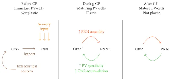
Figure 17: Critical period. ... 36
Figure 18: The appearance of PNNs in wild-type and Crtl1 knockout animals. ... 37
Figure 19: Three possible ways in which PNNs may act to restrict plasticity. ... 39
Figure 20: Otx2-PNN feedback loop for critical period plasticity. ... 41
Figure 21: Distribution of aggrecan-based PNNs compared to AD-typical neurofibrillary degeneration in the human cortex. ... 43
Figure 22: Schematic representation of the spinal cord injury. ... 45
Figure 23: Chondroitinase ABC treatment of the PNN... 48
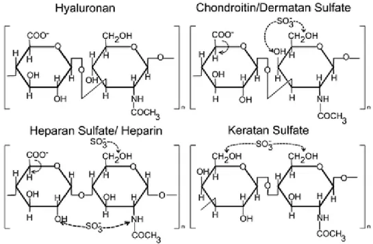
Figure 24: Structure of disaccharide unit of GAG members. ... 50
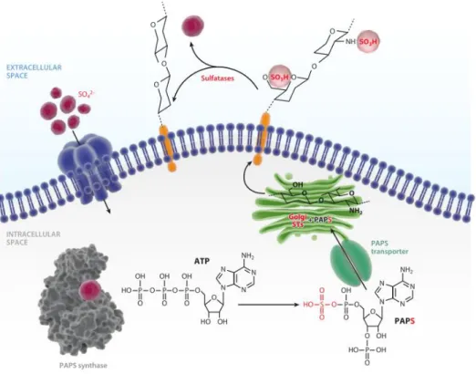
Figure 25: The sulphation cycle in mammalian cells. ... 51
Figure 26: Heparan sulphate structure. ... 52
Figure 27: Structure of chondroitin sulphate disaccharide units. ... 54
Figure 28: Subcellular localization of proteochondroitin/dermatan Sulphate. ... 55
Figure 29: Conventional scheme for CS biosynthetic machineries. ... 57
Figure 30: A schematic diagram of pathways for biosynthetic modification of CS/DS chains. ... 59
Figure 31: Action of hyaluronidase and chondroitinase... 62
Figure 32: Immunohistochemical localization of chondroitin sulphate-D in the developing mouse brain after birth. ... 66
Figure 33: The semaphorin family. ... 71
Figure 34: The sema domain: common feature of the extracellular regions of semaphorins and plexins. ... 73
Figure 35: Structure of neuropilin-1 and neuropilin-2. ... 77
Figure 36: Growth cone guidance. ... 78
Figure 37: schematic representation of dorsal root ganglion (DRG). ... 79
Figure 38: Schematic diagram of semaphorin 3A structure. ... 82
Figure 39: Crystal structure of Sema3A and Sema3A in complex with Neuropilin1 and plexinA1. ... 83
Figure 40: Model for Semaphorin 3A Signalling complex. ... 84
Figure 41: Main signal transduction pathways activated by class 3 semaphorin binding to neuropilins. ... 87
Figure 42: Schematic view of possible roles for Sema3A in PNN related plasticity. ... 88
Figure 43 : Schematic representation of the aim of the thesis project. ... 91
Figure 44: Sema3A heterologous expression in HEK293-6E cells. ... 111
Figure 45: Sema3A cleavage by furin. ... 112
Figure 46: Sema3A WT purification. ... 114
Figure 47: Sema3A-90 binds to GAG via specific sequences located in the C-ter domains. ... 116
Figure 48: Deletion of cluster 1 and 2. ... 118
Figure 49: Sema3A mutants purification. ... 120
Figure 50: Comparison between complete and partial substitution of cluster 2 basic amino acids. ... 121
Figure 51: Surface plasmon resonance (SPR) and GAG-surface preparation for interaction analysis. ... 124
Figure 52: Sema3A WT-GAG analysis in SPR. ... 126
Figure 53: Disaccharide composition of commercial CS-E and CS-D. ... 128
12
Figure 55 : Sema3A-65 binding to GAGs. ... 130
Figure 56: Sema3A Mutants-CSE interaction analysis in SPR. ... 131
Figure 57: Sema3A requires a minimal motif of tetrasaccharide to bind to CS-E. ... 133
Figure 58: Sema3A cleavage by furin. ... 135
Figure 59 : Schematic representation of QCM-D principle measuring frequency and dissipation. ... 136
Figure 60: different steps of GAG film formation in QCM-D. ... 138
Figure 61: Sema3A WT and mutants - GAGs analysis in QCM-D ... 140
Figure 62 : Effect of Sema3A WT and mutants on neurites outgrowth of dissociated DRG neurons in culture. 141 Figure 63: Extraction of GAGs from rat brain. ... 144
Figure 64: Reverse-phase ion-pair high-performance liquid chromatography (RPIP-HPLC). ... 145
Figure 65: Disaccharide composition of CS in rat brain. ... 146
Figure 66: Biotinylation of rat brain CS and their immobilisation on SPR sensor ship. ... 147
Figure 67: Nrp1 purification and ChABC digestion. ... 150
Figure 68: Theoretical Sema3A isoforms and their approximate MWs. ... 154
Figure 69: A schematic model constructed from literature data and our results on Sema3A signalling regulation at extracellular level in PNNs. ... 161
List of tables
Table 1: Typical proteoglycans expressed in the central nervous system. ... 17Table 2: Changes in ECM components after traumatic brain injury (TBI). ... 28
Table 3: mRNA expression of PNN components in adult rat deep cerebellar nuclei (DCN) and large excitatory deep cerebellar nuclei (LEDCN). ... 38
Table 4: Overview of the most important findings regarding the different experimental approaches using human postmortem tissue, in vitro trials, or animal models to investigate the protective action of ECM components. ... 43
Table 5: Effect of chondroitinase ABC treatment on neuronal plasticity. ... 47
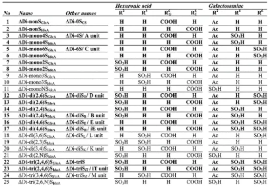
Table 6: Structure of disaccharide units derived from chondroitin sulphate and dermatan sulphate by Chondroitinase digestion. ... 53
Table 7: CS-GAG proportions: changes with development of rat brain. ... 63
Table 8: Disaccharide composition of CS/DS-GAG chains in buffer 1, 2, 3, and 4 extracts from adult rat brain. 64 Table 9 : CS-GAG proportions: changes with brain cortex lesion of adult rat. ... 65
Table 10: Influence of CS variants on neuronal growth and guidance... 67
Table 11: Generated mutations in Sema3A. ... 98
Table 12: Kinetic parameters resulting from the fit to 1:1 Langmuir binding model with mass transfer of Sema3A WT, Mut 1 and Mut 2 – CS-E/HS interaction in SPR. ... 126
Table 13: Frequency and dissipation shift resulting from injecting Sema3A-90 WT, Mut-1 and Mut-2 over HS, CS-E and CS-D films in QCM-D. ... 139
List of appendices
Appendix 1: Chondroitin sulphates and their binding molecules in the central nervous system. ... 175Appendix 2: pTT22SSP4 expression vector. ... 176
Appendix 3: Amino acids sequence of expressed Sema3A protein. ... 177
Appendix 4 : Nrp1-FC-His expression vector ... 178
Appendix 5: Amino acids sequence of expressed Nrp1 protein. ... 178
Appendix 6 : Strategy of the beads approach. ... 179
Appendix 7: Sulfo-NHS plus EDC (carbodiimide) crosslinking reaction scheme. ... 179
Appendix 8: Examples of elution profile in Ni-NTA and SP-sepharose of cell surface-associated protein and culture medium of all mutants. ... 180
13
Introduction
The central nervous system (CNS) is made of brain and spinal cord. It is responsible for the integration of information sent by all the physiological systems composing the living organism and provision of reaction accordingly. In addition to control the function of other systems, the CNS has to manage its own functions such as learning and memory. This makes it the most complex system in the organism. The functional units of the CNS are neurons and glial cells. Information transmission and storage are performed by the ability of the CNS in establishing, withdrawing and modifying the connections between neurons. This modulation of connection between neurons in response to environmental stimuli is called neuronal plasticity. Neuronal plasticity is one of the most important mechanisms in setting up the CNS function and maintaining its integrity during development as well as in the adult. This neuronal plasticity is ensured in part by the extracellular matrix (ECM) which occupied a significant part in the CNS. ECM is important in all tissues; in the CNS it plays a role at the same level as the neural cells. The complexity of neural ECM can further be stratified into a specialised structure called perineuronal nets (PNNs). These PNNs are the key regulators of neuronal plasticity and regeneration. How these PNNs regulate plasticity is an open question which is not completely elucidated yet. Compared to classical ECMs, PNNs are enriched in a type of glycosaminoglycan (GAG), chondroitin sulphates (CSs) which are important in recruiting of signalling molecules, such as semaphorin 3A (Sema3A), and modulating their downstream signal. As such, PNNs could regulate plasticity. In this introduction, we will go through all these highlighted elements starting by neuronal ECM which is the cradle of this project to introduce PNNs. Then, we will detail the PNN and its potential roles in CNS before focusing our description on the main molecules of the project: CS and Sema3A to finish by CS-Sema3A interaction which is the aim of the project.
14
1. Extracellular matrix in the central nervous system
ECM is the cement which connects cells to form a tissue. It is composed of plethora of molecules secreted by the surrounding cells. In addition to maintain the tissue structure, it is also involved in various physiological and pathological mechanisms. Its composition varies according to cell type constituting the tissue, which enables it to exert a multitude of functions. Moreover, ECM is the entrance and exit gate of all signalling molecules. It represents 50-70% of the dry body mass1.
It is believed three centuries ago that fibers are alive and constitute the main component of the tissue, this is designated as ―fiber theory‖2
. In 1830, Johannes Müller submitted ―Bindegewebe‖ concept, a German term meaning connective tissue. It was then postulated that connective tissue is composed of cells and their products. It is only one century later the term ―extracellular matrix‖ came into use and its components started to be analysed with the apparition of electron microscopy and x-ray diffraction2. Collagen is the first analysed molecule and solubilised for the first time by Nageotte at the Collège de France in Paris3. At the same time, medical specialties such as rheumatology started to be interested to the GAG4. With the development of chemical method and cell biology, other molecules composing ECM were discovered2.
In this introduction, we focus on neural ECM which includes the background of this thesis project. It is only in 1971 that the existence of brain ECM is reported5. Neuronal ECM is unique by its organisation, composition and diversity. It constitutes 20% of the total volume of adult brain, showing its quantitative importance in the CNS6. Neural ECM is important in early development and persists in the adult. It is involved in vital functions of the CNS such proliferation, migration, differentiation, plasticity and regeneration of neural cells. In adult CNS, ECM maintains the homeostasis of the brain by playing sometimes opposite roles. However in pathological conditions, ECM undergoes changes in its composition and a reorganisation of its structure. These changes are either the cause or the consequence of the pathology7 8. These changes can occur to overcome the abnormalities or in contrary to promote the spread of the cognitive impairment. For that, neural ECM thus potentially offers a multitude of therapeutic target choices given the diversity of its composition and functions. In addition, it is a much more accessible environment for therapeutic molecules than the
15 intracellular environment where thousands of signalling pathways are triggered and cross-talk. However given its diversity, complexity and interaction between the different components, it is unlikely to assign a single role to a given molecule to target it. Several researches have been carried out which allowed characterising and understanding more and more the neural ECM and its components. Despite that, there are still many questions to decipher.
1.1 Neural ECM components
Neural ECM is secreted by neurons and glia9. In contrary to ECM of other tissues, neural ECM contains less fibrous proteins (such as collagen, laminin, elastin…) and more proteoglycans (PGs)10. The ratio of GAGs to collagen is around 10:111. The major components of neural ECM are hyaluronic acid, PGs, link proteins and tenascins12. Features of each of these components are detailed below. It is worth noting that in addition to these major classes, other important neural ECM components are also present such as the glycoprotein, reelin, involved in considerable development processes13 and ECM receptors, integrins.
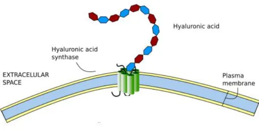
1.1.1 Hyaluronic acid
Hyaluronic acid (HA) (Figure 1), also known as hyaluronan, is a large linear polysaccharide composed of repeating disaccharide units: glucuronic acid (GlcA) and N-acetylglucosamine (GlcNAc) [-β(1,4)-GlcA-β(1,3)-GlcNAc-]n14. It constitutes the only
non-sulphated member in the GAG family and non-covalently linked to a core protein15. In physiological conditions, HA is made of 2000-25000 disaccharide units which corresponds to 1000-10000 kDa and 2-25 µm of length16. HA is synthetized by one of three hyaluronan synthases (HAS: 1-3) and degraded by hyaluronidases. HAS enzymes are transmembrane and synthesise HA chains of different length at different speed and are expressed in different neural tissues17. HA chain is extruded through the plasma membrane into ECM while it is being synthesized18.
HA was long regarded as ―goo‖ holding cells together to form tissue. However, its roles are much more than that15. It constitutes the backbone of the ECM on which other ECM molecules aggregate and assemble, thus forming the ECM net. Because of its large size, random-coil structure, and negatively charged and hydrophobic faces due to the carboxyl
16 groups and a cluster of hydrogen atoms, respectively, HA influences greatly physicochemical and hydrodynamic properties of the tissue15 16 18 19. These characteristics confer it an important use in tissue engineering20. Moreover, HA interacts with CD44, and thereby influences cell adhesion and migration18.
Figure 1: Diagram of hyaluronic acid synthesis by the hyaluronic acid synthase.
(Modified from Escudero, 200921).
1.1.2 Proteoglycans
Proteoglycan (PG) is the key component of neuronal ECM. It is made of a core protein linked covalently to one or more GAG chains. GAG chain(s) of PGs contributes to their biological function, while the core protein determines localisation and promotes the interaction with other ECM molecules22. The GAG chain can be either heparan sulphate (HS) or chondroitin sulphate (CS) giving rise to HSPG or CSPG, respectively. Hybrid PGs containing both HS and CS chains are also reported. Small amount of keratan sulphate PGs (KSPG) are found in the CNS23. In 1985, HSPGs were the first isolated and characterised adult CNS PG by Klinger et al.24 25 26. In 1990, Herdon et al. have identified 25 CSPGs and HSPGs in rat brain27. These PGs are distributed over the different level of ECM (ECM compartmentalisation is discussed in section 1.4). Some of them were found in the soluble fraction of the brain (secretory). Others are transmembrane or glycosylphosphatidylinositol (GPI)-anchored28. Major species of PGs expressed in the CNS are summarized in Table 1. An overview of these typical PGs structure is presented in the next paragraphs.
17
Table 1: Typical proteoglycans expressed in the central nervous system.
Nature Name Core protein size (kDa) Number of GAG chains Type CSPG Aggrecan 224 ~100 secretory VersicanV0 370 17-23 secretory VersicanV1 262 12-15 secretory VersicanV2 180 5-8 secretory Brevican 97 0-5 secretory Neurocan 133 3 secretory Phosphacan 173 3-4 secretory RPTPβ 253 3-4 transmembrane Decorin 36 1 secretory Biglycan 38 1-2 secretory NG2 252 0-3 transmembrane HSPG Syndecan (4 members) ~ (21, 22, 45 and 32, respectively) 3-8 transmembrane Glypican (6 members) ~ 53 2-4 GPI-anchored
1.1.2.1 Chondroitin sulphate proteoglycans
CSPGs are the most abundant PG in the mammalian CNS28 and are represented mostly by lecticans family including aggrecan, versican, neurocan and brevican. Lecticans are determined by globular domains in N- and C-termini. The N-ter part contains HA-binding sites, allowing the lecticans to bind to HA. The C-ter part, which interacts with tenascins, contains complement regulatory protein (CRP)-like domain, lectin and two epidermal growth factor (EGF)-like repeat. The central part includes attachment sites for CS29 (Figure 2).
Figure 2: Domain structures of lecticans.
All lecticans contain N-ter G1 domains and C-ter G3 domains. Only aggrecan contains the G2 domain. The G1 domain consists of an Ig-like loop and two link modules, whereas the G2 domain consists only of two link modules. The G3 domain consists of one or two EGF repeats, a C-type lectin domain and CRP-like domain. All lecticans contain chondroitin sulphate chains (yellow) in the central domain. Aggrecan also contains keratan sulphate chains (pink) in the N-ter part of the central domain (from Y. Yamaguchi.2000)30.
18 Receptor-type protein-tyrosine phosphate β (RPTPβ) is a transmembrane PG. It is made of a N-ter carbonic anhydrase-like domain, a fibronectin type III domain, a CS attachment site, a transmembrane domain and finally two intracellular tyrosine phosphatase domains. Phosphacan results from alternative splicing of RPTPβ mRNA. It does not contain the intracellular and transmembrane domains, which makes it a secretory protein12 (Figure 3).
Figure 3: Structure of receptor-type protein-tyrosine phosphate β (RPTPβ) and phosphacan.
RPTPβ has a N-ter carbonic anhydrase-like domain (CA), a fibronectin type III domain (FN), CS attachment regions and two intracellular tyrosine phosphatase domains (D1 and D2). Phosphacan is a soluble form that lacks the intracellular tyrosine phosphatase domains (modified from Galtry and Fawcett. 2007)12.
Biglycan and decorin are small leucine-rich PG secreted in ECM. The majority of the core protein (70%) is represented by leucine-rich repeats flanked by cysteine-rich clusters that may oxidize to form disulfide bond. They usually contain one or two CS chains in the N-ter31 (Figure 4).
Figure 4: Structure of decorin and biglycan
Neural/glial antigen 2 (NG2) is a transmembrane protein, also known as melanoma-associated chondroitin sulphate proteoglycan (MCSP). Its core protein is made of a large ectodomain, transmembrane region and short cytoplasmic domain (Figure 5). The
ectodomain is composed of the globular N-ter, the globular C-ter and the rod-like central region which contains CS (CSPG repeats). Intracellular domain interacts with intracellular
Decorin Biglycan N-ter Leucine-rich repeats Cysteine-rich clusters CS chain Cysteine-rich repeats
19
ligands and triggers signalling pathways involved in several functions such as proliferation, migration and modulation neuronal network32.
Figure 5: Structure of NG2.
1.1.2.2 Heparan sulphate proteoglycans
The two most reputable HSPG in the CNS are syndecan and glypican. Syndecan is transmembrane protein family including four members (Syndican-1, -2, -3 and -4). Syndecan members are composed of a large ectodomain, a transmembrane domain and a short cytoplasmic domain (Figure 6). The ectodomain is poorly conserved among different family members, while the intracellular domain is highly conserved among all family members. Syndecan-3 is the longest member containing at least seven HS chains. In addition to HS chains, syndecan-1 bears CS chains. In syndecan-1 and -3, HS chains are located at the N- and C-ter ends of the ectodomain, while in syndecan-2 and -4, HS chains are tethered to the N-ter only33.
20 Glypican is the other HSPG family in the CNS constituted of six members. They are anchored into the plasma membrane through GPI anchors
. T
he N-ter domains of glypicans adopt an elongated α-helical structure. It undergoes a furin-like convertase cleavage to generate two chains that remain connected by disulfide bonds34. HS chains are mainly located at the C-ter part of the ectodomain, close to the membrane35 (Figure 6).A significant amount of evidence that PG is an important molecule in ECM has been accumulated, during development as well as in maintaining of adult brain homeostasis. As most of the molecules composing ECM, PGs contribute in the assembly and structure of ECM. In embryonic brain, HSPG, for example, promotes the fibroblast growth factor (FGF)-2-mediated proliferation of neuroepithelial cells36. CSPGs, as phosphacan and neurocan, are involved in promoting or inhibiting neurite outgrowth and cell adhesion, depending on cell type37 38. CSPGs interact with a plethora of molecules involved in synaptogenesis, axon guidance and migration. In the mature brain, Versican V2 which constitutes the most abundant CSPG, is described as the major inhibitor of axonal growth39. Brevican inhibits neurite outgrowth of cerebellar granule neurons. It is also suggested to control the infiltration of axons and dendrites into the mature glomeruli40. PGs are also involved in neurodegenerative disease such as Alzheimer disease (AD) and CNS injury. Both HSPG and CSPG are found in amyloid plaques in AD28. Otherwise, many studies have reported the inhibitory effect of CSPGs on axon regeneration after glia scars41. After injury CSPGs expression is upregulated around the lesion area42. HSPG appears to be increased after CNS injury, but its effect is less investigated than that of CSPG28.
1.1.3 Tenascin-C and -R
Tenascins are a family of glycoproteins found in the ECM of several tissues, composed of five members: tenascin-C, -X, -R, -Y and –W43. Only tenascin-C and -R are reported in the CNS, playing important roles in cell proliferation, migration and differentiation, axonal guidance, synaptic plasticity and myelination44. Tenascin-C (TN-C) is the first discovered member. It is a ~ 1800 kDa protein, assembled from six monomers linked covalently with disulfide bonds. The monomer consists of a tenascin assembly domain, a cysteine-rich domain, 14.5 epidermal growth factor (EGF)-like domains, 8 constitutive fibronectin-type III homologous domains and a fibrinogen-like domain45 (Figure 7.A). TN-C expression is regulated during development and in the adult brain. It is highly expressed in
21 early development in the CNS (day 10 in mouse CNS) by different type of cells, mainly immature astrocytes and restricted population of immature neurons43. TN-C acts either by its interaction with cell surface receptors such as integrins or by modulation of ECM components such as CSPGs46. It is known to be involved in progenitor cells proliferation and migration and neurite outgrowth47. Its role in neuronal plasticity is also reported. Indeed, it was identified as one of molecules mediating learning and synaptic plasticity48.
Figure 7: Structures of tenascin-C and tenascin-R monomers.
TN-C: tenascin-C, TN-R: tenascin-R, TA: amino-terminal tenascin assembly, EGF: epidermal-growth
factor-like domain, FN: fibronectin-factor-like domain, and FG: fibrinogen-factor-like domain (modified from Reinhard et al., 201649).
Tenascin-R (TN-R) is exclusively expressed in the CNS. TN-R is 180 kDa protein from which a 160 kDa form is generated by a proteolytic cleavage. Expression of this two isoforms changes during CNS development.TN-R is assembled from two or three monomers linked with disulfide bonds. As TN-C, the monomer is also composed of a tenascin assembly domain, cysteine-rich N-ter region, 4.5 EGF-like domains, 8 fibronectin type III repeats (FN III repeats), and a carboxyl-terminal region and fibrinogen-like domain49 (Figure 7.B). TN-R undergoes a post-translational modification consisting in addition of three distinct sulphated oligosaccharides. Among these, one is a CS oligosaccharide which may mediate TN-R interaction with TN-C and fibronectin to inhibit neurite outgrowth50. It is expressed by certain subpopulation of neurons and oligodendrocytes in particular cortical region and laminae51. TN-R plays sometimes opposite roles, which earned it the name of ―Janusin‖ in reference to Roman god with two faces, according to the type of targeted cells and receptors, and the time of interaction. TN-R plays on one hand an important role in oligodendrocytes differentiation52. On the other hand, it is involved in generation of GABAergic neurons. These different effects are mediated by its different domains. The FN6–8 domains inhibit neuronal
22 stem cells proliferation and differentiation into astrocytes at the expense of neurons, while the EGF domain enhances their differentiation into neurons at the expense of astrocytes and oligodendrocytes52. TN-R is also qualified as a neuroprotector after brain injury by modulation microglia function. On one hand EGF-like repeats inhibit adhesion and migration of microglia through a protein kinase A-dependent mechanism. On another hand, fibronectin 6–8 repeats promote adhesion and migration of the primary microglia through a protein kinase C-dependent mechanism53.
Retina is considered as an excellent model to analyse proliferation and differentiation of neuronal cells as well as axonal guidance and growth. For this purpose, TN-C and TN-R roles were investigated in optic nerve and retinal neurodegeneration. In the optic nerve, huge amount of TN-C is secreted by astrocytes and TN-R is mainly expressed in oligodendrocytes49. Expression of these tenascins is found dysregulated in retinal ischemia. In optic nerve damage, TN-C and TN-R have opposing roles in regeneration of optic nerve fibers. TN-C is chemoattractive, whereas TN-R is inhibitory and chemorepulsive44.
1.1.4 Hyaluronan and proteoglycan binding link proteins
Hyaluronan and proteoglycan binding link protein (HAPLN) family contains four members (HAPLN 1-4). As their name indicate, they stabilise the interaction between HA and CSPG. HAPLNs are 38-43 kDa and made of Ig-like V-type, link 1 domain and link 2 domain
(Figure 8). The structure of HAPLNs is homologous to that of G1 domain of lecticans which also binds to HA. Genomic structure revealed that these HAPLN2 and HAPLN4 genes were physically linked to the genes encoding brevican and neurocan, respectively supporting thus the hypothesis of common evolutionary origin from an ancestral gene respectively54. HAPLN2 and HAPLN4 are restricted in expression to the CNS, hence their other names are the brain-specific link proteins: Bral1 and Bral2, respectively54. HAPLN1 is 40 kDa protein initially identified in articular cartilage, hence its other name is cartilage link protein (Crtl1). It interacts with HA and aggrecan. It is also expressed in the CNS where it is crucial for the highly organisation of the specialised matrix, the PNNs that we will discuss in detail in the later chapter. Animals lacking HAPLN1 in the CNS attenuate PNNs55. Similar results are observed in animal lacking HAPLN4 which is mainly expressed in cerebellum and brain stem. Moreover, it affects the localization of brevican56. HAPLN3 is not detected in the CNS.
23
Little is known about HAPLNs family. The 3D structure is not yet solved and their interaction with HA and CSPG still poorly characterised as well as its functional role in the CNS.
Figure 8: Structure of hyaluronan and proteoglycan binding link protein1 (HAPLN1). IGv: immunoglobulin-like domain and LINK: Link domain.
1.2
Foetal and adult neural ECM components
ECM composition changes with aging, responding thus to brain functions needs (Figure 9). Expression of some components is upregulated, while it is downregulated for others. These components can also play an inhibitory as well as an activatory role depending of the conditions. Alternative mRNA splicing of some ECM proteins leads to numerous combinations of isoforms, thereby increasing the functional diversity at different stages of development and in the adult CNS. Embryonic neural ECM is very dynamic and highly plastic. Indeed, this is required to control different mechanisms occurring in developing CNS such as proliferation, migration, differentiation and synaptogenesis. CNS is the most complex organ responsible of multiple vital functions. In human, its development starts at 4 weeks and it continues until after birth. All these need a dramatical remodelling of ECM components.
HA is the most abundant GAG in foetal rat brain (>60 %) and it decreases after birth57. During development of the chick, HA is concentrated in the intermediate zone which gives rise to the white matter58. HA is crucial in maintaining neural progenitor cells in an undifferentiated state59. Whereas in adult animals, it localizes around myelinated fibers in white matter and it is more diffuse60. It is also found in grey matter in PNNs that are involved in neuronal plasticity61.
HSPG and CSPG function in different way in developing and adult brain. It has been observed that HS represents 25 % of all GAG in the foetal rat brain, this makes it the most abundant Sulphated GAG57. In the postnatal brain, it represents only 10 % of all GAGs. CS represents 10 % of all GAGs in the foetal brain, and it becomes the most abundant GAG 20 days after birth57. Neurocan and RPTPβ/phosphacan are the most abundant CSPGs in
24 developing CNS where they are required for cells adhesion and neurite outgrowth. They are exerting an opposite effects on neurite outgrowth. Neurocan and phosphacan inhibit the neurites outgrowth of cortical and dorsal root ganglion (DRG) neurons38, while phosphacan promotes the neurite outgrowth of mesencephalic and hippocampal neurons62. In adult CNS, versican is the major CSPG63. It inhibits the neurite outgrowth of granular neurons40. CSPGs in adult brain play an important role in neuronal plasticity. Indeed, they are related to synaptic activities. Lacking in neurocan reduces late-phase hippocampal long term potentiation (LTP)64. Interestingly, after birth CS chains of neurocan change in size and in sulphation pattern23. Furthermore, lacking RPTPβ/phosphacan enhances LTP and impairment in memory task65.
HSPGs interact with several proteins present in developing brain to achieve their functions. HSPG-FGF is one of the well characterised interactions. Binding to HSPG i) stabilises and protects FGF from proteolysis66 ii) concentrates FGF locally and thus enhances binding to their receptor, and iii) induces the oligomerization of FGF, thereby receptor dimerization and signalling67. FGF1 and FGF2 are required for proliferation, migration and differentiation of neuronal precursor cells. First, HSPG binds to FGF2 to promote neuronal precursors cells proliferation. Then, HSPG switches rapidly its potentiating activity from FGF2 to FGF1 to promote differentiation of neural cells36. HSPGs are associated to human mesenchymal stem cells (hMSCs) lineage-specification to neural progenitors68. They are also involved in the development of specific synaptic connectivity patterns important for neural circuit function69. The four members of syndecan are expressed differently in developing and adult brain. Syndecan-2 is highly expressed in adult brain and concentrated in synapses, while syndecan-3 is highly is expressed in development and concentrated in axons70. Syndican-4 is dynamically expressed in the early stages of zebrafish embryonic neurogenesis where it inhibits neural proliferation71.
TN-C and TN-R are highly expressed during early development of the CNS. TN-C is expressed at early stages of developing brain, while TN-R is expressed later during development and its expression is restricted to the oligodendrocytes43. TN-C isoforms are secreted by numerous neural cells during development to accomplish different processes such as migration43. Despite expression of TN-C persists in the adult brain, it is restricted to well defined areas where neuronal plasticity and regeneration are still possible such as the nuclei of hypothalamus and olfactory system, respectively43. TN-R is detected at one week in postnatal
25 mice and the peak is reached at 2-3 weeks and remains stable in adulthood72. TN-R is considered as a key component of adult CNS matrix. Indeed, it modulates adult but not developmental neurogenesis in the olfactory bulb73. Furthermore, TN-R is transiently expressed in peripheral nervous system (PNS) in the sciatic nerve of mice embryos (E14-18) and neonatal74. HAPLN1 expression is reported during late embryonic and early postnatal75.
Figure 9: ECM changes at CNS synapses.
Synapses are embedded into an ECM meshwork (blue) composed of hyaluronan, chondroitin sulphate proteoglycans (CSPGs), tenascins, and others. The composition of the ECM changes during development. For example, neurocan, versican V1, and tenascin-C are abundant in the immature CNS, whereas tenascin-R, versican V2, and Bral1 are prominent in the mature CNS. The mature ECM is thought to restrict dendritic spine motility and lateral diffusion of α-amino-3-hydroxy-5-methyl-4-isoxazolepropionic acid (AMPA) receptors (AMPAr) (from barros et al., 201113).
1.3 Role of neural ECM
ECM plays a crucial physical barrier for the cells that it surrounds by preventing the infiltration of external agents. It is also a structural support for the cells and maintains the tissue integrity. ECM allows the communication between adjacent cells, but also with distant cells by promoting the transport of different molecules. It offers an adequate environment for enzymes responsible of post-synthesis modifications, signalling molecule cleavage for
26 activation or inhibition, and degradation. In addition to its structural role, ECM is involved in plethora of physiological and pathological functions.
It is important to mention that cell control by ECM is not one-way. Indeed, the cells are the principal actor of ECM formation and reorganisation. They contribute actively in producing ECM components and the enzyme responsible of its maintenance and restructure, responding thus to changes of conditions in the tissue.
Importance of ECM is reported earlier in development. Regulated spatial and temporal distribution of the components in developing ECM makes it very dynamic. Several studies indicate that the ECM affects all aspects of nervous system development. TN-C-deficient mice show a delay of the developmental program of neural stem due to changes in growth factor responsiveness76. ECM components support different migratory trajectories of neural crest cells. Versican isoforms V0 and V1 implanted micromembranes in chick embryos leads to attraction of neural crest cells, while micromembranes of aggrecan retain migratory cells near the implant and thereby perturb their spatiotemporal migratory pattern. Interestingly, this inhibitory effect of aggrecan is mostly due to the GAGs chains (CS and KS)77. These observations suggest that neural crest cells may migrate on versican-containing matrix and avoid aggrecan-containing matrix. Pattern expression of these two proteins could be a mechanism of guidance for neural crest cells migration. Neural crest migration is also influenced by laminin. Laminin α5 mutant mice exhibit abnormalities in neural cell migration which demonstrates in expanded neural crest streams. This suggests that laminin α5 may restrict migration into narrow streams78. Mice lacking laminin α2 display a detachment of embryonic neural stem cell (NSCs) apical process from ventricular zone79. This phenomenon is also observed when β1 integrin function is disrupted. Laminin and integrin thus play a role in anchoring embryonic NSCs in the ventricular surface and maintaining the physical integrity of the neocortical niche79. Moreover, conditional β1-integrin gene deletion in neural crest cells causes severe developmental alterations of the peripheral nervous system leading to lethality of mice after birth80. ECM provides an adequate microenvironment which controls the NSCs behaviour11 13. ECM shapes the niche of stem cells and contributes actively in their maintenance, differentiation and migration.
ECM plays an important role in myelination of axons13 (Figure 10). Myelination is a vitally important process for the proper function of neurons. It involves the accumulation of
27 myelin around axons. Myelin is formed by oligodendrocytes in the CNS and Schwann cells in the PNS. Myelination results in the concentration of voltage-gated sodium channels at the nodes of Ranvier, regenerating thus action potential81. One of remarkable role of myelination is to enable a very fast action potential propagation in vertebrate81. In PNS, ECM components notably laminin and collagen promote myelination of peripheral nerves by regulating Schwann cells proliferation, adhesion and spreading82 83 as well as neurite outgrowth83. In CNS, β1 integrin is required for myelination by promoting myelin sheaths outgrowth through AKT activation84. On the contrary, CSPGs are myelination inhibitors by inhibiting oligodendrocytes process outgrowth and myelination85. Roles of ECM in the CNS and PNS are innumerable. It influences all the cerebral process in physiological and pathological conditions and from the development to adult.
Figure 10: ECM and myelination.
The ECM surrounding nodes of Ranvier may regulate the local concentration of cations and clusters voltage-gated sodium channels, which allow for saltatory electrical conductivity. Several proteoglycans, tenascin-R, laminin and dystroglycan contribute to the formation of nodal matrices. Nav, voltage-gated channel; Naþ, sodium cations (from Barros et al., 201113).
Neural ECM undergoes several changes in structure and composition in response to a trauma in the CNS. Expression of certain component is upregulated, while it is downregulated for the other as showed in Table 2. These changes can both promote or inhibit the recovery of function. Traumatic brain injury (TBI) induces oxidative stress, leading to the release of reactive oxygen species (ROS). These ROS degrade HA and generate small HA fragments. These biologically active fragments thus modulate angiogenesis86. TBI upregulates the expression of domains B and D of fibronectin type III in tenascin-C, thus promoting axonal regeneration and repair processes87. However, TBI also results in sulphation pattern
28 modification of GAG chains carried by PGs. These changes induce the inhibition of axon regeneration, guidance and neuronal plasticity88. Role of the sulphation pattern after brain injury is discussed in PNNs section (section 2.7). All aspects of ECM after TBI are reviewed in detail by George Nand Geller HM, 201889.
Table 2: Changes in ECM components after traumatic brain injury (TBI).
29 1.4 Types of ECM in the CNS
ECM in the CNS is organised in three different compartments (Figure 11), with difference in composition and function. Basal lamina (basement membrane) surrounds the blood vessels and the entire pial surface of the CNS. At the electron microscopic level, the basal lamina is composed of an electron-dense layer called the lamina densa (composed of type IV collagen) and an electron-lucid layer called the lamina lucida (consisting of laminin, dystroglycan and associated proteins)6. Basal lamina is involved in neurogenesis, CNS injury repair90 and nerve regeneration90 91. It constitutes a key component in maintaining the integrity of the blood–brain barrier (BBB). Basal lamina is required early in CNS development where it is important for the maturation of endothelial cells required for the BBB92.
Figure 11: extracellular matrix within the central nervous system (CNS).
The three major compartments of the extracellular matrix in the CNS are the basement membrane, perineuronal net and neuronal interstitial matrix. The basement membrane is found surrounding cerebral blood vessels, the perineuronal net is a dense matrix immediately surrounding neuronal cell bodies and dendrites, and the neuronal interstitial matrix occupies the space between neurons and glial cells (from Lau L.W et al., 20138).
Interstitial matrix corresponds to molecules that fill the space between CNS cells in parenchyma. Interstitial matrix is the classical matrix of the CNS whose components and function are described above (section 1.1 and 1.3, respectively). Two different levels of
30 interstitial matrix can be distinguished: loose matrix and membrane-associated matrix. Loose matrix (diffuse matrix) consists of unbound or loosely attached ECM that can be extracted easily with physiological saline (150 mM NaCl). Membrane-associated matrix consists of ECM molecules bound to the plasma membrane and will only be detached or solubilised with higher salt concentration (1000 mM NaCl) and detergent (0.5% tritonX-100)93. In some regions of the CNS, interstitial matrix becomes more complex to give rise to the condensed matrix structure called perineuronal nets (PNNs).
PNNs are the third type of ECM in the CNS. As its name indicates, PNN is a specific and highly organized structure found only on the surface of neurons. PNN is important in controlling neuronal plasticity61. PNN components are solubilised only with the combination of 1 M NaCl, 0.5% tritionX-100 and 6 M of urea93.
2.
Perineuronal nets (PNNs)
Camillo Golgi (1843–1926) is known by its discovery of Golgi apparatus which carries his name. However, long before he highlights the existence of Golgi apparatus in the CNS in 1888. He described a particular reticular structure surrounding several neuronal populations, impregnated by his staining ―black reaction‖. He first mentioned his observation in Enciclopedia Medica in 1882. In 1892, he announced the existence of reticular network enwrapping cell bodies and proximal dendrites in a publication of Accademia dei Lincei. In 1888, during a conference in the Società Medico-Chirurgica on the discovery of the Golgi apparatus, Golgi officially stated this reticular structure on neuronal surface as ―pericellular net‖. He described Golgi apparatus as an internal reticular structure of the nerve cell and pericellular net as an external reticular structure. This external structure is described as ―delicate covering, mainly reticular in structure, but also in the form of little embraced tiles or an interrupted envelope which covers the body of all the nerve cells continuing along their protoplasmic extensions up to the second and third order arborisations‖ (Figure 12). At the same time, pericellular net was also observed by other neurobiologists among them Santiago Ramón y Cajal. However, since Golgi and Cajal conflicted about the organization of the nervous system, reticular theory vs neuron doctrine, he postulated then that the pericellular nets were staining artefact claiming that ―The pericellular net is not nervous in nature. We do
31 not think it is neuroglial because there is never a continuity with the ramifications of Deiters‘ cells and it has not any morphological or histological characteristic of these cells. If we can propose another solution, waiting for a new one, we should say that this net is due to a coagulation of a substance in the pericellular fluid‖. After these words of the giant of neuronal theory, pericellular nets were fell into oblivion till 1980s9495. Cajal was correct in one sense that the pericelluar nets are coagulation of a substance in the pericellular fluid (i.e. aggregation of ECM molecules). However, he was wrong to disregard the nets as staining artefact. The composition of PNNs started to be elucidated (glycoconjugates96, hyaluronate-protein aggregates97 and tenascin98) as well as their roles. The number of papers about PNNs has steadily increased since. Discovery of PNNs is reviewed in more detail by Celio and
Blümcke (1994 )99, Celio et al.,(1998)94 and Spreafico et al., (1999)95.
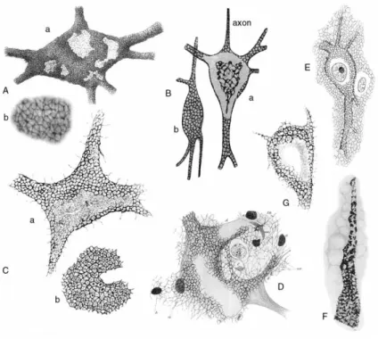
Figure 12: Perineuronal nets as drawn by various authors at the turn of the century.
(A) Nerve cell with reticular covering (anterior horn of cat spinal cord) (a) and (b) enlarged detail of (a)
illustrating the fine texture of the perineuronal net. (B) Two cerebral cells with short axons (adult cat), stained with reduced silver nitrate after fixation in formol acetone: (a) cell with ascending axon viewed in the equatorial plane; (b) cell with descending axon viewed in the superficial plane. (C) Nerve cell derived from the anterior horns of the dog spinal cord (a). The cartwheel pattern (raggere di Donaggio), formed by thin filaments radiating from a central spot, recognizable within the meshes of the perineuronal net (b). These cartwheel structures probably represent shrunken synaptic endings, which occupy the meshes of the net. (D) Cell with Golgi‘s net and a diffuse net (anterior horn of the spinal cord of a calf embryo), stained according to Bethe‘s method (Ehrlich‘s methylene blue and ammonium molybdate). (E) Cortical cell of an adult dog, stained according to a modification of Bethe‘s method. (F) Alterations within the perineuronal net of a human cortical cell, derived from a patient with paralytic dementia. (G) Cell derived from the nucleus of the vagus (medulla oblongata) of Lacerta muralis. Within the meshes of the peripheral apparatus (or perineuronal net), a typical cartwheel pattern radiating from a central small spot is evident. Filaments originating from the cell surface connect with the surrounding stroma (from Celio et al., 199894).
32 2.1 Composition and organisation of PNNs
PNNs are derived from ECM, thus all PNN components can be found in classical ECM. However, the amount and organisation of PNN components makes it different from the interstitial ECM. It is also important to know that PNN composition and structure vary in time and space. Expression of PNN components such as brevican, phosphacan and TN-R is inhomogeneous in distinct areas of the spinal cord. Moreover, the amount of CS varies significantly in different spinal cord PNNs. This variation is correlated to expression of the fast-spiking neurons marker, Kv3.1b subunit of the potassium channel. Fast-spiking neurons contains more amount of CS in their PNNs100.
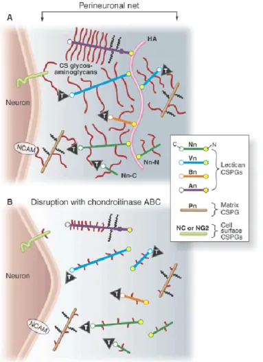
Figure 13: Structure of the PNN.
PNN is composed of hyaluronan (HA) synthetized by hyaluronase synthase. Chondroitin sulphate proteoglycans (CSPGs) interact with HA and this interaction is stabilised by the link protein. Tenascin-R bridges different CSPGs. It is involved in the stabilisation of the PNN.
Identified PNN components so far are: HAS and its synthesized product HA, CSPGs (aggrecan, neurocan, brevican, versican and phosphacan), link proteins (Crtl1 and Bral2) and TN-R93 101 (Figure 13). HA constitutes the backbone of PNNs. Traditionally, HA can attach to the cell surface either by binding to its receptor such as CD44 or by remaining attached to HAS while being extruded. However, as no expression of CD44 or other HA receptors on the PNNs-bearing neurons have been identified102, PNN is likely remained attached to neurons