RESEARCH OUTPUTS / RÉSULTATS DE RECHERCHE
Author(s) - Auteur(s) :
Publication date - Date de publication :
Permanent link - Permalien :
Rights / License - Licence de droit d’auteur :
Institutional Repository - Research Portal
Dépôt Institutionnel - Portail de la Recherche
researchportal.unamur.be
University of Namur
Adsorption of titanium dioxide nanoparticles onto zebrafish eggs affects colonizing
microbiota
Brinkmann, Bregje W.; Beijk, Wouter F.; Vlieg, Redmar C.; van Noort, S. John T.; Mejia,
Jorge; Colaux, Julien L.; Lucas, Stéphane; Lamers, Gerda; Peijnenburg, Willie J.G.M.; Vijver,
Martina G.
Published in:
Aquatic Toxicology
DOI:
10.1016/j.aquatox.2021.105744
Publication date:
2021
Document Version
Publisher's PDF, also known as Version of record
Link to publication
Citation for pulished version (HARVARD):
Brinkmann, BW, Beijk, WF, Vlieg, RC, van Noort, SJT, Mejia, J, Colaux, JL, Lucas, S, Lamers, G, Peijnenburg,
WJGM & Vijver, MG 2021, 'Adsorption of titanium dioxide nanoparticles onto zebrafish eggs affects colonizing
microbiota', Aquatic Toxicology, vol. 232, 105744. https://doi.org/10.1016/j.aquatox.2021.105744
General rights
Copyright and moral rights for the publications made accessible in the public portal are retained by the authors and/or other copyright owners and it is a condition of accessing publications that users recognise and abide by the legal requirements associated with these rights. • Users may download and print one copy of any publication from the public portal for the purpose of private study or research. • You may not further distribute the material or use it for any profit-making activity or commercial gain
• You may freely distribute the URL identifying the publication in the public portal ?
Take down policy
If you believe that this document breaches copyright please contact us providing details, and we will remove access to the work immediately and investigate your claim.
Aquatic Toxicology 232 (2021) 105744
Available online 24 January 2021
0166-445X/© 2021 The Authors. Published by Elsevier B.V. This is an open access article under the CC BY-NC-ND license
(http://creativecommons.org/licenses/by-nc-nd/4.0/).
Adsorption of titanium dioxide nanoparticles onto zebrafish eggs affects
colonizing microbiota
Bregje W. Brinkmann
a,*
, Wouter F. Beijk
a, Redmar C. Vlieg
b, S. John T. van Noort
b,
Jorge Mejia
c, Julien L. Colaux
c, St´ephane Lucas
c, Gerda Lamers
d, Willie J.G.M. Peijnenburg
a,e,
Martina G. Vijver
aaInstitute of Environmental Sciences (CML), Leiden University, Leiden, the Netherlands bLeiden Institute of Physics (LION), Leiden University, Leiden, the Netherlands
cNamur Institute of Structured Matter (NISM), Synthesis, Irradiation and Analysis of Materials Platform (SIAM), University of Namur, Namur, Belgium dInstitute of Biology (IBL), Leiden University, Leiden, the Netherlands
eNational Institute of Public Health and the Environment (RIVM), Center for Safety of Substances and Products, Bilthoven, the Netherlands
A R T I C L E I N F O
Keywords:
Host-microbiota interactions Cascading effects
Particle-induced X-ray emission Two-photon multifocal microscopy EcoPlate
A B S T R A C T
Teleost fish embryos are protected by two acellular membranes against particulate pollutants that are present in the water column. These membranes provide an effective barrier preventing particle uptake. In this study, we tested the hypothesis that the adsorption of antimicrobial titanium dioxide nanoparticles onto zebrafish eggs nevertheless harms the developing embryo by disturbing early microbial colonization. Zebrafish eggs were exposed during their first day of development to 2, 5 and 10 mg TiO2 L−1
(NM-105). Additionally, eggs were exposed to gold nanorods to assess the effectiveness of the eggs’ membranes in preventing particle uptake, localizing these particles by way of two-photon microscopy. This confirmed that particles accumulate onto zebrafish eggs, without any detectable amounts of particles crossing the protective membranes. By way of particle-induced X-ray emission analysis, we inferred that the titanium dioxide particles could cover 25–45 % of the zebrafish egg surface, where the concentrations of sorbed titanium correlated positively with concentrations of potassium and correlated negatively with concentrations of silicon. A combination of imaging and culture- based microbial identification techniques revealed that the adsorbed particles exerted antimicrobial effects, but resulted in an overall increase of microbial abundance, without any change in heterotrophic microbial ac-tivity, as inferred based on carbon substrate utilization. This effect persisted upon hatching, since larvae from particle-exposed eggs still comprised higher microbial abundance than larvae that hatched from control eggs. Notably, pathogenic aeromonads tolerated the antimicrobial properties of the nanoparticles. Overall, our results show that the adsorption of suspended antimicrobial nanoparticles on aquatic eggs can have cascading effects across different life stages of oviparous animals. Our study furthermore suggests that aggregation dynamics may occur that could facilitate the dispersal of pathogenic bacteria through aquatic ecosystems.
1. Introduction
Many vertebrate and invertebrate animals, including chordates, ar-thropods, echinoderms, cnidarians and mollusks, lay eggs that develop
ex vivo in the water column. Their developing embryos are surrounded
by envelopes of varying complexity, ranging from simple coats in cni-darians and nematodes, to acellular membranes with or without external jelly coating in chordates, arthropods, echinoderms and mol-lusks (Mazzini et al., 1984). In addition to the inner vitelline membrane,
eggs of cephalopods, pterygotan insects and teleost fish are surrounded by an external chorion membrane. Together, the chorion and vitelline membranes protect the externally developing embryos against envi-ronmental stressors in the water column, including mechanical forces, predators, and pathogens (McMillan, 2007).
Research on teleost fish eggs has shown that the chorion constitutes an effective physical barrier against the uptake of particulate pollutants. In medaka fish eggs, for instance, microplastic particles were found to adhere to the villi on the outside of the chorion, and could not be * Corresponding author.
E-mail address: [email protected] (B.W. Brinkmann).
Contents lists available at ScienceDirect
Aquatic Toxicology
journal homepage: www.elsevier.com/locate/aqtox
https://doi.org/10.1016/j.aquatox.2021.105744
detected within eggs (Beiras et al., 2018). In zebrafish eggs, which do not comprise villi, microplastic particles from the water column attached to the chorion surface in a size-dependent manner. Relatively large microplastic particles, with a primary size of 10-20 μm, adhered in
patches to the outside of the chorion, whereas smaller microplastic particles, with a primary size of 1-5 μm, covered the complete chorion
surface (Batel et al., 2018). After hatching, only a few of these fluo-rescently labeled particles could be detected on the skin or in the in-testinal tracts of larvae, suggesting that the uptake of the particles across the chorion surface was minor.
Similar to these micro-scaled particles, nanoparticles composed of polystyrene (Van Pomeren et al., 2017a; Lee et al., 2019; Duan et al., 2020), copper, zinc oxide, cobalt(II,III) oxide, nickel oxide (Lin et al., 2011; Brun et al., 2018), silicon dioxide (Fent et al., 2010), titanium dioxide, silver, gold and aluminum oxide (Osborne et al., 2013; B¨ohme
et al., 2015), were found to accumulate on or in chorion structures of
zebrafish eggs. A few studies have tracked particles moving through pores in the chorion membrane (Browning et al., 2009; Lee et al., 2013), or have identified nanoparticles in sections of zebrafish larvae that had been exposed to the particles prior to hatching (Lee et al., 2013, 2019). However, these first indications for potential uptake of particles across the chorion and vitelline membrane are hard to interpret due to limi-tations of the applied detection techniques, including the potential dissociation of the internalized fluorescent dyes that were used to localize particles (Schür et al., 2019), the potential impacts of sample fixation and sectioning on particle distribution, and the low z-confine-ment of the non-destructive imaging techniques that were used alternatively.
In addition to adverse effects of nanoparticles that may cross the protective membranes of zebrafish eggs, effects of particles that adsorb, and thereby accumulate, onto the chorion, could harm developing em-bryos. Such effects have for example been reported by Duan et al.
(2020), who showed that micro- and nanoplastic particles can block the
pores of zebrafish eggs, possibly lowering the oxygen supply to the embryo, resulting in hypoxia. The attached particles moreover caused delayed hatching of the embryos. Furthermore, indirect effects of par-ticle adsorption have been described by Batel et al. (2018), who demonstrated that toxic compounds can leach out of chorion-attached polystyrene particles, onto the developing embryos. A parallel concern, posed by B¨ohme (2015), is the release of toxic metal ions into zebrafish eggs from chorion-attached metal nanoparticles. A fourth and hypothetical effect of chorion-attached nanoparticles, is the potential of these particles to interfere with establishing host-microbiota in-teractions, in particular in case the adsorbed particles exhibit antimi-crobial activity. The chorion constitutes the first site of miantimi-crobial colonization in developing zebrafish. After hatching, the chorion mi-crobial community is assumed to play a key role in colonization of the initially sterile, pre-hatch embryo (Llewellyn et al., 2014). Hence, im-pacts of antimicrobial nanoparticles on chorion-attached microbial consortia may have cascading effects on the composition and func-tioning of larval microbiota. This is of concern, since already at the larval stage, microbiota is essential to zebrafish health, promoting epithelial proliferation, beta cell expansion and normal neurobehavioral development (Rawls et al., 2004; Hill et al., 2016; Phelps et al., 2017). In this study, we investigate the hypothesized effects of nanoparticles on establishing zebrafish-microbiota interactions, focusing on the im-pacts of titanium dioxide nanoparticles (nTiO2). These particles are amongst the most commonly applied antimicrobial metal nanoparticles on the market (Eduok and Coulon, 2017), and have already been found to adsorb rapidly onto zebrafish eggs (Shih et al., 2016). We take a three-step approach to study potential adverse effects of nTiO2 on zebrafish colonizing microbiota. Firstly, we investigate how nTiO2 ac-cumulates on zebrafish eggs, quantifying the total amount of titanium sorbed on zebrafish eggs using Particle-Induced X-ray Emission (PIXE) analysis (Lozano et al., 2012). We furthermore assess the effectiveness of the chorion barrier against particle uptake by localizing gold nanorods
(nAu) on zebrafish eggs by way of two-photon microscopy. This detec-tion method is non-destructive, highly sensitive and independent of potentially dissociating particle dyes. Secondly, we explore how nTiO2 affects the survival of microbiota on the zebrafish chorion, combining culture-independent imaging techniques and culture-based microbi-ology techniques. Thirdly, we infer if impacts on egg microbiota can have cascading effects to microbiota of the larval life stage when particle exposure is discontinued, based on approximation of total microbiota abundance and carbon substrate utilization profiles of eggs and larvae. Altogether, these experiments shed light on potential consequences of nanoparticle adsorption on establishing host-microbiota interactions in aquatic oviparous animals.
2. Material and methods
2.1. Nanoparticle dispersions
Titanium dioxide nanoparticles (nTiO2) of series NM-105, with pri-mary particles sizes around 15–24 nm, in rutile (12–19 %) and anatase (81–88 %) crystalline phase (Rasmussen et al., 2014), were purchased from the European Commission’s Joint Research Centre (Ispra, Italy). For particle localization experiments (paragraph 2.5), cetyl-trimethylammonium bromide (CTAB)- and polyvinylpyrrolidone (PVP)-capped gold nanorods (nAu) with a diameter of 10 nm, a length of 45 nm and peak surface plasmon resonance at 850 nm, were purchased from Nanopartz (Loveland, USA). For analyses of microbiota composi-tion (paragraph 2.8), silver nanoparticles with a primary particle size of 15 nm (series NM-300 K; Klein et al., 2011), kindly provided by RAS AG (Regensburg, Germany), were used as a positive control.
Nanoparticle dispersions were prepared in egg water (60 mg L−1 Instant Ocean artificial sea salts in demi water; Sera GmbH, Heinsberg, Germany). Stock dispersions with a concentration of 100 mg TiO2 L−1, 5 mg Au L−1 (CETAB-capped nAu), 0.5 mg Au L−1 (PVP-capped nAu), and 100 mg Ag L−1, were stabilized for 10 min in an ultrasonic bath (USC200 T; VWR, Amsterdam, The Netherlands) at an acoustic power of 12 W, determined following the sonicator calibration standard operation pro-cedure (SOP) delivered in EU FP7 project NANoREG (v 1.1; Jensen et al., 2018). Immediately thereafter, stock dispersions were diluted to obtain exposure concentrations 0, 2, 5 and 10 mg TiO2 L−1; 0, 0.03 and 0.1 mg Au L−1; and 0 and 0.25 mg Ag L−1.
Primary particle size and shape of nTiO2 were characterized by way of transmission electron microscopy (TEM). We applied 5 μL of a 10 mg
TiO2 L−1 nTiO2 dispersion onto a 200 mesh carbon-coated copper TEM grid, and the grid was incubated for > 24 h to dry. Thereafter, images from the grid were acquired at 150,000 times magnification using a 100 kV JEOL (Tokyo, Japan) 1010 transmission electron microscope. The width and length of 30 particles were measured using ImageJ (v. 2.0.0;
Abr`amoff et al., 2004).
The stability of the nTiO2 dispersions was assessed based on zeta potential (ZP) and hydrodynamic size measurements (z-average size) after 0, 2 and 24 h of incubation in egg water, using a Zetasizer Ultra instrument (Malvern Panalytical, Malvern, UK). We applied the Smo-luchowski formula to derive ZP approximations from electrophoretic mobility measurements. The refractive index and absorption value were set to 2.49 and 0.01 respectively, according to Rasmussen et al. (2014). We performed 10 runs for each of the 3 repeated measurements of hy-drodynamic size per sample (n = 3). All other procedures were adopted from the standard operating procedure developed for dynamic-light scattering (DLS) analyses in NANoREG (Jensen, 2018).
The sedimentation rate of nTiO2 aggregates was determined in 15 mL, 16.9 mm-diameter conical tubes comprising 9 mL of 2, 5 and 10 mg TiO2 L−1 (n = 3). Following 0, 2, and 24 h of incubation, the center 3 mL of the column was sampled. The samples were acidified by adding 0.3 mL of 96 % H2SO4, 0.3 mL of 85 % H3PO4 and 0.3 mL of 65 % HNO3, and digested by ultrasonication for 1 h at 50 ◦C in an ultrasonic bath. Tita-nium concentrations in acid-digested samples were measured by
inductively coupled plasma optical emission spectrometry (ICP-OES), using a 5100 ICP-OES spectrometer (Agilent Technologies, Santa Clara, USA).
For characterization of primary particle characteristics and stability of the applied batch of nAu and nAg, we refer to Van Pomeren et al. (2019) and Brinkmann et al. (2020), respectively.
2.2. Nanoparticle exposures
Zebrafish eggs were obtained by crossing AB/TL wildtype zebrafish, following standard procedures described in ‘The zebrafish book’
(https://zfin.org). Adult zebrafish were housed at Leiden University’s
zebrafish facility at 28 ◦C in a 14 h:10 h light:dark-cycle. Animal welfare was surveyed by the Animal Welfare Body of Leiden University, and complied with Dutch national regulation on animal experimentation (‘Wet op de dierproeven’ and ‘Dierproevenbesluit 2014’), and European animal welfare regulations (EU Animal Protection Directive 2010/63/ EU).
Zebrafish eggs were exposed to nanoparticle dispersions in the 24- well plate setup introduced for nanotoxicity testing by Van Pomeren
et al. (2017b). Ten eggs were exposed to 2 mL of exposure medium per
17.5 mm-diameter well from 0 to 1 day post-fertilization (dpf) at 28 ◦C. Considering the UV-induced toxicity of nTiO2 (Jovanovi´c, 2015), well plates were placed under a blacklight (F15W/T5/BL359; Sylvania, Amsterdam, The Netherlands) at a 12 h:12 h light-dark regime. The blacklight had a UV-A irradiance optimum around 380 nm of 3–6
μW cm−2 nm-1 (Fig. S1). Exposures to nAu were performed without UV-irradiation, in the dark.
Eggs that had been exposed from 0 to 1 dpf to nTiO2, nAg or no nanoparticles, were used to investigate potential cascading effects (paragraph 2.7 and 2.8). To this end, the eggs were rinsed thrice with egg water immediately following the 0–1 dpf exposure. Subsequently, the eggs were transferred to 24-well plates, comprising 10 eggs and 2 mL egg water per well. Incubation took place in the dark at 28 ◦C until hatching. Possibly as a side-effect of the initial UV-radiation from 0 to 1 dpf, part of the embryos of all treatment groups died or did not hatch from 1 to 3 dpf (Table S1). As a consequence, three instead of four biological replicates were included in analyses of microbiota carbon substrate utilization (paragraph 2.8). Chorions of embryos that did not hatch naturally before the end of day 3 were removed manually. Thereafter, egg water was refreshed, and incubation was continued until analyses of larval microbiota at 5 dpf.
In order to verify whether the exposure concentrations of nTiO2 were sublethal to zebrafish larvae, 3 dpf-larvae were exposed for 48 h to 0, 10, 30, 35, 50 and 100 mg TiO2 L−1 in the 24-well setup described above. Following 24 h of exposure, dead larvae were removed from the wells and exposure media were refreshed. Mortality was scored at 24 h and 48 h of exposure. Following the same procedure, the exposure concentra-tion of nAg (0.25 mg Ag L−1), used as positive control, has already been found to be sublethal to zebrafish larvae (Brinkmann et al., 2020).
2.3. Quantification of sorbed titanium
The total amount of sorbed titanium (Ti) of zebrafish eggs was quantified by way of PIXE analysis. This approach allowed to detect 17 additional elements, which were phosphorus, chloride, potassium, sul-fur, sodium, magnesium, manganese, calcium, silicon, iron, cobalt, chromium, zinc, aluminum, nickel, copper and vanadium. To prepare the sample material for analysis, 5–40 zebrafish eggs of each exposure concentration were gently rinsed thrice with 5− 10 mL egg water immediately following exposure in order to remove loosely attached particles (n=6). Subsequently, the eggs were frozen in 1 mL egg water at − 20 ◦C (5 of 6 replicates) or they were frozen without egg water at − 20 ◦C (1 of 6 replicates), and were lyophilized overnight.
The lyophilized zebrafish eggs were applied onto a cylindrical sam-ple holder consisting of three layers: a vitreous carbon substrate at the
bottom, followed by a ~ 40 nm-thick layer of gold (theoretical thickness =77 ± 8 μg cm−2; mean ± standard error of the mean (SEM)), and a final piece of carbon tape on top. The sample material was deposited inside of a 3 mm-diameter hole in the center of the carbon tape. The gold layer, which was applied onto the carbon substrate using a physical vapor deposition system, allowed to determine the thickness of the applied sample material.
For analysis, the sample holder was placed into a vacuum chamber. An incident 2.5 MeV proton beam with a diameter of ~1 mm was directed at the analysis chamber in a 30◦angle. A magnetic filter was applied to remove the flux of backscattered protons. X-rays were collected by an Ultra-LEGe detector mounted at a 135◦ angle to the incident beam direction with a magnetic filter to remove the flux of backscattered protons. Backscattered particles were collected by two passivated implanted planar silicon detectors mounted at 165◦and 135◦ to the incident proton beam. Measurements on 4 Certified Reference Materials (MicaMg, MAN, BEN, and DRN), performed at 2.5 MeV, indicated that the energy resolution of the system was excellent, aver-aging 126.6 ± 1.5 eV (mean ± SEM) measured on the Fe Kα line.
Elastic backscattering spectrometry spectra were reduced with DataFurnace software (calculation code: ndf v.9.6i, graphical interface: windf 9.3.94) (Barradas and Jeynes, 2008), and PIXE spectra were analyzed using GUPIX software (by J.L. Campbell; v. 2.2.0; http://pixe.
physics.uoguelph.ca/gupix/main/). Masses were corrected for the
elemental masses in 1 mL egg water, according to the elemental com-positions of Instant Ocean salts determined by Atkinson and Bingman (1996). Elemental masses were expressed as ng per zebrafish egg, assuming that the dry mass of a zebrafish egg at 1 dpf approximates 70
μg (Hachicho et al., 2015).
2.4. Calculation of aggregate coverage on eggs
In order to estimate the total surface area of zebrafish eggs that could be covered by nTiO2 aggregates at the measured concentrations of sor-bed nTiO2, we first calculated the maximum number of aggregates (N) that could theoretically attach to zebrafish eggs in one monolayer of aggregates in a hexagonal close-packing (packing factor = π
2√̅̅3), assuming that both the zebrafish eggs and aggregates are spherically shaped: N = 4πregg 2 πraggregate2 × π 2√̅̅̅3 (1)
The radius of the aggregates (raggregate) was derived from DLS mea-surements (paragraph 2.1) using the hydrodynamic (z-average) size estimates obtained at the start of exposure, and the radius of zebrafish eggs (regg) was derived from confocal microscopy images (paragraph 2.6). Subsequently, the mass of this nTiO2-aggregate monolayer (mN) was calculated following:
mN =
4 3πraggregate
3 ×N ×ρ
aggregate (2)
where the effective density of aggregates (ρaggregate) was calculated
based on the average area fraction of aggregates covered by particles (Aparticle) in TEM pictures (Fig. S2) and the particle density (ρparticle) of 4.26 g cm−3 determined by Teleki et al. (2008), following:
ρaggregate =Aparticle 3
×ρparticle (3)
Finally, the fraction of the surface area of zebrafish eggs that was covered by nTiO2 aggregates was inferred by dividing the mass of one monolayer of nTiO2 aggregates by the measured mass of nTiO2 per egg. For comparison with previous research, the same calculation was per-formed for primary particles, using the primary particle radius deter-mined by TEM (paragraph 2.1) and primary particle density deterdeter-mined by Teleki et al. (2008), instead of aggregate radius and effective aggregate density.
2.5. Localization of nanoparticles on eggs
We localized nAu on zebrafish eggs by way of two-photon micro-scopy to assess the effectiveness of the chorion barrier against nano-particle uptake. These rods can be detected with high sensitivity at an excitation wavelength of 850 nm, owing to their plasmon-enhanced two- photon luminescence (TPL) at this wavelength. Zebrafish eggs were mounted onto a glass slide with 1.5 % low melting agarose (CAS 39346− 81-1; Sigma-Aldrich, Darmstadt, Germany) comprising 200
μg mL−1 of the anesthetic tricaine (3-amino-benzoic acid; Sigma- Aldrich) in egg water. As soon as the agarose had solidified, samples were excited with a femtosecond pulsed Ti:Sa laser (Chameleon Ultra; Coherent, Santa Clara, CA, USA), operating in spiral scanning mode (n = 14, A = 8.912 μm, σG =21.166 μm; Van den Broek et al., 2013) at 4 Hz
per frame, with an exposure time of 0.2 s. The microscope was equipped with a 25×/1.10 NA objective (CFI75 Apochromat 25XC W; Nikon, Tokio, Japan).
Excitation spectra were acquired between 670 nm and 900 nm at 1 nm-resolution. Laser power was set to 1.44 mW per focus. In order to obtain z-projections, optical sections of control, 0.03 mg Au L−1 exposed and 0.1 mg Au L−1 exposed samples were obtained at an excitation wavelength of 830 nm and with a 0.3 μm pitch over a total sample
thickness of 234 μm. The exposure time was set to 0.2 s for control and
0.1 mg Au L−1 exposed samples, and to 0.1 s for 0.03 mg Au L−1 exposed samples. Transmitted light images were obtained using a light emitting diode. Maximum intensity projections of optical sections were computed using ImageJ.
2.6. Microscopy-based analysis of zebrafish egg microbiota
The survival of microbiota on eggs was studied using culture- independent imaging techniques, in combination with culture-based microbiota isolation and identification (paragraph 2.7). For imaging analysis, we used the BacLight fluorophore mixture (Molecular Probes, Eugene, Oregon), comprising the green fluorescent dye Syto-9 (0.12
μM), labelling all microbes, and the red fluorescent dye propidium
io-dide (0.73 μM), labelling microbes with damaged cell membranes. Eggs
were stained for 15 min at room temperature in 15 mL conical tubes comprising 1–1.5 mL of the fluorophore mixture in egg water (3–6 eggs per tube). Afterwards, eggs were rinsed thrice with 5− 10 mL egg water. Fluorescently labeled and rinsed eggs were mounted on a glass bottom petri dish (WilCo-dish; Willco Wells, Amsterdam, The Netherlands), by covering the eggs with 1.5 % low melting agarose (Sigma-Aldrich) containing 400 μg mL−1 tricaine (Sigma-Aldrich) in egg water. In total, 45 eggs were imaged of the 0 and 10 mg TiO2 L−1 exposures, and 24 eggs were imaged of the 2 and 5 mg TiO2 L−1 exposures.
Labelled microbes were detected using an inverted LSM 880 confocal laser scanning microscope (Zeiss, Oberkochem, Germany) equipped with EC Plan-Neofluar 20×/0.50 NA M27 objective (Zeiss). Syto-9 was excited at 488 nm and detected using a 505–530 BP filter. Propidium idodide was excited sequentially at 543 nm and was detected in a separate channel using a 560 LP filter. From each egg, fifteen consecu-tive, 2.27 μm-thick optical slices were acquired, starting at the center
bottom of the eggs. Each of the optical slices had a surface of 322.56 μm
×322.56 μm. Line averaging was set to 4 to reduce noise.
Image stacks were imported into MATLAB (v. R2020a) using the TIFF-Stack module developed by Muir and Kampa (2015), and were processed in five steps using the DipImage plugin (v. 2.9; www.diplib.
org/dipimage). Firstly, we applied a mask to the scanned volume, in
order to exclude autofluorescence from yolk globules. The mask corre-sponded to the volume of a sphere with the same radius as zebrafish eggs (0.6 mm), which was shifted two 2.27 μm-thick optical sections
down-wards relative to the eggs. Secondly, optical sections were combined by way of maximum intensity projection. Thirdly, projections were con-verted to binary images, applying a threshold equal to the mean signal of the concerning projection, plus thrice the standard deviation of this
signal. Fourthly, at all locations where both Syto9 (all microbes) and propidium iodide (dead microbes) were detected, the signal in the Syto9 projection was set to 0. As a result, signal in Syto9 projections corre-sponded to live microbes. Finally, binary images were exported in TIFF format to ImageJ, where impulse noise was removed from images using a median filter with a radius of 1 pixel. The total area fraction covered by live microbes (Syto9 projections) or dead microbes (propidium iodide projections) was quantified using ImageJ. Regions with signal that clearly did not correspond to the size and shape of microbes, were omitted. The median surface (and interquantile range (IQR)) of the omitted regions was 0.21 % (IQR = 0− 0.71 %) in controls, 0.36 % (IQR =0− 0.57 %) at 2 mg TiO2 L−1, 0.40 % (IQR = 0–1.1 %) at 5 mg TiO2 L−1 and 0.21 % (IQR = 0− 0.60 %) at 10 mg TiO2 L−1.
2.7. Culture-based analysis of egg and larval microbiota
In addition to culture-independent imaging analysis (paragraph 2.6), we determined the number and identity of bacterial isolates on nutrient- rich Lysogeny broth (LB) medium as an approximation of microbiota abundance and composition. Microbiota was isolated from zebrafish eggs and larvae using a tissue homogenizer (Bullet Blender model Blue- CE; Next Advance, New York, USA). From each exposure, 3 embryos or 3 larvae were transferred to a 1.5 mL SafeLock microcentrifuge tube (Eppendorf, Nijmegen, The Netherlands) comprising 200 μL autoclaved
egg water and 6 zirconium oxide beads (1.0 mm-diameter; Next Advance). After initial cooling on ice for 2 min, performed to anaes-thetize embryos and larvae, samples were homogenized for 15 s at speed 7. The homogenate was cooled on ice for 10 s immediately thereafter, to minimize potential damage to microbes due to heat generated in the tissue homogenizer. Subsequently, the homogenization and cooling steps were repeated seven times, to obtain a total homogenization time of 2 min. The sample homogenate was diluted 10, 100 and 1000 times in autoclaved egg water. Of each dilution, and of the non-diluted homog-enate, 50 μL was plated on solid LB growth medium. After two days of
incubation at 28 ◦C, colony-forming units (CFUs) were counted as a measure of live microbiota abundance. Incubations were continued for 2 more days, and any additional colonies were counted. Dilutions comprising 22–240 CFUs were used to assess microbiota abundance.
In order to identify what bacterial species occurred on zebrafish eggs, 10 CFUs isolated from nTiO2-exposed and non-exposed zebrafish eggs (1 dpf) were selected for 16S rRNA-based identification (n = 3). We aimed to identify as many different species as possible, and hence selected equal numbers of CFUs of each of the colony morphologies that appeared on LB plates. Colonies were selected from 1:10 dilutions of control samples, and 1:100 dilutions of exposed samples, comprising similar CFU counts (76 ± 21 CFUs per plate; mean ± SEM), to exclude potential dilution effects on microbial composition. Of each selected colony, a 1505-nt fragment of the 16S rRNA gene was amplified using universal bacterial primers (27F and 1492R). Polymerase-chain re-actions, Sanger sequencing and species identification were performed as we described before (Brinkmann et al., 2020).
For two reasons, the above methods used to isolate, quantify and identify zebrafish microbes, likely underestimate microbiota abundance and diversity. Firstly, the isolation method is detrimental to a small fraction of the microbiota, as determined in our previous study (
Brink-mann et al., 2020). Secondly, some bacteria may require different
nu-trients than present in LB medium, longer incubation times than 4 days, or alternative incubation conditions. For this reason, both the number and identity of microbial isolates as determined in this study, should be interpreted as a relative rather than an absolute representation of zebrafish larvae associated microbiota across the different experimental treatments.
2.8. Carbon-substrate utilization profiles of eggs and larval microbiota
carbon substrate utilization of zebrafish microbiota. These plates are commercially available and comprise 96-wells with 31 different lyoph-ilized carbon substrates and 1 blank in triplo. In addition to the carbon substrates, each EcoPlate well comprises the redox dye tetrazolium vi-olet that can be used to quantify the utilization of the corresponding carbon substrates colorimetrically at a wavelength of 590 nm.
Microbiota for EcoPlate analyses was isolated from 12 larvae of each biological replicate (n=3, three tubes with four larvae each), following the procedure described in paragraph 2.7. Larvae homogenate was pooled and diluted with 14.4 mL sterile egg water. We inoculated Eco-Plates with 100 μL of diluted microbiota per well, and incubated the
plates in the dark at 28 ◦C. In order to test if zebrafish enzymes contribute to color development on EcoPlates, homogenate of germ-free zebrafish larvae at 5 dpf, obtained following Brinkmann et al. (2020), was inoculated on an additional EcoPlate. The optical density (OD) of EcoPlates was measured daily, from 1 to 4 days of incubation, at 590 nm with an Infinite M1000 plate reader (Tecan, M¨annedorf, Switzerland).
Ecoplate substrate utilization profiles of zebrafish larvae microbiota were compared between the different experimental conditions, based on the rate of color development between 1 and 2 days of incubation, expressed in OD units per hour. During this part of the incubation, color development increased linearly (Fig. S3). Additionally, four functional community parameters were derived from EcoPlate well-color devel-opment. Firstly, the average well color development (AWCD) was calculated following (Garland and Mills, 1991):
AWCD =
∑S i=1
ODi− ODc
31 (4)
where ODi is the well color development for the ith carbon substrates S, and ODc is the color development in the control well without carbon substrate. In some cases, where ODi<ODc, this produced negative
values, which were set to 0. Secondly, Ecoplate carbon substrate
utilization richness (R) was calculated as the sum of all substrates where ODi >0.25, according to Sillen et al. (2015):
R = ∑
S
{i | (ODi>0.25)}
S(i) (5)
Thirdly, color development was normalized, and the Shannon di-versity index (H’) was calculated as a measure of the didi-versity in utilized carbon substrates, following:
pi= ∑S i=1 ODi− ODc AWCD (6a) H’= − ∑ S i=1 (pilnpi) (6b)
where pi represents the normalized color development. Fourthly, to characterize the evenness of carbon utilization across the different substrates, the Pielou’s evenness index (J’) was calculated as:
J’ = H
’
ln(R) (7)
2.9. Statistical analyses
Statistical analyses were performed in R (v. 3.6.3; www.r-project.or g). Mean and SEM are presented, calculated using the ‘bear’ package (v. 2.8.7; pkpd.kmu.edu.tw/bear). In case data did not follow a normal distribution, median and IQR values are presented. Figures were plotted using Python (v. 3.6.5) with packages ‘numpy’ (v.1.19.1), ‘pandas’ (v.0.23.3), ‘matplotlib’ (v.3.1.3), and ‘scipy’ (v.1.5.0).
An overview of the applied statistical models is presented in Table 1. Only significant interactions between explanatory variables were included in statistical models. In case explanatory variables were
Table 1
Performed statistical analyses. Abbreviations: ANOVA, analysis of variance; AWCD, average well-color development; CLSM, confocal laser scanning microscopy; CFU, colony-forming unit; DLS, dynamic light scattering; log, logarithmic; PERMANOVA, permutational analysis of variance; PIXE, particle-induced X-ray emission ; sqrt, square root; ZP, zeta potential.
Experimental
analysis Response variable Statistical model Explanatory variables Significant interaction Transformation
DLS Hydro-dynamic size Two-way ANOVA
Incubation time × exposure
concentration yes none
ZP Two-way ANOVA Incubation time + exposure concentration no none *
PIXE
TiO2 concentration per
egg Two-way ANOVA Exposure concentration** N.A. log
Elemental
concentrations per egg Spearman rank correlations N.A. N.A. none
CLSM Total microbial cover Dead or live microbial Nested ANOVA Exposure concentration N.A. sqrt
cover Nested ANOVA Fraction (dead or live) N.A. log (at 0 mg TiO2 L
−1), none (at 2 mg TiO 2 L−1),
sqrt (at 5 and 10 mg TiO2 L−1***)
CFU
abundances CFU count Two-way ANOVA Life stage + exposure concentration no log
EcoPlate
Carbon substrate profile PERMANOVA Life stage × exposure concentration yes none
AWCD Nested ANOVA Life stage + exposure concentration no none
Richness Nested ANOVA Life stage + exposure concentration no none
Shannon diversity index Nested ANOVA Life stage + exposure concentration no none
Evenness Nested ANOVA Life stage + exposure concentration no log (data without nAg) none (data with nAg)
*Only if one outlier with positive ZP was excluded, residuals followed a normal distribution. As this did not affect the results of the ANOVA test, the outlier was not
excluded from the model.
**Exposure concentration was included as a factor, in order to obtain a normal distribution of model residuals. Despite log transformation of the data, the
distri-bution of variance over the different exposure concentrations remained slightly unequal (F3,20 =3.61, p = 0.03), and hence results should be treated with care.
***Residuals from the 10 mg TiO
2 L−1 model deviated slightly from a normal distribution according to a Shapiro-Wilk test for normality (W = 0.98, p = 0.03). As
factorial, two-way ANOVA tests were combined with Tukey’s HSD post- hoc test. Spearman rank correlations between elemental concentrations of the 18 elements determined by PIXE analysis, were computed using the ‘Hmisc’ package (v. 4.4− 0). For CLSM and EcoPlate data, data included repeated measurements from the same exposure well (CLSM data) or EcoPlate. We accounted for this in a nested ANOVA design using the ‘nlme’ (v.3.1–144) package, including exposure well or EcoPlate as the random variable. Cluster analyses of EcoPlate data were performed using the ‘vegan’ package (v.2.5-6), based on Bray-Curtis dissimilarity matrices, computed using the vegdist function. We compared dissimi-larity matrices by way of Permutational multivariate analysis of vari-ance (PERMANOVA) with 999 permutations, using the adonis function. Permutations were restricted to the exposure treatment groups and were not allowed to occur among replicates of the same exposure. Clusters were visualized in non-metric multidimensional scaling (nMDS) plots, obtained using the metaMDS function.
For all models, model assumptions were checked by inspecting the model’s diagnostic Q-Q plot and residuals vs. fit plot. Equality of vari-ance between groups was tested using Levene’s test from the ‘car’ package (v. 3.0–7). For cluster analyses, the betadisper function was used to test if multivariate spread across matrices was similar. The Shapiro- Wilk test for normality was performed to test if residuals followed a normal distribution. In several cases, square-root, logarithmic or rank transformation was required to ensure this (Table 1). Skewness was quantified using the ‘e1071’ package (v. 1.7–3).
3. Results
3.1. Nanoparticle shape, size and stability
Primary nTiO2 particles had irregular, angulated shapes with a width of 21.3 ± 1.2 nm and a length of 28.0 ± 1.5 nm (n = 30) (Fig. 1a). Following dispersion, the particles formed aggregates with a hydrody-namic size of 1.31 ± 0.17 μm, irrespective of the exposure concentration
(Table 2). Over the 24-h exposure time, the size of the aggregates changed (F1,23 = 14.6, p = 0.0009) in a concentration-dependent manner (F1,23 = 11.3, p = 0.003), reaching hydrodynamic sizes of 1.22 ± 0.38 μm, 2.50 ± 0.89 μm, and 4.08 ± 1.08 μm at the end of
exposure, at the nominal exposure concentrations of 2, 5 and 10 mg TiO2 L−1, respectively (F1,23 =6.3, p = 0.02). The sedimentation anal-ysis indicated that all aggregates had reached the bottom of the well in 14.0 h, 11.4 h and 10.5 h at the 2, 5 and 10 mg TiO2 L-1 exposures, respectively (Fig. S4). This suggests that particle stability decreased at increasing exposure concentrations. Accordingly, the zeta potential of aggregates was concentration-dependent (F1,23 =33.3, p = 6.1 10-6), averaging − 19.1 ± 0.30 mV, − 14.2 ± 0.92 mV, and − 10.0 ± 1.7 mV at nominal exposure concentrations of 2, 5 and 10 mg TiO2 L−1,
respectively. At all concentrations, the zeta potential of the aggregates did not change over time (F1,23 =1.5, p > 0.05) (Table 2).
3.2. Sorption of nanoparticles to zebrafish eggs
Akin to the aggregation of nTiO2, sorption of nTiO2 to zebrafish eggs was concentration-dependent (F3,20 =1.0 103, p < 2.0 10−16; Fig. 1b). No titanium could be detected in zebrafish eggs that were not exposed to nTiO2 (with a limit of detection of 3.6 ± 1.0 ng per egg). A lower con-centration of titanium was detected at the exposure concon-centration of 2 mg TiO2 L−1, averaging 30.4 ± 9.0 ng per egg, than at the highest two exposure concentrations, averaging 155 ± 34 ng at 5 mg TiO2 L−1 and 154 ± 21 ng at 10 mg TiO2 L−1 (p=9.0 10-6). The latter two concen-trations did not differ significantly (p > 0.05), suggesting that sorption of nTiO2 had saturated at this concentration of sorbed Ti, averaging 153 ± 19 ng Ti (257 ± 32 ng TiO2) per egg. Based on the size of the nTiO2 aggregates determined by DLS (Table 2), the effective density of ag-gregates (0.328 g cm−3) determined from TEM pictures (Fig. S2), and the radius of zebrafish eggs determined from CLSM images (0.6 mm), aggregates of this amount of nTiO2 could cover 25.2 % of the egg surface in a hexagonal close-packing. In non-aggregated form, the correspond-ing number of primary particles could cover 44.8 % of the zebrafish egg surface.
Fifteen other elements were detected on zebrafish eggs, including phosphorus, chloride, potassium, sulfur, sodium, magnesium, manga-nese, calcium, silicon, iron, cobalt, chromium, zinc, aluminum and nickel (Table S2). Of all these elements, only the concentration of po-tassium correlated positively with the concentration of titanium (rs = 0.45, n=24, p = 0.03). There was a slight but insignificant correlation between concentrations of titanium and phosphorus (rs =0.33, n=24, p =0.12). The concentrations of silicon per egg correlated negatively with concentrations of titanium (rs=–0.46, n=24, p = 0.02) (Fig. 1c).
Fig. 1. Quantification of the sorption of titanium (Ti) and correlated elements on zebrafish eggs. Subplots show a TEM picture of the TiO2 nanoparticles (a),
quantification of total ad- and/or absorbed Ti at the nominal exposure concentrations (circles) (b), and quantification of phosphorus (P, squares), potassium (K, triangles) and silicon (Si, diamonds) (c). Mean and SEM concentrations of all quantified elements (n=6) are presented in Table S2.
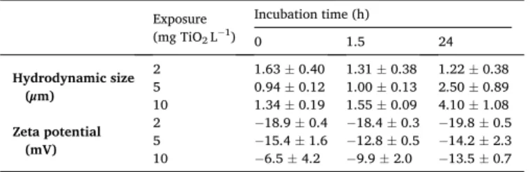
Table 2
Mean hydrodynamic size and zeta potential of nTiO2 aggregates over one day of
incubation in egg water. Mean and standard error of the mean are presented (n =
3). Exposure (mg TiO2 L−1) Incubation time (h) 0 1.5 24 Hydrodynamic size (μm) 2 1.63 ± 0.40 1.31 ± 0.38 1.22 ± 0.38 5 0.94 ± 0.12 1.00 ± 0.13 2.50 ± 0.89 10 1.34 ± 0.19 1.55 ± 0.09 4.10 ± 1.08 Zeta potential (mV) 2 −18.9 ± 0.4 − 18.4 ± 0.3 − 19.8 ± 0.5 5 −15.4 ± 1.6 − 12.8 ± 0.5 − 14.2 ± 2.3 10 −6.5 ± 4.2 −9.9 ± 2.0 −13.5 ± 0.7
3.3. Internalization of nanoparticles by zebrafish eggs
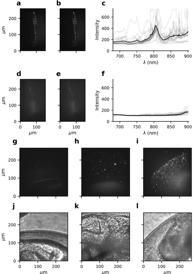
In order to investigate if nanoparticles can be internalized by zebrafish eggs, we localized nAu on zebrafish eggs following 24 h of exposure by way of two-photon multifocal microscopy (Fig. 2). We firstly used CETAB-stabilized nanorods to study how nAu appears in two-photon images. Due to their high stability, these particles can be identified based on their TPL optimum at an excitation wavelength around 850 nm, resulting from plasmon resonance. nAu appeared as bright spots in two-photon images (Fig. 2 a–c). Background fluores-cence, as detected in images of non-exposed eggs, was visible as a blur comprising dim spots that lacked the characteristic emission optimum (Fig. 2d-f). We subsequently used this information to localize PVP- coated nanorods with much lower toxicity, at increasing exposure con-centrations. It was not possible to localize these rods based on their emission optima directly, because aggregation of these rods, resulting from their lower stability, can shift the wavelength of emission optima. In z-projections of zebrafish eggs exposed to increasing concentrations of nAu, increasing amounts of nAu could be detected on the surface of zebrafish eggs (Fig. 2g-l). Inside of the zebrafish eggs, a background blur was visible at the location of the head of the developing embryo. However, no bright spots corresponding to excited nAu could be detected inside of zebrafish eggs (Fig. 2i, Fig. S5).
3.4. Survival of microbiota on zebrafish eggs
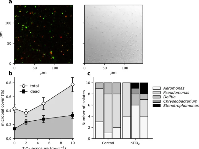
Using fluorescence microscopy, ovoid and rod-shaped microbes could be identified on the surface of zebrafish eggs (Fig. 3a). Without exposure to nTiO2, the microbes were spread homogeneously over the chorion, covering 0.43 ± 0.06 % of the total chorion surface. A 2-fold higher surface area of the chorion was covered by live microbes (0.29 ±0.06 %) than by dead microbes (0.14 ± 0.01 %). However, this dif-ference was not significant (F1,79 =1.48, p > 0.05), possibly due to the required log-transformation of the data on coverage. Exposure to nTiO2 resulted in a higher total cover of live and dead microbes on zebrafish eggs (F1,127 =7.77, p = 0.006) (Fig. 3b; Table S3). It moreover affected to ratio between the cover of dead and live microbes on the chorion. At 2 mg TiO2 L−1, dead microbes covered a larger surface area of zebrafish eggs than live microbes (F1,41 =8.58, p = 0.006). In contrast, at 5 and 10 mg TiO2 L−1, equal surface areas were covered by dead and live mi-crobes (F1,41 =1.52, p > 0.05 and F1,79 =0.30, p > 0.05, respectively).
The bacteria that could be isolated from zebrafish eggs belonged to five different genera (Fig. 3c, Table S4). Most (33) of the isolated 16S rRNA sequences had high sequence similarity (98–100 % identity) to sequences of identified bacterial species included in the NCBI database. The seven sequences with lower similarity (93–98 % identity) corre-sponded to the same bacterial species as the sequences with high simi-larity. Eggs of both nTiO2-exposed and non-exposed conditions comprised bacteria of the genera Aeromonas, Pseudomonas and Delftia. The aeromonads had highest 16S rRNA sequence similarity to the spe-cies A. hydrophila, A. sobria. A. salmonicida, A. veronii, or A. diversa; the pseudomonads were most similar to P. mosselii, P. putida, P. fluorescens,
P. entomophila, P. hunanensis, or P. cf. monteilii; and the Delftia bacteria
had highest sequence similarity to either D. lacustris or D. tsuruhatensis. In addition to these genera, the bacteria Chryseobacterium massiliae and
Stenotrophomonas maltophilia could be identified among bacterial
iso-lates from exposed eggs.
3.5. Cascading effects on microbiota of zebrafish larvae
All of the applied concentrations of nTiO2 were sublethal to zebrafish larvae during 24 h of exposure (Fig. S6). Given the saturation of nTiO2 on zebrafish eggs at an exposure concentration of 5 mg TiO2 L−1, we selected this embryonic exposure scenario to study potential cascading effects on larval microbiota abundance and composition. As a positive control, the highest sublethal concentration of nAg (0.25 mg Ag L−1)
was selected, which has previously been confirmed to exert high anti-microbial activity. In accordance with confocal microscopy results, exposure to nTiO2 resulted in a significantly higher number of colony- forming units (CFUs) per egg than incubation in egg water without nanoparticles (F1,13 = 22.95, p = 0.00035) (Fig. 4, Table S3). From surrounding water without nTiO2, a median number of 8.2 102 CFUs mL−1 (IQR = 4.2 102-1.8 103 CFUs mL−1, n = 23) could be isolated at 6 hpf, which corresponded to 1.6 102 CFUs egg−1 (IQR = 83− 3.7 102 CFUs∙egg−1, n = 23). Larvae comprised higher numbers of CFUs than eggs (F1,13 =50.90, p = 3.5 10-6). This difference in microbial abun-dance between the exposed and non-exposed group persisted until 5 dpf (Fig. 4; Table S3).
We used EcoPlates to assess the functional composition of hetero-trophic zebrafish microbiota based on carbon substrate utilization. The activity of zebrafish enzymes did not interfere with this analysis, since carbon substrate utilization by germ-free larvae resulted in a negligible average well-color development (AWCD) of 0.007 ± 0.0008 in com-parison to carbon substrate utilization of microbially colonized larvae (AWCD = 0.45 ± 0.01). In contrast to microbiota abundance, the functional composition of the zebrafish egg microbiota did not differ between exposed and non-exposed groups, as determined based on carbon substrate utilization (F1,28 =0.68, p > 0.05; Fig. 5a, Fig. S8a). Larvae had a functionally distinct microbiota composition (F1,28 = 11.21, p = 0.001), which diverged slightly following exposure to nTiO2, as indicated by a significant interaction between life stage and exposure (F1,28 =5.63, p = 0.001; Fig. 5a, Fig. S8a). However, compared to the effects of embryonic exposure to sublethal concentrations of nAg, im-pacts of sublethal concentrations of nTiO2 on the functional microbiota composition of zebrafish larvae were minor (Figs. S8-S9). Accordingly, exposure to nTiO2 did not result in differences in AWCD (0.45 ± 0.01; F1,31 =0.27, p > 0.05; Fig. 5b), the diversity in utilized carbon substrates (F1,31 =0.57, p > 0.05; 2.75 ± 0.017 in larvae and 2.63 ± 0.041 in eggs, F1,31 =15.0; p = 0.0005; Fig. 5c), and the richness in utilized carbon substrates (14.5 ± 0.34; F1,31 =1.34, p > 0.05; Fig. 5d). For comparison, although nAg exposed and non-exposed larvae had similar microbiota abundance (Fig. S7), exposure to nAg resulted in significantly lower AWCD of larval microbiota (0.33 ± 0.02; F2,22 =18.13, p < 0.0001; Fig. S8b). No difference in the diversity in utilized carbon substrates (2.77 ± 0.02; F2,22 =1.51, p = 0.24; Fig. S8c) and richness of utilized carbon substrates (14.85 ± 0.40; F2,22 =0.055, p > 0.05; Fig. S8d) of larval microbiota could be detected following exposure to nAg. The evenness of the egg and larval microbiota carbon substrate utilization could only be compared with care, as residuals did not follow a normal distribution, even following log or rank transformation. However, the carbon substrate utilization profile of embryos appeared to be slightly more even (1.00 ± 0.004) than that of larvae (1.02 ± 0.006; F1,31 =8.78,
p = 0.006). The evenness of larval microbiota carbon substrate
utiliza-tion was not affected by exposure to nTiO2 or nAg (1.03 ± 0.007; F2,22 = 2.65, p > 0.05; Fig. S8e).
4. Discussion
Teleost fish embryos, developing ex vivo in the water column, are protected against particulate pollutants by two acellular envelopes: the inner vitelline and the outer chorion membrane. Despite the efficacy of this physical barrier against particles in the environment, as confirmed in several studies (B¨ohme, 2015; Van Pomeren et al., 2017a; Batel et al., 2018; Brun et al., 2018; Lee et al., 2019; Duan et al., 2020), the adsorption and enrichment of particles on the chorion surface has been related to several adverse effects. These include the obstruction of gas exchange across the chorion surface (Duan et al., 2020), and the leaching of toxic chemicals and ions out of chorion-attached particles into the eggs (B¨ohme, 2015; Batel et al., 2018). Given the antimicrobial nature of many metal nanoparticles, and the importance of early mi-crobial colonization to larval development (Rawls et al., 2004; Hill et al.,
Fig. 2. Two-photon microscopy detection of gold nanorods on zebrafish eggs at 1 day post-fertilization. Subplots show representative images of a zebrafish embryo
following 24-h exposure to 0.03 mg Au L−1 CETAB-coated gold nanorods (a), with 20 regions of interest (ROIs) (b) used to obtain excitation spectra (c); a zebrafish
embryo that had not been exposed to nanorods (d), with 20 ROIs (e) that were used to obtain excitation spectra (f); and z-projections of embryos that had been exposed for 24 h to no nanorods (g), 0.03 mg Au L−1 PVP-coated gold nanorods (h), and 0.1 mg Au L−1 PVP-coated gold nanorods (i). Transmitted light images j, k,
and l correspond to emitted light images g, h and i, respectively. The black line in graph c and f depict the mean intensity of the intensity of all 20 ROIs, indicated with grey lines.
chorion-attached microbial community constitutes a hitherto unex-plored adverse effect of concern. In this study, we took a three-step approach to investigate this, focusing on the impacts of titanium
dioxide nanoparticles on zebrafish embryos. Firstly, we inferred if the particles accumulated on the chorion surface; secondly, we studied how this affected the microbial community that establishes on the chorion surface; and thirdly, we examined if this resulted in any cascading ef-fects on the larval microbial community.
4.1. Internalization of nanoparticles by zebrafish eggs
As a first step, we assessed if nanoparticles can cross the external membranes of zebrafish eggs, employing the advantages of two-photon microscopy for the localization of nAu. This approach builds on previous knowledge obtained using fluorescently labeled particles, or using destructive analyses such as Laser Ablation Inductively Coupled Plasma Mass Spectrometry (LA-ICP-MS), enabling live imaging without the risk of fluorescent dye dissociation, with less autofluorescence background than with the use of fluorophores, and providing a high z-confinement with large penetration depth (Van den Broek et al., 2013). Using this approach, we did not detect any particles inside of zebrafish eggs, yet observed a high and concentration-dependent accumulation of nano-particles on the chorion surface. Considering the high sensitivity of two-photon microscopy detection of nAu, this suggests that the particles did not cross the chorion surface. Moreover, in view of the lower sta-bility of nTiO2 than nAu, resulting in larger aggregate sizes, it is even less likely that nTiO2 aggregates can cross the chorion pores. These findings agree with previous results, showing an accumulation of polystyrene nanoparticles (Van Pomeren et al., 2017a; Duan et al., 2020), silicon nanoparticles (Fent et al., 2010), copper nanoparticles (Brun et al., 2018), titanium dioxide, silver, gold, and aluminum oxide nanoparticles
Fig. 3. Impacts of nTiO2 on microbial communities establishing on zebrafish eggs. Subplots show a CLSM picture dead (red) and live (green) microbes (a, left picture)
on the surface of a zebrafish egg (a, right picture); the impact of 24 h-exposure to nTiO2 on total microbial cover (white diamonds) and dead microbial cover (black
squares) on the surface of zebrafish eggs (n=24–45; mean ± SEM are presented) (b); and the identity of bacteria isolated from zebrafish eggs exposed for 24 h to no nanoparticles (‘Control’; left bars) and 5 mg nTiO2 L−1 (‘nTiO2’; right bars). Each bar represents 10 bacterial isolates from 1 biological replicate. The corresponding
BLAST results are included in Supplementary Table S4.
Fig. 4. Total count of colony-forming units associated per zebrafish embryo (1
dpf; white bars) or per zebrafish larva (5 dpf; grey bars) following embryonic exposure (0-1 dpf) to egg water without nanoparticles (‘Control’; non-hatched, white fill), and egg water comprising nTiO2 (5 mg TiO2 L−1; hatched fill). Bars
represent means with standard error of the mean (n = 3). Asterisks indicate significant differences (p < 0.001).
(Osborne et al., 2013; B¨ohme et al., 2015) onto or inside of zebrafish chorions, with limited or no detectable uptake of particles by the embryos.
Using PIXE to quantify elemental concentrations of Ti, we observed a concentration-dependent accumulation of nTiO2 onto zebrafish eggs, reaching a maximum of 154.3 ± 19.2 ng Ti (257.4 ± 32.0 ng TiO2) per egg at a nominal exposure concentration of 5 mg TiO2 L−1. Previous studies have shown that small (1-5 μm) microplastic particles (Batel
et al., 2018) and metal nanoparticles (B¨ohme, 2015; Shih et al., 2016) can cover the zebrafish chorion completely. Although our estimates of the chorion coverage are lower, we nevertheless estimated that a rela-tively large surface of the chorion could be covered by TiO2 aggregates (25.2 %) or primary particles (44.8 %), as compared to the surface of the chorion that is occupied by microbes (<1%). Studies applying more optimal sorption conditions, obtained even higher particle coverages. In an experimental setup where eggs were exposed to nanoparticles in a rotating glass vial, Shih et al. (2016) reached a maximum of 6 tightly bound layers of nTiO2 in a hexagonal close-packing of primary particles. Comparably, Lin et al. (2015) calculated that 2–17 layers of primary TiO2 particles attached to cells of algae Raphidocelis subcapitata in a hexagonal close-packing.
The accumulation of TiO2 coincided with changes in the elemental concentrations of silicon and postassium of zebrafish eggs. Concentra-tions of Ti correlated negatively with silicon, which could result from competition in binding sites of Ti and silicon on the chorion, or from lower internalization rates of silicon by the TiO2-exposed embryos. In view of indications for the contribution of silicon to immune and in-flammatory responses, osteogenesis, and the formation of connective tissue (Nielsen, 2014), the latter scenario merits further investigation.
A positive correlation was found between concentrations of titanium and potassium of zebrafish eggs. A similar, yet insignificant trend was observed for the concentration of phosphorus. Of all detected elements by PIXE (Table S2), potassium constitutes the most abundant element in bacteria, followed by phosphorus (Novoselov et al., 2013). This suggests that the higher concentrations of both potassium and phosphorus were detected on zebrafish chorions as a result of the accumulation of bacteria on zebrafish chorions.
4.2. Survival of microbiota on zebrafish eggs
To address our second research question, we characterized the ef-fects of nTiO2 on microbial consortia that establish on the chorion of zebrafish eggs. In accordance with the reported antimicrobial activity of nTiO2, we detected a higher cover of dead microbes on the chorion surface of zebrafish eggs following exposure to nTiO2. However, imag-ing analyses also showed that exposure concentrations above 2 mg TiO2 L−1 resulted in an overall increase in total microbial abundance. For this reason, the antimicrobial effects of nTiO2 could not have been identified using culture-dependent microbial techniques only. For future research, this shows the importance to support culture-dependent analysis of microbiota composition with culture-independent tech-niques to assess microbiota composition, specifically using microbial profiling techniques that are based on RNA, or include initial removal of relic DNA, to allow to distinguish dead from live microbiota (Knight et al., 2018).
Similar to our results, Zhai et al. (2019) detected a concentration-dependent increase in soil microbiota abundance following 24 h-exposure to nTiO2, with higher microbial abundance at 500 and 2000 mg TiO2 kg−1 soil than at 0 and 1 mg TiO2 kg−1 soil. Nevertheless, these effects were of transient nature, and the adverse effects of nTiO2 on microbial abundance gradually appeared from 15 to 60 days of exposure. In the present study, such adverse effects of pro-longed exposure to nTiO2 might explain why the acute growth-promoting effects of nTiO2 did not result in higher AWCD over the two days longer Ecoplate incubations.
Following 24 h of exposure to 5 mg TiO2 L−1, over 16 times more CFUs were isolated from TiO2-exposed embryos than from control em-bryos. One explanation to this difference could be potential growth- promoting effects of nTiO2 on certain bacterial isolates; only four gen-eration cycles are required to obtain a 16-times difference in microbial abundance. An alternative and non-exclusive explanation follows from our DLS results. Above exposure concentrations of 2 mg TiO2 L−1, we measured a more neutral zeta potential, and observed a corresponding increase in the hydrodynamic size of aggregates over time. This suggests that particle stability was lower at exposure concentrations above 2 mg TiO2 L−1. Accordingly, imaging analyses indicated that only at exposure concentrations above 2 mg TiO2 L−1, exposure to nTiO2 resulted in
Fig. 5. Carbon substrate utilization characteristics of microbiota isolated from zebrafish embryos (1 dpf) and zebrafish larvae (5 dpf) following embryonic exposure
(0-1 dpf) to egg water without nanoparticles (non-hatched), and egg water comprising nTiO2 (5 mg TiO2 L−1; hatched fill). Subplots represent carbon substrate
utilization profiles (a), total well color development (TWCD) (b), Shannon index (c), richness (d), and evenness (e). Each biological replicate, comprising three measurement replicates, is depicted with a unique marker shape. Bars represent means with standard error of the mean (n = 3). Asterisks indicate significant differences (**, p < 0.01; ***, p < 0.001).
higher microbial abundance on zebrafish eggs. Combined, these results may imply that the increased microbial abundance at exposure con-centrations above 2 mg TiO2 L−1 resulted from the formation of heter-oaggregates of microbes and TiO2 in the water column, which subsequently settled onto zebrafish eggs. In agreement with this reasoning, the total number of CFUs in the water column without exposure to TiO2 (1.6 102 CFUs egg−1; IQR = 83− 3.7 102 CFUs egg−1, n =23), can explain a large part of the difference in CFU counts between non-exposed eggs (52 ± 23 CFUs egg−1) and nTiO
2-exposed eggs (8.5 102 ±4.2 102 CFUs egg−1). Heteroaggregation dynamics of nTiO
2 with microbes have already been described for the unicellular alga
R. subcapitata (Lin et al., 2015). Moreover, the aggregation and subse-quent sedimentation of microbes with suspended particles is a well-described phenomenon in the water column of pelagic water bodies, known as marine snow (Suzuki and Kato, 1953) or lake snow (Grossart and Simon, 1998).
4.3. Cascading effects on microbiota of zebrafish larvae
The third part of our study concerned the potential consequences of altered embryonic microbiota to zebrafish larvae health. Specifically, we inferred if nTiO2-induced changes in chorion-attached microbiota resulted in different larval microbiota abundance and composition. Considering the potential functional redundancy of microbes, we used carbon substrate utilization as a functional measure of microbiota composition. We found that the higher microbial abundance on zebra-fish eggs that had been exposed to nTiO2, persisted until the 5 dpf-larval stage. This indicates that microbes which grow on zebrafish eggs as a result of nTiO2 exposure, possibly due to the adsorption of aggregates consisting of microbes and nTiO2 onto the chorion surface, can colonize the larvae upon hatching. In our experiment, this did not result in different functional composition of larval microbiota, as determined based on carbon-substrate utilization. However, there is a risk that hazardous microbes are transferred to eggs following heteroaggregate formation in aquatic ecosystems. In the Baltic Sea, for instance, poten-tially pathogenic Vibrio bacteria have been identified on microplastic particles (Kirstein et al., 2016). Of comparable concern, we identified
Aeromonas species on zebrafish eggs which are known to be pathogenic
to fish (Fern´andez-Bravo and Figueras, 2020), which were one of the species A. hydrophila (infecting salmonids and eel), A. salmonicida (infecting salmonids), A. sobria (infecting tilapia and common carp) or
A. veronii (infecting catfish, common carp, and eel). More broadly,
aeromonads have been associated with infections in echinoderms, mollusks, copepods, crocodiles, frogs, rabbits, cats, dogs, horses and humans (Fern´andez-Bravo and Figueras, 2020). The presence of these aeromonads on nTiO2-exposed eggs suggests that these bacteria are able to cope with the particles’ antimicrobial properties. Therefore, it is possible that pathogenic aeromonads are transferred throughout aquatic ecosystems, and to aquatic eggs, via attachment to suspended nTiO2 particles and aggregates.
Although the nAg-exposed eggs of the positive control developed functionally different larval microbiota than zebrafish eggs that had not been exposed to nanoparticles, larval microbiota of nTiO2-exposed eggs did not differ from non-exposed eggs in carbon substrate utilization. This lower impact of nTiO2 on microbiota composition, as compared to nAg, at concentrations that are sublethal to the host, has been reported for soil microbes (Ahmed et al., 2020), intestinal microbiota of mice (Chen et al., 2017), and human gut simulator microbiota (Agans et al., 2019). Nevertheless, even though we exclusively isolated the bacteria
C. massiliae and S. maltophilia from nTiO2-exposed eggs, which could indicate that the composition of exposed and non-exposed egg micro-biota differed taxonomically, also micromicro-biota of eggs did not differ in carbon substrate utilization profiles. This may imply that nTiO2 -expo-sure can induce shifts in microbiota composition that do not result in different microbiota functioning, as a result of functional redundancy. Interestingly, Zhai et al. (2019) obtained similar results for the impacts
of nTiO2 on soil microbiota. This remarkable similarity in the impacts of nTiO2 on microbiota from different environments warrants further research, investigating the generalizability of these results.
On a similar note, comparing the particle-specific protection of the external structures surrounding the embryos of different taxa merits further investigation. At a large phylogenetic distance to teleost fish, for instance, it can be questioned if egg coatings facilitate early microbial colonization, given the bactericidal or bacteriostatic activity of the jelly layer of amphibian eggs (Mazzini et al., 1984). Even at low phylogenetic distances, a remarkable diversity of egg surfaces exists. Demersal fish, for example, generally produce eggs with much thicker and harder chorion structures than pelagic fish (McMillan, 2007). Moreover, cho-rions can have diverse kinds of protrusions, such as threads, fibrils, or spikes, increasing their surface area. Additionally, differences between the environments where these eggs develop – including turbidity, water column depth, currents, salinity, and the presence of other pollutants or stressors – will affect the interactions between suspended particles and microbiota colonizing aquatic eggs externally. In view of this diversity in the external surface and environment of aquatic eggs, the adverse effects of nanoparticle adsorption on the eggs of oviparous animals, as exam-ined in this study, may contribute to variation in nanoparticle sensitivity across different taxa. Further characterization thereof may help to refine the assessment of environmental risks of nanoparticle exposure on early life stages of oviparous animals.
5. Conclusions
Early host-microbiota interactions are of key importance to the development of zebrafish larvae. In this study, we tested the hypothesis that the adsorption of antimicrobial titanium dioxide nanoparticles on zebrafish eggs can perturb microbial communities that establish on the egg surface, indirectly affecting the subsequent colonization of hatched larvae. Our results confirmed that both nanoparticles and microbes accumulated on the surface of zebrafish eggs, whereas no particles could be detected inside of the eggs. At increasing exposure concentrations, this resulted in higher dead and total microbial abundance, and altered concentrations of potassium and silicon. These impacts of titanium di-oxide nanoparticles on egg microbiota had cascading effects on later life stages, as indicated by a persisting increased microbial abundance of larvae. Combined with measurements on particle stability, this overall stimulatory effect of nTiO2 on egg- and larval microbial abundance can potentially be explained by the formation of heteroaggregates of mi-crobes and nanoparticles that settle on the egg surface. Given the tolerance of pathogenic aeromonads against antimicrobial properties of titanium dioxide particles, these dynamics potentially come at the expense of increased dispersal of pathogenic bacteria through aquatic ecosystems, as well as across different life stages of oviparous animals.
Data availability statement
Data is available via Mendeley Data doi:10.17632/2d4hcr5cb5.1.
CRediT authorship contribution statement
Bregje W. Brinkmann: Conceptualization, Investigation,
Supervi-sion, Formal analysis, Writing - original draft, Writing - review & edit-ing. Wouter F. Beijk: Investigation, Methodology, Writing - review & editing. Redmar C. Vlieg: Investigation, Methodology, Writing - review & editing. S. John T. van Noort: Supervision, Funding acquisition, Writing - review & editing. Jorge Mejia: Investigation, Methodology, Writing - review & editing. Julien L. Colaux: Investigation, Method-ology, Writing - review & editing. St´ephane Lucas: Supervision, Funding acquisition. Gerda Lamers: Methodology, Writing - review & editing. Willie J.G.M. Peijnenburg: Supervision, Writing - review & editing. Martina G. Vijver: Supervision, Funding acquisition, Writing - review & editing.