HAL Id: tel-03220102
https://tel.archives-ouvertes.fr/tel-03220102
Submitted on 7 May 2021HAL is a multi-disciplinary open access archive for the deposit and dissemination of sci-entific research documents, whether they are pub-lished or not. The documents may come from teaching and research institutions in France or abroad, or from public or private research centers.
L’archive ouverte pluridisciplinaire HAL, est destinée au dépôt et à la diffusion de documents scientifiques de niveau recherche, publiés ou non, émanant des établissements d’enseignement et de recherche français ou étrangers, des laboratoires publics ou privés.
nephropathy : new diagnostic test and identification of
the immunodominant epitopes
Kristel Zaghrini
To cite this version:
Kristel Zaghrini. THSD7A, the second autoantigen in membranous nephropathy : new diagnostic test and identification of the immunodominant epitopes. Cell Behavior [q-bio.CB]. COMUE Université Côte d’Azur (2015 - 2019), 2019. English. �NNT : 2019AZUR4026�. �tel-03220102�
“You give but little when you give of your possessions. It is when you give of yourself that you truly give.”
Kahlil Gibran, The Prophet
Acknowledgments
The work presented in this thesis would not have been possible without the effort of several wonderful people. I sincerely appreciate the inspiration, support and guidance of all those who made this PhD possible.
First and foremost, I would like to extend my sincere gratitude to Dr. Gérard Lambeau for his dedicated help, advice, inspiration, encouragement and continuous support throughout my PhD. You believed in me like nobody else did, you taught me how good scientific research is done and you allowed me to grow as a research scientist. Your unwavering enthusiasm and your mission for providing high–quality work has finely shaped my career. I doubt that I will ever be able to convey my appreciation fully, but I owe you my eternal gratitude.
I would like to thank the members of the PhD committee: Dr. Andreas Schedl, Dr. Sonia Berrih–Aknin, Dr. Marco Prunotto, Dr. Hanna Debiec and Dr. Véronique Braud for having accepted to participate in this committee and evaluate my work. I thank Dr. Sonia Berrih– Aknin and Dr. Marco Prunotto for their reports and their insightful comments. I also thank my PhD advisors Dr. Andreas Kistler and Dr. Andreas Schedl for their cooperation and for evaluating the work progress during my PhD.
I also gratefully acknowledge the Laboratory of Excellence SIGNALIFE program for the PhD fellowship and Konstanze Beck for her generous administrative assistance.
I thank all our collaborators particularly Dr. Pierre Ronco, Dr. Laurence Beck and Dr. Jack Wetzels for their tremendous help in the inclusion of patients in this study and for their valuable advices and discussions.
I would also like thank all the lab members for all their priceless support and for making my stay at IPMC more pleasurable. Joana, thank you for your scientific input and for all the memorable travel journeys that we had together. Your friendship shall always be remembered. Guillaume, thank you for the numerous scientific discussions, and for your cheerfulness in the difficult times. Christine, thank you for your kindness, your positive attitude and for the moral support you provided me. Barbara, thank you for the help in collecting the patients’ clinical data. Agnès, thank you for your gentleness, your humor and your enthusiasm for research. Alice, Franck and Joëlle, thank you for your kindness and for the stimulating discussions in the last stage of my PhD. I acknowledge the previous lab members especially Vesna for her friendliness and for sharing her research expertise, Joel for his helpfulness and for his assistance at the beginning of my PhD, Louise for her kindness and Sarah for her amiability and friendship. I thank also the staff of IPMC and specially the colleagues in IPMC R+2 for their cooperation and insightful discussions.
Last but not least, I owe my deepest gratitude to my family. For mom and dad who raised me, supported me, taught me and loved me. Without your help, I wouldn’t have become who I am. To you I dedicate this work. For my brother and sister, who provided me with endless support and encouragement, your infallible love and trust has always been my strength. Finally, a special thanks for Léopold my loving and patient partner, your faithful support got me through the last stages of the PhD.
Table of Contents
List of figures ... 1
List of tables ... 3
Abbreviations ... 5
1
Introduction
– Membranous nephropathy ... 7
1.1 Autoimmune diseases ... 7 1.2 Membranous Nephropathy ... 8 1.2.1 Kidney physiology ... 8 1.2.2 Glomerulonephritides...10 1.2.3 Definition of MN ...11 1.2.4 History of MN ...12
1.2.5 Multiple clinical forms of MN ...13
1.2.6 Genetics of MN ...19
1.2.7 Natural history of MN ...19
1.2.8 Clinical features of MN ...20
1.2.9 Treatment of MN ...21
1.3 Physiopathology of MN ...22
1.3.1 Immunoregulation and inflammation in MN ...22
1.3.2 Immune complexes ...24
1.3.2.1 Mechanisms of deposition of immune complexes ...24
1.3.2.2 The antigens in MN ...26
1.3.2.3 The different antibodies in various forms of MN ...38
1.3.4 Epitopes recognized by anti–PLA2R1 and –THSD7A ...48
1.3.5 Epitope spreading in MN antigens ...51
1.3.6 Animal models of MN ...55
1.4 Diagnosis and prognosis of MN ...57
1.4.1 Biopsy staining ...57
1.4.2 Detection of autoantibodies ...59
1.4.3 Comparison of the detection assays: Western Blot, IIFT and ELISA ...61
1.4.4 Diagnostic value of antibodies in MN ...62
1.4.5 Monitoring value of antibodies in MN ...64
1.4.6 Prognosis value of antibodies in MN ...65
2
Introduction
– THSD7A ... 67
2.1 The thrombospondin type–1 repeat (TSR) superfamily ...67
2.2 The Thrombospondin family ...70
2.2.1 Structure of Thrombospondins ...70
2.2.2 Function of thrombospondins ...72
2.2.3 Thrombospondin–1 ...72
2.3 Structure and biology of THSD7A ...74
2.3.1 THSD7A family members ...74
2.3.2 Structural properties of THSD7A ...75
2.3.3 Functional properties of THSD7A ...77
3
Aims of the study ... 81
4
Results ... 83
4.1 Development of an ELISA to identify THSD7A–associated MN patients (Article 1) ...83
4.1.2 Identification of THSD7A–associated MN patients ...85
4.1.3 Clinical characteristics of THSD7A–associated MN patients ...86
4.2 Identification of THSD7A epitopes in MN (Unpublished data) ... 133
4.2.1 Identification of THSD7A epitopes by limited proteolysis ... 134
4.2.2 Identification of THSD7A epitopes by site–mutagenesis ... 138
4.2.3 From immunodominant epitopes to epitope spreading in THSD7A ... 147
4.3 Epitope profiling of PLA2R1 at baseline predicts outcome of MN (Article 2) ... 159
5
Discussion and perspectives ... 179
5.1 Clinical features and etiology of THSD7A–associated MN ... 179
5.2 Immunological phase of THSD7A–associated MN ... 185
5.2.1 Epitope profile ... 185
5.2.2 Epitopes and immunodominance ... 186
5.2.3 Epitope spreading in THSD7A ... 187
5.2.4 In Vivo Experimental Epitope Spreading for THSD7A... 191
5.3 Pathological effector phase ... 195
5.3.1 Pathogenicity of anti–THSD7A ... 195
5.3.2 Shedding of THSD7A ... 196
5.3.3 Complement activation in MN ... 199
5.3.4 Clinical advances for THSD7A–associated MN... 202
5.3.5 THSD7A epitopes as biomarkers of MN ... 202
5.3.6 Additional biomarkers of disease activity in MN ... 204
5.3.7 Towards a serology–based classification of MN disease? ... 204
5.4 Conclusion ... 207
List of figures
Figure 1.1 Physiology of the glomerulus in representative modification of the GBM in MN
Figure 1.2 Possible scenarios for immune complex formation and deposition in MN
Figure 1.3 Schematic structures of the four antigens implicated in MN
Figure 1.4 Mechanism of activation of complement pathways
Figure 1.5 Schematic representation of the epitope spreading mechanism on megalin and PLA2R1
Figure 1.6 Renal biopsy staining in PLA2R1– and THSD7A–associated MN patients Figure 2.1 Superfamily of thrombospondin repeats (TSR) family
Figure 2.2 Prediction of the three–dimensional structure of TSP–1–like and C6–like
domains in THSD7A
Figure 2.3 Schematic illustration of the structural organization of the five members of the Thrombospondin family
Figure 2.4 Overall domain structure of thrombospondin–1 Figure 2.5 In silico model structure of THSD7A
Figure 4.1 Western blot of proteolysis of THSD7A by three proteases
Figure 4.2 Patients' autoantibody reactivity of THSD7A and PLA2R1 after treatment with increasing concentrations of DTT by western blot
Figure 4.3 Strong proteolysis of THSD7A by combining DTT and enzymes
Figure 4.4 Alignment of TSP–1 like and C6–like domains in THSD7A Figure 4.5 Design and expression of soluble constructs of THSD7A
Figure 4.6 Epitopes regions recognized by autoantibodies of THSD7A–associated MN
Figure 4.7 Relationship between anti–THSD7A titer and positivity towards increasing
number of epitopes
Figure 4.8 Optimization of the competition assay for THSD7A
Figure 4.9 Immunodominant epitope competition assays with THSD7A epitope domains by ELISA using full THSD7A for 13 THSD7A–positive patients
Figure 4.10 Competition assay between THSD7A full antigen and the THSD7A constructs D1–D2 and D9–D10 for patient MN1
Figure 4.11 Absence of cross–reactivity between D1–D2 and D9–D10
Figure 4.12 Distribution of epitope immunodominance in THSD7A–associated MN
patients
Figure 4.13 Distribution of anti–THSD7A titer according to the type of immuno–
dominance or not
Figure 5.1 Epitope reversal during follow–up in MN13
Figure 5.2 Immune response and THSD7A epitope domains recognized by rabbit sera over the time course of immunization with human THSD7A
Figure 5.3 Shedding of THSD7A in vitro
List of tables
Table 1.1 Classification of the different MN forms and their characteristics
Table 4.1 Epidemiological and clinical characteristics of patients stratified according to their epitope profile
Table 4.2 Clinical characteristics of THSD7A–associated patients from different
immunodominant epitope groups
Table 4.3 Comparison of the clinical characteristics of THSD7A–associated MN
patients of the combined immunodominant group and the "non– immunodominant" epitope group
Table 4.4 Comparison of the clinical characteristics of patients with low antibody titer in the immunodominant and non–immunodominant epitope groups
Abbreviations
ACE Angiotensin–Converting Enzyme AchR Acetylcholine Receptor 1
ANCA Anti–Neutrophilic Cytoplasmic Autoantibody
AR Aldose Reductase
BSA Bovine Serum Albumin
C6 Complement Component 6
CD Cluster of Differentiation
CFH Complement Factor H
CTLD C–Type Lectin Domain
COMP Cartilage Oligomeric Matrix Protein CysR Cystein–Rich Domain
Dsg Desmoglein
DTT Dithiothreitol
ECM ExtraCellular Matrix E. coli Escherichia coli
EGF Epidermal Growth Factor
eGFR estimated Glomerular Filtration Rate ELISA Enzyme–Linked Immunosorbent Assay
EM Electron Microcopy
ESRD End–Stage Renal Disease FAK Focal Adhesion Kinase FNII Fibronectin type II domain
GBM Glomerular Basement Membrane Gp330 Glycoprotein 330 (megalin)
GWAS Genome–Wide Association Study HLA Human Leukocyte Antigen
LY75 Lymphocyte Antigen 75 (also called DEC–205)
MRC2 C–type Mannose Receptor 2 (also called Endo180) MuSK Muscle–Specific Kinase
HEK293 Human Embryonic Kidney 293 cells
HN Heymann Nephritis
HUVEC Human Umbilical Vein Endothelial Cells
IgG Immunoglobulin G
IIFT Indirect ImmunoFluorescence Test JAK2 Janus kinase 2
KDIGO Kidney Disease Improving Global Outcomes LBD Ligand Binding Domain
LRP4 Lipoprotein receptor–related protein–4
MAC Membrane Attack Complex
MBL Mannose Binding Lectin
MG Myasthenia Gravis
MHC Major Histocompatibility Complex
MN Membranous Nephropathy
MusK Muscle–Specific Kinase NEP Neutral Endopeptidase
PDB–ID Protein Data Bank– Structure Identifier
PLA2R1 Phospholipase A2 Receptor 1 (also called M–type receptor) PM2.5 Particulate Matter 2.5 µm
PTM Post–translational Modification
RU Relative Unit
SNP Single Nucleotide Polymorphism SOD2 Superoxide Dismutase 2
sPLA2 secreted PhosphoLipase A2
TGF– β Transforming Growth Factor–beta
THSD7A Thrombospondin type 1 Domain–containing 7A
TSP–1 Thrombospondin –1
1 Introduction – Membranous
nephropathy
1.1 Autoimmune diseases
Protecting and defending the host against infectious agents is the chief function of the immune system. There are two cases where the deficiency of the immune system can lead to pathologies (Wang et al., 2015). In the first, the deficient immune system is incapable of protecting host against pathogens. In the second, the immune system fails to distinguish between self and non–self–molecules and the breach of tolerance leads to an autoimmune disease.
Autoimmune diseases affect 3–5% of the general population, with autoimmune thyroid disease and type I diabetes being the most common forms. Noteworthy, there are around 100 different autoimmune diseases that can be either organ–specific as in the case of primary biliary cirrhosis where the autoantibodies react to autoantigens localized in a specific tissue, or can also be organ non–specific as in systemic lupus erythematosus where the autoantibody reactivity targets several tissues (Yu et al., 2014).
It has been shown that the development of an autoimmune reaction can be triggered by environmental factors and depends on genetic predisposition (Wang et al., 2015). The environmental factors include microorganisms, xenobiotics, infectious agents and nutrition and can influence the microbiota whereas the genetic factors include gene mutations, HLA susceptible loci and epigenetic mechanisms. These factors stimulate the autoimmune response which will lead to tissue damage. The epidemiology of the different autoimmune diseases varies with age, sex, ethnicity and geographic localization with an increased frequency of autoimmune responses in women, and typical large female–to–male ratios ranging from 10:1 to 1:1.
Circulating autoantibodies targeting various structures of the cell are a common characteristic of autoimmune diseases and mediate tissue damage through different mechanisms. First, the autoantibodies can lead to destruction of the cell through
complement activation and antibody–dependent cell–mediated cytotoxicity as in the case of autoimmune thyroid disease which is mediated by anti–thyroid autoantibodies (Rodien et al., 1996). A second important pathogenic mechanism is the immune complex–mediated
damage as in systemic lupus erythematosus.
The immune complexes produced in response to soluble antigens are usually eliminated by phagocytosis. If the clearance fails mainly because of the large amounts of immune complexes, tissue damage can occur. Furthermore, the circulating immune complexes can deposit and cause glomerulonephritis, vasculitis or even arthritis (Lleo et al., 2010). Autoantibodies may also bind to cell surface receptors and either activate or block different signaling pathways. In Myasthenia Gravis, autoantibodies target different key protein components of the postsynaptic neuromuscular junction (like the acetylcholine receptor AchR, the muscle–specific tyrosine kinase MUSK or the low–density lipoprotein receptor– related protein 4 LRP4) and disrupt the neuromuscular transmission by different mechanisms, dependent or not from activation of the complement pathway (Berrih-Aknin et al., 2014).
1.2 Membranous Nephropathy
1.2.1 Kidney physiology
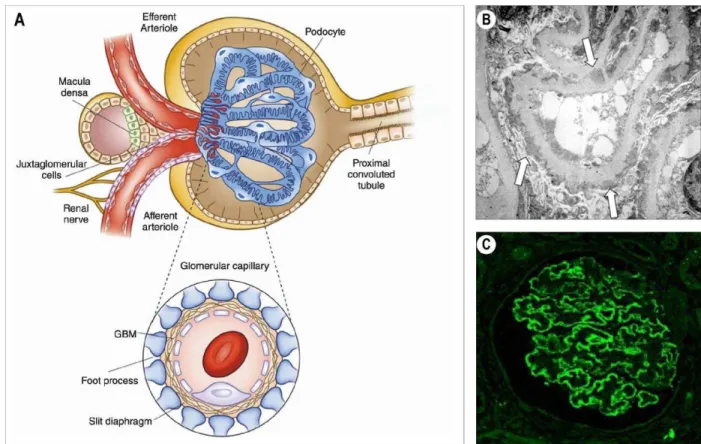
The kidney is the main filtrating organ in mammals. The basic functional unit in the kidney is the nephron which regulates the excretion of metabolic wastes, re–uptake of salt, water and important components such as nutrients, vitamins or proteins. In addition to its role in fluid homeostasis, the kidney is essential for the control of blood pressure, vitamin D synthesis and bone mineralization (Scott et al., 2015). An adult human kidney contains between 1 to 2.5 million of nephrons. Each nephron comprises a glomerulus where blood filtration occurs and a segmented tubular resorption compartment ending into the collecting duct (Figure 1.1–A). Every day, 150 to 180 liters of blood are filtered through the glomerular capillaries. During the tubular transport, the primary urine is modified, and most of the fluid is reabsorbed before becoming the final urine, approximately 1.5 liters per day (Puelles et al., 2011).
Figure 1.1 – Physiology of the glomerulus in representative modification of the GBM in MN. (A) Structure of the renal glomerulus and the filtration barrier. Adapted from (Pollak et al., 2014) with copyright permission from “Clinical Journal of American Society of
Nephrology: CJASN”. (B) Electron microscopy showing the immune deposits (arrows) in
subepithelial space of the glomerulus in membranous nephropathy. Adapted from (Lai et al., 2015) with copyright permission from “Elsevier”. (C) Immunofluorescence staining of
glomerulus showing granular deposits the glomerular basement membrane. Adapted from (Debiec et al., 2014) with copyright permission from “SpringerLink”.
The glomerulus (after the Latin word glomus for a ball of yarn) is the most complex biological membrane system that allows plasma filtration with total restriction to high– molecular–weight proteins and blood cellular components, generating the primary urinary ultrafiltrate (Haraldsson et al., 2008). This membrane, first proposed by Karl Ludwig in the 1800s, (Jarad et al., 2009) is termed as the glomerular filtration membrane. It assembles three different layers organized as follows: the podocytes, intimately wrapped around the glomerular capillaries and sharing with the glomerular endothelial cells an extracellular matrix known as the glomerular basement membrane (GBM) (Figure 1.1–A)
(Scott et al., 2015). The foot processes of two adjacent podocytes interdigitating and forming the slit diaphragm consisting of multiple adhesion proteins such as nephrin, podocin, etc.
(Jarad et al., 2009). In mammals, the slit diaphragm can be compared to a dialysis membrane with a cut–off value of around 60 kDa, close to the molecular mass of albumin, the most abundant protein in our blood. Any dysfunction of the glomerulus will lead to pathologies collectively called glomerulonephritides.
1.2.2 Glomerulonephritides
Glomerulonephritides comprise a group of rare kidney diseases affecting about 20% of chronic kidney disease cases. The most common types of glomerulonephritides include IgA nephropathy, membranous nephropathy, membranoproliferative glomerulonephritis, minimal change disease and focal segmental glomerulosclerosis. Rare forms of glomerulonephritides include dense deposit disease and C3 glomerulonephritis (Floege et al., 2016). Interestingly, although the Anti–Neutrophilic Cytoplasmic Autoantibody (ANCA)
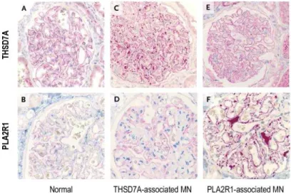
disease is not a glomerulonephritis but a vasculitis that affects kidney function by targeting the small vascular endothelial cells near the podocytes (Jennette et al., 2017). The clinical course of the disease varies from coincidental detection of proteinuria and increased serum creatinine levels in asymptomatic patients to weight gain and edema in nephrotic patients. The definitive diagnosis of glomerulonephritis is done after a kidney biopsy and immunohistological examination (Figure 1.1–BC).
Clinical manifestations in the different age categories can be an indicator of the type of glomerulonephritides: minimal change disease and focal segmental glomerulosclerosis are more common in children and young adults while membranous glomerulonephritis occurs
in older subjects. Regional differences are also important in distribution of glomerulonephritides: while the incidence of IgA nephropathy is higher in Asian subjects, focal segmental glomerulosclerosis is higher in the USA and Canada (Floege et al., 2016; Johnson et al., 2003). Of interest, the annual incidence of glomerulonephritides in a Europe is as follows: 2.5/100,000 case per adult for IgA nephropathy, 1.2/100,000 case per adult for membranous glomerulonephritis, between 0.6 and 0.8/100,000 case/adult for minimal change disease and for focal segmental glomerulosclerosis and 0.2/100,000 case/adult for membranoproliferative glomerulonephritis (McGrogan et al., 2011).
1.2.3 Definition of MN
Membranous nephropathy (MN) is a rare autoimmune kidney disease with a worldwide incidence of 1/100,000 new cases per year (Maisonneuve et al., 2000). The disease affects patients of all ages, ethnic groups but is quite surprisingly for an autoimmune disease, more common in men than women (ratio 2:1) (Ronco et al., 2017). However, MN is more likely to occur in individuals older than 50 years and is less in the pediatric population.
MN is defined by the increased thickness of the glomerular basement membrane (GBM), due to the presence of large amount of immune complexes sitting there (Figure 1.1 – BC)
(Farquhar et al., 1957; Glassock, 2010; Mellors et al., 1957; Movat et al., 1959). In the common idiopathic form of the disease, the immune complexes consist predominantly of IgG4 autoantibodies colocalized with a known or unknown target antigen and are associated with the complement factor C3 in a peripheral capillary loop pattern on immunofluorescence and in electron–dense subepithelial deposits on electron microscopy.
Definitive diagnosis of MN relies on subepithelial immune–complex deposits on renal biopsy visible by electron microscopy (Figure 1.1–B) (Churg et al., 1973; Lai et al., 2015). The early stage I, is characterized by small immune deposits scattered in the subepithelial space. The stage II is characterized by spikes of the GBM around the subepithelial deposits. The stage III is characterized by a basement membrane element surrounding the immune deposits and the stage IV is characterized by complete incorporation of the immune deposits into the GBM.
1.2.4 History of MN
In 1957, David Jones described membranous nephropathy as a distinct morphological entity from minimal change lesions (previously called lipoid nephrosis), membranoproliferative glomerulonephritis (previously called lobular glomerulonephritis) and focal segmental glomerulosclerosis (or probably chronic moderate glomerulonephritis)
(Jones, 1957). The histopathological patterns of membranous nephropathy showed GBM thickening visualized as spike–like extrusions seen on light microscopy, a fine granular capillary wall deposition of IgG and complement seen by immunofluorescence in addition to electron dense subepithelial immune deposit seen by electron microscopy (Figure 1.1–BC)
(Beck, 2017; Bell, 1946; Ma et al., 2013; SP. Makker, 2011).
In 1959, Heymann and colleagues. discovered that an intraperitoneal injection of a crude kidney extract from Sprague Dawley rats (prepared in Freund’s adjuvant supplemented with heat–killed Mycobacterium tuberculosis H37Ra in Lewis rats) induced clinical and pathological lesions similar to what is seen in human membranous nephropathy (Heymann et al., 1959). Several weeks after the immunization, rats developed immune deposits in the kidneys in addition to proteinuria, hypoalbuminemia and hyperlipidemia.
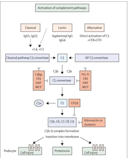
Although Heymann suggested that the immune deposits were due to an autoimmune production against the glomerular tissue, these deposits were long thought to result from glomerular trapping of circulating immune complexes in the blood. In 1978, nearly simultaneously, Couser and colleagues and Van Damm and colleagues showed that an intrinsic antigen present on the podocytes can serve as target for the circulating antibodies rather than the hypothesis of circulating antigen captured by the kidney (Couser et al., 1978; Van Damme et al., 1978). Further studies revealed in 1982 that megalin (gp330) (a membrane glycoprotein of the renal proximal tubular brush border) was the main pathogenic antigen in Heymann nephritis (HN) (Kerjaschki et al., 1982). Proteinuria was shown to be dependent on complement activation (C3, C6 and C5b–9) which in turn causes podocyte injury, cytoskeletal reorganization and loss of the slit diaphragm molecular structure (Cybulsky et al., 1986).
This animal model, now called the rat Heymann nephritis model of MN, provided further impetus to identify the autoantigens involved in the different forms of human MN.
However, while megalin was not identified as an autoantigen in human MN and despite intensified efforts, the identification of the human antigens remained elusive for more than 20 years. Indeed, it is only in 2002 that NEP was identified by Debiec and colleagues. as the first human MN antigen in a very rare alloimmune form of MN (Debiec et al., 2002). This was followed in 2009 by the identification of PLA2R1 as the major autoantigen for about 70% of patients with adult MN (Beck et al., 2009) and in 2014 by the identification of THSD7A as a second autoantigen for 2–5% of another group of patients with adult MN
(Tomas et al., 2014). The detailed description of these antigens is provided in chapter 1.3.1.2 of this manuscript.
1.2.5 Multiple clinical forms of MN
MN can be defined as primary (or idiopathic) in about 85% of all MN cases because there is no obvious cause of the disease (hence idiopathic). In the remaining 15% of other cases, MN can be defined as secondary to a "primary" disease affecting another organ, including various cancers, infections, other autoimmune diseases, etc. (see below for a full definition).
– Primary (idiopathic) MN and possible causes (etiologies)
The term idiopathic or primary is used when no obvious cause of MN can be determined after careful examination of the patient, analysis of patients’ history and after all laboratory tests regularly performed to assess glomerulonephritis and causes have remained negative (search for cancers, other autoimmune diseases, diabetes, etc.)
(Table 1.1).
The etiology of primary MN in general or when associated with anti–PLA2R1 or anti– THSD7A autoantibodies remains in fact largely unknown. Genetically, aside from some rare cases in which more than one member of the same family is affected, MN is not a hereditary transmitted disease (Bockenhauer et al., 2008; Scolari et al., 1998). A genetic polymorphism is detected in PLA2R1 and HLA–D class II MHC alleles and confers a high degree of susceptibility to idiopathic MN, up to 80–fold (Coenen et al., 2013; Stanescu et al.,
2011). However, the pathway behind this predisposition and loss of the self–tolerance to the
podocytic antigen is not yet elucidated.
As MN is highly prevalent in the elderly, a number of other diseases have been found to be associated with MN, without being able to demonstrate causality. It is thus unclear whether these diseases are simply co–incidental or at the origin of MN (hence providing a primary cause of secondary MN), or may constitute aggravating factors facilitating the progression of MN to severe cases. For example, MN has been associated with many infections by viruses such as HIV, HBV or HCV (Gupta et al., 2015; Xie et al., 2015), cancers (Cambier et al., 2012; Lefaucheur et al., 2006), autoimmune diseases (Thyroïdites, rhumatoïd arthritis, antiphospholipid antibodies, etc.) (Asami et al., 2016), other nephritis (Alport, IgA nephropathy, etc.) (Nishida et al., 2015), sarcoidosis (Stehlé et al., 2015) or syndromes like Sjogren's (Yoshida et al., 1996) or paraneoplastic ones.
Molecular mimicry has been proposed to be a trigger for anti–PLA2R1 autoimmune reactivity but this remains to be demonstrated (Fresquet et al., 2015). Indeed, Fresquet and colleagues has proposed that an immune response against a viral or bacterial agent may turn into an autoimmune response by cross–reactivity with a self–component, here PLA2R1. Using bioinformatic analysis against microbial protein databases, they identified a protein identity between the sequence LTLENCK in the N–terminal CysR domain of
PLA2R1 and the corresponding sequence in the D–alanyl–D alanine carboxypeptidase,
a bacterial cell wall enzyme common in several bacterial species including Clostridium species.
Recently, an increased incidence of MN has been observed in some regions of China which has been attributed to the environmental air pollution with PM2.5 particles (Xu et al., 2016). The molecular mechanisms linking pollution and MN have not yet been identified but interactions between the human genetics and the environment have been proposed
(Zhang et al., 2018). In this context, it is suggested that expression of PLA2R1 in the lungs can be upregulated in response to environmental pollution and airway damage. This could trigger an interaction with the antigen presenting cells in the inflamed lungs and lead to the autoimmune reaction against PLA2R1 expressed in the podocyte (Salant, 2019).
As for the THSD7A–associated MN, investigating the possible events that trigger the immune system is currently limited by the low prevalence of this disease, thus rendering
data collection and analysis even more problematic than for PLA2R1–associated MN. Nonetheless, a recent still unpublished study identified a small peptide in the N–terminal region of THSD7A that shares homology with the above immunodominant epitope peptide in PLA2R1. The homology modeling revealed two important areas for antibody binding suggesting that a pathogenic epitope structure may be present in both THSD7A and PLA2R1 and act as a common MN entry point (Rhoden et al., 2017). The above studies and different scenarios remain so far quite speculative. Analysis of large cohorts of THSD7A– associated and PLA2R1–associated MN patients with detailed natural history and serum samples available before the onset of MN disease are still needed to identify the real causes of primary MN and the nature of autoantibodies before worsening of the disease, as it was shown for Systemic Lupus Erythematosus or Pemphigus vulgaris (Arbuckle et al., 2003; Li et al., 2003). Furthermore, the above possible scenarios should be demonstrated by using animal models where such "nephritogenic" peptides or full antigens are injected and processed by the immune system to raise an autoimmune response and induces MN disease.
– Secondary MN
MN should be referred to as secondary MN when there is not only an association with another disease but when it is demonstrated that this latter disease is the underlying cause of MN, as evidenced by targeted therapy of the disease leading to both the cure of the disease and remission of MN. Secondary MN have been found to be "linked" to chronic viral diseases (Hepatitis B and C), cancers (Couser et al., 1974), medications (nonsteroidal anti–
inflammatory agents) and autoimmune diseases (systemic lupus erythematosus, ANCA etc.) (Table 1.1)(Larsen et al., 2013; Ronco et al., 2015).
As discussed above, the association between MN (with a primary unknown cause) and other diseases and the causal link between the two diseases (hence secondary) is complicated. Diagnosis of primary versus secondary MN is not always straightforward, thus histopathological features are used to define the form of MN (Huang et al., 2013; Larsen et al., 2013). In contrast to what is known for idiopathic MN, subendothelial and mesangial immune deposits are found in secondary MN. In the case of Lupus– or HIV– associated MN, tubulo–reticular inclusions in endothelial cells can also be detected. Furthermore, an
important difference between the two forms of MN resides in the immunoglobulin subclasses found in immune deposits. In primary MN, IgG4 is the main IgG subclass present in immune deposits, especially at late stages, while IgG1 and IgG3 subclasses can be occasionally detected at the early stages. Thus, primary MN is proposed to be an IgG4– mediated disease. In contrast, in secondary forms of MN, IgG1, IgG2 and IgG3 are predominant and a full house IgG deposition (A, M, G) as well as C1q can be detected in the different stages of the disease (Ma et al., 2013). A detailed description of the IgG subclass predominance can be found in chapter 1.3.1.3. of this manuscript.
– MN by maternal alloimmunization
Very rare cases of MN can affect newborn subjects who present with severe renal lesions and nephrotic range proteinuria, in addition to respiratory distress syndrome and hypertension. These cases are related to a rare alloimmune form of MN where the mother has bi–allelic mutations in the gene coding for NEP (neutral endopeptidase) and are natural knock–out for this gene. The fetus, having inherited a functional allele of the gene from the father, is heterozygous and expresses NEP during development. During the pregnancy (or following a previous miscarriage), the mother is immunized against NEP and develops antibodies which are then transferred through the placenta into the blood flow of the fetus and reach the embryonic kidneys. Thirteen days after birth, the antibody titer decreases in the neonate’s circulation and the disease enters into remission, confirming the passive transfer of antibodies from the mother to the fetus, similarly to what occurs in the passive Heymann nephritis model (Debiec et al., 2002; Vivarelli et al., 2015).
Interestingly, in these cases of alloimmune MN, two distinct scenarios were documented: in the first scenario where the main IgG subclass is IgG1, a direct effect of anti–NEP antibodies has been shown on NEP enzymatic activity as well as a pathogenic role due to complement activation. In the second scenario, antibodies were mainly of IgG4 subclass with slight amount of IgG1. Here, the complement pathway was not activated and the IgG4 antibodies had weak inhibitory effect on the NEP enzymatic activity.
Table 1.1 – Classification of the different MN forms and their characteristics. (*) percentage of prevalence calculated based on data in Larsen et al. 2013.
MN Human forms Antigen (Mass) Prevalence (%) Targeted population Sex ratio Male/female Antibody subclass Role of complement Pathogenicity of antibodies (based on active immunization or
passive transfer)
References
Primary 85
(of all cases) (Beck, 2017; Ponticelli et al., 2014)
PLA2R1-associated MN PLA2R1 (180 kDa) 70 (of primary cases) Mostly adults, (age ≥50 years) 2:1 M:F IgG4>IgG1, IgG3 Likely (C3 and C5b-9 in immune deposits)
Yes, passive transfer based on MN relapse of a human kidney transplanted in a
PLA2R1-ab positive patient No available experiments in rodents (endogenous PLA2R1 not expressed in
mouse and rat podocytes)
(Beck et al., 2009; Hofstra et al., 2012; Segawa et al., 2010; Yang et al., 2016)
THSD7A-associated MN THSD7A (250 kDa) 3 (of primary cases) Mostly adults, 1.3:1 M:F IgG4>IgG1 >IgG3 Likely
Yes, passive transfer based on MN relapse of a human kidney transplanted in a
THSD7A-ab positive patient see also mouse model below
(Tomas et al., 2014; Tomas et al., 2016; Wang et al., 2018) Double negative unknown 27 Mostly adults, (age ≥50 years) 2:1 M:F IgG4>IgG1
>IgG3 Likely Yes, likely (Hoxha et al., 2015)
Secondary 15
(of all cases) IgG, IgA, IgM
(Beck, 2010; Glassock, 1992; Jefferson et al., 2003) Childhood cBSA cationic BSA (66 kDa) <1 (of secondary cases) Children <5
years IgG1, IgG4 Likely
Yes, immunization of dogs, mice, rats and rabbits with cBSA. IgG deposits and complement activation in immunized
rabbits
(Debiec et al., 2011; Ronco et al., 2015)
Hepatitis B (other infections)
? 14* Children/
adults IgG1>IgG4 Likely not tested (Larsen et al., 2013)
Sarcoidosis ? 5* Children/
adults IgG1>IgG4 Likely not tested (Larsen et al., 2013)
Neoplasms ? 15* Children/
adults IgG1>IgG4 Likely not tested (Larsen et al., 2013)
Other autoimmune diseases (SLE, thyroiditis, etc.) multiple 58* Children/
adults IgG1>IgG4 Likely not tested (Larsen et al., 2013)
Alloimmune NEP <1% Neonates IgG1>IgG4 Yes Yes, passive transfer
from mothers' anti-NEP antibodies
(Debiec et al., 2002; Vivarelli et al., 2015)
MN animal models Antigen (Mass) Prevalence (%) Targeted population Sex ratio Male/female Antibody subclass Role of complement Pathogenicity of antibodies (based on active immunization or passive transfer) References Rat Heymann Nephritis Megalin
(LRP2) NA Rodents rat IgG
Yes complement
activation
Yes, active model of Fx1A to rats and passive transfer of anti-Fx1A
in rabbits
(Feenstra et al., 1975; Heymann et al., 1959; Kerjaschki et al., 1997; Salant et
al., 1988) Heymann's mouse model mouse kidney extract
NA Mouse mouse IgG Yes Yes, active model (Meyer-Schwesinger et al., 2011)
NC3
collagen NA Mouse mouse IgG Yes Yes, passive model (Zhang et al., 2012)
THSD7A mouse model THSD7A NA Mouse (Balb/C) Human total IgG Rabbit IgG
Yes Yes, passive transfer of human or
1.2.6 Genetics of MN
Long before PLA2R1 was identified as the major autoantigen in MN, the association of MHC class II and HLA–DQA1 with primary MN has been documented (Klouda et al., 1979; Vaughan et al., 1989). These findings are in accordance with the known risk of autoimmune disease linked to HLA–DQ and HLA–DR (Stanescu et al., 2011) (Chapter 1.1).
More recently, a genome–wide association study (GWAS) in 556 patients from three different Caucasian populations of idiopathic MN identified 2 loci associated with high risk of idiopathic MN (Stanescu et al., 2011). On the chromosome 2q24 containing the gene coding for PLA2R1 (Ancian et al., 1995), the SNP (Single Nucleotide Polymorphisms) rs4664308 was shown to be the target of an autoimmune response. Also, on the chromosome 6p21 containing the gene coding for the HLA–DQA1, the SNP rs2187668 was significantly associated with MN. Interestingly, the 4–fold increase in risk with genetic polymorphism in PLA2R1 augments to 80–fold when combined with homozygosity for HLA–DQA1. The explanation for this increase is still unknown. It is noteworthy that the SNP rs2187668 was strongly associated with MN in Caucasian patients but not in Afro–American patients
(Saeed et al., 2014). A sequencing analysis of PLA2R1 in a cohort of 95 patients with either circulating anti–PLA2R1 or PLA2R1 antigen staining detected in kidney deposits led to the identification of 18 SNP variants in PLA2R1, of which 2 were not described previously, 7 were rare variants (<1%) and 9 were common variants. They confirmed 6 of the common polymorphisms as significantly associated with MN (Coenen et al., 2013).
In Han Chinese patients, a GWAS in 261 patients with idiopathic MN identified DRB1*15:01 and DRB1*03:01 as two independent risk alleles for MN (Cui et al., 2017). Another study also showed that HLA–DRB1*15:01 and HLA–DRB3*02:02 alleles are independently and strongly associated with MN in general and with PLA2R1–associated MN in the Chinese population (Le et al., 2017).
1.2.7 Natural history of MN
Understanding the natural history of MN is important to determine if treatment of the disease is appropriate or useful (Glassock, 2010). In 1993, Schieppati et al. studied the natural history of 100 patients with biopsy–proven idiopathic MN diagnosed between 1974
and 1992 and followed for a minimum of 6 months (Schieppati et al., 1993). The patients only received symptomatic treatment consisting of diuretic and antihypertensive drugs. The study showed that a significant fraction of MN patients receiving only symptomatic treatment seem to have a benign course.
After a five year follow up period, 65% of the patients reached partial or complete remission of proteinuria while 16% developed ESRD and required dialysis. The probability of maintaining normal renal function was 88% after 5 years and 73% after 8 years. Additionally, they showed that patients younger than 50 and females had a better prognosis (62% of females and 59% of men had complete or partial remission).
In a more well–defined cohort of 328 patients with only nephrotic syndrome from Spain
(Glassock, 2010; Polanco et al., 2010), 32% of patients reached spontaneous remission and most of the remissions occurred in the first two years of follow–up. The study also showed that a progressive decrease of proteinuria by at least 50% during the first year was an indicator of spontaneous remission. Female gender, low serum creatinine and treatment with conservative treatments also tended to associate with remission.
These observations showed that initiating treatment upon diagnosis is not essential except in cases of severe nephrotic syndrome and that it is important to identify new biomarkers of disease severity to identify the patients who are going to develop renal failure and who urgently require the use of immunosuppressive therapy. Some of these observations form the basis for the recommendations found in the KDIGO guidelines for membranous nephropathy, as further developed below (KDIGO Clinical Practice Guidelines)
1.2.8 Clinical features of MN
Clinically, 80% of patients with MN suffer from nephrotic syndrome, i.e. with proteinuria >3.5g/day. It is only in 41% of cases that edema is observed, meaning that MN can manifest in patients without any sign of disease. Hypertension is detected in 55% of cases (Schieppati et al., 1993) and venous thromboembolism occurs in 7% of MN patients, more frequently in the first two years after diagnosis (Lionaki et al., 2012). As mentioned earlier, signs of MN include: proteinuria, hypoalbuminemia, hyperlipidemia, and edema
(Ma et al., 2013). The outcome of the disease varies from spontaneous remission to end stage renal disease (ESRD). Spontaneous remission occurs in 40% of cases within the first two years of presentation. This is usually linked to low proteinuria levels, female sex and age below 50 years. The remaining patients can be divided into those with persistent proteinuria fluctuating between nephrotic and sub–nephrotic ranges or those who reach ESRD at a median age of 5–10 years after diagnosis (Ponticelli et al., 2014; Ronco et al., 2017). In patients receiving renal transplantation, MN recurrence occurred in 42% and 35% before and after the identification of PLA2R1 and its value as a biomarker of disease activity respectively (Dabade et al., 2008; Quintana et al., 2015).
1.2.9 Treatment of MN
Before the identification of the autoantigens in MN, a series of treatment strategies have been used with different drugs aimed at non–specifically suppressing the immune system (Ruggenenti et al., 2007). Steroids were the first drugs used in the treatment of MN
(syndrome, 1979) but later showed poor efficiency as it is not superior to symptomatic treatment alone (Radhakrishnan et al., 2012). More powerful treatments including alkylating agents and steroids were shown effective (Ponticelli et al., 1998) however the cytotoxic effect of these agents discouraged nephrologists to use these treatments
(Ruggenenti et al., 2007). Furthermore, calcineurin inhibitors showed beneficial signs in the induction of remission. However, the necessity for long treatment periods raised concerns for nephrotoxicity (Cattran et al., 2001). Among other immunosuppressive treatments, Rituximab showed encouraging results in the treatment of MN as early as 2001 and is now one of the most commonly used immunosuppressors in MN (Remuzzi et al., 2002; Ruggenenti et al., 2015). MN patients at high risk achieve remission and have improved renal function after treatment with rituximab. Furthermore, treatment with Rituximab in complement with the antiproteinuric treatment did not affect safety in the randomized controlled trial of GEMRITUX (Dahan et al., 2017).
Cattran and colleagues. validated a predictive, semi–quantitative algorithm model of idiopathic MN (Cattran et al., 1997). They showed that proteinuria and creatinemia during the first six months after diagnosis was important to predict the outcome of the disease. This model categorized patients based on proteinuria and kidney function: low risk patients
(normal renal function and proteinuria <4 g/day), medium risk patients (normal renal function and proteinuria >4 g/day and <8g/day) and high–risk patients (with or without renal insufficiency and proteinuria >8 g/day).
Currently, the evaluation and treatment of MN patients follow the Kidney Disease– Improving global outcomes (KDIGO) guidelines established in 2012 (Radhakrishnan et al., 2012). It recommends clinicians to initiate a symptomatic treatment using anti–
hypertensive drugs such as angiotensin–converting enzyme (ACE) inhibitors or angiotensin receptor blockers for an initial 6 months period. The initiation of immunosuppressive treatment in patients with nephrotic syndrome is recommended when at least one of the listed conditions is fulfilled:
– Proteinuria superior to 4g/day and remains at over 50% of the baseline value, and does not decrease progressively after the initiation of the conservative treatment of at least 6 months;
– The presence of severe or life–threatening symptoms related to the nephrotic syndrome;
– A 30% or more increase in serum creatinine levels within 6 to 12 months from the time of diagnosis with an estimated glomerular filtration rate (eGFR) not lower than 25– 30 ml/min per 1.73m2 and not explained by any superimposed complication.
1.3 Physiopathology of MN
1.3.1 Immunoregulation and inflammation in MN
The initiation of an autoimmune response can be the result of a loss of tolerance at the central or peripheral level due to the modification in expression or conformation of the autoantigen (including post–translational modifications generating neoepitopes on the self– protein) or to epitope molecular mimicry. In MN, the following evidence support a role of various immune cells in raising the autoimmune response, with a possible role for a loss of peripheral tolerance. The increase in Th2 cytokines stimulates B lymphocytes to produce IgG4 antibodies targeting the specific antigens involved in MN (Hirayama et al., 2002; Kuroki et al., 2005). Detection of circulating autoantibodies secreted by plasma cells arising from B lymphocytes indicates that the B lymphocytes have a major role in the pathogenesis
of the disease (Wang et al., 2011). Cui et al. used predictive algorithms to predict T–cell
epitopes within the CTLD1 and CTLD7 domains of PLA2R1, potentially linking the T–cell and B–cell epitopes of PLA2R1 (Cui et al., 2017). Besides the B–T cell education system to
produce antibodies, other immune cells like Tregs play a fundamental role in the control of tolerance (Gratz et al., 2014; Sakaguchi et al., 2010). The imbalance between the self–
specific effector T cells that attack tissues and the Treg cells that normally controls these latters leads to autoimmune diseases. Recent studies have shown that patients with active MN have low levels of Treg cells (Roccatello et al., 2016; Rosenzwajg et al., 2017). A successful treatment with Rituximab leads to depletion of B cells and a concomitant increase of Treg cells before clinical remission of proteinuria. Inversely, the percentage of Tregs remains low in patients not responding to Rituximab or receiving only a symptomatic treatment.
Very little is known about the inflammatory cell infiltration in the glomeruli of patients with MN. B cells infiltrates have been described in membranous nephropathy mostly in the tubulointerstitium part of the nephron but not in the glomeruli (Segerer et al., 2008). These infiltrated B cells along with T cells and dendritic cells form tertiary lymphoid organs in the inflamed tissue with a complex cell organization similar to that of the secondary lymphoid organs, suggesting a sustained local production of autoantibodies possibly associated with chronic inflammation. On the other hand, cell infiltration in the glomeruli was shown to be two–fold higher in cancer–associated MN as compared to idiopathic MN patients
(Lefaucheur et al., 2006). Within the glomeruli, the mesangial cells constitute a particular cell type that strongly respond to the local inflammation and may participate to MN physiopathology. In vitro cultured rat mesangial cells have been shown to express PLA2R1 and release secreted PLA2–IIA in response to TNF–alpha or other inflammatory stimuli
(Beck et al., 2003). In vivo, in the rat Thy–1 glomerulonephritis model, an overexpression of
PLA2R1 and sPLA2s has been observed in the glomeruli, together with invasion by immune cells (Beck et al., 2006). However, the role of mesangial cells in local inflammation possibly leading to accelerated podocyte injury, as well as the expression and role of PLA2R1 and sPLA2s in these cells, all in the context of the Heymann rat nephritis model and of human MN, is simply unknown. Finally, in line with the role of PLA2R1 in cellular senescence (Augert et al., 2009; Vindrieux et al., 2013), it is interesting to note the increased staining of the senescence marker p16(INK4A) in the kidney biopsy of patients
with glomeulonephritis, suggesting a role for somatic cellular senescence in the progression of the disease (Sis et al., 2007).
1.3.2 Immune complexes
In most cases of MN, i.e. idiopathic MN, immune deposits consist of immune complexes between podocyte proteins and IgG autoantibodies and various factors from the complement pathway. In this section, we will introduce the different hypotheses explaining the formation of these deposits in addition to a thorough review of the different antigens and IgG subclasses involved.
1.3.2.1 Mechanisms of deposition of immune complexes
Back to the initial studies, Heymann proposed that the deposition of immune complexes in the active glomerulonephritis rat model resulted from binding of free circulating autoantibodies to a "fixed antigen" located in situ in the glomerulus and not from a soluble antigen already present in serum and pre–forming circulating immune complexes (Okuda et al., 1965; SP. Makker, 2011). The concept of circulating immune complexes as a source of the glomerular deposits proposed by Germuth and Dixon et al. was adapted to explain the granular deposits in active Heymann nephritis model (Dixon et al., 1961; Germuth et al., 1955). Heymann’s hypothesis was experimentally proven later by Van Damme, Couser and
colleagues. Extending the rat Heymann passive model, they demonstrated by ex vivo bloodless kidney perfusion that injection of autoantibodies free of antigen rapidly formed in situ immune complexes with a fixed podocyte antigen thus supporting the "fixed antigen" model (Figure 1.2) (Couser et al., 1978; Van Damme et al., 1978). In rats, the antigenic target called “megalin” is highly expressed at the surface of the proximal tubular cells and also at the sole foot processes of the podocytes (Kerjaschki et al., 1983). Upon binding of the IgG to megalin at the surface of the podocyte membrane, the membrane–bound antigen would undergo shedding by proteolysis (Chapter 5.3.2.) and subsequent cross–linking with
circulating antibodies and immobilization in the GBM, thus escaping clearance by endocytosis. Repeated cycles of this mechanism would lead to large immune deposits
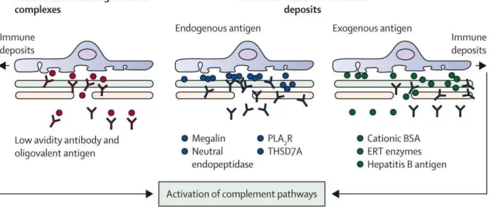
Figure 1.2 – Possible scenarios for immune complex formation and deposition in MN. Three possible scenarios would occur to explain the formation and accumulation of the immune complexes. On the left, pre–formed circulating immune complexes would accumulate and form deposits in the GBM. In the center, endogenous proteins expressed on the podocyte (Megalin, NEP, PLA2R1, THSD7A) can be directly targeted by circulating autoantibodies. On the right, circulating antigens can be first "planted" in the GBM and this is followed by the in–situ binding of circulating free antibodies. This is the case for cBSA, enzyme replacement therapy or the hepatitis B antigen. The accumulated antibodies would then activate the complement pathways (classical, alternative or lectin pathway) and induce podocyte injury and proteinuria. Adapted from (Ronco et al., 2015) with copyright permission from “Elsevier”.
Nowadays, three potential scenarios have been proposed for subepithelial immune complex formation in the various forms of primary and secondary MN (Figure 1.2) (Glassock, 2009; Ronco et al., 2015). In the first scenario that follows the Heymann hypothesis, an “intrinsic antigen” is endogenously expressed at the foot processes of the podocytes and serve as a target for circulating antibodies to form immune complex deposition in situ. This scenario applies for the four identified integral membrane glycoproteins of podocytes including megalin in the Heymann nephritis model (Heymann et al., 1959), NEP in neonatal MN (Debiec et al., 2002), as well as PLA2R1 (Beck et al., 2009)
and THSD7A (Tomas et al., 2014) in adult MN. The second scenario corresponds to a “planted antigen” extrinsic to the glomerulus (Figure 1.2). The autoantigen, from a variety of sources, leaves the circulation and gets trapped in the subepithelial space. This is the case for cationic bovine serum albumin (cBSA) in secondary MN (Debiec et al., 2011). Deposition of circulating cBSA along the anionic glomerular capillary wall is followed by anti–cBSA IgG binding and leads to immune complex formation in situ. The third scenario involves the deposition of pre–formed “circulating immune complexes” (Figure 1.2). This mechanism was only documented in rare cases of MN secondary to infection by the hepatitis B virus (HBV). HBV antigens were detected free or IgG–associated in the serum and the glomerular capillary walls of two MN patients (Takekoshi et al., 1979). However, this scenario was never confirmed in corresponding animal models of MN. It should be pointed out that the above scenarios are not exclusive one to another, and they may "co– operate" to form immune deposits.
1.3.2.2 The antigens in MN
Membranous nephropathy is a remarkable autoimmune disease in which an experimental animal model, the Heymann's model, was established much before we knew the antigens and the underlying molecular mechanisms responsible for the various forms of human MN. Indeed, the molecular pathogenesis of the Heymann's model was established long before the identification of any human antigens, and actually most of what we know today about human MN was in fact established in the Heymann's model and could be predictable from this model.
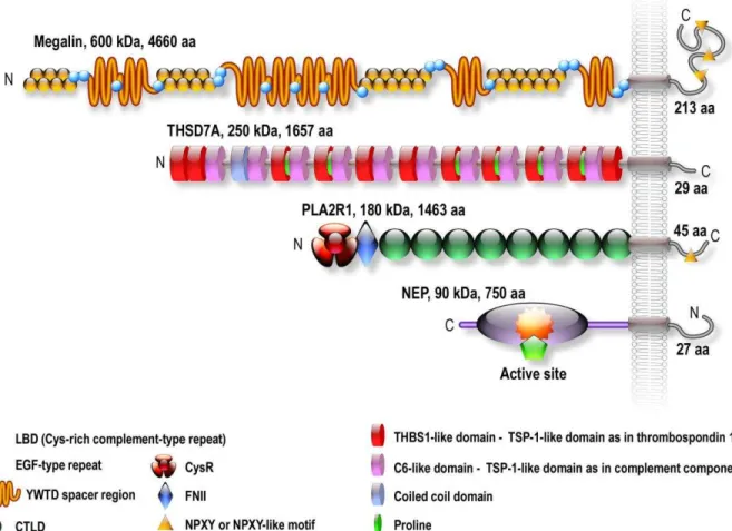
Walter Heymann and his colleagues established the first experimental model of MN in the late 50s, and it was about 20 years later that megalin (gp330 or Lrp2) was identified as the main nephritogenic antigen in rat podocytes (Figure 1.3) (Heymann et al., 1959; Kerjaschki et al., 1982). Unfortunately, translation of the Heymann nephritis model to the human physiopathology was not possible as it turned out that megalin is not the autoantigen in human MN. Furthermore, megalin is mostly located in the proximal brush border in humans and was recently found to be involved in another rare autoimmune kidney disease called "Anti–Brush Border Antibody" (ABBA) disease (Larsen et al., 2018).
It was in 2002, 20 years after the identification of megalin in the rat model, that the seminal work of Debiec and colleagues. identified NEP as the first human antigen in the rare alloimmune form of MN (Figure 1.3) (Debiec et al., 2002). However, NEP was not the antigen for the more frequent form of "sporadic" idiopathic adult MN and it was only in 2009, exactly 50 years after the Heymann rat model was established, that PLA2R1 was identified as the major autoantigen in adult MN for about 70–80% of MN patients
(Figure 1.3) (Beck et al., 2009). THSD7A was then identified as a second autoantigen for another group of patients of about 3% (Figure 1.3) (Tomas et al., 2014). Together, the antigens and autoantigens have now been identified for about 80% of MN patients. However, we still miss the autoantigen(s) for the remaining group of about 20% of patients with adult MN, who are "double negative" for PLA2R1 and THSD7A, and these patients are thus still "orphans" for the antigenic target of their MN disease.
– Megalin in Heymann Nephritis
In the early 80s, megalin was identified as the principal pathogenic antigen in Heymann nephritis (Kerjaschki et al., 1982). Also known as gp600, gp330 or LRP2, megalin is a giant type 1 transmembrane glycoprotein of 600 kDa that belongs to the low–density lipoprotein receptor superfamily (LDLR) (Figure 1.3). In rat kidneys, megalin is located in large amounts in the renal proximal tubule but also in the podocytes (Kerjaschki et al., 1983). In human kidneys, megalin is highly expressed in the proximal tubules, and in fact lower levels in podocytes (Prabakaran et al., 2011).
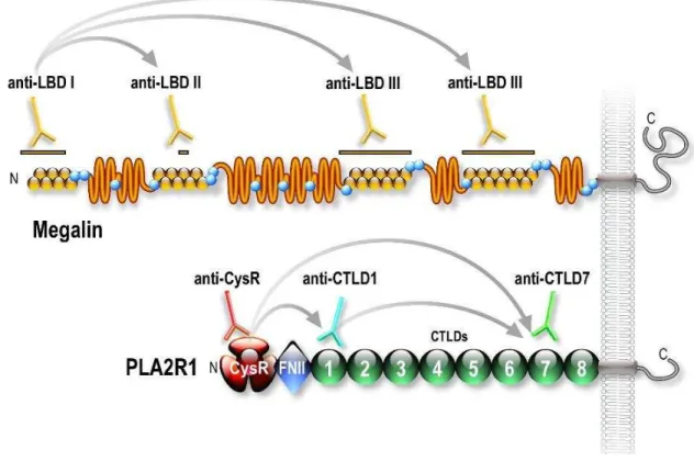
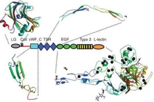
Figure 1.3 – Schematic structures of the four antigens implicated in MN. Megalin is the antigen of the Heymann nephritis model. PLA2R1 and THSD7A are the antigens in 70– 80% and 3% of patients with idiopathic MN, respectively. NEP is the antigen responsible for a rare alloimmune form of MN. The antigens are ranked by size, megalin being the largest antigen with a molecular mass of 600 kDa, followed by THSD7A, 250 kDa, PLA2R1 180 kDa and NEP, 90 kDa. Megalin contains four ligand binding domains (LBD) with each domain containing one ligand binding repeat flanked with epidermal growth factor–type repeats. Megalin also contains 3 NPXY internalization motifs. THSD7A is formed by an alternation of TSP–1 repeat and C6–like repeats with a short intracellular tail but no obvious internalization signal. PLA2R1 is composed of a cysteine–rich domain followed by a type–II fibronectin domain and eight C–type lectin domains (CTLDs) and a cytoplasmic tail with an NPXY internalization motif.
Although the investigation of the Heyman nephritis model contributed greatly to our understanding of the pathophysiology of human MN, it was limited by the fact that megalin was not the main autoantigen in patients’ immune deposits (Table 1.1).
– The neutral endopeptidase (NEP)
The neutral endopeptidase (NEP) is the first human podocytic antigen identified in a rare form of alloimmune–associated MN in the neonates. NEP (also known as Neprilysin or Enkephalinase or CD10) is a transmembrane zinc–containing metalloproteinase of 90– 110 kDa (Figure 1.3). It is expressed in the kidney in podocytes, in the brush borders of the proximal tubules and in the vascular walls of the kidney. It is also expressed in various tissues and cells including brain, smooth muscle cells, cardiomyocytes and neutrophils. In the brain, NEP degrades the β–amyloid peptide involved in the Alzheimer’s disease. NEP also degrades many peptides such as substance P, glucagon and oxytocin. In the kidneys, NEP cleaves angiotensin II, bradykinin and natriuretic peptides, thereby participating in the regulation of blood circulation in nephrons (P. Judge, 2015).
In neonate alloimmune MN, the mother has a homozygous null mutation in the MME (Metallomembrane Endopeptidase) gene while the father has a functional gene. During pregnancy or following a previous miscarriage, the mothers’ immune system is exposed to the NEP expressed by the fetus and initiates a typical immune reaction against NEP. The maternal IgGs (mainly of IgG1 or IgG4 subclass) cross the placenta, bind to fetal glomerular podocytes expressing NEP and cause antenatal renal disease (Table 1.1). Kidney biopsy staining of the infant shows an unusual form of glomerulonephritis with distended Bowman’s spaces and collapsed capillary tufts.
It is important to mention that although the life span of the maternal IgG is short, the antenatal nephron loss may lead to chronic renal failure in the adult life (Debiec et al., 2002; Debiec et al., 2004; Vivarelli et al., 2015). These findings also demonstrate that a podocyte antigen can be at the origin of formation of immune deposits in human MN (following the above scenario for in situ antigen). However, alloimmune cases of MN are very rare, and NEP is not the autoantigen in adult primary MN.
– The phospholipase A2 receptor 1 (PLA2R1)
Secreted phospholipases A2 are abundant in snake and bee venoms and are responsible for many toxicities towards preys (Gutiérrez et al., 2013). This includes neurotoxic, myotoxic, pro–inflammatory and cardiovascular toxic effects. The molecular mechanisms involved in these toxic effects are multiple and were proposed to be due to either the complex enzymatic activity of venom sPLA2s or to direct binding to putative soluble and membrane–bound proteins (Kini et al., 1989).
In the early 90's, two novel receptors for sPLA2s were discovered in the team of Dr. Lambeau. A first type of sPLA2 receptors of around 40–50 kDa was identified in rat brain neurons (hence called N–Type) and binds neurotoxic sPLA2s (Lambeau et al., 1989). This N–type receptor has not been cloned and thus its structure, endogenous ligands and function are still unclear. Several brain proteins have been identified by others since then but their relationship to N–type receptors is unclear (Šribar et al., 2014). A second sPLA2 receptor of 180 kDa was discovered in rabbit skeletal muscle cells in 1990 and called the M –type phospholipase A2 receptor (now called PLA2R1) (Lambeau et al., 1990). The M –
type receptor was purified, partially sequenced and finally cloned from rabbit and human species in our laboratory (Ancian et al., 1995; Lambeau et al., 1994) and also from mouse and bovine species by Shionogi Pharmaceuticals in Japan (Higashino et al., 1994; Ishizaki et al., 1994).
Molecular cloning has revealed that PLA2R1 belongs to the C–type lectin superfamily and share a number of structural properties with 3 other proteins from the mannose receptor lectin subgroup: the macrophage mannose receptor (MRC1), Endo180 (MRC2) and DEC205 (LY75) (East et al., 2002). PLA2R1 is a large type I transmembrane glycoprotein of 180 kDa and consists of a large extracellular domain, a single transmembrane segment and a short cytoplasmic tail (Figure 1.3).
The extracellular domain of PLA2R1 consists of an N–terminal Cysteine–rich (CysR) domain, a fibronectin type II domain (FNII) and 8 different C–type Lectin (CTLD) domains in tandem (Ancian et al., 1995; Ancian et al., 1995; Lambeau et al., 1994).
Human PLA2R1 was cloned from kidney tissue (Ancian et al., 1995), but it was not known that the protein is specifically expressed in the podocytes until it was identified as the target autoantigen in MN. However, PLA2R1 is also expressed in proximal tubules and
overall (Vindrieux et al., 2014), its relative and precise cellular distribution along the nephron and in the different sections of the kidney is still incomplete. Depending on the species, PLA2R1 is also expressed by immune tissues and cells such as spleen, macrophages and type II alveolar cells in the lungs, but also liver, pancreas, small intestine and colon including Paneth cells (Cupillard et al., 1999; Schewe et al., 2016), thyroid, testis, ovaries and placenta (Ancian et al., 1995; Granata et al., 2005; Higashino et al., 1994). Of note, large differences in the expression of PLA2R1 exist among species, and PLA2R1 is not expressed in mouse and rat kidney podocytes (Meyer-Schwesinger et al., 2015).
The biological roles of PLA2R1 are not well understood (Girard et al., 2014; Lambeau et al., 1999). PLA2R1 is likely a multifunctional and multiligand receptor. PLA2R1 has been shown to bind and inhibit, as well as quickly internalize and degrade multiple sPLA2s, and acts as an endogenous receptor for several sPLA2s (Ancian et al., 1995; Ancian et al., 1995; Cupillard et al., 1999; Lambeau et al., 1995; Nicolas et al., 1995; Rouault et al., 2007; Zvaritch et al., 1996). In line with these sPLA2 binding properties, it may play a role in controlling sPLA2 enzymatic activity in various inflammatory conditions like sepsis and asthma (Hanasaki et al., 1997; Nolin et al., 2016). PLA2R1 binds collagen, suggesting a role in cell adhesion (Ancian et al., 1995; Skoberne et al., 2014; Takahashi et al., 2015) and different types of sugars, indicating some functions as a lectin (Ancian et al., 1995; Lambeau et al., 1994). Finally, PLA2R1 has been proposed to function as a tumor suppressor gene by initiating replicative or oncogene–induced senescence through the p53 pathway or other signaling pathways (Augert et al., 2013; Vindrieux et al., 2013; Vindrieux et al., 2014).
After half a century of extensive investigations to discover the human autoantigens of idiopathic MN, a collaboration between the team of David Salant and Laurence Beck in Boston and our team has finally led to the identification of PLA2R1 as the major autoantigen in adult MN (Table 1.1) (Beck et al., 2009). It all started when Dr. Laurence Beck was testing the reactivity of serum from MN patients by western blot using crude extracts of human glomerular proteins. It is only under non–reducing that a strong and specific reactivity was found against a protein of about 180 kDa. From 37 patients with idiopathic MN, 27 (70%) recognized the 180 kDa band.
In contrast, serum from control donors or secondary MN failed to react with this protein. The antigen was highly glycosylated since treatment with N–glycosidase F caused