HAL Id: tel-01623408
https://hal.archives-ouvertes.fr/tel-01623408
Submitted on 30 Oct 2017
HAL is a multi-disciplinary open access archive for the deposit and dissemination of sci-entific research documents, whether they are pub-lished or not. The documents may come from teaching and research institutions in France or abroad, or from public or private research centers.
L’archive ouverte pluridisciplinaire HAL, est destinée au dépôt et à la diffusion de documents scientifiques de niveau recherche, publiés ou non, émanant des établissements d’enseignement et de recherche français ou étrangers, des laboratoires publics ou privés.
in live cells -a quantitative FRET-FLIM approach
Cornelia Ziegler
To cite this version:
Cornelia Ziegler. Imaging the assembly of the phagocyte NADPH oxidase in live cells -a quantitative FRET-FLIM approach. Biophysics. Université Paris Saclay, 2016. English. �tel-01623408�
T
HESE DE DOCTORAT
DE
L’U
NIVERSITE
P
ARIS
-S
ACLAY
PREPAREE A
L
’U
NIVERSITE
P
ARIS
S
UD
E
COLED
OCTORALE N°571
Sciences chimiques : molécules, matériaux, instrumentation et biosystèmes
Spécialité de doctorat : Chimie
Cornelia Susanne Ziegler
Imaging the assembly of the phagocyte NADPH oxidase in live cells
- a quantitative FRET-FLIM approach
Thèse présentée et soutenue à Orsay, le 14 mars 2016 :
Maitre de Conférences, UPMC
Maitre de Conférences, Université Joseph Fourier Professeur, Université Paris Diderot
Professeur, UPSud
Maitre de Conférences, UPSud
Composition du Jury :
Mme Bonneau Stéphanie Mme Paclet Marie Hélène Mr Ostuni Mariano Mr Van Tilbeurgh Herman Mme Erard Marie
Mr Nüße Oliver Professeur, UPSud
Rapporteur Rapporteur Examinateur Président du jury Directeur de thèse Co-directeur de thèse
Foremost, I would like to express my sincere gratitude to Mehran Mostavafi, the former head of the LCP, to give me the opportunity to make my PhD in his laboratory. In the same context, I would like to thank all members of the laboratory and the administration for their ongoing support during my stay.
I am deeply grateful to my thesis committee, especially the rapporteurs Stéphanie Bonneau and Marie-Hélène Paclet, as well as the examinateurs Mariano Ostuni, and Herman Van Tilbeurgh to accept my thesis.
I would also like thank Marc Tramier (IGDR, Rennes) for his collaboration to give me access to his laboratory and research facilities. Likewise, I would like to thank Dominique Durand (Université Paris Sud) for kindly and straightforward providing me the structure files which were indispensable for the structural modelling.
My sincere thanks also go to Fabienne Mérola, who gave me the chance to join her group and furthermore for the stimulating discussions and scientific input to my work.
Special thanks goes to my director Marie Erard for her continuous support in both scientific and private life, for the immense scientific input to my project, the advice, and patient explanations. She contributed greatly to the successful and pleasant time of my PhD project. Likewise I would like to thank my co-director Oliver Nüße most sincerely for his guidance and strong scientific contribution to this project, the helpful discussions and his patience.
I am as well thankful to Sophié Dupré for her gentle advice and ongoing help during my project, as well as to Hélène Pasquier for her support. Special thanks goes to Yasmina Bousmah, her never-ending help for my practical lab work and for maintenance of the lab.
I would also like to thank warmly my fellow PhD students, namely Leila Bouchab for introducing me to the practical flow cytometer work and for her friendship, Myriam Moussaoui for her cheerfulness, with which she delighted the weekends and nights we spent together in the lab during the writing of our thesis, Ana-Laura Luna-Barron for the very good time we spent together and for her motivation, Damien Clavel for the rare but great evenings without ever talking about our work, Dahdjim Betolngar for facilitating my start in France, and finally Zhimin Song for his cheerfulness.
It is my personal intention to thank my husband Sven Böckmann for his love and his immense support in every respect. There are too many things to say. Without him in my back, this project would not have been possible. Likewise, I would like to thank my son Julian Erasmus to light up my days with his smile and to master the hard time of my writing with the help of his daddy. Last but not least I would like to thank Antony Campbell, University of Cardiff, Wales, advisor of my master thesis, to instil the self-confidence and motivation in me to do a PhD and for his ongoing support.
Contents
1 Introduction ... 5
1.1 Quantitative fluorescence imaging of protein-protein interactions in the NADPH oxidase complex ... 6
1.2 Aim of the thesis ... 8
1.3 Fluorescence and Förster Resonance Energy Transfer ... 9
1.3.1 Fluorescence – the physical background ... 9
1.3.2 Förster resonance energy transfer... 11
1.4 NADPH Oxidase ... 15
1.4.1 The phagocyte NADPH oxidase ... 17
1.4.1.1 Resting state, activation, and deactivation ... 18
1.4.1.2 Structure of the NADPH Oxidase ... 20
2 Material and Methods ... 30
2.1 Buffers ... 30
2.2 Methods ... 30
2.2.1 Bacterial cell culture ... 30
2.2.2 Plasmid preparation ... 31
2.2.3 Plasmid cloning ... 32
2.2.4 Production and purification of recombinant proteins ... 32
2.2.5 Eukaryotic cell culture ... 34
2.2.6 Transient transfection of COS cells ... 34
2.2.7 Western Blot ... 35
2.2.8 Luminescence Assay ... 38
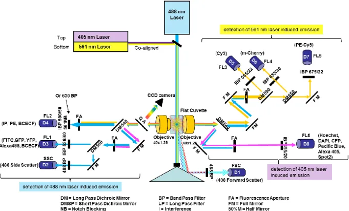
2.2.9 Flow Cytometry ... 40
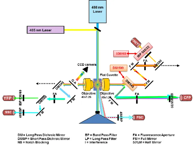
2.2.10 Microscopy ... 46
2.2.10.1 Total Internal Reflection Fluorescence (TIRF) Microscopy ... 46
2.2.10.2 FRET-FLIM ... 47
2.2.11 Fluorescence cross correlation spectroscopy ... 51
2.2.11.1 FCCS setup ... 55
2.2.12 Data acquisition and analysis with SymPhoTime software ... 56
2.2.13 Molecular structure visualisation and alignment ... 56
2.2.14 Statistical analysis ... 56
3 Results – methodology development for FRET quantification ... 57
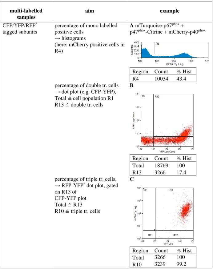
3.1 Flow Cytometry ... 57
3.1.1 Elucidating the transfection efficiency by flow cytometry ... 57
3.1.2.1 Optimization of the cytometer for FRET demands ... 60
3.1.2.2 Developing a FRET analysis method ... 62
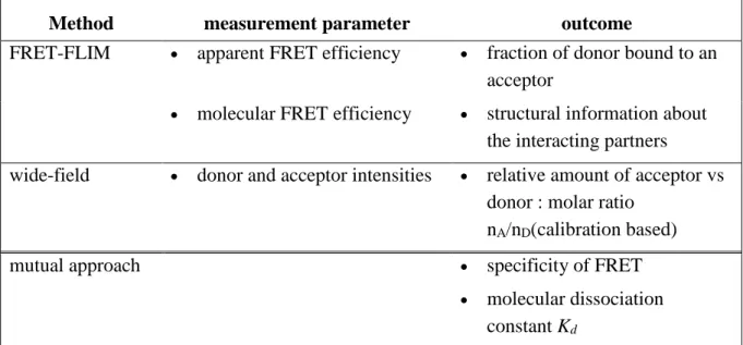
3.2 Quantification of protein-protein interactions in live cells ... 66
3.2.1 Introduction to the methodology ... 66
3.2.2 Extraction of FRET parameters from fluorescence lifetime imaging microscopy data 67 3.2.3 Fluorescence intensity wide field microscopy ... 69
3.2.3.1 Determination of the donor and acceptor intensity ... 69
3.2.3.2 Determination of the ratio of the amount of acceptor/donor ... 73
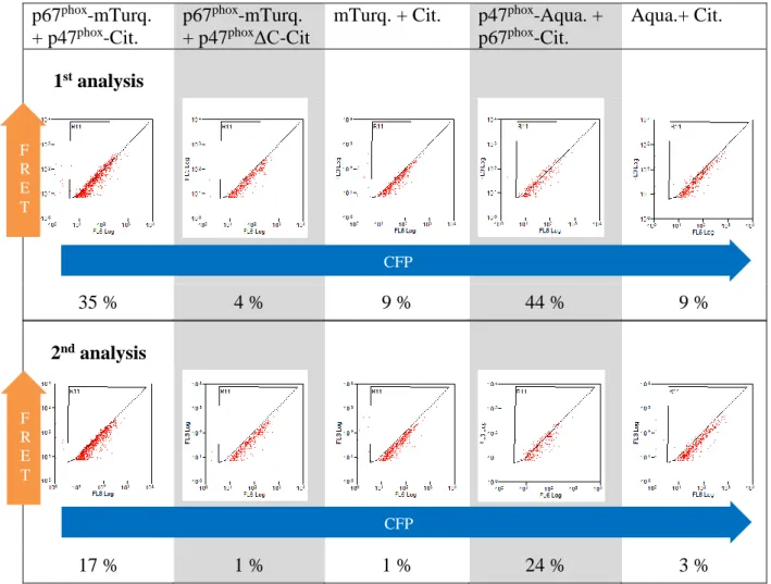
3.2.4 Extraction of the quantitative data and specificity of the interaction ... 75
3.2.5 Estimation of the dissociation constant Kd ... 79
4 Results – Interactions between NADPH oxidase subunits ... 81
4.1 Proving functionality of the cytosolic subunits ... 81
4.1.1 Verification of the size of the expressed protein by Western Blot ... 81
4.1.2 Verifying the activatability of the complex ... 89
4.1.2.1 Tracking the arrival of the complex on the membrane ... 89
4.1.2.2 Measuring the hydrogen peroxide production ... 95
4.2 Interactions of the cytosolic subunits ... 97
4.2.1 Hetero-dimeric interactions between the subunits ... 97
4.2.1.1 Interaction of p67phox with p47phox ... 97
4.2.1.2 p67phox interacts with p40phox ... 102
4.2.1.3 p47phox interacts with p40phox ... 105
4.2.1.4 Homo-dimerization of the subunits ... 107
4.2.1.5 Intramolecular interactions ... 110
4.2.2 Results obtained from CFP-YFP FCCS ... 116
5 Discussion ... 119
5.1 Discussion of the quantitative approach ... 120
5.1.1 Interpretation of the bound fraction ... 120
5.1.1.1 Factors influencing the apparent bound fractions of proteins ... 122
5.1.1.2 Methodology specific biasing factors on the bound fraction ... 127
5.1.1.3 Conclusion on the apparent bound fraction ... 131
5.1.2 Dissociation constant estimated with FRET-FLIM and wide-field microscopy approach ... 133
5.2 Discussion of the structural information obtained by FRET-FLIM ... 134
5.2.1 Intra-molecular interactions ... 135
5.2.2 Inter-molecular Interactions ... 142
6 Conclusion ... 149
8 Appendix ... 153
8.1 Protein production and purification ... 153
8.1.1 Fluorescent proteins ... 153
8.1.2 Cytosolic subunits ± FP tag ... 154
8.2 Determination of the intensity working frame ... 156
8.3 Derivation of the Kd resolved with the bound fraction ... 157
8.4 Influence the bound fraction on the Kd ... 158
8.5 Correction of the spectral blead-through in FCS with FLCS ... 159
9 List of abbreviations ... 161
10 List of figures ... 162
1
Introduction
Proteins take on a variety of tasks in living organisms like structural, enzymatic and regulatory functions. The majority is not acting as individual proteins but in complex with other proteins – for the human proteome a share of 80 % is estimated. These interactions are mainly dynamic and allow the cells to react to environmental changes and stimuli. Signalling cascades are a good example of highly regulated transient protein interactions, in which conformational changes of at least one partner leads to the next step of the cascade. To understand these fundamental processes of life, numerous techniques were developed to discover and study protein-protein interactions such as protein microarrays, X-ray crystal structure, affinity tagging and pull-down assays, immunoprecipitation, peptide arrays or yeast two-hybrid and fluorescence microscopy (Dwane and Kiely, 2011). Most of these techniques are performed in vitro on purified proteins or on a lysed cell population except the two last that are compatible with live cell. The native environment of the proteins is important, since it is influencing the spatiotemporal dynamics of any process: It can differ from in vitro studies (Ganesan and Zhang, 2012). In addition, most of the quantitative data on protein interactions such as molecular affinities are obtained with in vitro studies and may differ by several orders of magnitudes from those prevailing under intracellular conditions (Cardarelli et al., 2009, Foo et al., 2012). Therefore, there is a strong need of truly in situ quantitative and dynamic approaches of these protein interactions. Here, we will concentrate on fluorescence imaging approaches and more particularly on those that allows their quantitative investigation.
1.1 Quantitative fluorescence imaging of protein-protein interactions in the NADPH oxidase complex
Powerful bioanalytical imaging techniques based on fluorescence resonance energy transfer (FRET) and Green Fluorescent Proteins (GFPs) are increasingly used in pharmacological and clinical research (Aoki et al., 2012, Lang et al., 2006, Lu and Wang, 2010, Prinz et al., 2008) as well as in environmental sciences and biotechnologies (Larrainzar et al., 2005). Donor and acceptor fluorescent proteins (FP) are linked to the proteins of interest. When these fusion proteins interacts, the fluorophores come into close proximity, which allows the energy transfer (distance between FPs < 10 nm). FRET imaging provides an optical readout of the interaction events within complex media such as living cells (Sun et al., 2011b).
There are several possibilities to measure FRET: With regard to the fluorescence properties of the donor and acceptor fluorophore, the effect of FRET is different: It implies a shortening of the fluorescence lifetime and intensity of the donor and an increase of the fluorescence intensity of the acceptor upon donor excitation. Consequently, FRET can either be measured with approaches based on intensity changes (acceptor photobleaching, ratiometric measurement, and “3-cubes” approach) or on the decrease of donor fluorescence lifetime (fluorescence lifetime imaging microscopy (FLIM)) (Padilla-Parra and Tramier, 2012).
FRET has been used in numerous biological systems addressing molecular interactions, probing the conditions and even the location of these interactions. Over 8000 articles report FRET experiments in biomedicine. With respect to “quantitative FRET measurements”, there are different levels, which can be reached depending on the methodology: As a basic step, it can mean differentiation between FRET positive vs. negative outcome (Banning et al., 2010), and further between specific vs. unspecific FRET (Kenworthy and Edidin, 1998, Abankwa and Vogel, 2007, Day, 2014); or more advanced, it provides information about the stoichiometry (Hoppe et al., 2002, Hoppe and Swanson, 2004, Beemiller et al., 2006, Beemiller et al., 2010) and the structure of the interacting partners (Preus and Wilhelmsson, 2012, Banerjee and Pal, 2007). Finally, it allows even a full characterization of a protein-protein binding by a determination of the fraction of donor-fused protein to the acceptor-fused partner (bound fraction, β) of the donor-fused protein to its (Meyer et al., 2006, Cardarelli et al., 2009) and of the dissociation constant (Kd) (Cardarelli et al., 2009). Within this project we aim to demonstrate, how far a FRET-FLIM based quantitative approach can go in live cells using the newly engineered cyan fluorescent protein (CFP) variants mTurquoise (Goedhart et al., 2010) and Aquamarine (Erard et al., 2013):
Indeed, although CFP is used in most of FRET studies (> 60 %) as donor in combination with a yellow fluorescent protein (YFP) as an acceptor, quantitative FRET-FLIM studies have remained limited until now by the suboptimal performance of the donor fluorophore (ECFP). ECFP is characterized by a low brightness and low photostability, a complex photophysics including multi-exponential fluorescence decays, and a strong environmental sensitivity (Villoing et al., 2008). In the past years, the structure-based photophysical engineering of CFPs led to the development of improved variants in several laboratories including our group ((Merola et al., 2013) for a review). mTurquoise (ECFP T65S, S72A, H148D, S175G, A206K) and Aquamarine (ECFP T65S, H148G) used during my PhD have a fluorescence quantum yield close to 90% leading to an improved brightnessi, a single-exponential fluorescence decay, an improved photostability, and an insensitivity to their environment (Goedhart et al., 2010, Erard et al., 2013). These donors with a single-exponential emission decay (in contrast to FPs with multi-exponential decay) combined with FLIM detection methods allow in principle accurate quantifications of interacting vs. non-interacting populations and the analysis of subunit stoichiometry within multi-protein complexes (Miyawaki, 2011, Berezin and Achilefu, 2010, Kumar et al., 2011). We wanted to explore the feasibility of an intracellular quantitative approach based on the new CFP variants mTurquoise and Aquamarine, previously demonstrated using the less widely used green GFP donor (Cardarelli et al., 2009). FRET studies were already used to clarify interactions in dynamic processes with respect to time and localization during phagocytosis like G-protein activation relative to actin movement (Hoppe and Swanson, 2004) or the G-protein dependent cyclic regulation of phosphatidylinositol-3-kinase activity (Beemiller et al., 2010). For testing our approach, we chose the NADPH oxidase complex.
The NADPH oxidase complex (NOX) is responsible of the oxidative burst of phagocytic cells like neutrophils that is a major element in antimicrobial defence: The superoxide anion produced by the NADPH oxidase is the precursor of most other ROS that are later produced in the phagosome (Segal, 2005). However, an overactivation of the NADPH oxidase may lead to oxidative stress responsible of tissue damages, as in chronic inflammatory diseases such as rheumatoid arthritis (El-Benna et al., 2009). To keep this potentially harmful enzyme under tight control, it is composed of several subunits that are maintained apart, or pre-assembled as inactive complexes, in the resting cell. The gp91phox and p22phox subunits are integral membrane proteins that form the flavocytochrome b558. In response to cell stimulation, the other regulatory cytosolic components, p67phox, p47phox, p40phox and the small GPase Rac translocate
to the flavocytochrome and induce superoxide production (Nauseef, 2004). Due to the difficulty to cultivate and transfect primary neutrophils, most of our knowledge about the assembly and activation of the NADPH oxidase comes from biochemical studies (Nauseef, 2004, Nordenfelt and Tapper, 2011, Nüsse, 2011). Furthermore the mechanisms responsible for the ending of the ROS production and for the NADPH oxidase deactivation are still unknown (DeCoursey and Ligeti, 2005). Recently video microscopy experiments combined with subunits tagged with fluorescent proteins gave new insights on the dynamic behaviour of the subunits in the complex ((Tlili et al., 2012, Li et al., 2009, Tian et al., 2008, Faure et al., 2013). For example, it has be shown that p67phox needs p47phox to translocate at the phagosomal membrane and that the interaction between p47phox and p67phox was disrupted just after the phagosome closure whereas the ROS production was prolonged for several minutes (Ueyama et al., 2007, Li et al., 2009, Faure et al., 2013, Tlili et al., 2012). These first observations raised numerous questions on the spatio-temporal re-organization of the subunits for sustained ROS production that can only be solved with imaging approaches. Deciphering the sequence of the NADPH oxidase assembly and activation, as well as the conditions of its subsequent deactivation, would be the key of new diagnosis or therapeutic strategies for many inflammatory and degenerative diseases.
1.2 Aim of the thesis
The aim of the thesis was first to probe the feasibility of the CFP variants mTurquoise and Aquamarine for quantitative live cell studies of protein-protein interactions with FRET-FLIM. We want to test out the maximum reachable quantification level and to compare it to other quantitative imaging approaches such as FCCS.
Second, we intended to develop a new imaging approach for studying the interactions between the cytosolic subunits of the phagocyte NADPH oxidase in time and space. These interactions have been shown by biochemical techniques. Our approach aimed at demonstrating these interactions in live cells. Furthermore, we were seeking structural information on the complex formed by the oxidase subunits under live cell conditions.
1.3 Fluorescence and Förster Resonance Energy Transfer
FRET allows the monitoring of dynamic processes of transient protein-protein interactions with spatial resolution in live cells. Therefore, it is a very useful tool for biosciences. Among a variety of techniques suited for FRET measurements we chose the lifetime based FRET-FLIM, which features high accuracy, fluorophore concentration independency, and no necessity for cross talk correction (Padilla-Parra and Tramier, 2012). The current paragraph addresses the theoretical background of this method.
1.3.1 Fluorescence – the physical background
To facilitate the understanding of the FRET phenomenon and the FLIM approach, a short introduction in the physics of fluorescence will be given.
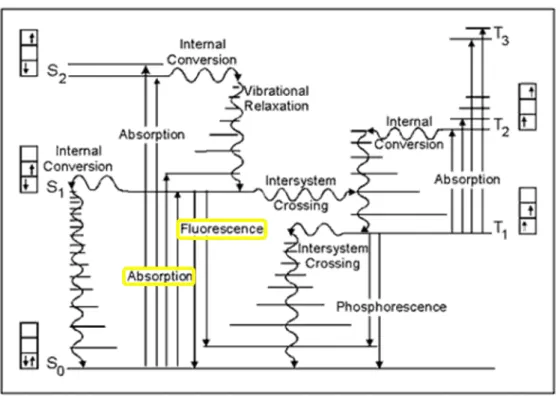
Fluorescence is, together with phosphorescence, a luminescence phenomenon. The excitation is initiated by the absorption of a photon. The relaxation from the excited state happens via radiative and non-radiative processes summarized in the Jablonsky-Perrin diagram (Figure 1) and is detailed below.
Figure 1 Jablonski diagram of the excitation and de-excitation pathways of a fluorophore (Smith, 2009)
On the electron level, the excitation leads to a transition of an electron from an occupied or non-bonding orbital to an unoccupied one, whereby in principle the electron spin is conserved (= singlet state transition; S0 → S1 or S2). The probability of a transition is given by the
Equation 1 𝐴(𝜆) = log(𝐼𝜆
0 𝐼𝜆).
where 𝐼𝜆0 and 𝐼𝜆 are the intensities of the light before and after crossing the absorbing probe. Under certain circumstancesii, the absorbance is linearly proportional to the concentration, which is described by the Beer-Lambert Law:
Equation 2 𝐴(𝜆) = ε(λ) ∙ l ∙ c
with ε(λ), the molar extinction coefficient, l, the length of the absorption path, and c, the concentration of the absorbing probe.
The relaxation from the excited state happens via radiative and non-radiative processes. The radiative transition S1 → S0 is called the fluorescence emission. The emitted photons have less
energy than the excitation photons, which results in a shift of the excitation to the emission spectrum towards longer wavelengths. The difference in the maxima of the excitation and emission is called Stokes shift.
Other non-radiative de-excitation mechanisms can compete with the photon emitting fluorescence pathway (Figure 1): Internal conversion denotes a transition to a lower state with remaining spin orientation (S2 → S1or S1 → S0), while the vibrational relaxation describes the
de-excitation from a high vibrational level to the lowest one (high level S1 or 2 → low level S1 or 2). The latter path is less energetically favoured and is in competition with the fluorescence
pathway and intersystem crossing to the triplet state (S1 → T1), which is as well a non-radiative
relaxation process. The triplet state mainly de-excitates trough non-radiative mechanisms at room temperature.iii
Fluorescence is only one of numerous de-excitation paths and the fraction of excited molecules, which de-excite through this way is the fluorescence quantum yield Φ, defined by the function of the rate constants of the radiative kr and non-radiative processes knr:
Equation 3 Φ =𝑘 𝑘𝑟
𝑟+𝑘𝑛𝑟
With regard to the time, the photon absorption and electron transition to the excited state is a very fast process. In contrast, the electron can stay in the excited state from a few tens of ps to a few hundreds of ns before a relaxation (via photon emission, internal conversion, or
ii validate for homogeneous, low concentrated solutions with negligible scattering/variance of the
ε(λ)/self-emission, which are excited by monochromatic light
iii At low temperatures, the radiative path (phosphorescence) is preferred (T
1 → S0). In contrast, with increasing
temperature a reverse intersystem crossing (T1 → S1) followed by a radiative relaxation to the ground state can
occur, which is called delayed florescence. At high illumination power, a photon absorption of a molecule in T1
intersystem crossing) takes place. This time is the fluorescence lifetime τ, which is characteristic for a specific molecule and is as well defined by the rate constants:
Equation 4 𝜏 =𝑘 1
𝑟+𝑘𝑛𝑟
Experimentally, the value of the fluorescence lifetime is calculated upon analysis of fluorescence emission decay after a very short excitation pulse. The fluorescence intensity I(t) of a fluorophore follows an exponential decay law:
Equation 5 𝐼(𝑡) = 𝑘𝑟[𝐹𝑃∗]0∙ 𝑒− 𝑡 𝜏= 𝐼0∙ 𝑒− 𝑡 𝜏 where [𝐹𝑃∗]
0 is the fluorophore concentration in the excited state and I0 is the fluorescence intensity at time point 0 (Valeur, 2006).
1.3.2 Förster resonance energy transfer
Förster resonance energy transfer (FRET) is an additional de-excitation pathway. It denotes the energy transfer via long-range dipole-dipole transition between a so-called donor and an acceptor fluorophore. It can only take place when the donor emission spectrum overlaps with the acceptor excitation spectrum, allowing energetically coupled (resonant) transitions. The rate constant of energy transfer kT is given by:
Equation 6 𝑘𝑇(𝑟) = 𝑘𝐷[𝑅0𝑟] 6 =𝜏1 𝐷 0 [ 𝑅0 𝑟] 6
with the emission rate constant of the donor kD, the lifetime of the donor without energy transfer τD0, the distance between donor and acceptor r and the Förster radius R0iv. The latter one is defined by: Equation 7 𝑅06 =9000(𝑙𝑛10)𝜅 2Φ 𝐷 0 128𝜋5𝑁 𝐴𝑛4 ∫ 𝐼𝐷(𝜆)𝜀𝐴(𝜆) ∞ 0 𝜆4𝑑𝜆
with the spatial orientation factor κ², the fluorescence quantum yield of the donor (without energy transfer) Φ𝐷0, the refractive index of the medium nv, and the Avogadro constant NA. The integral depends on the spectral overlap and the intensity of the acceptor absorption: 𝐼𝐷(𝜆) is
iv For r = R
0, the probability of a spontaneous relaxation and an energy transfer are equally probable (kD = kT),
which corresponds to an energy transfer of 50 %
v refractive index of the cytosol: 1.36 – 1.39 CURL, C. L., BELLAIR, C. J., HARRIS, T., ALLMAN, B. E.,
HARRIS, P. J., STEWART, A. G., ROBERTS, A., NUGENT, K. A. & DELBRIDGE, L. M. D. (2005) Refractive index measurement in viable cells using quantitative phase-amplitude microscopy and confocal microscopy. Cytometry Part A, 65A, 88-92, VAN MANEN, H.-J., VERKUIJLEN, P., WITTENDORP, P., SUBRAMANIAM, V., VAN DEN BERG, T. K., ROOS, D. & OTTO, C. (2008) Refractive Index Sensing of Green Fluorescent Proteins in Living Cells Using Fluorescence Lifetime Imaging Microscopy. Biophysical
Journal, 94, L67-L69, CHOI, W., FANG-YEN, C., BADIZADEGAN, K., OH, S., LUE, N., DASARI, R. R. &
the normalized emission spectrum of the donor, i.e. ∫ 𝐼0∞ 𝐷(𝜆)𝑑𝜆 = 1 and 𝜀𝐴(𝜆) is the molar
extinction coefficient of the acceptor.
The spatial orientation factor κ² is given by the angle between donor (green) and acceptor (orange) transition moments θDA and the angles between these transition moments and the axis donor-acceptor θD and θA (Figure 2):
Equation 8 𝜅2 = (cos 𝜃𝐷𝐴− 3 cos 𝜃𝐷 ∙ cos 𝜃𝐴)2
Figure 2 Angles involved in the definition of the orientation factor κ²
The dipole moment of the donor (green) and acceptor (orange) is displayed with flashes. The axis between donor and acceptor is the separation vector. The angles between this vector and the dipole orientation of donor and acceptor θD and θAgives the dipole orientation while θADgives the angle between the two dipole moments. κ² can have values between 0 and 4 depending on the relative orientation of the transient dipole moments of the donor and the acceptor (perpendicular oriented dipoles no FRET possible, coaxial aligned dipoles perfect condition for FRET). Figure 3 A displays the influence of the spatial orientation on the Förster distance on the example of Aquamarine/Citrine couple: For very small κ² values, the influence on the Förster distance is high, but for values 0.5 the changes are moderate. It is possible to make some approximations about the κ² value under certain conditions. If both chromophores can be considered to move freely during their excited state, an isotropical orientation can be assumed and the value of κ² is 2/3. For free GFP variants, the rotational diffusion is too slow (15-20 ns) to adopt this commonly used value of 2/3, though (Heikal et al., 2000). In our case it may be more appropriate to take κ² = 0.476, a value calculated for molecules with frozen rotatory motion (Demchenko, 2008, Grailhe et al., 2006, Steinberg, 1968).
A B
Figure 3 Förster distance and molecular FRET efficiency depend on the spatial orientation factor κ² A: The Förster distance R0 is calculated for different values of κ² (n = 1.4 , Φ = 0.76 ).
B: Mathematical simulation of the variation of Emol. with the distance for κ² = 0.476
In conclusion, several conditions must be fulfilled to make the energy transfer possible: (i) The donor emission spectrum has to overlap with the acceptor excitation spectrum; (ii) the distance between the fluorophores has to be small (< 10 nm) (iii) the spatial orientation of the fluorophores towards each other must be favourable.
The molecular FRET efficiency, Emol., can be described either as a function of the rate constants, or by the donor –acceptor distance and the Förster radius:
Equation 9 𝐸𝑚𝑜𝑙. =𝑘 𝑘𝑇 𝑟+𝑘𝑛𝑟+𝑘𝑇 = 1 1+(𝑟 𝑅0) 6
As the equation shows, Emol. depends on R0, which – in turn – depend on κ² (Equation 7), hence the spatial orientation of the fluorophores influences the molecular FRET efficiency, as illustrated in Figure 3 B: Based on a R0 = 49 Å (calculated with κ² = 0.476, n = 1.4, Φ = 0.76), we simulated the variation of Emol. by assuming distance values between 0 – 200 Å. Due to the uncertainty on κ², a measured Emol. does not allow a precise prediction of the distance between the fluorophores but on the other hand if a non-favourable orientation can be excluded, the absence of FRET allows to estimate the distance above 2*R0.
The fluorescence lifetime of the donor alone (τD) is expressed as in Equation 4 and the one of
the donor in presence of the acceptor (τDA) can be expressed as follow:
Equation 10 𝜏1
𝐷 = 𝑘𝑟+ 𝑘𝑛𝑟 and
1
𝜏𝐷𝐴 = 𝑘𝑟+ 𝑘𝑛𝑟+ 𝑘𝑇
Introducing the expression of τD and τDA in Equation 9 gives the following expression of Emol.: R0 R0
Equation 11 𝐸𝑚𝑜𝑙. = 1 −𝜏𝐷𝐴𝜏
𝐷
(Valeur, 2006, Lakowicz, 2006)
This expression highlight the fact that FRET measurements always necessitate the comparison of two situations without FRET (donor alone) and with FRET (donor with acceptor).
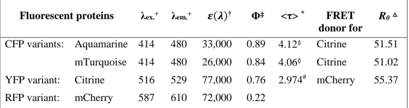
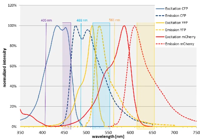
For my project, I used two newly engineered variants of the CFP Aquamarine and mTurquoise, Citrine as an improved variant of the YFP, and mCherry as a red FP (RFP) variant. They can be composed to three different FRET pairs, Aquamarine or mTurquoise + Citrine, and Citrine + mCherry. The photophysical properties, which are important for FRET are listed in Table 1 for the mentioned FPs.
Fluorescent proteins λex.+ λem.+ 𝜺(𝝀)† Φ‡ <τ> * FRET
donor for
R0△
CFP variants: Aquamarine 414 480 33,000 0.89 4.12◊ Citrine 51.51 mTurquoise 414 480 26,000 0.84 4.06◊ Citrine 51.02 YFP variant: Citrine 516 529 77,000 0.76 2.974# mCherry 55.37
RFP variant: mCherry 587 610 72,000 0.22 Table 1 Fluorescent proteins used in this project and their main properties
+ excitation and emission maximum [nm], respectively † extinction coefficient [M-1cm-1]
‡ quantum yield
* fluorescence lifetime [ns]
△ Förster radius [Å], calculated with a κ² of 0.476 (Steinberg, 1968) ◊ in solution, 20 °C and pH 7.4 (Erard et al., 2013)
1.4 NADPH Oxidase
The NADPH oxidases are an enzyme family designated to catalyse the reduction of oxygen to superoxide anions, a precursor for other reactive oxygen species (ROS) such as hydrogen peroxide and hypochlorous acid. Their members have the transmembrane subunit NOX (subfamily NOX 1 – 5vi and DUOX 1 and 2) in common, which is the catalytic core of the enzyme (Figure 4): NADPH binds to its C-terminus where it is oxidised. The electron is transported via FAD and the two haem groups (coordinated between the transmembrane helices TM 3 and TM 5vii) to the other side of the membrane, where it reduces molecular oxygen to superoxide anions (Sumimoto, 2008, Bedard and Krause, 2007).
A B
Figure 4 General structure and function of the NOX
The structure and function of the NOX subunit is highly conserved in all known NADPH oxidase family members. A: The NOX consists of the N-terminal membranous part consisting six transmembrane helices and two haem groups, which are coordinated between TM 3 and 5, and the C-terminal cytosolic “dehydrogenase domain” with its NADPH and FAD binding sites. (Bedard and Krause, 2007)
B: Electron transfer from the oxidation of NADPH to NADP+ to FAD, which is reduced in turn to FADH 2. The
latter one delivers its electron via the two haem groups (H1 and H2) to the other side of the membrane, where it finally reduces O2 to superoxide anions (O2⦁-) (Quinn and Gauss, 2004)
The different members of the NOX family are found in various tissues and cells, as it is summarized in Table 2 (Bedard and Krause, 2007). Based on tissue expression studies, divers physiological (Table 2) and pathological roles (Figure 5) of the subfamilies were suggested.
vi NOX4 produces hydrogen peroxide, though
vii DUOX 1/2 contain an additional N-terminal TM, hence the haem groups are coordinated between TM 4 and
However – beside the host defence function of the phagocyte NADPH oxidase and the involvement in biosynthetic processes like the thyroid hormone synthesis of DUOX2 and the otoconia formation of NOX3 – their exact function is not elucidated yet (Krause, 2004, Altenhöfer et al., 2014, Katsuyama et al., 2012).
Subfamily High level
expression
Intermediate to low level expression
Suggested (main) functions
NOX1 Colon Smooth muscle, endothelium,
uterus, placenta, prostate, osteoclasts, retinal pericytes
host defence (colon), stimulation of cell proliferation NOX2 Phagocytes B lymphocytes, neurons ,
cardiomyocytes, skeletal muscle, hepatocytes,
endothelium, hematopoietic stem cells, smooth muscle
host defence
NOX3 Inner ear Fetal kidney, fetal spleen, skull bone, brain
otoconia formation and as a consequence the vestibular sensing of the equilibrium
NOX4 Kidney, blood
vessels
Osteoblasts, endothelium, smooth muscle, hematopoietic stem cells, fibroblasts,
keratinocytes, melanoma cells, neurons
oxygen sensing in the kidney cortex, regulation of cell proliferation
NOX5 Lymphoid
tissue, testis
Endothelium, smooth muscle, pancreas, placenta, ovary, uterus, stomach, various fetal tissues
ROS-dependent signalling and regulation of
transcription factors (testis); lymphocyte differentiation
DUOX1 Thyroid Airway epithelia, tongue
epithelium, cerebellum, testis
pulmonary host defence DUOX2 Thyroid Salivary and rectal glands,
gastrointestinal epithelia, airway epithelia, uterus, gall bladder, pancreatic islets
thyroid hormone synthesis
Table 2 Overview of the tissue/cell specificity of the NOX family members and their suggested functions (Krause, 2004, Bedard and Krause, 2007, Katsuyama et al., 2012)
Figure 5 NADPH oxidases are associated with a variety of diseases
NOX1 (green) is associated with vascular diseases such as atherosclerosis, ischemic retinopathy, ischemia reperfusion of the heart, liver fibrosis, and perhaps with tumour (melanoma) angiogenesis. Involvement in inflammatory bowel disease and carcinogenesis was suggested.
Hyperactivity of NOX2 (pink) seems to be involved in a plethora of inflammatory (e.g. allergic asthma) and vascular diseases including arteriosclerosis and ischemic and reperfusion disorders such as stroke and heart attack, while its hypo- or in-activity is associated with the Chronic Granulomatous Disease (CGD), a severe immune deficiency syndrome.
NOX3 (violet) is involved in the synthesis of the otoconia, which are indispensable for the vestibular sensing system of the equilibrium. It is as well associated with cisplatin induced hearing loss.
The role of DUOX1 remains unclear, it may be involved in bladder or pulmonary immune defence.
DUOX2 is necessary for the synthesis of thyroid hormones. (Altenhöfer et al., 2014, Brown and Griendling, 2009)
1.4.1 The phagocyte NADPH oxidase
The phagocyte NADPH oxidase (in the following called NOX2 complex) is a major part of the host response to bacterial and fungal infections as well as to inflammations. Additionally, ROS are involved in signalling cascades of the host response, cell death of the phagocyte, and affects extracellularly the surrounding tissues and induce stress responses in microbes.
The importance of the respiratory burst – as the immense production of superoxide anions by the active oxidase is called – becomes apparent in case of activity-restricting genetic mutations of the oxidase leading to the Chronic Granulomatous Disease (CGD), which is characterized by a severe immune deficiency syndrome (chronic infections, impaired wound healing) (Nunes et al., 2013, Dupré-Crochet et al., 2013, Brown and Griendling, 2009). However, a hyperactivity
of the oxidase leads to excessive ROS production, which is associated with tissue damaging and oxidative stress. This imbalance is assumed to be involved in inflammatory, vascular, and ischemic diseases (Figure 5, pink). Furthermore, the oxidative stress induced by NOX2 complex might play a negative role in cerebrovascular dysfunctionsviii leading to hypertension, hypercholesterolemia, and advanced aging, as well as in neurodegenerative disorders including Alzheimer’s disease (Brown and Griendling, 2009, Drummond et al., 2011, Altenhöfer et al., 2014). These facts point out the importance of a tight regulation of the activity of the oxidase.
1.4.1.1 Resting state, activation, and deactivation
The NADPH oxidase consists of five subunits, two membranous ones gp91phox (also called NOX2) and p22phox, which build a stable complex, the flavocytochrome b558, and three cytosolic subunits (p47phox, p67phox, and p40phox). In the resting state of the neutrophil, the flavocytochrome b558 is mainly localized in the membranes of specific granules and to a minor part in the plasma membrane, while the three cytosolic subunits are assumed to be organized in a ternary complex (El-Benna et al., 2008).
The in vivo activation of the oxidase can be achieved by particle stimuli (opsonised bacteria or fungi), which are phagocytised, or by soluble activators such as pro-inflammatory agents. Both mechanisms lead to NADPH oxidase activation by protein kinases (e.g. PKC, MAPKix) induced phosphorylations of all subunits of the NADPH oxidase, which finally lead to the translocation of the cytosolic complex to the membranous subunits (Dupré-Crochet et al., 2013, Nauseef and Borregaard, 2014). Furthermore, phagocytosis induces a flavocytochrome b558 recruitment to the plasma membrane of which the phagosome will be built (El-Benna et al., 2008).
To establish the active NADPH oxidase, the small GTPase Rac has to assemble with the oxidase subunits. Its translocation is independent of the cytosolic subunits, though. In the resting state, Rac is in tight complex with the GDP dissociation inhibitor (RhoGDI), which renders Rac soluble and inhibits an interaction with phospholipids. A dissociation of RhoGDI is followed by translocation of Rac to the membrane, most probably due to electrostatic and hydrophobic interactions with the membrane (Pick, 2014).
The cytosolic subunits play different roles during activation. P47phox is extensively phosphorylated mainly on the C-terminal part (amino acid 303 – 379x) (El-Benna et al., 2009), whereby the phosphorylation of the serine residues S303/304/328/379 of p47phox were shown to be critical for the enzyme activation (el Benna et al., 1994, Benna et al., 1996, Faust et al.,
viii Angiotensin II dependent
ix which kinase is activated is stimulus dependent
1996, Ago et al., 1999, Ago et al., 2003, Marcoux et al., 2010, Meijles et al., 2014), while the phosphorylation of S345 was shown to be an important priming mechanism for further phosphorylations (El-Benna et al., 2008)xi. The phosphorylation induced conformational
changes allow the binding to p22phox and to the plasma membrane. The specificity for the plasma membrane is derived by two phospholipid binding sitesxii, one for phosphatidylinositol (3, 4) diphosphate (PI(3,4)P2) and the other one phosphatidic acid or phosphatidylserine. On its
way it recruits the cytosolic subunits to the membranous subunits (El-Benna et al., 2008, Nunes et al., 2013). The recruitment of p67phox by p47phox allows an interaction with gp91phox, which induces conformational changes in the flavocytochrome (Paclet et al., 2000), enabling the electron transport from NADPH to FAD (Nisimoto et al., 1999). The role of p40phox is not completely understood yet. This subunit is not necessary for the NADPH oxidase activation in vitroxiii. In live cells, it was shown to have phospholipid binding properties with a preference for PI(3)P rich phagosomal membrane and enhances the ROS production (Tian et al., 2008, Nunes et al., 2013, Suh et al., 2006). The importance of the interaction of p40phox to PI(3)P for the stability of the NADPH oxidase complex on the phagosomal membrane became apparent by a point mutation in the p40phox binding domain to PI(3)P of a CGD patient (Matute et al., 2009). Regarding the role of p40phox during translocation of the cytosolic subunits, it was shown that during FcγR induced activation, p40phox needs the binding to p67phox to be recruited to the
membranous subunits (Tian et al., 2008), however for phagocytosis induced oxidase assembly it is speculated that p40phox assists p47phox in its carrier function during phagocytosis induced
oxidase assembly (Ueyama et al., 2011). Rac is supposed to act as a carrier for p67phox like
p47phox, and additionally to induce conformational changes in p67phox, which trigger the binding to gp91phox (Pick, 2014).
The maintenance of the complex in the active state was often assumed to be a continuous turnover (association and dissociation) of the cytosolic subunits on the membranous complex (DeCoursey and Ligeti, 2005). However, recent studies of our group give evidence that p47phox and Rac dissociate from the membranous complex after activation of the NADPH oxidase (Tlili et al., 2012) maybe due to an disruption of the tail-to-tail binding of p47phox and p67phox (Li et al., 2009), while p67phox is staying in the membrane complex (Tlili et al., 2012) most probably with the help of p40phox (Song and Dupré-Crochet, unpublished data).
xi In fact, all subunits are phosphorylated during activation which increases the affinity of the flavocytochrom
b558 towards the cytosolic subunits; the exact role of the phosphorylations of p40phox and p67phox remains
unclear, though.
xii localized on the PX domain, see below
xiii In live cells an activation of the NADPH oxidase without p40phox is as well possible, but leads to strong
The inactivation process is a crucial step in the NADPH oxidase regulation to prevent tissue damaging due to exaggerated superoxide anion productionxiv. Still, little is known about the
mechanism of NADPH oxidase inactivation. Suggested conformational changes of Rac and dephosphorylation of p47phox leading to a dissociation of these subunits from the complex and hence to its destabilization and inactivation (DeCoursey and Ligeti, 2005), are in contrast to the observed ongoing activity after the dissociation of p47phox and Rac (Tlili et al., 2012, Faure et al., 2013). Both hypothesis (continuous turnover versus stable complex) might be tested by the difference in required Ca²+ signalling: While the turnover may demand sustained Ca2+ supply during the active phase, a stable complex may stay active without Ca2+ (Dupré-Crochet et al., 2013).
It is also possible that other modifications (e.g. phosphorylation/dephorphorylation) of subunits of the NADPH oxidase induce conformational changes, which result in a dissociation of the cytosolic subunits.
1.4.1.2 Structure of the NADPH Oxidase
The structure of a protein is important for the understanding of its interactions with e.g. other proteins, small molecules, or lipids and hence its function. The most detailed information is provided by X-ray crystal structures. In the case of the NADPH oxidase, no complete crystal structure, neither for the entire complex nor for the single subunits – beside the one of p40phox (Honbou et al., 2007) – is available. The structural knowledge about the oxidase is mainly derived from binding studies, low resolution techniques such as small angle X-ray scattering (SAXS), and crystal or NMR structures of single domains. Computational modelling approaches constructed in silico full length structures of p22phox, p47phox and p67phox, which are
– beside of the one of p22phox – mainly based on the available resolved structures of different
domains (Durand et al., 2006, Durand et al., 2010, Meijles et al., 2012, Meijles et al., 2014). In the following, each subunit will be described with respect to its domains in context to its functions and interactions.
xiv ROS is associated with a plethora of diseases, such as atherosclerosis, ischemic stroke, Parkinson disease, and
1.4.1.2.1 The membranous subunits of the NADPH oxidase
The membranous subunits gp91phox and p22phox build together a stable, constitutively expressed
complex, the flavocytochrome b558. This complex is important for the stability of gp91phox
(Porter et al., 1994, Yu et al., 1997).
gp91phox
The membranous glycoprotein gp91phox (570 aa, 65.3 kDa) is the catalytic core of the NADPH oxidase: It allows the electron transport from the NADPH oxidase molecules to molecular oxygen via FAD and two haem groups.
Figure 6 Structure of cytochrome b558
The cytochrome b558 consists of gp91phox (left), with its N-terminal catalytic core built of six transmembrane
α-helices, and with the C-terminally located binding sites for FAD and a NADPH, and of p22phox (right), with
its transmembrane domains, N-terminus and a cytosolic C-terminus holding a PRR domain, which includes a PxxP motive. Red, dashed lines: interactions after activation. Red ovals indicates predicted binding sites for p47phox, cyan dots show glycosylation sites (modified from (Groemping and Rittinger, 2005)).
The protein can be distinguished in (Figure 6): N-terminal membranous part, consisting of:
six transmembrane (TM) α-helices I-VI, linked by loops A-E two haem groups coordinated between TM helix 3 and 5 C-terminal cytosolic part including:
FAD and NADPH binding sites
binding site for the regulatory cytosolic subunits
The N-terminus allows the electron transport through the membrane, which is accomplished by the two haem groups, which are coordinated between His101 and His115 of the TM helix 3 and His209 and His222 of TM helix 5 (Figure 4, right) (Biberstine-Kinkade et al., 2001, Finegold et al., 1996). At these haem groups the final reduction of molecular oxygen to superoxide anions takes place. Additionally, several binding sides for p67phox (helix 2 (Picciocchi et al., 2011)), p47phox (B loop (DeLeo et al., 1995, Biberstine-Kinkade et al., 1999)), and FAD (helix 2 (Picciocchi et al., 2011) and D-loop (Li et al., 2005)) were suggested. Another study is claiming that the main binding sites for p47phox, p67phox, and Rac are solely located on the C-terminal part of gp91phox, though (von Löhneysen et al., 2010).
The C-terminal cytosolic part is homologous to ferrodoxin-NADP+ reductase (FNR) and comprises the binding sites for FAD and NADPH oxidase (Segal et al., 1992, Rotrosen et al., 1992, Ravel and Lederer, 1993). In the resting state, the NADPH binding site is masked by the α-helical loop (Li et al., 2005). During activation it is set free and allows the interaction with p47phox with aa 484-500xv (Biberstine-Kinkade et al., 1999, Li et al., 2005) and aa 555-564
(Adams et al., 1997).
xv However, a Δ488-497 mutant did not affect the NADPH oxidase assembly, only the electron transfer from
NADPH to FAD was impeded YU, L., CROSS, A. R., ZHEN, L. & DINAUER, M. C. (1999) Functional
Analysis of NADPH Oxidase in Granulocytic Cells Expressing a ▵488-497 gp91 phox Deletion Mutant.; one
explanation might be that Asp500 was conserved which was shown to be indispensable for the p47phox
translocation LI, X. J., GRUNWALD, D., MATHIEU, J., MOREL, F. & STASIA, M.-J. (2005) Crucial Role of Two Potential Cytosolic Regions of Nox2, 191TSSTKTIRRS200 and 484DESQANHFAVHHDEEKD500, on NADPH Oxidase Activation. Journal of Biological Chemistry, 280, 14962-14973.
p22phox
P22phox (195 aa, 21 kDa) is constitutively in complex with gp91phox and serves as a maturation
assistant and stabilizer of gp91phox and as a membrane anchor for the complex of the three
cytosolic subunits.
The detailed structure of p22phox is still discussed controversially. It is assumed in consensus that it is built of N-terminal TM helices (2 – 4 were proposed (Dahan et al., 2002, Groemping and Rittinger, 2005, Campion et al., 2009, Meijles et al., 2012)) and a minor structured cytoplasmic C-terminus which comprises a proline-rich region (PRR) with its PxxP (proline – Xaa – Xaa – proline) motive, the binding site for the tandem SH3 domains of p47phoxxvi(Figure 6 and Figure 8) (Groemping and Rittinger, 2005). The localization of the N-terminus (extracellular or cytosolic) is as well not finally clarified (Dahan et al., 2002, Campion et al., 2009, Meijles et al., 2012).
1.4.1.2.2 The cytosolic subunits of the NADPH oxidase
p47phox
The cytosolic subunit p47phox (390 aa, 44.7 kDA) is called the organizer of the NADPH oxidase
since it is crucial both for activation induced translocation of the cytosolic complex and its anchoring to the membrane (Groemping and Rittinger, 2005, Sumimoto, 2008).
Figure 7 Domain structure of p47phox
From the N- to the C-terminus: PX domain binds intra-molecularly to the SH3B domain in the inactive state preventing membrane anchoring, which is set free by phosphorylations in active state, SH3A and SH3B are blocked in the inactive state by internal interactions with the auto-inhibitory region (AIR) and allow the assembly with p22phox after activation induced phosphorylations. The PRR domain is a binding site both for p67phox and
p40phox, in vitro the affinity towards p67phox is higher. Blue arrows: intramolecular interactions in the inactive state;
green lines: constitutive intermolecular interactions; red, dashed lines: interactions after activation.
Regarding the function of the subunit, four main domains are described (from N-terminus to C-terminus, Figure 7):
phagocyte oxidase (PX) domain, aa 4 – 121
tandem Src homology (SH3) domains (SH3A, aa 159 – 214 and SH3B, aa 229 – 284) auto-inhibitory region (AIR), aa 282 – 340
proline rich region (PRR), aa 342 – 368, including a PXXP motif between aa 358 – 372 The phagocyte oxidase (PX) domain is found both in p47phox and p40phox. It is crucial for the membrane binding properties (due to its phosphoinositide-binding capacity) of these subunits and thus important for the translocation of the cytosolic complex to the membrane. In the case of p47phox it binds specifically phosphatidylinositol-3,4-bisphosphate [PI(3,4)P2] (Sumimoto,
2008, Groemping and Rittinger, 2005, Stampoulis et al., 2012). In the inactive state, however, this domain is blocked by intra-molecular interactions with the SH3B domain. Due to phosphorylation of the serine residues S303/304/328 during activation, it is set free (Ago et al., 2003, Marcoux et al., 2010).
Figure 8 Interaction of p47phox and p22phox
The binding sites of p22phox are marked with arrows. The crucial aa in the PRR motive are Pro152, Pro156
(interacting with SH3A of p47phox) and Arg158 (interacting with SH3B of p47phox). The C-terminal αhelix of
p22phox (aa 149-168) is important for the affinity of the two subunits (Nobuhisa et al., 2006), it enhances the affinity
of the two subunits by a factor of 10 (Ogura et al., 2006).
The tandem SH3 domains of p47phox are masked by intramolecular interactions with the AIR
during resting statexvii and interact with the C-terminus of p22phox after activation by embedding
the PRR of p22phox between its two SH3 domains (Figure 8). This latter interaction is important
for the assembly of the cytosolic complex with the membrane-bound subunits NOX2 and p22phox (in the cell-free system p47phox is dispensable for the activation in the presence of excess amounts of p67phox and Rac, though) (Sumimoto, 2008).
The PRR mediates the interaction with the two other cytosolic subunits: The SH3B of p67phox binds with high affinity to the PXXP motif of p47phox and to its C-terminal α-helix (Sumimoto,
2008). The SH3 domain of p40phox is another interaction partner of the PRR (Grizot et al., 2001b, Massenet et al., 2005, Fuchs et al., 1995, Wientjes et al., 1996, Ito et al., 1996)) However, p47phox has a higher affinity towards p67phox, which is derived from the specific binding mode of Arg368 (part of PXXP motif of p47phox) to p67phox. Furthermore, the affinity of SH3 of p40phox is decreased in the presence of the flanking PX and PB1 domains (Massenet et al., 2005). Live cell studies and immunoprecipitation could not confirm an interaction between p40phox and p47phox (Ueyama et al., 2007).
xvii However, it is also described that the SH3 domains can only either block the PX domain or be blocked by
AIR ITO, T., NAKAMURA, R., SUMIMOTO, H., TAKESHIGE, K. & SAKAKI, Y. (1996) An SH3 domain-mediated interaction between the phagocyte NADPH oxidase factors p40phox and p47phox. FEBS Letters, 385, 229-232, MARCOUX, J., MAN, P., CASTELLAN, M., VIVÈS, C., FOREST, E. & FIESCHI, F. (2009) Conformational changes in p47phox upon activation highlighted by mass spectrometry coupled to
hydrogen/deuterium exchange and limited proteolysis. Ibid.583, 835-840, MARCOUX, J., MAN, P., PETIT-HAERTLEIN, I., VIVES, C., FOREST, E. & FIESCHI, F. (2010) p47(phox) Molecular Activation for Assembly of the Neutrophil NADPH Oxidase Complex. Journal of Biological Chemistry, 285, 28980-28990.
The p47phox C-terminus (PRR domain and downstream up to the very end) seems to be involved
in rendering the subunit in the inactive state by interacting with the AIR. Phosphorylation of serine S379 interrupts this interaction and makes the phosphorylations of the AIR possible (Marcoux et al., 2010).
p67phox
P67phox (526 aa, 59.8 kDa) is the activator of the NADPH oxidase since it is directly interacting with the gp91phox.
Figure 9 Domain structure of p67phox
From the N- to the C-terminus: 4 TPRs, providing the Rac binding site; the activation domain (AD) is indispensable for the activation of the NADPH oxidase (an extension of the AD to aa 190-210 was suggested by (Maehara et al., 2010)); a PRR motif and a SH3A domain (in both cases, no interaction partners are identified at present); the PB1 domain provides the binding site for p40phox; the SH3B domain is interacting with p47phox. Green lines:
intermolecular interactions in inactive state; red, dashed lines: interactions after activation.
It is structured as follows (N- to C-terminus, Figure 9) (Groemping and Rittinger, 2005, Sumimoto, 2008):
four tetratricopeptide repeats (TPR) motifs, aa 3-36/36-70/70-104/120-153 activation domain (AD), aa 201-210
two SH3 domains, aa 243-298 and aa 460-515
Phox and Bem1 (PB1) domain between the SH3 domains, aa 351-429
The TPR motif enables the interaction with Rac (Groemping and Rittinger, 2005). The binding of Rac is accomplished by the connection loops between TPR1 & TPR2, TPR2 & TPR3 as well as the hairpin intersection between the last two TPR motifs. (Sumimoto, 2008)
The activation domain is known to be crucial for the activation of oxidase complex, whereby Val204 is indispensable for the oxidase activation (Han et al., 1998, Sumimoto, 2008). A direct interaction with the flavocytochrome and perhaps an involvement in the electron transfer is assumed (Groemping and Rittinger, 2005). The region N-terminal of the AD is essential for the reconstitution of the NADPH oxidase complex (in vivo and in vitro), since it provides interaction sites for gp91phox leading to an extended AD (aa 190-210) (Maehara et al., 2010).
Neither for the PRR nor for the SH3A was a binding partner identified (Groemping and Rittinger, 2005). However, in cells the SH3A domain is important for the superoxide production. Therefore, its role might be to promote the oxidase assembly (Maehara et al., 2009). The PB1 domain is strongly interacting with the PB1 domain of p40phox (Groemping and
Rittinger, 2005, Sumimoto, 2008).
The SH3B domain binds to the PRR domain of p47phox as described in before. Thus this domain is indirectly responsible for the membrane translocation of p67phox.
Oligomerization of p67phox has not been shown, but N- (TPR motif) to C-terminal dimerization were suggested. However, Rac-p67phox binding studies did not reveal any interference when C-terminal fragments of p67phox were present (Groemping and Rittinger, 2005).
p40phox
Despite the complete structural characterisation of p40phox by crystal structure, the role of p40phox (339 aa, 39 kDa) is not completely understood, but it is known to contribute to the membrane translocation of the ternary cytosolic complex due to its binding specificity of the phagosomal membrane (Sumimoto, 2008).
Figure 10 Structure of p40phox
P40phox comprises a PX domain, which allows the binding to the phagosomal membrane in the active state and
which is masked by an intramolecular interaction with the PB1 domain in the resting state. The latter also provides the binding site for p67phox, whereas the SH3 domain is interacting with the PRR of p47phox
The following domains are described for p40phox (Figure 10): PX domain, aa 18-136
SH3 domain, aa 173-228 PB1 domain, aa 237-329
The PX domain is capable to bind PI(3)P, a phosphoinositide, which is accumulated in the phagosomal membrane and hence enhancing the complex recruitment to the phagosome (Groemping and Rittinger, 2005). In the inactive state, the PX domain is masked by intramolecular interaction with the PB1 domain (Honbou et al., 2007, Ueyama et al., 2007).
The SH3 domain is the binding site for the PRR of p47phox, as described before and to the
PPR motif of p22phox, but with lower affinity than for p47phox (in vitro) (Tamura et al., 2007). A
contribution of this domain to the activation of the NADPH oxidase was suggested from live cell experiments (Suh et al., 2006, Chessa et al., 2010).
The PB1 domain of p40phox binds to the PB1 domain of p67phox (crystallised by (Wilson et al., 2003)), while the C-terminal tail of p40phox is as well indispensable for the interaction with p67phox (Lys355) (Groemping and Rittinger, 2005)
1.4.1.2.3 The cytosolic complex in active and inactive state
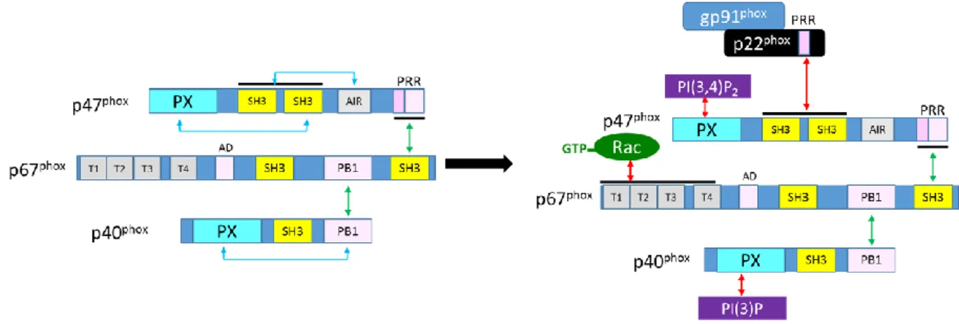
The proposed models of the complex of the cytosolic subunits of the NADPH oxidase display schematically the known interaction sites of the different components (Figure 11), as they were described in the previous chapters. Briefly, p67phox serves as an adaptor since it binds to the PRR domain of p47phox via its SH3B domain and concomitantly to the PB1 domain of p40phox by its own PB1 domain. Intramolecular interactions in p47phox and p40phox hinder the membrane binding capacities of the respective PX domains and a p22phox binding of the tandem SH3 domains of p47phox. During activation, these intramolecular interactions are broken by multiple phosphorylation steps in case of p47phox and probably by hydrogen peroxide in case of p40phox, hence allowing the translocation and anchoring of the cytosolic complex to the membrane and via p22phox to the flavocytochrome b558. Rac translocates independently to the membranous complex. Its binding is assumed to contribute to the correct localization and fixation of the complex, which finally makes the electron transfer from the NADPH oxidation to the other side of the membrane possible, where molecular oxygen is reduced to superoxide anions. During the activation process, the intramolecular interactions of the ternary complex are preserved, but during ongoing activity p47phox and Rac may leave the complex, while p67phox and p40phox are
Figure 11 Model of the cytosolic complex in resting and active state
The inactive state (left) shows the C-terminal intermolecular interaction sites of the different subunits with green flashes and the intramolecular interactions of p47phox and p40phox with blue flashes. During activation, the latter
open up to allow the interactions with the membranous subunits and the phospholipids (right, red flashes). During activation, Rac dissociates from its inhibitor RhoGDI entailing an exchange of GDP with GTP, which makes an interaction to p67phox possible (modified from Sumimoto 2008). (Not all interactions are shown in this figure to
improve the readability. A more complete picture is can be find in Nunes 2013)
However, due to the lack of a complete crystal structure of the complex, the information about the spatial organisation of the complex remains rudimentary.
Within this project we built a new model of the inactive cytosolic complex by aligning the spatial information of the FRET-FLIM studies derived from live cell experiments with the available structural data such as crystal structures, SAXS studies, and computational modelling. This model will contribute to a better understanding of the structure of the ternary cytosolic complex of the NADPH oxidase.
2
Material and Methods
2.1 Buffers HEPES buffer (pH 7.4) [mM] PBS (pH 7.4) [mM] TBS-T (pH 7.4) [mM] NaCl 140 KCl 2.7 NaCl 150 KCl 5 KH2PO4 1.5 Tris HCl 7.7 MgCl2 1 Na2HPO4 8.1 Tween20 0.10% CaCl2 2 NaCl 137 HEPES 10 glucose 0.10% serum (decomplemented) 1%Buffers for plasmid preparation, standard protocol
Buffer P1 [mM] Buffer P2 Buffer P3
Tris HCl (pH 8.0) 25 NaOH 0.2 M K2CH3CO2 5 M
EDTA 10 SDS 1 % CH3CO2H 1.7 M
RNAse 20
µg/ml
2.2 Methods
2.2.1 Bacterial cell culture
Bacterial cell lines (Table 3) were purchased from invitrogen (Thermo Fisher Scientific, Waltham, MA).
Strain Genotype
Escherichia coli DH5α F– Φ80lacZΔM15 Δ(lacZYA-argF) U169 recA1 endA1 hsdR17 (rK–, mK+) phoA supE44 λ– thi-1 gyrA96 relA1
Top10 F– mcrA Δ(mrr-hsdRMS-mcrBC) Φ80lacZΔM15 ΔlacX74 recA1 araD139 Δ(ara leu) 7697 galU galK rpsL (StrR) endA1 nupG Table 3 Bacterial cell lines
Bacteria were grown from a single CFU over night (o.n.) in LB-broth containing the corresponding selection antibiotics with shaking (150 rpm) at 37 °C.
2.2.2 Plasmid preparation
Plasmids were prepared either with a non-commercial protocol (in the following called standard protocol) or with a commercial endotoxin free preparation kit (EndoFree Plasmid Maxi Kit, QIAGEN, Venlo, Netherlands). After preparation, the plasmid solutions were aliquoted and stored at -20 °C.
Standard protocol (mini-preparation):
Bacteria were grown o.n. in 5 ml LB broth. The suspension was centrifuged (8 min at 3000 g) and the supernatant discarded. The remaining pellet was resuspended with 200 µl buffer P1 by vortexing. For lysing the bacteria 200 µl of buffer P2 were added and mixed by gentle inversion of the tube, then incubated for 5 min at room temperature (RT). The reaction was stopped by the addition of 200 µl of buffer P3, which results in a fluffy white precipitation. After another centrifugation step (15 min, 13,000 rpm), the supernatant was transferred to a fresh tube. The containing DNA was precipitated by the addition of 420 µl of pure isopropanol and gentle shaking. This step is followed two more centrifugation steps (5 min, 13,000 g) and an intermediate washing step with 420 µl of 70 % ethanol. The received DNA palled was dried (approximately 5 – 10 min, 65 °C) and resuspended with 40 µl water. The protocol gave a typical yield of 40 µg DNA.
Endotoxin-free protocol (maxi-preparation)
The supplier’s protocol was followed: A bacteria pre-culture was started from a single CFU and 5 ml LB broth containing the appropriate selective antibiotic (shaking incubation at 300 rpm for 8 h at 37 C). 200 µl of the pre-culture was used to inoculate 100 ml of fresh LB medium containing the selective antibiotic, which was incubated o.n. at 37 °C and shaking at 300 rpm. The next day, the bacteria suspension was centrifuged at 3,000 g for 30 min at 4 °C. The received pellet was resuspended with 10 ml of the kit buffer P1 by vortexing, then 10 ml of the kit lysis buffer P2 was added and mixed by inversion. After an incubation of 5 min at RT, 10 ml of pre-cooled (4 °C) kit buffer P3 was added and gently mixed by inversing. The suspension was poured into the barrel of the QIAfilter Cartridge and incubated for 10 min. Afterwards, the suspension was filtered through the cartridge. 2.5 ml of the kit buffer ER was added to the filtrate, mixed by inversion, and then incubated on ice for 30 min. This solution was filtered through a buffer QBT equilibrated QIAGEN-tip by gravity flow. The flow-through was discarded. The tip was washed two times with 30 ml of buffer QC. Importantly, the following steps were performed using endotoxin-free plastic ware. The DNA was eluted from the tip with 15 ml buffer QN and precipitated by adding 10.5 ml isopropanol. After a centrifugation step (3,000 g, 1 h, 4 °C), the received pellet was washed with 40 ml of 70 % ethanol and centrifuged
again (3,000 g, 20 min). The final DNA pellet was air-dried for 5 – 10 min and redissolved in 500 µl of endotoxin-free buffer TE. The protocol gave a typical yield of 400 µg DNA.
2.2.3 Plasmid cloning
A library of the cytosolic subunits of the NADPH oxidase tagged with different FPs (overview of the linkers is given in Table 4), both on N- and C-terminus was built by molecular cloning (Table 5). Therefore, the bacterial vector and insert were digested with the selected restriction enzymes. DNA vectors and fragments were purified on 1 % agarose gels (DNA gel extraction performed with E.Z.N.A.® Gel Extraction Kit (Omega Bio-Tek, Norcross, GA)) and ligated using a T4 DNA ligase (all enzymes were purchased from New England Biolabs, Ipswich, MA). If necessary, DNA fragments were amplified or plasmids were mutated by T100TM thermal cycler (Bio-Rad Laboratories Inc., Hercules, CA). The variants of the plasmids EGFP-N1 was mutated by removing the start codon (ATG). Primers were purchased from Eurogentec (part of Kaneka corp., Osaka, Japan), PfuTurbo DNA polymerase from Agilent Technologies (Santa Clara, CA).
tagged subunit Linker Length
[aa]
p67phox-Citrine/mTurquoise, C-ter. linker of the tandem PRARDPPVAT 10
mTurquoise-p67phox SGLRSRAQAS 10
p47phox-Citrine/Aquamarine, C-ter. linker of the tandem LPVAT 5 Citrine-/mCherry-p40phox and N-ter. linker of the tandem SGLRSRA 7 Citrine-p67phox-mTurquoise, N-ter. linker (C-ter. see above) SGLELKLATM 10 Citrine-p47phox-Aquamarine, N-ter. linker (C-ter. see above) SGLRSRAQAY 10 mCherry-p40phox-Citrine, C-ter. linker (N-ter. see above) PRARDPPVAT 10 Table 4 Overview of the linkers between the subunit and the FP
2.2.4 Production and purification of recombinant proteins
The determination of the ratio of the amount of acceptor/donor demands a calibration of the fluorescence intensity to the concentration of the FP of interest, as it is described in paragraph 3.2.3.2. The purification of these proteins were not performed by myself but by Y. Bousmah, however a brief description of the protocol can be found in appendix 8.1.1.
Likewise, I gave a short summery of the procedure in appendix for the preliminary in vitro activation assay of the tagged vs. wild type subunits (performed by T. Bizouarn, paragraph 5.1.1.1) for which the subunits p67phox ± FP, p47phox ± FP, and Rac were purified by P. Machillot (appendix 8.1.2).