HAL Id: hal-03049799
https://hal.inrae.fr/hal-03049799
Submitted on 10 Dec 2020
HAL is a multi-disciplinary open access
archive for the deposit and dissemination of
sci-entific research documents, whether they are
pub-lished or not. The documents may come from
teaching and research institutions in France or
abroad, or from public or private research centers.
L’archive ouverte pluridisciplinaire HAL, est
destinée au dépôt et à la diffusion de documents
scientifiques de niveau recherche, publiés ou non,
émanant des établissements d’enseignement et de
recherche français ou étrangers, des laboratoires
publics ou privés.
Distributed under a Creative Commons Attribution| 4.0 International License
sheds light on Musa phylogeny and origin
Matthieu Chabannes, Marc Gabriel, Abderrahmane Aksa, Serge Galzi,
Jean-françois Dufayard, Marie-line Iskra-caruana, Emmanuelle Muller
To cite this version:
Matthieu Chabannes, Marc Gabriel, Abderrahmane Aksa, Serge Galzi, Jean-françois Dufayard, et
al.. Badnaviruses and banana genomes: a long association sheds light on Musa phylogeny and origin.
Molecular Plant Pathology, Wiley, 2020, �10.1111/mpp.13019�. �hal-03049799�
Mol Plant Pathol. 2020;00:1–15. wileyonlinelibrary.com/journal/mpp
|
11 | INTRODUCTION
Although integration is not a requisite in the life cycle of pararet-roviruses, integrated pararetrovirus sequences do occur in the nu-clear genomes of several plant species, including banana (Harper et al., 1999; Lockhart & Jones, 2000; Ndowora et al., 1999), citrus (Yu et al., 2019), dahlia (Pahalawatta et al., 2008), fig (Laney et al.,
2012), grape (Bertsch et al., 2009), petunia (Richert-Poggeler et al., 2003), pineapple (Gambley et al., 2008), potato (Hansen et al., 2005), rice (Kunii et al., 2004), tobacco, and tomato (Gregor et al., 2004; Jakowitsch et al., 1999; Staginnus et al., 2007). Integration into host chromosomes probably occurs in the nucleus (Hull, 2002) via non-homologous end-joining (NHEJ) during repair of host DNA breaks, as reported for several endogenous viral elements (EVEs) (Feschotte Received: 8 June 2020
|
Revised: 7 October 2020|
Accepted: 7 October 2020DOI: 10.1111/mpp.13019
O R I G I N A L A R T I C L E
Badnaviruses and banana genomes: a long association sheds
light on Musa phylogeny and origin
Matthieu Chabannes
1| Marc Gabriel
1| Abderrahmane Aksa
1| Serge Galzi
1|
Jean-François Dufayard
2| Marie-Line Iskra-Caruana
1| Emmanuelle Muller
1This is an open access article under the terms of the Creative Commons Attribution License, which permits use, distribution and reproduction in any medium, provided the original work is properly cited.
© 2020 The Authors. Molecular Plant Pathology published by British Society for Plant Pathology and John Wiley & Sons Ltd 1CIRAD, UMR BGPI, University of
Montpellier, Montpellier SupAgro, Montpellier, France
2CIRAD, UMR AGAP, University of Montpellier, CIRAD, INRA, Montpellier SupAgro, Montpellier, France
Correspondence
Emmanuelle Muller, CIRAD, UMR BGPI, University of Montpellier, Montpellier SupAgro, 34398 Montpellier, France. Email: emmanuelle.muller@cirad.fr
Abstract
Badnaviruses are double-stranded DNA pararetroviruses of the family Caulimoviridae. Badnaviral sequences found in banana are distributed over three main clades of the genus Badnavirus and exhibit wide genetic diversity. Interestingly, the nuclear ge-nome of many plants, including banana, is invaded by numerous badnaviral sequences although badnaviruses do not require an integration step to replicate, unlike animal retroviruses. Here, we confirm that banana streak viruses (BSVs) are restricted to clades 1 and 3. We also show that only BSVs from clade 3 encompassing East African viral species are not integrated into Musa genomes, unlike BSVs from clade 1. Finally, we demonstrate that sequences from clade 2 are definitively integrated into Musa genomes with no evidence of episomal counterparts; all are phylogenetically dis-tant from BSVs known to date. Using different molecular approaches, we dissected the coevolution between badnaviral sequences of clade 2 and banana by comparing badnavirus integration patterns across a banana sampling representing major Musa speciation events. Our data suggest that primary viral integrations occurred millions of years ago in banana genomes under different possible scenarios. Endogenous bad-naviral sequences can be used as powerful markers to better characterize the Musa phylogeny, narrowing down the likely geographical origin of the Musa ancestor. K E Y W O R D S
Badnavirus, banana streak virus, endogenous pararetrovirus, host and virus coevolution, Musa
& Gilbert, 2012; Holmes, 2011). These integrated sequences are rel-ics of quite ancient infection events, so their presence is not nec-essarily associated with current infection, especially when present only as partial viral sequences or nonfunctional sequences with null mutations.
Usually, pararetroviral integrations have no deleterious impact on their host plants because they are untranslatable sequences. However, in some cases, integrated sequences contain a functional full-length viral genome that can be activated, leading to systemic infection of the host plant. Also known as infective integration, ex-amples include Petunia vein clearing virus (genus Petuvirus) in petu-nia (Richert-Poggeler & Shepherd, 1997), Tobacco vein clearing virus (genus Solendovirus) in tobacco (Gregor et al., 2004), and banana streak viruses (BSVs; genus Badnavirus) in banana (Gayral et al., 2008; Harper et al., 1999; Ndowora et al., 1999). BSVs in banana are by far the most economically significant examples; indeed, banana— one of the oldest domesticated crops in the world—is ranked as the world's sixth most important food crop in terms of gross production value after cassava, potato, rice, wheat, and maize (FAOStat, 2014), and first among fruit crops.
Most modern banana cultivars arose via traditional selection processes (Perrier et al., 2011). The seedy progenitors of all domes-ticated banana cultivars are Musa acuminata (A genome) and Musa
balbisiana (B genome) and, to a much lesser extent, Musa schizocarpa
(S genome) and Musa textilis/Musa maclayi (T genome) (Carreel et al.,
2002; Daniells et al., 2001a). M. acuminata exhibits large diversity based on morphological and molecular characters, and up to nine different subspecies are known (Christelova et al., 2011; Daniells et al., 2001a). M. balbisiana shows comparatively narrower diver-sity, with a more restricted centre of origin (Perrier et al., 2011). Interestingly, infective endogenous BSV sequences (eBSV)—found exclusively in the M. balbisiana B genome to date—belong to three distinct BSV species: Banana streak GF virus, Banana streak IM virus, and Banana streak OL virus (Chabannes et al., 2013; Gayral et al., 2008; Iskra-Caruana et al., 2010). Several reports have noted the presence of partial badnaviral sequences also in M. acuminata and/ or M. balbisiana genomes (Geering et al., 2001; Ndowora et al., 1999), but most significant was the description of 33 distinct groups of ba-nana endogenous viruses (BEV) related to either M. acuminata or
M. balbisiana genomes (Geering et al., 2005).
The genomes of badnaviruses contain three main open reading frames (ORFs), with the largest encoding a movement protein, a capsid protein, an aspartic protease, a reverse transcriptase (RTase), and a ribonuclease H (RNAse H). Badnavirus genetic diversity (based on partial sequences of the RTase and RNase H genes) in banana appears large and complex, as viral sequences generated to date are distributed over three different clades within the diversity of the genus Badnavirus (Gayral & Iskra-Caruana, 2009; Harper et al., 2005) (Figure 1). Importantly, the 11 full-length sequenced episomal BSV species responsible for banana streak disease described to date
F I G U R E 1 Maximum-likelihood phylogeny based on RTase/RNase H region. Statistical aLRT SH-like branch supports given above
nodes when >0.6. Virus or sequences names (GenBank numbers): BSUCV (AJ968464), BSUDV (AJ968465), BSUFV (AJ968469), BSUGV (AJ968470), BSUHV (AJ968472), Banana streak Cavendish virus (BSCAV, HQ593111), Banana streak MY virus (BSMYV, AY805074), Banana
streak GF virus (BSGFV, AY493509), Banana streak IM virus (BSIMV, HQ659760), Banana streak OL virus (BSOLV, AJ002234), Banana streak VN virus (BSVNV, AY750155), Banana streak UA virus (BSUAV, HQ593107), Banana streak UI virus (BSUIV, HQ593108), Banana streak UJ
virus (BSUJV, AJ968501), Banana streak UK virus (BSUKV, AJ968504), Banana streak UL virus (BSULV, HQ593109), Banana streak UM virus (BSUMV, HQ593110), Bougainvillea chlorotic vein banding virus (BCVBV, EU034539), Cacao swollen shoot Togo B virus (CSSTBV, L14546),
Cacao swollen shoot CD virus (CSSCDV, JN606110), Cacao swollen shoot Togo A virus (CSSTAV, AJ781003), Citrus yellow mosaic virus (CiYMV,
AF347695), Commelina yellow mottle virus (ComYMV, X52938), Dioscorea bacilliform AL virus (DBALV, X94576-581), Dioscorea bacilliform SN
virus (DBSNV, DQ822073), Kalanchoe top-spotting virus (KTSV, AY180137), Sugarcane bacilliform IM virus (SCBIMV, AJ277091), Sugarcane bacilliform MO virus (SCBMOV, M89923), Sugarcane bacilliform Guadeloupe A virus (SCBGAV, FJ824813), Sugarcane bacilliform Guadeloupe D virus (SCBGDV, FJ439817), Taro bacilliform virus (TaBV, AF357836)
belong only to clades 1 and 3; clade 3 encompasses East African BSV species exclusively (Geering et al., 2005, 2011; Harper & Hull, 1998; James et al., 2011). All other (partial) badnavirus sequences described in banana are in clade 2. However, controversially, Harper et al. (2005) associated banana streak disease in Uganda (banana gen-otype East African Highland [EAH] AAA) with badnaviral sequences belonging to clade 2. With the previous International Committee on Taxonomy of Viruses (ICTV) demarcation criteria of an 85% nucle-otide divergence threshold (now fixed at 80%), they proposed five novel BSV Uganda BSUXV species (X = species descriptor): BSUCV, BSUDV, BSUFV, BSUGV, and BSUHV. Subsequently, examining different evolutionary parameters of episomal and endogenous sequences, Gayral and Iskra-Caruana (2009) suggested that BSV sequences from clade 2, including the five BSUXV species, exist as endogenous sequences only, unlike BSV sequences from clade 3, which are episomal.
Here, we focused our research on banana badnaviral clade 2 sequences to categorize their exact nature (integrated and/or ep-isomal) and estimate their distribution in the family Musaceae. Performing Southern blot and immunocapture PCR (IC-PCR) tests on a wide sample survey of 109 samples of EAH AAA banana collected in Uganda, we extended sampling to include diploid banana plants representative of the diversity of the family Musaceae. Interestingly, the different BEV integration patterns observed indicate that ba-nana genomes have been colonized extensively over time under different possible scenarios. BEVs could thus serve as phylogenetic markers to precisely map Musa phylogeny. This coevolution between
clade 2 badnaviral sequences and banana allows us to better esti-mate the age of different integration events and narrow the likely geographical origin of the Musa ancestor.
2 | RESULTS
2.1 | Detection of BSV sequences in Ugandan EAH
AAA banana samples
We estimated the distribution and prevalence of BSV species in 109 samples representative of local banana diversity collected from EAH AAA bananas in Uganda. All samples were first subjected to DNase I-treated IC-PCR using specific primers (Table 1) to detect episomal forms of the main circulating BSVs, banana streak OL virus (BSOLV), banana streak GF virus (BSGFV), banana streak IM virus (BSIMV), and banana streak MY virus (BSMYV), belonging to spe-cies of clade 1. Among 91 EAH AAA plant samples exhibiting typical disease symptoms, only 34 (c.1:3) were infected by the BSVs from clade 1 tested here: 32 plants by BSOLV and two by BSIMV (Table 2). Symptomless plants were also infected, one by BSOLV and four by BSIMV. In total, 33/109 plants were infected with BSOLV and six with BSIMV, with only one plant coinfected with both viruses. No samples were infected with either BSMYV or BSGFV.
Because the polyclonal antiserum used for IC-PCR did not reli-ably detect BSVs from clade 3, every sample was subjected to di-rect PCR using specific primers (Table 1) to detect banana streak
TA B L E 1 PCR primers used to detect badnaviral species of the different clades in banana plants
Specificity Forward primer (5′–3′) Reverse primer (5′–3′)
Tm (°C) Number of PCR cycles Amplicon size (bp)
IC-PCR for species of clade 1
BSOLV ATCTGAAGGTGTGTTGATCAATGC GCTCACTCCGCATCTTATCAGTC 62 30 522
BSGFV ACGAACTATCACGACTTGTTCAAGC TCGGTGGAATAGTCCTGAGTCTTC 62 30 475
BSIMV TGCCAACGAATACTACATCAAC CACCCAGACTTTTCTTTCTAGC 62 25 383
BSMYV TAAAAGCACAGCTCAGAACAAACC CTCCGTGATTTCTTCGTGGTC 62 30 588
PCR for species of clade 2
BEV UC CTGAAGAATGCACCAGCAATATTC TCCATACATCCGTCAGTTTCGAG 60 35 542
BEV UD GGTACAGAACAATTYATTGCTGTGTAC CCAGTTGGGCTTGTTTTTGAATAC 60 35 362
BEV UF ACTGCTTCAAAGGTACGGAACAAT CTAGATCCGGGAGATTTTGTACCA 60 35 450
BEV UG TGGACGACTGCTTTCGAGGT CATACATCCGTCCACCTCTAGGA 60 35 506
BEV UH GCATCTTCATAAAATGCTAGAAATCTGT TCTGGGGGTGGTACTTCTAGATCA 60 35 381
BEV NGA CCGTGTTCCAAAGGAAAATGGAC CATGCATCCGTCTGTTTCAAGTATTATG 60 35 524
BEV P CAAAGGTACAGAACAATTTATAGCAGTC GGAGTAAAGTGGTCCTAACAGCCTT 60 35 348
BEV Q CCTGGTATTCTCAGAGAATGAAGAA GTCTTCGGGGGGAACTTCTAAG 60 35 418
PCR for species of clade 3
BSUIV AAGAAGARCATGCGGAGCATTT GCATCCTCTGGGGGTATTGC 55 35 401
BSUJV CAGAAGCCTTTATAGCAGTTTACATT TCCTGCTTCATCCTTCTGATAATT 55 35 412
BSUKV GATGATATTCTGGTTTTTTCAGAAACTA CAGTTTCAATTACTATGTATGCATCTGA 55 35 448
BSULV TGGTTTTCTCAGAAACTGARGAAGA ATGTATGCATCTTGAGGGGGTAT 53 35 424
UI virus (BSUIV), banana streak UJ virus (BSUJV), banana streak UK virus (BSUKV), banana streak UM virus (BSUMV), and banana streak UL virus (BSULV). Of 91 plants with symptoms, 82 appeared infected by at least one of the five BSVs tested from clade 3. No samples were infected by BSUKV. Of the four other BSVs in this clade, BSUJV is strongly under-represented in our samples, with only two plants harbouring BSUJV sequences compared to 20, 34, and 34 for BSUIV, BSUMV, and BSULV, respectively (Table 2). Two symp-tomless banana plants were infected with BSVs from clade 3, both carrying BSUIV. The finding that BSVs from clade 3 are detected in most plants with symptoms, and absent from most symptomless EAH AAA plants, supports the episomal nature of viruses in these five BSV species.
Similarly, direct PCR using specific primers identified BSV se-quences from clade 2. All samples, regardless of symptoms, con-tained all five BSV sequences belonging to clade 2 (Table 2). Their systematic presence in every EAH AAA banana tested suggests that sequences from all five species are probably integrated into the
M. acuminata genome.
2.2 | Are badnaviral sequences from clades 2 and 3
integrated in Ugandan EAH AAA genotypes?
To establish whether badnavirus sequences corresponding to spe-cies from clades 2 and 3 are episomal, integrated or both, Southern blot analyses were performed. Probes corresponding to BSUDV and BSUFV (clade 2), and BSUIV, BSULV, and BSUMV (clade 3) were hybridized to undigested and digested genomic DNA of three ba-nana samples with symptoms and three samples without symptoms (Figures 2, S1, and S2) representative of the whole EAH AAA diver-sity. Figure 2 illustrates results obtained with BSUDV and BSUMV probes. The three samples with symptoms were negative for BSVs from clade 1, one was infected with BSUIV (sample 5), another with BSUMV (sample 7), and the third was coinfected with BSULV and BSUIV (sample 8). As expected, hybridization with the BSUMV probe (Figure 2a) yielded a signal only with sample 7, testing posi-tive with BSUMV primers. The upper band corresponds either to viral
concatemers, as described by Geijskes et al. (2004), or, more probably, to episomal viral DNA present in the heavy plant genomic DNA, with the two lower bands corresponding to open circular and supercoiled viral double-stranded (ds)DNA (the lowest band). The viral sequence (which was not available when the experiments were carried out) does not harbour a recognition sequence for either KpnI or Alw441; the hybridization pattern was, therefore, similar to that of undigested DNA (Figure 2a). Similar patterns were observed with BSUIV and BSULV probes in samples testing positive with the corresponding primers (Figures S1 and S2). Thus, the three BSV species belonging to clade 3 do not seem to be integrated in the samples tested, represent-ing the five clone sets of the EAH AAA genotypes, or in Cavendish (AAA), Pahang (AA), and PKW (BB) control plants. Additionally, rolling circle amplification (RCA) performed on one BSUIV and two BSUMV infected banana samples gave products of high molecular weight in all cases. RCA products were digested and the sum of the fragments obtained was in each case of the order of 7.5 kb (Figure 3). For one of the samples, the fragments were cloned and sequenced, confirming the presence of BSUMV (data not shown).
In contrast, and corroborating PCR results, the BSUDV probe hybridized with all (digested and undigested) M. acuminata samples tested, including virus-free banana plants cultivars Cavendish AAA and Pahang AA (Figure 2b). We also observed high molecular weight signals on undigested DNA (>15 kb) without the two lower bands (open circular/supercoiled viral dsDNA, cf. Figure 2a) associated with multiple band patterns; the cumulative size of all bands was >7 kb per sample on digested DNA. This result indicates that BSUDV is integrated into the genomes of all genotypes tested except PKW. Similar patterns were observed with a BSUFV probe (Figure S1). We also performed a parallel immunosorbent electron microscopy experiment on eight symptomless banana samples containing only clade 2 sequences (also analysed in the Southern blot experiment) and on nine samples with symptoms containing clade 3 and clade 2 sequences (including samples 7 and 8 in Figure 2, and sample 9 in Figure S1). As expected, viral particles were observed only for the samples with symptoms containing clade 3 sequences (Figure S3).
Consequently, and given the absence of corresponding episo-mal virus in the samples tested so far and identity with any known
TA B L E 2 Badnavirus diversity in EAH AAA banana samples from Uganda
Banana samples
Clade 1 Clade 2 Clade 3
BSOLV BSIMV BSMYV BSGFV
BSUCV, BSUDV, BSUFV, BSUGV,
BSUHV BSUIV BSUJV BSUKL BSULV BSUMV
With typical BSV symptoms (91)
32 2 0 0 91 20 2 0 34 34
Symptomless or doubtful
symptoms (18) 1 4 0 0 18 2 0 0 0 0
Note. Numbers refer to the number of plants.
viral species far below the threshold of 80%, we definitively con-clude that clade 2 BSUDV and BSUFV in our tested banana sam-ples are exclusively endogenous badnaviral-related sequences. According to the PCR results (Table 2), we can extend this state-ment to other clade 2 species and reclassify these BSUXV species under the general term BEV UX, as defined by Geering et al. (2005).
2.3 | Are clade 2 badnaviral sequences present
in the Musa diversity?
To gain a better overview of the presence/spread of clade 2 bad-naviral sequences in banana genomes over evolutionary time, we looked for clade 2 BEVs in diploid banana plants representing sub-species of M. acuminata and M. balbisiana, as well as other sub-species from the genus Musa and the two other genera of Musaceae (Ensete and Musella).
We first analysed diversity of clade 2 badnaviral sequences using PCR primers specific for different clade 2 BEVs (Table 1). BEV-NGA corresponds to a distinct BEV integrated in the genome of M.
acum-inata subsp. malaccensis ‘Pahang’ (D’Hont et al., 2012). BEVs UC and
UF generated amplification products from all M. acuminata samples (AA1–AA14 and AA26) and all M. balbisiana samples (BB15–BB20) (Table 3). BEV UG primers gave the same results, with the exception of four M. acuminata samples (AA4, AA5, AA14, and AA26), where no amplification was obtained. Primers specific to BEVs UD, UH, and NGA amplified products from all M. acuminata samples. In contrast, no am-plification was obtained for the five M. balbisiana samples. Other spe-cies of the family Musaceae yielded very diverse results. Interestingly,
M. laterita (M22) contained all six BEVs tested, M. ornata (M25) five
BEVs (only BEV UD missing), M. basjoo (M24) and M. itinerans (M27) harboured both BEV UC and BEV UG, and BEV UH was detected in
M. schizocarpa (M21) (Table 3). Furthermore, no BEVs from clade 2
were amplified in M. textilis (M23) and M. coccinea (M28), or from sam-ples of the other two Musaceae genera (M32 and M34) (Table 3).
All PCR product sequences were aligned to generate a phyloge-netic tree (Figure 4). Our analysis included BEVs that are closely re-lated to our sequences from among the different subgroups defined by Geering et al. (2005). As expected, all amplified sequences belong to clade 2 and most sequences grouped with the reference sequence of the amplified BEVs. Notably, all BEVs identified here can be assigned
F I G U R E 2 Southern blot hybridization of genomic DNA from
EAH AAA banana plants using clade 3 (BSUMV; a) and clade 2 (BSUDV; b) badnaviral sequences as probes. Total genomic DNA (undigested, KpnI-digested, Alw441-digested): 1, Pisang Klutuk Wulung (PKW) (BB genotype); 2, Cavendish (CAV) (AAA genotype); 3, Pahang (AA genotype); 4–9, EAH AAA plants (4, 6, 9, healthy samples [symptomless and no bacilliform particles]; 5, BSUIV-infected; 7, BSUMV-BSUIV-infected; 8, BSUIV and BSULV coinfected). M, 1 kb ladder from Invitrogen
F I G U R E 3 Agarose gel analysis of rolling circle-amplified
(RCA) DNA. Undigested (lanes 1, 4, and 5), KpnI-digested (lane 2),
BamHI-digested (lane 3), and PstI-digested (lane 6) RCA products
derived from BSUIV (lanes 1 and 2) and BSUMV (lanes 3, 4, 5, and 6) infected banana plants. M, 1 kb ladder from Invitrogen
to a BEV subgroup described by Geering et al. (2005) (Figure 4) and exhibit >85% nucleotide sequence similarity with other sequences from the same subgroup. BEV UD is close to BEV5 with 92.7%–98.8% nucleic acid identity, BEV UH to BEV13 (94.3%–97.4% identity), BEV NGA to BEV2 (96.3%–98.3% identity), and BEV F to BEV1 (86.2%– 98.8% identity). Sequences amplified by BEV UC primers were divided into two subgroups: one resembles BEV9 (90.4%–98.5% identity) and the other BEV8 (samples M24C and M27C, 89% identity). Similarly, sequences amplified by BEV UG primers are related to BEV11 (97.4%– 98.8% identity) in sequences originating from M. balbisiana and to
BEV12 (95.7%–99.1% identity) in all other sequences. Interestingly, two new sequence groups were revealed due to weak specificity of the first BEV UD and UF primer sets designed; named P and Q, re-spectively, both are related to known BEVs, that is, BEV25 for BEV P (93%–95.5% identity) and BEV14 for BEV Q (92% identity). New BEV P- and Q-specific primers were then used to rescreen all sam-ples. Interestingly, only M. balbisiana samples harbour BEVs P and Q (Table 3). As observed in the phylogenetic tree (Figure 4), BEV UF is divided into two subgroups; one is more closely linked to BEV UD (nu-cleotide identity c.85%), which could be derived from BEV UF.
TA B L E 3 PCR amplification results
Species Plant samples
BEV-specific primers
BEV UC BEV UD BEV UF BEV UG
BEV UH BEV NGA BEV P BEV Q M. acuminata AA1/errans + + + + + + − − AA2/zebrina + + + + + + − − AA3/siamea + + + + + + − − AA4/malaccensis + + + − + + − − AA5/malaccensis + + + − + + − − AA6/truncata + + + + + + − − AA7/burmannica + + + + + + − − AA8/microcarpa + + + + + + − − AA9/malaccensis (ITC0250) + + + + + + − − AA10/banksii + + + + + + − − AA11/siamea + + + + + + − − AA12/malaccensis (ITC0399) + + + + + + − − AA13/burmannicoides + + + + + + − − AA14/siamea + + + − + + − − AA26/banksii + + + − + + − − M. balbisiana BB15/Honduras + − + + − − + + BB16/Cameroun + − + + − − + +
BB17/Pisang Klutuk Wulung + − + + − − + +
BB18/Butuhan + − + + − − + +
BB19/Lal Velchi + − + + − − + +
BB20/Papouasie Nouvelle Guinée
+ − + + − − + +
Other species of the
family Musaceae M21/M. schizocarpaM22/M. laterita −+ −+ +− −+ ++ +− −− −−
M23/M. textilis − − − − − − − − M24/M. basjoo + − − + − − − − M25/M. ornata + − + + +/− + − − M27/M. itinerans + − − + − − − − M28/M. coccinea − − − − − − − − M32/Ensete ventricosum − − − − − − − − M34/Musella lasiocarpa − − − − − − − −
2.4 | Clade 2 badnaviral sequences are spread
widely in the Musa diversity
To further characterize BEVs in the Musa diversity, we hybrid-ized banana genomic DNA of the same samples with probes corresponding to individual BEVs (Figure 5). Genomic DNA was digested by HindIII, which does not cleave BEVs UC, UD, UF, NGA, and P probe sequences but recognizes a single conserved site in
BEVs UG, UH, and Q. No signal was observed for samples corre-sponding to other genera of the family Musaceae (M32 and M34) (data not shown).
Importantly, no bands corresponding to episomal viruses were observed in any of the samples. Overall, Southern blot hybridizations and PCR results were very consistent, although a few samples gener-ated hybridization signals while the corresponding PCR was negative (e.g., samples AA4, AA5, AA14 plus AA26 with the BEV UG probe). A
F I G U R E 4 Maximum-likelihood
phylogeny of clade 2 badnavirus sequences based on RTase/RNase H region. Statistical aLRT SH-like branch supports given above nodes when >0.6. BEV sequences (Geering et al., 2005) close to our BEV groups are included. BSV species from clades 1 and 3 are used as outgroups. AA1–M27 refer to sample number. Samples: blue, AA; red, BB; green, M. Sequences used for comparative analysis (GenBank numbers): BEV1-PWil1 (AY028702), BEV1-Bank10 (AY189385), BEV2-Bank19 (AY189390), BEV5-Mal3 (AY189398), BEV8-Mal15 (AY189395), BEV9-Batu20 (AY189422), BEV11-PKW8 (AY189442), BEV12-OBLE3 (AY189412), BEV13-Shiz2 (AY189378), BEV14-PKW12 (AY189436), BEV25-Batu10 (AY189420), BEV-NGA integrated in Musa acuminata subsp. malaccensis ‘Pahang’ (from genomic scaffold 305, HE806766)
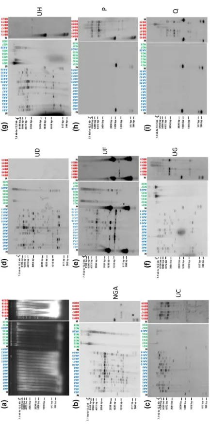
FI G U R E 5 So ut he rn b lo t hy br id iz at io n o f t ot al g en om ic D N A ( di ge st ed b y Hi nd III ) f ro m p la nt s o f t he f am ily M us ac ea e u si ng d iv er se c la de 2 b ad nav ira l s eq ue nc es a s p ro be s. S am pl es A A 1– 14 a nd A A 26 c or re sp on d t o M . ac um ina ta g en ot yp es , s am pl es B B1 5– 20 t o M . bal bi sia na g en ot yp es , s am pl es M 21 –2 5, M 27 , a nd M 28 t o o th er s pe ci es f ro m t he f am ily M us ac ea e. ( a) A ga ro se ge l e le ct ro ph or es is . ( b) –( i) H yb rid iz at io n r es ul ts f or B EV p ro be s U C ( b) , U D ( c) , U F ( d) , U G ( e) , U H ( f), N G A ( g) , P ( h) , a nd Q ( i). M , 1 k b l ad de r f ro m I nv itr og en
mismatch at the primer recognition site seems the most likely expla-nation. In addition, although membranes were washed stringently, we cannot rule out slight cross-hybridization yielding a very weak signal, as explained further below. The BEV NGA probe hybridized with all
M. acuminata samples, showing several bands ranging from high to low
molecular weight, revealing several BEV NGA sequences in those sam-ples. Although individuals displayed different patterns, they all shared two conserved fragments at 1 kb and c.3 kb (Figure 5b). In four M.
bal-bisiana samples (BB15–BB17 and BB20), a similarly weak signal was
seen at c.1 kb. This signal is probably due to cross-hybridization with BEV UF because no PCR amplification was obtained with BEV NGA primers on M. balbisiana samples, hybridization patterns were similar to those of BEV UF but fainter, and BEV UF shares c.85% identity with BEV NGA. Geering et al. (2005) also observed the absence of BEV 2 (similar to BEV NGA) in M. balbisiana. We also observed hybridization signals corresponding to high molecular weight (>12 kb) bands in sam-ples of other species of the genus Musa (M22, M24, M25, and M27). Similar to the NGA probe, we observed hybridization signals both for
M. acuminata and M. balbisiana with BEV UC, UF, UG, and UH probes
(Figure 5c,e–g). Hybridization patterns differed for all M. acuminata samples for each given probe and between probes, whereas hybrid-ization patterns in M. balbisiana plants were much more conserved, with fewer integrations detected except for BEV UC, for which poly-morphic patterns were observed (Figure 5c). Again, hybridization signals corresponding to high molecular weight (>12 kb) bands were present in some species other than M. acuminata and M. balbisiana be-longing to the genus Musa. Finally, corroborating PCR amplification re-sults, the three remaining probes (BEV-UD, -P, and -Q) exhibited very striking pattern differences between M. acuminata and M. balbisiana samples, with BEV UD hybridizing only with M. acuminata individuals (Figure 5d), and BEV P (Figure 5h) and Q (Figure 5i) probes hybridizing only with M. balbisiana samples.
3 | DISCUSSION
3.1 | Badnaviral sequences from clade 2 are
integrated in the banana genome
Previous studies differed on whether BEV UC, UD, UF, UG, and UH species corresponded to integrated and/or episomal viruses (Gayral & Iskra-Caruana, 2009; Harper et al., 2005). To address this ques-tion, we performed DNA analysis on EAH AAA triploids collected in BSV-affected areas in Uganda as well as on a diverse sampling encompassing BSV-free diploids M. acuminata and M. balbisiana and other Musa species from the family Musaceae.
Our data revealed BEV integration in M. acuminata and M.
balbi-siana genomes, with hybridization signals observed for plants with
symptoms (EAH AAA samples) and without symptoms (EAH AAA samples plus all the Musa diversity), consistent with the conclu-sion that BEV sequences are integrated in all our samples. For the first time, we show that some BEV sequences are shared between
M. acuminata and M. balbisiana genomes.
Our Southern blot results (Figure 5) indicate the level of plant ge-nome colonization, and suggest coevolution between virus and ba-nana. All M. acuminata banana samples showed different integration patterns, suggesting a large degree of badnavirus sequence coloni-zation via either multiple integration waves or genome duplications and evolution of one or a few initial integration events. Interestingly, each given probe suggested a wide polymorphism of badnavirus se-quence integration in all M. acuminata banana samples, probably re-flecting genetic and geographical M. acuminata subspecies diversity linked to various environmental pressures (Perrier et al., 2011). Our data are consistent with preliminary analysis by D’Hont et al. (2012), who reported the presence of different BEVs belonging to clade 2 at several loci distributed on 10/11 chromosomes of the genome of
M. acuminata ‘Pahang’.
The patterns observed for M. balbisiana are less complex, sug-gesting either fewer integrations or very few genome duplication events with a much less diverse profile. This low polymorphism of badnavirus sequence integration could be due to limited genetic di-versity among M. balbisiana banana species (Carreel, 1994; Gayral et al., 2010). Overall, our data corroborate preliminary data pub-lished by Geering et al. (2001), who reported integrations in all sam-ples, but fewer in M. balbisiana than in M. acuminata. Importantly, we demonstrate here for the first time that several BEVs are inte-grated in other species of the genus Musa, that is, M. basjoo, M.
itin-erans, M. laterita, M. ornata, and M. schizocarpa, (sections Eumusa or Rhodochlamys), but not in more phylogenetically distant samples
cor-responding to Callimusa and Australimusa sections or other genera of the family Musaceae (Table 3 and data not shown). This suggests that, for some BEVs, initial integrations occurred after formation of the family Musaceae but before speciation of the genus Musa (see below).
BEVs UC and UG appear to be widely disseminated within
M. acuminata and M. balbisiana, unlike BEV UF, which is abundant
in M. acuminata but poorly represented in M. balbisiana. Our results also show the absence of BEVs UD, UH, and NGA in M. balbisiana genomes, the absence of BEVs P and Q in M. acuminata genomes and the presence of all BEVs (except P and Q) in some other species of the family Musaceae.
Thus, our results differ from those of Harper et al. (2005), who suggested that BEVs UC, UD, UF, UG, and UH exist as episomal BSV particles, and support the hypothesis proposed by Gayral and Iskra-Caruana (2009), ascribing an exclusively integrated status to all BEVs of clade 2. Our present results allow us to ascribe the diversity of BSV exclusively to clades 1 (seven species to date: BSOLV, BSGFV, BSMYV, BSIMV, BSCAV, BSUAV, and BSVNV) and 3 (five species to date: BSUIV, BSULV, BSUMV, BSUJV, and BSUKV).
3.2 | What can BEVs tell us about the badnavirus/
banana coevolution?
Badnaviral sequences linked to banana plants are distributed over three main clades (Figure 4). Surprisingly, they are as diverse as all
the other viruses of the genus Badnavirus with which they share a same common ancestor. Interestingly, the clade to which these se-quences belong is associated with a particular status (episomal and/ or integrated) as a result of specific interactions between the virus and its banana host.
Clade 1 encompasses BSVs that are either episomal sensu stricto or both episomal and integrated (Iskra-Caruana et al., 2014) as ob-served for BSOLV, BSGFV, BSIMV, and BSMYV, where integrations restricted to M. balbisiana genomes occurred only after speciation of
M. acuminata/M. balbisiana, but before diversification of M. balbisi-ana (Chabannes et al., 2013). Although those eBSV exhibit a strong
rearranged structure, with inverted and duplicated sequences at-testing to past integration, pseudogenization has not progressed to the point where they can no longer reconstitute an infectious viral genome (Chabannes et al., 2013; Chabannes & Iskra-Caruana, 2013; Iskra-Caruana et al., 2010). Because nucleic acid identity between a given eBSV and the corresponding BSV is >99%, it is likely that episomal BSOLV, BSGFV, and BSIMV observed now are due mainly, or exclusively, to the awakening of an endogenous counterpart (Chabannes et al., 2013; Gayral et al., 2008).
Because no episomal virus belonging to clade 2 has ever been detected in banana plants, we assume that the episomal BEV coun-terpart has long since disappeared. BEVs are thus relics of ances-tor viruses that existed previously in wild plant populations, before speciation of the genus Musa and long before domestication and trade. Consequently, they are older than the BSVs and eBSV of clade 1, that is, the BSOLV, BSMYV, BSIMV, and BSGFV integrated in
M. balbisiana genomes, with integration estimated to have occurred
c.640,000 years ago (Gayral et al., 2010). Whether integrated BEV sequences confer a selective advantage by contributing towards plant virus resistance (via transcriptional or posttranscriptional gene silencing) is still an open question, but would explain both the disap-pearance of the counterpart episomal viruses and current integrated sequences resulting from pseudogenization. As described for other viruses (Feschotte & Gilbert, 2012), BEVs have become part of the genetic material of the banana host.
BSVs of clade 3 result from recent viral evolution in East Africa. Karamura et al. (1996) described a widespread BSV epidemic in Uganda on the East African Highland M. acuminata banana group (EAH AAA) (Kubiriba et al., 1997). The endemic presence of BSV in Uganda is probably the result of vegetative propagation of in-fected plants rather than vector transmission because cultivated cultivars are seedless and the rate of disease spread by mealybugs is slow (Daniells et al., 2001b; Kubiriba et al., 2001). Furthermore, BSVs generally do not have a severe impact on bunch production, particularly when cultural conditions are good. In the 109 EAH AAA samples analysed here, we observed a prevalence of species BSUIV, BSULV, and BSUMV from clade 3 (Table 2), whereas species BSUJV and BSUKV are poorly represented or absent, respectively, in agreement with the findings of Harper et al. (2005) (after cor-rection of BSV species names mislabelled in the latter article; the species BSUHV, BSUIV, BSUJV, BSUKV, and BSULV described by
Harper et al., 2005, were respectively submitted to GenBank as BSUIV, BSUJV, BSUKV, BSULV, and BSUMV). Importantly, no se-quences corresponding to BSUIV, BSULV, and BSUMV have so far been found integrated in banana plants (Figure 2a). The diversity of viruses from clade 3 observed nowadays is therefore more likely due either to introduction of infected banana cultivars from dif-ferent locations or to host shifts between banana and other plants (Iskra-Caruana et al., 2014).
F I G U R E 6 Schematic phylogenetic tree of BEV fixation
events relative to speciation events within the family Musaceae constructed using nodes supported from the latest Musa phylogenies (Christelova et al., 2011; Janssens et al., 2016; Li et al., 2010). Banana samples from different sections of the genus
Musa and other genera of the family Musaceae are represented.
Samples characterized during this work are in red. M. acuminata and M. balbisiana subspecies are represented by samples AA2 and AA7, and BB17, respectively. Green arrows, BEV fixation events; X, number of chromosomes
3.3 | What can BEVs tell us about the banana
phylogeny?
BEVs phylogeny suggests two close (in terms of parsimony) scenarios of banana genome integration: (a) a unique integration event before emergence of Musaceae, associated with several gene duplications and losses in some Musa subgroups; and (b) multiple integrations, whereby badnavirus ancestors of BEVs integrated into the banana ge-nome in at least three waves corresponding to distinct periods of ba-nana evolution, as proposed by Yu et al. (2019) for other endogenous pararetroviruses in Citrinae. Synteny analyses between the M.
acumi-nata Pahang (D’Hont et al., 2012) and PKW (Wang et al., 2019)
ge-nomes found no evidence that any BEV-containing loci were identical. This is not too surprising considering that the number of initial integra-tions is low, and that only half of the genomes of these two sequenced plants are being examined, because each derives from a duplication of an initial haploid plant. On the other hand, because genomes A and B diverged 4.5 million years ago (Lescot et al., 2008), the initial integra-tion loci may have diverged sufficiently so as to no longer be identi-fied during synteny analyses. Therefore, based on recently published banana phylogenies (Christelova et al., 2011; Janssens et al., 2016; Li et al., 2010), we propose a speculative scheme (Figure 6) depicting each BEV integration event for scenario (b) with regards to the main speciation events reported within the family Musaceae.
The presence of BEVs UC, UF, and UG within both M. acuminata and M. balbisiana genomes, as well as other species of the genus
Musa (M. basjoo, M. itinerans, M. laterita, or M. ornata), could
repre-sent the first and second integration waves (Figure 7a,b). Indeed, BEV UF integration may have followed that of BEV UC and BEV UG because M. basjoo and M. itinerans genomes do not harbour BEV UF integrations (Table 3). Interestingly, amongst Musa sections, BEVs are present only in Rhodochlamys (M. laterita and M. ornata) and Eumusa (M. acuminata, M. balbisiana, M. schizocarpa, M. basjoo, and M. itinerans) and are absent from both Australimusa (M.
texti-lis) and Callimusa (M. coccinea)—phylogenetically very distant from Rhodochlamys and Eumusa (Christelova et al., 2011; Li et al., 2010).
According to estimates of species divergence times within the family Musaceae (Christelova et al., 2011), integrations of BEVs UC, UF, UG took place between c.28 and 50 million years ago, corresponding to speciation events within Rhodochlamys/Eumusa and the age of the genus Musa, respectively. Importantly, based on the known geographical distribution of the main sections of the genus Musa (De Langhe et al., 2009) and the M. acuminata and
M. balbisiana subspecies (Perrier et al., 2011), the absence of BEV
in Australimusa and Callimusa genomes, and their presence in all
M. acuminata and M. balbisiana subspecies, indicates that initial
badnavirus infections of the Musa ancestor and viral integration of BEVs UC, UF, and UG occurred in South/South-East continental Asia (Figure 7a). Using the BEV markers developed here, we can
F I G U R E 7 Geographical distribution of main sections of
the genus Musa and subspecies Eumusa along with proposed BEV fixation events. A first wave of BEV fixation (BEV UC and UG) occurred in the Rhodochlamys and Eumusa ancestor (a). A second wave of integration fixed BEV UF in the
M. balbisiana/M. acuminata/Rhodochlamys ancestor but not in
the M. itinerans/M. basjoo ancestor (b). The boundaries of the
M. itinerans/M. basjoo ancestor are uncertain. Finally, a third wave
of BEV fixation occurred in M. acuminata (BEV UD, UH, and NGA) and M. balbisiana (P and Q) ancestors prior to diversification of both subspecies (c). M. balbisiana and M. acuminata diversification routes are illustrated by black and red arrows, respectively. All BEV integration and fixation events proposed took place in South/ South-East continental Asia. The figure was inspired by Simmonds (1962), De Langhe et al. (2009), and Perrier et al. (2011)
further restrict the origin of the Eumusa/Rhodochlamys ancestor to the south mainland of Asia, thus narrowing considerably the area defined initially by De Langhe et al. (2009).
Alternatively, virus ancestors of BEVs UC, UF, and UG could have integrated independently and randomly into different Musa genomes. This hypothesis is less parsimonious because it requires that at least three distinct viruses (BEVs UC, UF, and UG) integrated into different Musa genomes over time and in different geographical areas.
The presence of BEVs UD, UH, and NGA only in the genome of
M. acuminata, in contrast to the absence of BEVs P and Q (present
only in M. balbisiana), suggests a third wave of integrations occur-ring after M. acuminata and M. balbisiana speciation (Figures 6 and 7c) in scenario (b). Considering the close phylogenetic relationship of BEVs UF and UD (Figure 4), we propose that BEV UD emerged after a duplication of BEV UF in M. acuminata only. Interestingly, Southern blot hybridization patterns clearly indicate that this in-tegration occurred after speciation of M. acuminata/M. balbisiana but before species diversification. This third wave of integration seems likely to have been a host response to different viral pres-sures, namely BEVs P and Q for M. balbisiana and BEVs NGA, UD, and UH for M. acuminata, which come from different geographical
TA B L E 4 Description of plant material used in this study
Genus or section in the
family Musaceae Species/subspecies Common name Genome Accession number
Number in that study
Eumusa M. acuminata subsp. errans Agutay AAw ITC1028 (NEU0033) AA1
Eumusa M. acuminata subsp. zebrina Zebrina AAw ITC1177 (NEU0029) AA2
Eumusa M. acuminata subsp. siamea Pa (Rayong) AAw ITC0672 (NEU0024) AA3
Eumusa M. acuminata subsp. malaccensis Pahang AAw ITC0609 (NEU0013) AA4
Eumusa M. acuminata subsp. malaccensis THA018 AAw ITC1067 (NEU0034) AA5
Eumusa M. acuminata subsp. truncata Truncata AAw (NEU0027) AA6
Eumusa M. acuminata subsp. burmannica Long Tavoy AAw ITC0283 (NEU0016) AA7
Eumusa M. acuminata subsp. microcarpa Bornéo AAw ITC0253 (NEU0028) AA8
Eumusa M. acuminata subsp. malaccensis Malaccensis AAw ITC0250 AA9
Eumusa M. acuminata Wikago AAcv NEU0102 (ITC0888) AA10
Eumusa M. acuminata subsp. siamea Khae (Phrae) AAw ITC0660 (NEU0025) AA11
Eumusa M. acuminata subsp. malaccensis AAw ITC0399 AA12
Eumusa M. acuminata subsp.
burmannicoides
Calcutta 4 AAw ITC0249 (NEU0017) AA13
Eumusa M. acuminata subsp. siamea Pa (Songkhla) AAw ITC0408 (NEU0043) AA14
Eumusa M. acuminata subsp. banksii Madang AAw ITC0254 AA26
Eumusa M. balbisiana Honduras BBw ITC0247 (NEU0049) BB15
Eumusa M. balbisiana Cameroun BBw ITC0246 (NEU0050) BB16
Eumusa M. balbisiana Pisang Klutuk Wulung BBw ITC1063 (NEU0056) BB17
Eumusa M. balbisiana Butuhan BBw ITC0565 BB18
Eumusa M. balbisiana Lal Velchi BBw NEU0051 BB19
Eumusa M. balbisiana Papouasie New
Guinea
BBw ITC0626 BB20
Eumusa M. schizocarpa SS ITC856 M21
Rhodochlamys M. laterita NEU0008 M22
Australimusa M. textilis TT ITC1072 (NEU0001) M23
Eumusa M. basjoo NEU0060 M24
Rhodochlamys M. ornata NEU0007 M25
Eumusa M. itinerans M27
Callimusa M. coccinea ITC0287 (NEU0003) M28
Ensete Ensete ventricosum M32
Musella Musella lasiocarpa M34
Note. ITC numbers, accession codes from the International Transit Center, Catholic University, Leuven, Belgium; NEU numbers, accession codes from
the Banana collection of CIRAD Neufchateau, Guadeloupe, France; AAw or AAcv, wild or cultivar of diploid acuminata bananas; BBw, wild diploid
balbisiana bananas; M, other species from the family Musaceae. Sections Eumusa, Rodochlamys, Callimusa, and Australimusa correspond to subsections
areas (Perrier et al., 2011) (Figure 7b,c). Interestingly, within the genus Musa, PCR and Southern blot profiles of BEV species sug-gest a close relationship between M. acuminata subspecies and banana species from section Rhodochlamys (M. ornata M25 and
M. laterita M22), in agreement with published Musa phylogeny
(Christelova et al., 2011; Li et al., 2010). Similarly, profiles of
M. basjoo (M24) and M. itinerans (M27) are barely distinguishable,
again corroborating their closeness in Musa phylogeny (Li et al., 2010). Notably, both exhibit intermediate BEV profiles compared with M. acuminata and M. balbisiana, supporting their equidistant position from the latter in the phylogenetic tree of Musaceae (Li et al., 2010).
Duroy et al. (2016) previously demonstrated eBSV to be rele-vant phylogenetic markers to illustrate the M. balbisiana phylogeo-graphic story. In light of our data, and given the high polymorphism of BEVs within M. acuminata species, BEV patterns can be added to the arsenal of phylogenetic markers to describe and complete Musa phylogeny.
4 | EXPERIMENTAL PROCEDURES
4.1 | Plant material
Leaf samples from 109 East African Highland banana plants repre-senting five clone sets encompassing EAH AAA banana genetic di-versity (Nakitembe [32], Musakala [19], Nakabulu [16], Nfuuka [23], and Nbide [beer cultivar, 19]; Karamura & Pickersgill, 1999) were col-lected in Uganda in 2009; 91 samples showed typical banana leaf streak mosaic symptoms, indicating BSV infection.
We also analysed 30 samples encompassing different M.
acumi-nata (AA1–AA14, AA26) and M. balbisiana (BB15–BB20) diploid
sub-species, along with other species from the genus Musa (M21–M25, M27, and M28) and other genera of the family Musaceae (M32 and M34) (Table 4). Samples were collected in the Banana field collection of CIRAD Neufchateau, Guadeloupe, France.
4.2 | Extraction of genomic DNA from banana
Genomic DNA was extracted from fresh or frozen banana leaf tissue using the method of Gawel and Jarret (1991).
4.3 | Immunocapture PCR detection
BSV species were detected by IC-PCR according to a procedure adapted from Le Provost et al. (2006) using specific BSOLV, BSGFV, BSIMV, and BSMYV primers (Table 1) and a polyclonal an-tiserum raised against a cocktail of purified BSV species and SCBV species (kindly provided by B.E.L. Lockhart). To avoid contamina-tion by plant genomic DNA, samples were treated with RNase-free
DNase I (Promega). DNase mix (3 µl of 10 × buffer [400 mM Tris. HCl pH 8, 100 mM MgSO4, 10 mM CaCl2], 3 µl of DNase I [1 U/µl],
and 24 µl of water) was added to coated tubes and incubated for 1 hr at 37 °C. The supernatant was removed and the tubes washed once with water. DNase I was inactivated by incubation at 95 °C for 10 min.
4.4 | PCR, cloning, and sequencing
Primers in the RTase/RNase H region of badnavirus ORF III were designed to specifically detect each Ugandan badnaviral species of clades 2 and 3 (Table 1). PCR was performed with 1 U GoTaq DNA polymerase according to the manufacturer's instructions (Promega) and the following thermal cycling conditions: 1 cycle at 94 °C for 4 min; 35 cycles at 94 °C for 30 s, Tm (Table 1) for 30 s, 72 °C for 30 s; followed by 1 cycle at 72 °C for 10 min. PCR fragments were sequenced by Beckman Coulter Genomics (UK).
PCR products used as probes for Southern blot hybridization were gel-purified using the Wizard SV Gel and PCR Clean-up System (Promega) and cloned into the pGEM-T Easy vector according to the manufacturer's instructions (Promega).
4.5 | Southern blot hybridization
Total genomic DNA (40 µg per sample) was digested overnight with 1 U/μg DNA for each enzyme in a final volume of 200 µl; HindIII for samples representative of the Musa diversity, and KpnI or Alw441 for EAH banana samples. Samples were separated by electrophoresis on 1% agarose gels and capillary-transferred overnight to Hybond N+ membrane (Amersham Biosciences) in 20 × saline-sodium citrate (SSC) buffer. Nucleic acids were fixed onto the membrane using a UV crosslinker (70,000 μJ/cm2), then prehybridized in 20 ml of buffer
(50 mM Tris.HCl pH 8, 25 mM ETDA, 5 × SSC, 1% sodium dode-cyl sulphate [SDS], 2.5 × Denhardt's solution and 2 mg denatured salmon sperm DNA) and incubated for 3 hr at 65 °C.
Fragments corresponding to RTase/RNaseH regions of each species of clade 3 (BSUIV, BSULV, BSUMV) and each group of clade 2 (BEVs UC, UD, UF, UG, UH, P, Q, NGA) were released from pGEM-T Easy plasmids using EcoRI digestion and used as probes. DNA probe (50 ng) was labelled with [α-32P] dCTP using a random
priming protocol (Prime-a-Gene kit, Promega). Labelled probe was then added to 20 ml of hybridization solution (as above + 5% dex-tran sulphate) and incubated overnight at 65 °C. To remove non-specific hybridization signal, membranes were washed at 65 °C for 10 min, twice in SSC with 0.1% SDS solution and once in 0.5 × SSC with 0.1% SDS. Additional washes, if required, used 0.2 × SSC with 0.1% SDS solution. Membranes were wrapped in transparent plas-tic (Scel O Frais) and screen scanned after overnight exposure on a filmless autoradiography Typhoon FLA 9000 imaging system (GE Healthcare).
4.6 | Rolling circle amplification
DNA was amplified using a TempliPhi Amplification kit (GE Healthcare) following the protocol described by James et al. (2011). Reaction products were digested using 2 U of different restriction endonucleases (Promega), according to the manufacturer's instruc-tions, and then separated by electrophoresis in 1% agarose gels.
4.7 | Phylogenetic analysis
Badnaviral sequences were aligned using the MAFFT software algo-rithm (Katoh & Standley, 2013). Phylogenetic trees were constructed using the maximum-likelihood method with PhyML 3.0 (Guindon et al., 2010) and visualized using Darwin 5 software (Perrier et al., 2003). The robustness of trees was tested with aLRT-SH-like statis-tical support (Anisimova et al., 2011). The new sequences produced during this work have GenBank accession numbers KJ720037– KJ720154 and KJ734678–KJ734703.
ACKNOWLEDGMENTS
We are very grateful to Jerome Kubiriba from the National Agricultural Research Organisation (NARO), Jim Lorenzen (previ-ously IITA), and farmers in Uganda for assistance with field sam-pling. We would like to thank the Guadeloupe Centre de Ressources Biologiques Plantes Tropicales and especially Nilda Paulo de la Reberdière and Danièle Roques for providing plant material. We also thank CIRAD for funding.
DATA AVAIL ABILIT Y STATEMENT
The data that support the findings of this study are openly available in NCBI GenBank at https://www.ncbi.nlm.nih.gov/nucle otide, ref-erence numbers KJ720037–KJ720154 and KJ734678–KJ734703.
ORCID
Matthieu Chabannes https://orcid.org/0000-0001-5754-5982
Marie-Line Iskra-Caruana https://orcid.
org/0000-0003-4486-2449
Emmanuelle Muller https://orcid.org/0000-0003-3292-4809
REFERENCES
Anisimova, M., Gil, M., Dufayard, J.-F., Dessimoz, C. & Gascuel, O. (2011) Survey of branch support methods demonstrates accuracy, power, and robustness of fast likelihood-based approximation schemes.
Systematic Biology, 60(), 685–699.
Bertsch, C., Beuve, M., Dolja, V.V., Wirth, M., Pelsy, F., Herrbach, E. et al. (2009) Retention of the virus-derived sequences in the nuclear ge-nome of grapevine as a potential pathway to virus resistance. Biology
Direct, 4, 21.
Carreel, F. (1994) Etude de la diversité génétique des bananiers (genre
Musa) à l'aide des marqueurs RFLP. Paris, France: Institut National
Agronomique Paris-Grignon.
Carreel, F., Gonzalez de Leon, D., Lagoda, P., Lanaud, C., Jenny, C., Horry, J.P. et al. (2002) Ascertaining maternal and paternal lineage within
Musa by chloroplast and mitochondrial DNA RFLP analyses. Genome,
45, 679–692.
Chabannes, M., Baurens, F.-C., Duroy, P.-O., Bocs, S., Vernerey, M.-S., Rodier-Goud, M. et al. (2013) Three infectious viral species lying in wait in the banana genome. Journal of Virology, 87, 8624–8637. Chabannes, M. & Iskra-Caruana, M.L. (2013) Endogenous
pararetro-viruses—a reservoir of virus infection in plants. Current Opinion in
Virology, 3, 615–620.
Christelova, P., Valarik, M., Hribova, E., De Langhe, E. & Dolezel, J. (2011) A multi gene sequence-based phylogeny of the Musaceae (banana) family. BMC Evolutionary Biology, 11, 103.
D’Hont, A., Denoeud, F., Aury, J.M., Baurens, F.C., Carreel, F., Garsmeur, O. et al. (2012) The banana (Musa acuminata) genome and the evolu-tion of monocotyledonous plants. Nature, 488, 213–217.
Daniells, J., Jenny, C., Karamura, D.&Tomekpe, K. (2001a) Musalogue: A
catalogue of Musa germplasm. Diversity in the genus Musa. Montpellier,
France: Compiled by Arnaud, E. and Sharrock, S. International Network for the Improvment of Banana and Plantain.
Daniells, J.W., Geering, A.D.W., Bryde, N.J. & Thomas, J.E. (2001b) The effect of Banana streak virus on the growth and yield of dessert ba-nanas in tropical Australia. Annals of Applied Biology, 139, 51–60. De Langhe, E., Vrydaghs, L., De Maret, P., Perrier, X. & Denham, T. (2009)
Why bananas matter: An introduction to the history of banana do-mestication. Ethnobotany Research and Applications, 7, 199–216. Duroy, P.-O., Perrier, X., Laboureau, N., Jacquemoud-Collet, J.P. &
Iskra-Caruana, M.L. (2016) How endogenous plant pararetroviruses shed light on Musa evolution. Annals of Botany, 117, 625–641.
FAOStat (2014) FAO production statistics for banana and plantain 2012. Rome, Italy: FAO.
Feschotte, C. & Gilbert, C. (2012) Endogenous viruses: insights into viral evolution and impact on host biology. Nature Reviews Genetics, 13, 283–296.
Gambley, C.F., Geering, A.D., Steele, V. & Thomas, J.E. (2008) Identification of viral and non-viral reverse transcribing elements in pineapple (Ananas comosus), including members of two new badnavi-rus species. Archives of Virology, 153, 1599–1604.
Gawel, N.J. & Jarret, R.L. (1991) A modified CTAB DNA extraction pro-cedure for Musa and Ipomoea. Plant Molecular Biology Reporter, 9, 262–266.
Gayral, P., Blondin, L., Guidolin, O., Carreel, F., Hippolyte, I., Perrier, X. et al. (2010) Evolution of endogenous sequences of banana streak virus: what can we learn from banana (Musa sp.) evolution? Journal of
Virology, 84, 7346–7359.
Gayral, P. & Iskra-Caruana, M.-L. (2009) Phylogeny of Banana streak virus reveals a recent burst of integrations in the genome of banana (Musa sp.). Journal of Molecular Evolution, 69, 65–80.
Gayral, P., Noa-Carrazana, J.C., Lescot, M., Lheureux, F., Lockhart, B.E., Matsumoto, T. et al. (2008) A single Banana streak virus integration event in the banana genome as the origin of infectious endogenous pararetrovirus. Journal of Virology, 82, 6697–6710.
Geering, A.D., Olszewski, N.E., Dahal, G., Thomas, J.E. & Lockhart, B.E. (2001) Analysis of the distribution and structure of integrated
Banana streak virus DNA in a range of Musa cultivars. Molecular Plant Pathology, 2, 207–213.
Geering, A.D.W., Olszewski, N., Harper, G., Lockhart, B., Hull, R. & Thomas, J.E. (2005) Banana contains a diverse array of endogenous badnaviruses. Journal of General Virology, 86, 511–520.
Geering, A.D., Parry, J.N. & Thomas, J.E. (2011) Complete genome se-quence of a novel badnavirus, banana streak IM virus. Archives of
Virology, 156, 733–737.
Geijskes, R.J., Braithwaite, K.S., Smith, G.R., Dale, J.L. & Harding, R.M. (2004) Sugarcane bacilliform virus encapsidates genome concatam-ers and does not appear to integrate into the Saccharum officinarum genome. Archives of Virology, 149, 791–798.
Gregor, W., Mette, M.F., Staginnus, C., Matzke, M.A. & Matzke, A.J. (2004) A distinct endogenous pararetrovirus family in Nicotiana
tomentosiformis, a diploid progenitor of polyploid tobacco. Plant Physiology, 134, 1191–1199.
Guindon, S., Dufayard, J.-F., Lefort, V., Anisimova, M., Hordijk, W. & Gascuel, O. (2010) New algorithms and methods to estimate maxi-mum-likelihood phylogenies: assessing the performance of PhyML 3.0. Systematic Biology, 59, 307–321.
Hansen, C.N., Harper, G. & Heslop-Harrison, J.S. (2005) Characterisation of pararetrovirus-like sequences in the genome of potato (Solanum tuberosum). Cytogenetic and Genome Research, 110, 559–565.
Harper, G., Hart, D., Moult, S., Hull, R., Geering, A. & Thomas, J. (2005) The diversity of Banana streak virus isolates in Uganda. Archives of
Virology, 150, 2407–2420.
Harper, G. & Hull, R. (1998) Cloning and sequence analysis of banana streak virus. Virus Genes, 17, 271–278.
Harper, G., Osuji, J.O., Heslop-Harrison, J.S. & Hull, R. (1999) Integration of banana streak badnavirus into the Musa genome: molecular and cytogenetic evidence. Virology, 255, 207–213.
Holmes, E.C. (2011) The evolution of endogenous viral elements. Cell
Host & Microbe, 10, 368–377.
Hull, R. (2002) Matthew’s plant virology. San Diego: Academic Press. Iskra-Caruana, M.-L., Baurens, F.-C., Gayral, P. & Chabannes, M. (2010)
A four-partner plant–virus interaction: enemies can also come from within. Molecular Plant-Microbe Interaction, 23, 1394–1402.
Iskra-Caruana, M.L., Duroy, P.-O., Chabannes, M. & Muller, E. (2014) The common evolutionary history of badnaviruses and banana. Infection,
Genetics and Evolution, 21, 83–89.
Jakowitsch, J., Mette, M.F., van Der Winden, J., Matzke, M.A. & Matzke, A.J. (1999) Integrated pararetroviral sequences define a unique class of dispersed repetitive DNA in plants. Proceedings of the National
Academy of Sciences of the United States of America, 96, 13241–13246.
James, A.P., Geijskes, R.J., Dale, J.L. & Harding, R.M. (2011) Molecular characterisation of six badnavirus species associated with leaf streak disease of banana in East Africa. Annals of Applied Biology, 158, 346–353.
Janssens, S.B., Vandelook, F., De Langhe, E., Verstraete, B., Smets, E., Vandenhouwe, I. et al. (2016) Evolutionary dynamics and biogeog-raphy of Musaceae reveal a correlation between the diversifica-tion of the banana family and the geological and climatic history of Southeast Asia. New Phytologist, 210, 1453–1465.
Karamura, D.A., Karamura, E.B. & Gold, C.S. (1996) Cultivar distribution in primary banana growing regions of Uganda. MusAfrica, 9, 3–5. Karamura, D. & Pickersgill, B. (1999) A classification of the clones of
East African highland banana (Musa) found in Uganda. Plant Genetic
Resources Newsletter, 119, 1–6.
Katoh, K. & Standley, D.M. (2013) MAFFT multiple sequence alignment software version 7: improvements in performance and usability.
Molecular Biology and Evolution, 30, 772–780.
Kubiriba, J., Legg, J.P., Tushemereirwe, W. & Adipala, E. (2001) Disease spread patterns of banana streak virus in farmers’ fields in Uganda.
Annals of Applied Biology, 139, 31–36.
Kubiriba, J., Tushemereirwe, W. & Karamura, E.B. (1997) Distribution of banana streak virus in Uganda. MusAfrica, 11, 17.
Kunii, M., Kanda, M., Nagano, H., Uyeda, I., Kishima, Y. & Sano, Y. (2004) Reconstruction of putative DNA virus from endogenous rice tungro bacilliform virus-like sequences in the rice genome: implications for integration and evolution. BMC Genomics, 5, 80.
Laney, A.G., Hassan, M. & Tzanetakis, I.E. (2012) An integrated badnavirus is prevalent in fig germplasm. Phytopathology, 102, 1182–1189.
Le Provost, G., Iskra-Caruana, M.-L., Acina, I. & Teycheney, P.-Y. (2006) Improved detection of episomal banana streak viruses by multiplex immunocapture PCR. Journal of Virological Methods, 137, 7–13. Lescot, M., Piffanelli, P., Ciampi, A.Y., Ruiz, M., Blanc, G., Leebens-Mack,
J. et al. (2008) Insights into the Musa genome: syntenic relationships to rice and between Musa species. BMC Genomics, 9, 58.
Li, L.F., Hakkinen, M., Yuan, Y.M., Hao, G. & Ge, X.J. (2010) Molecular phylogeny and systematics of the banana family (Musaceae) inferred from multiple nuclear and chloroplast DNA fragments, with a special reference to the genus Musa. Molecular and Phylogenetic Evolution, 57, 1–10.
Lockhart, B.E.L. & Jones, D.R. (2000) Banana streak. In: Jones, D.R. (Ed.), Diseases of banana, abaca and enset. Wallingford, UK: CAB International, pp. 263–274.
Ndowora, T., Dahal, G., La Fleur, D., Harper, G., Hull, R., Olszewski, N.E. et al. (1999) Evidence that badnavirus infection in Musa can originate from integrated pararetroviral sequences. Virology, 255, 214–220. Pahalawatta, V., Druffel, K. & Pappu, H. (2008) A new and distinct
spe-cies in the genus Caulimovirus exists as an endogenous plant pararet-roviral sequence in its host, Dahlia variabilis. Virology, 376, 253–257. Perrier, X., De Langhe, E., Donohue, M., Lentfer, C., Vrydaghs, L., Bakry,
F. et al. (2011) Multidisciplinary perspectives on banana (Musa spp.) domestication. Proceedings of the National Academy of Sciences of the
United States of America, 108, 11311–11318.
Perrier, X., Flori, A. & Bonnot, F. (2003) Data analysis methods. In: Hamon, P., Seguin, M., Perrier, X. & Glaszmann, J.C. (Eds.) Genetic
Diversity of Cultivated Tropical Plants. Montpellier: Enfield Science
Publishers, pp. 43–76.
Richert-Poggeler, K.R., Noreen, F., Schwarzacher, T., Harper, G. & Hohn, T. (2003) Induction of infectious petunia vein clearing (pararetro) virus from endogenous provirus in petunia. EMBO Journal, 22, 4836–4845.
Richert-Poggeler, K.R. & Shepherd, R.J. (1997) Petunia vein-clearing virus: a plant pararetrovirus with the core sequences for an integrase function. Virology, 236, 137–146.
Simmonds, N.W. (1962) The evolution of the bananas. London: Longmans. Staginnus, C., Gregor, W., Mette, M.F., Teo, C.H., Borroto-Fernandez,
E.G., Machado, M.L. et al. (2007) Endogenous pararetroviral se-quences in tomato (Solanum lycopersicum) and related species. BMC
Plant Biology, 7, 24.
Wang, Z., Miao, H., Liu, J., Xu, B., Yao, X., Xu, C. et al. (2019) Musa
balbisi-ana genome reveals subgenome evolution and functional divergence. Nature Plants, 5, 810–821.
Yu, H., Wang, X., Lu, Z., Xu, Y., Deng, X. & Xu, Q. (2019) Endogenous pararetrovirus sequences are widely present in Citrinae genomes.
Virus Research, 262, 48–53.
SUPPORTING INFORMATION
Additional supporting information may be found online in the Supporting Information section.)
How to cite this article: Chabannes M, Gabriel M, Aksa A, et
al. Badnaviruses and banana genomes: a long association sheds light on Musa phylogeny and origin. Mol Plant Pathol. 2020;00:1–15. https://doi.org/10.1111/mpp.13019