Cellular and Molecular Analysis of Neuronal Structural Plasticity in the
Mammalian Cortex
by
Wei-Chung Allen Lee S.B., Chemical Engineering California Institute of Technology, 1998
A.B., Biochemistry and Government Bowdoin College, 1998
Submitted to the Department of Brain and Cognitive Sciences in Partial Fulfillment of the Requirements for the Degree of
Doctor of Philosophy in Neuroscience at the
Massachusetts Institute of Technology June 2006
© 2006 Massachusetts Institute of Technology. All rights reserved.
Signature of Author... Department of Brain and Cognitive Sciences May 15, 2006
Certified by... Elly Nedivi Fred and Carol Middleton Associate Professor of Neurobiology Thesis Supervisor
Accepted by... Matthew Wilson Picower Scholar & Professor of Neurobiology
Cellular and Molecular Analysis of Neuronal Structural Plasticity in the
Mammalian Cortex
by
Wei-Chung Allen Lee
Submitted to the Department of Brain and Cognitive Sciences on May 15, 2006 in Partial Fulfillment of the
Requirements for the Degree of Doctor of Philosophy in Neuroscience
ABSTRACT
Despite decades of evidence for functional plasticity in the adult brain, the role of structural plasticity in its manifestation remains unclear. cpg15 is an activity-regulated gene encoding a membrane-bound ligand that coordinately regulates growth of apposing dendritic and axonal arbors and the maturation of their synapses. Here we compare cpg15 expression during normal development of the rat visual system, with that seen in response to dark rearing, monocular retinal action potential blockade, or monocular deprivation. Our results show that: (1) cpg15 expression in visual cortex correlates with the electrophysiologically mapped critical period for development of eye-specific preference in the primary visual cortex. (2) Dark rearing elevates adult levels of expression. (3) A component of cpg15 expression is activity-dependent after the peak of the critical period. (4) At the peak of the critical period, monocular deprivation decreases cpg15 expression more than monocular TTX blockade. And (5) cpg15 expression is robust and regulated by light in the superficial layers of the adult visual cortex. This suggests that cpg15 is an excellent molecular marker for the visual system’s capacity for plasticity and predicts that neural remodeling normally occurs in the extragranular layers of the adult visual cortex. To examine the extent of neuronal remodeling that occurs in the brain on a daily basis, we used a multi-photon based microscopy system for chronic in vivo imaging and reconstruction of entire neurons in the superficial layers of the rodent cerebral cortex. Here, we show the first unambiguous evidence of dendrite growth and remodeling in adult neurons. Over a period of months, neurons could be seen extending and retracting existing branches, and in rare cases adding new branch tips. Neurons exhibiting dynamic arbor rearrangements were GABA positive non-pyramidal interneurons, while pyramidal cells remained stable. These results are consistent with the idea that dendritic structural remodeling is a substrate for adult plasticity and suggest that circuit rearrangement in the adult cortex is restricted by cell type-specific rules.
Thesis Supervisor: Elly Nedivi
Acknowledgements
Tremendous thanks to my advisor Elly Nedivi for teaching, nurturing, and having faith in a fledgling interested in the science of how the brain works.
I would also like to thank the past and present members of the Nedivi Lab, especially Marnie Phillips, Tadahiro Fujino, Corey Harwell, Ulrich Putz and Jeff Cottrell for their colleagueship, friendship, and for the unique, invigorating research environment they helped to create.
I would next like to thank my committee, Mark Bear, Martha Constantine-Paton, and Peter So, for their guidance. I would also like to thank my collaborators, Josh Sanes, Guoping Feng, Emery Brown, and particularly Peter So and Hayden Huang, whose assistance was invaluable. I would like to thank my parents, Han-San and Ming-Tzu Lee, for their life-long support and dedication to my scholarship.
Finally, this would not have been possible without the unwavering support, patience, love and understanding of my wife, Jenny.
Contents
Abstract...2
Acknowledgements...3
Table of Contents...4
Chapter 1: Introduction...6
Chapter 2: Extended plasticity of visual cortex in dark-reared animals may result from prolonged expression of cpg15-like genes...14
Chapter 3: Dynamic remodeling of dendritic arbors in GABAergic interneurons of adult visual cortex...44
Chapter 4: Conclusion...75
Appendix...84
But the functional specialization of the brain imposed on the neurones two great lacunae; proliferative inability and irreversibility of intraprotoplasmic differentiation. It is for this reason
that, once development was ended, the founts of growth and regeneration of the axons and dendrites dried up irrevocably. In adult centres the nerve paths are something fixed, ended,
immutable. Everything may die, nothing may be regenerated. It is for the science of the future to change, if possible, this harsh decree.
Chapter 1
IntroductionDevelopmental and adult plasticity
Within the astounding complexity of the brain, fundamental yet unknown mechanisms sub serve learning, memory, perception, behavior, reason, and consciousness. How the outside world influences the brain likely changes the mind. The capacity of the central nervous system (CNS) to modify connections in response to activity is termed plasticity. Plasticity is a prominent feature of CNS development (Constantine-Paton et al., 1990; Shatz, 1990; Goodman and Shatz, 1993), and in the adult, putatively underlies learning and memory (Bailey and Kandel, 1993; Bliss and Collingridge, 1993) and adaptive reorganization of primary sensory maps (Merzenich et al., 1988; Kaas et al., 1990; Allard et al., 1991; Kaas, 1991; Gilbert and Wiesel, 1992; Recanzone et al., 1993; Zarzecki et al., 1993; Armstrong-James et al., 1994; Diamond et al., 1994). Anatomical and physiological studies of developmental plasticity in the visual system have provided valuable insight into how activity drives the final patterning of neuronal connections (Constantine-Paton et al., 1990; Shatz, 1990; Katz and Shatz, 1996).
The critical period for susceptibility to environmental influences is one of the most influential models to emerge from studies of visual system development. In highly binocular animals such as cats, primates, and ferrets, electrophysiological plasticity during the critical period for development of eye specific preference in the primary visual cortex is accompanied by activity-driven segregation of geniculocortical afferents into ocular dominance columns (Hubel et al., 1977; Shatz and Stryker, 1978; Issa et al., 1999). There exist, however, many instances where no apparent anatomical plasticity is detectable when physiological plasticity clearly exists. Electrophysiological studies of the rodent visual system show that cortical neurons develop well
defined functional properties relating to eye preference and orientation selectivity (Parnavelas et al., 1981; Maffei et al., 1992; Fagiolini et al., 1994; Gordon and Stryker, 1996; Antonini et al., 1999) and are capable of undergoing shifts in eye preference during a critical period in development (Drager, 1978; Fagiolini et al., 1994; Gordon and Stryker, 1996; Guire et al., 1999; Frenkel and Bear, 2004); however, they are not anatomically segregated according to these properties akin to the feline and primate visual system. Since there is no segregation into eye-specific zones, anatomical changes in response to monocular deprivation (MD) during the critical period can be seen only with single-cell labeling and reconstruction (Antonini et al., 1999).
The capacity for change in response to manipulation of patterned visual input is normally restricted to a defined age window during postnatal development (Hubel and Wiesel, 1970; Hubel et al., 1977). However, in kittens or rats dark-reared (DR) past the end of the normal critical period, subsequent manipulation of visual input results in significant plasticity of cortical binocular connectivity measured by electrophysiological recordings (Cynader and Mitchell, 1980; Mower et al., 1981; Fagiolini et al., 1994; Guire et al., 1999), but not by altered segregation of ocular dominance columns (Mower et al., 1985). The complexity of interpreting dark rearing experiments underscores our poor understanding of the molecular mechanisms underlying critical period plasticity.
Physiological plasticity without large-scale anatomical rearrangements, as found during the critical period in species without ocular dominance columns, or in the superficial cortical layers of adult animals of all species, normal or DR, suggests that the capacity for rearrangement of thalamocortical afferents in layer 4 is not always the most appropriate measure of plasticity. Recent studies show that organization of the extragranular layers precedes the establishment of
cortical columns and serves to guide the anatomical remodeling in layer 4 (Ruthazer and Stryker, 1996; Crair et al., 1997; Crair et al., 1998; Trachtenberg et al., 2000). In fact, since plasticity of the cortical extragranular layers persists in the adult beyond the critical period for anatomical rearrangement in layer 4 (Daw et al., 1992; Diamond et al., 1994; Glazewski and Fox, 1996; Rioult-Pedotti et al., 1998; Feldman, 2000), remodeling of layer 4 may not be a requisite for plasticity, but in some species may only serve to set a foundation for further progressive refinement at higher levels of the cortical circuitry (Trachtenberg et al., 2000). The most attractive aspect of this interpretation is that it unifies the adult plasticity seen in extragranular layers with developmental plasticity during the critical period.
Little is known about the cellular mechanisms underlying developmental activity-evoked synaptic remodeling, and how it relates to plasticity in the adult. During development inappropriate connections are withdrawn, while appropriate connections develop elaborate terminal arbors (Guillery, 1972; Sretavan and Shatz, 1986; Antonini and Stryker, 1993). Here, an appropriate connection is defined as one where pre- and postsynaptic activity is correlated, also termed a Hebbian synapse (Stryker and Strickland, 1984; Fregnac et al., 1988; Shulz and Fregnac, 1992). The Hebbian hypothesis provides a theoretical basis for linking developmental and adult plasticity. Hebbian characteristics of synapses in layer IV of visual cortex during the critical period, are similar to those of synapses in the CA1 region of the adult hippocampus that are capable of undergoing long-term potentiation, a form of synaptic plasticity associated with explicit forms of learning and memory (Morris et al., 1986; Bach et al., 1995).
The capacity of the developing and adult cortex to detect coincident activity is in large part derived from the N-methyl-D-aspartate (NMDA)-type glutamate receptor (Kleinschmidt et al., 1987; Fox et al., 1989; Bear et al., 1990). When neighboring fibers are activated
synchronously, or the same fiber fires in rapid succession, depolarization of the postsynaptic membrane releases the Mg2+ block from NMDA channels, allowing Ca2+ entry (Bliss and Collingridge, 1993). Ca2+ influx through the NMDA receptor activates second messengers, including cAMP and a variety of kinases (Bliss and Collingridge, 1993). One effect of second messengers is activation of transcription factors that induce expression of downstream genes (Goelet et al., 1986; Sheng and Greenberg, 1990). Beyond the NMDA receptor and the second messenger pathways triggered via this receptor, the ensuing processes that actually modify cellular properties to achieve synaptic reorganization are still unresolved. Identification and characterization of the downstream effector genes induced by neuronal activity and transcription factor activation is crucial in elucidating the cellular processes that produce long-term plasticity.
Candidate plasticity genes (CPGs)
To isolate activity-regulated effector genes, Nedivi and colleagues used a highly sensitive subtractive and differential cloning procedure (Nedivi et al., 1993). This screen isolated over 360 candidate plasticity genes (CPGs). Sequence analysis shows that 120 of the cloned CPGs encode known proteins, approximately 70 correspond to expressed sequence tags, while more than 100 are novel. The known CPGs can be classified into distinct functional categories; for example, transcription factors, proteins participating in second messenger pathways, growth factors and structural proteins. The second largest category is that of membrane-, vesicle- and synapse-related proteins. The large representation by structural and synaptic proteins suggests that terminal restructuring and synaptic remodeling are likely to play a crucial role in functional modification of adult synapses.
One of the novel genes isolated in this screen was candidate plasticity gene 15 (cpg15) (Nedivi et al., 1996). cpg15 expression is regulated by light adult cat and rat visual cortex (Corriveau et al., 1999; Lee and Nedivi, 2002) and by whisker activity in the somatosensory barrel cortex (Harwell et al., 2005). cpg15 encodes a small protein putatively attached to the cell surface by a glycosylphosphatidylinositol anchor (Nedivi et al., 1998). Recently researchers found that a soluble secreted form of cpg15 functions as a survival factor for early embryonic cortical progenitors (Putz et al., 2005). In the developing Xenopus retinotectal system, cpg15 promotes elaboration of dendritic and axonal arbors, and synaptic maturation (Nedivi et al., 1998; Cantallops et al., 2000). Thus, cpg15 is an activity-regulated gene encoding a protein that coordinately regulates growth of apposing dendritic and axonal arbors and the maturation of their synapses, and its localization and regulation in the postnatal developing visual system are consistent with a role in activity-dependent plasticity.
Two-photon microscopy
In 1990, Denk, Webb and co-workers introduced two-photon excitation microscopy (Denk et al., 1990), a promising technology for imaging deep in tissue with minimal photodamage (Denk et al., 1994). This innovation relies on the fact that fluorophores can be excited by the simultaneous absorption of two photons each having approximately half the energy required for the excitation transition. Since the two-photon excitation probability is significantly less than the one-photon probability, it occurs at appreciable rates only in regions of high temporal and spatial photon concentration. High spatial photon concentration can be achieved by using a high numerical aperture objective to focus a laser beam to a
diffraction-limited spot. High temporal concentration of photons is made possible by high peak power mode-locked lasers.
In general, two-photon excitation allows 3-D biological structures to be imaged with resolution comparable to confocal microscopes but with a number of significant advantages: (1) Conventional confocal techniques obtain 3-D resolution by using a detection pinhole to reject out-of-focal-plane fluorescence. In contrast, two-photon excitation achieves a similar effect by limiting the excitation region to a sub-micron volume at the focal point. This capability of limiting the region of excitation instead of the region of detection is critical. Photodamage of biological specimens is restricted to the focal point. Since out-of-plane chromophores are not excited, they are not subject to photobleaching. (2) Two-photon excitation wavelengths are typically red-shifted to about twice the one-photon excitation wavelengths. The significantly lower absorption and scattering coefficients at these wavelengths ensure deeper tissue penetration. (3) The wide separation between the excitation and emission spectra ensures that the excitation light and the Raman scattering can be rejected without filtering out fluorescence photons. This sensitivity enhancement improves the detection signal to background ratio. Another major problem of deep-tissue imaging is that tissue is highly scattering for visible light, resulting in degradation of resolution and contrast when using normal light and confocal microscopy. Two-photon microscopy, although not immune to the scattering properties of tissue, is better suited for deep-tissue imaging.
Today, two-photon microscopy has found many applications in neurobiology. This technique has been used to study motor neuron function in the Zebrafish spinal cord (O'Malley and Kao, 1996). Its use in mammalian neurobiology has been applied in vitro to studies of dendritic spines in brain slices, exploring their structural plasticity (Engert and Bonhoeffer, 1999;
Maletic-Savatic et al., 1999), the role of calcium signaling in their function (Denk et al., 1995; Yuste and Denk, 1995; Svoboda et al., 1996; Mainen et al., 1999; Yuste et al., 1999; Sabatini and Svoboda, 2000), and trafficking of AMPA receptors to their surface (Shi et al., 1999). In vivo studies of dendritic spines and calcium signaling (Svoboda et al., 1997; Helmchen et al., 1999; Svoboda et al., 1999; Chen et al., 2000; Lendvai et al., 2000; Ohki et al., 2005) have recently been extended to long-term monitoring of dendritic spine (Grutzendler et al., 2002; Trachtenberg et al., 2002; Majewska and Sur, 2003; Holtmaat et al., 2005; Zuo et al., 2005a; Zuo et al., 2005b; Majewska et al., 2006) and axon terminal (De Paola et al., 2006; Stettler et al., 2006) structure and dynamics.
Summary
Here, we show that cpg15 expression in the rodent visual system reflects not just levels of activity, but the system’s capacity for plasticity. This suggests cpg15 may be a good molecular marker for plasticity. We used in vivo two-photon microscopy to directly test predictions generated from the expression pattern of cpg15. We asked the question: if cpg15 promotes process outgrowth in the rodent cortex as it does in the developing Xenopus retinotectal system, why is cpg15 robustly expressed and transcriptionally regulated in the superficial layers of the putatively ‘hard-wired’ adult visual cortex?
We found that neurons in the superficial layers of the adult visual cortex normally exhibit structural plasticity at the level of dendritic arbors, some of which may be driven by cpg15. Pyramidal neurons are relatively stable at basal levels of activity, whereas GABAergic non-pyramidal neurons undergo process retraction, elongation and branch tip addition. These results are consistent with the idea that structural remodeling may be substrate for adult plasticity and
suggest that circuit rearrangement in the adult cortex is greater than previously assumed and is subject to cell type-specific rules.
Chapter 2
Extended plasticity of visual cortex in dark-reared animals may result from
prolonged expression of cpg15-like genes
Abstract
cpg15 is an activity-regulated gene encoding a membrane-bound ligand that coordinately regulates growth of apposing dendritic and axonal arbors and the maturation of their synapses. These properties make it an attractive candidate for participating in mammalian visual system plasticity. Here we compare cpg15 expression during normal development of the rat visual system with that seen in response to dark rearing, monocular retinal action potential blockade, or MD. Our results show that onset of cpg15 expression in visual cortex is coincident with eye opening, and increases until the peak of the critical period at 4 weeks postnatal (P28). This early expression is independent of both retinal activity and visual experience. After P28, a component of cpg15 expression in the visual cortex, lateral geniculate nucleus (LGN), and superior
colliculus (SC) develops a progressively stronger dependence on retinally driven action
potentials. Dark rearing does not affect cpg15 mRNA expression in the LGN and SC at any age, but significantly affects its expression in visual cortex from the peak of the critical period and into adulthood. In dark-reared rats, the peak level of cpg15 expression in visual cortex at P28 is lower than in controls. Rather than showing the normal decline with maturation, these levels are maintained in dark-reared animals. We suggest that the prolonged plasticity in visual cortex seen in dark-reared animals may result from failure to down-regulate genes like cpg15 that could promote structural remodeling and synaptic maturation.
The critical period for susceptibility to environmental influence is one of the central concepts to emerge from studies of visual system development. The capacity for change in response to manipulation of patterned visual input is normally restricted to a defined age window during postnatal development (Hubel and Wiesel, 1970; Hubel et al., 1977). However, in kittens or rats DR past the end of the normal critical period, manipulation of visual input results in significant plasticity of cortical binocular connectivity measured by electrophysiological recordings (Cynader and Mitchell, 1980; Mower et al., 1981; Fagiolini et al., 1994; Guire et al., 1999). This prolonged functional plasticity is not accompanied by a prolonged plasticity measured by anatomical methods. Segregation of ocular dominance columns when visualized by autoradiography of geniculocortical afferents is incomplete in DR cats and is not altered by later visual manipulations (Mower et al., 1985). The complexity of interpreting dark rearingexperiments highlights our poor understanding of the molecular mechanisms underlying critical period plasticity.
Electrophysiological studies of the rodent visual system show that cortical neurons develop well defined functional properties relating to eye preference and orientation selectivity, although they are not anatomically segregated according to these properties as seen in cat and monkey visual cortex (Parnavelas et al., 1981; Maffei et al., 1992; Fagiolini et al., 1994; Gordon and Stryker, 1996; Antonini et al., 1999). Similar to cats and monkeys, cells in the rodent primary visual cortex are capable of undergoing shifts in eye preference during a critical period in development (Drager, 1978; Fagiolini et al., 1994; Gordon and Stryker, 1996; Guire et al., 1999). Anatomical studies show that in mice, MD during the critical period affects arbor growth of thalamocortical afferents (Antonini et al., 1999). These studies, in combination with new mouse genetic manipulation technologies, argue that the rodent visual projection can be a
valuable experimental system for examining the cellular and molecular mechanisms of developmental plasticity.
candidate plasticity gene 15 (cpg15) was isolated in a screen for seizure-induced genes in the rat hippocampus dentate gyrus (Nedivi et al., 1993). cpg15 expression is regulated by light in the adult rat visual cortex (Nedivi et al., 1996), and its localization and regulation in the developing cat visual system are consistent with a role in activity-dependent plasticity (Corriveau et al., 1999). cpg15 overexpression in the developing Xenopus optic tectum induces exuberant growth of tectal cell dendritic arbors (Nedivi et al., 1998), accompanied by enhanced retinal axon arbor growth and retinotectal synapse formation (Cantallops et al., 2000). cpg15’s effects on these different aspects of circuit formation are all non-cell autonomous, consistent with its glycosylphosphatidylinositol cell surface attachment (Nedivi et al., 1998; Cantallops et al., 2000). cpg15’s potential role in dendritic and axonal arbor restructuring and synaptic maturation led us to explore the effect of dark rearing on its expression and regulation. We monitored cpg15 mRNA levels in visual structures of the rat, before, during, and after the cortical critical period for development of eye specific preference. We compared the effects of dark rearing with those of retinal action potential blockade and MD. Our results suggest a mechanism whereby the visual cortex of DR animals maintains a capacity for delayed plasticity through elevated levels of genes like cpg15 that can enhance local circuit remodeling and new synaptic stabilization. These results may provide a clue to the intriguing discrepancy seen in DR animals between the capacity for change as measured by anatomical versus electrophysiological methods.
Results
To investigate a role for cpg15 in the activity dependent phase of rat visual system development, in situ hybridizations with a cpg15 probe were conducted on sections through visual structures starting at P10. LGN, SC, and visual cortex were monitored before and after eye opening, throughout the critical period for development of eye specific preference in the
binocular zone of primary visual cortex, and in the adult. At birth and up to P10, expression of cpg15 is undetectable in the neocortex (Nedivi et al., 1996). At P10, four days before eye
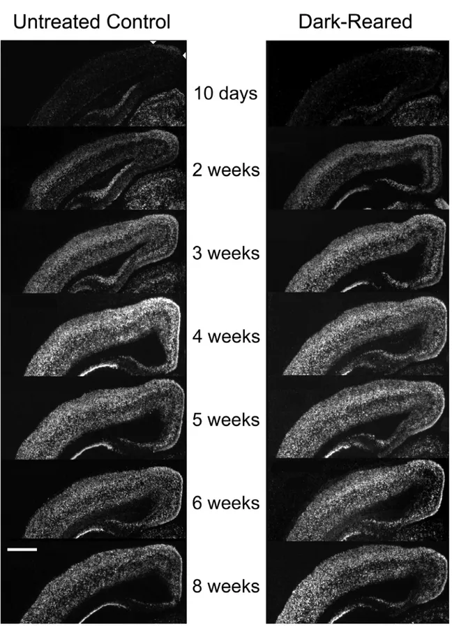
opening, low levels of cpg15 mRNA was detectable at the apex of the cortical hemispheres (Fig. 2-2). General onset of cpg15 expression in visual cortex occurred coincident with eye opening at two weeks postnatal with highest expression in layers 2/3, 4 and 6. Subsequently, cpg15 mRNA expression levels gradually increased, peaking at four weeks postnatal (P28), the height of the physiologically characterized critical period for development of eye specific preference in visual cortex. Subsequently, cpg15 mRNA levels declined to a lower basal adult level. These results show that cpg15 levels in rat visual cortex correspond well with the electrophysiologically mapped critical period.
Effect of dark rearing on cpg15 expression
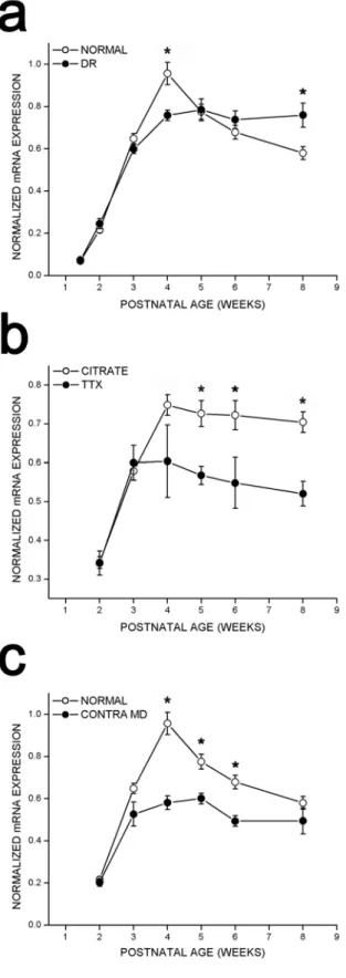
To test whether regulation of cpg15 expression was sensitive to visual input, rats were deprived of visual experience by dark rearing to various developmental ages. cpg15 mRNA levels were compared in DR and age-matched controls. Dark rearing did not alter the time of onset or the early increase in levels of cpg15 expression in visual cortex (Fig. 2-2). At P28, cpg15 mRNA levels in DR animals plateaued, but at a significantly lower level of expression than age-matched controls (Figs. 2-2 and 2-3a). This level of expression persisted in older DR animals, so that after eight weeks of dark rearing, cpg15 expression levels were significantly
higher than in their age-matched controls, where cpg15 expression normally declined after P28 (Figs. 2-2 and 2-3a). Differences in cpg15 mRNA levels between normal and DR animals were not seen in the LGN or SC (data not shown). The enhanced expression levels of cpg15 in visual cortex, seen with dark rearing, may be a molecular indicator of the prolonged plasticity specific to this region.
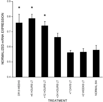
Short exposure to light can reportedly trigger the end of the delayed plasticity seen in DR animals (Mower et al., 1983; Philpot et al., 2001). To test if late onset of visual experience affects cpg15 expression, animals were DR to eight weeks then exposed to light for 6, 12, 24 hours, 7 days, or 14 days. In situ hybridizations show that with exposure to light of up to 12 hours cpg15 expression remains significantly higher than normal (Fig. 2-4). Following 24 hours of exposure to light, DR animals showed cpg15 mRNA expression levels comparable to those found in normally raised adults (Fig. 2-4). These experiments indicate that brief visual experience in DR adults can reset cpg15 expression to normal levels. This down-regulation of expression may be a molecular representation of the light induced end of the delayed plasticity in DR animals.
Effect of retinal action potential blockade on cpg15 expression
To investigate whether cpg15 mRNA expression in visual cortex is dependent on action potential driven activity from the retina, the Na+ channel blocker TTX was applied to the left retina of normally raised rats for three day periods, starting at different developmental times. Age-matched control animals were treated with citrate buffer applied to the left retina. cpg15 mRNA levels in visual cortex contralateral to the TTX treated eye were compared with those in visual cortex contralateral to the citrate treated eye in control animals. There was no change in
the onset of cpg15 expression in response to monocular retinal activity blockade, and there was no significant influence on cortical levels of cpg15 mRNA during early postnatal development (Fig. 2-5). Mean levels of cpg15 expression at 2, 3, and 4 weeks postnatal were not significantly different between the visual cortex contralateral to the blocked eye and visual cortex contralateral to citrate controls (Fig. 2-3b). By contrast, following the peak of the critical period and into adulthood, monocular retinal action potential blockade decreased cpg15 expression in visual cortex contralateral to the blocked eye (Figs. 2-3b and 2-5).
Monocular TTX blockade also decreased cpg15 expression in the contralateral LGN and SC starting at three weeks postnatal. For both LGN and superficial layers of the SC contralateral to the treated eye, the effect of TTX blockade became more pronounced with age (LGN not shown; for SC see Figures 2-5 and 2-6). These experiments indicate that in all visual structures tested, developmental regulation of cpg15 expression is divided into two phases. Early cpg15 expression is independent of retinally driven action potentials. During the critical period for development of eye specific preference in visual cortex, an activity-dependent component of cpg15 expression emerges, and the effect of retinal action potential blockade becomes progressively more pronounced with age.
Effect of MD on cpg15 expression
Studies have shown that total blockade of retinal activity produces a smaller shift in ocular dominance plasticity than that produced by MD (Rittenhouse et al., 1999). To investigate whether the effect of monocular activity blockade on cpg15 expression is less extreme than that of MD, we monocularly deprived normally raised rats for three-day periods, starting at different developmental times. cpg15 levels in visual cortex and SC contralateral to the sutured eye were
compared to the visual cortex and SC in normal animals. Similar to both DR and TTX treated animals, there was no change in the onset of cpg15 expression in the visual cortex of monocularly deprived rats (Fig. 2-3c). During the following 4 weeks of development, cpg15 expression in the visual cortex contralateral to the monocularly deprived eye was significantly less than in normal animals. Unlike animals whose retinally driven action potentials are blocked by TTX, at the peak of the critical period (P28) monocularly deprived animals exhibit levels of cpg15 expression significantly less than controls. With maturation the effect of MD becomes less significant, and at 8 weeks postnatal cpg15 expression in the deprived visual cortex is no longer different from controls. This trend is opposite to the effect of TTX, which becomes more significant as the animals mature. As in DR animals and in contrast to animals with retinal activity blockade, MD did not affect cpg15 levels in the contralateral SC.
Effect of early visual experience on adult cpg15 expression
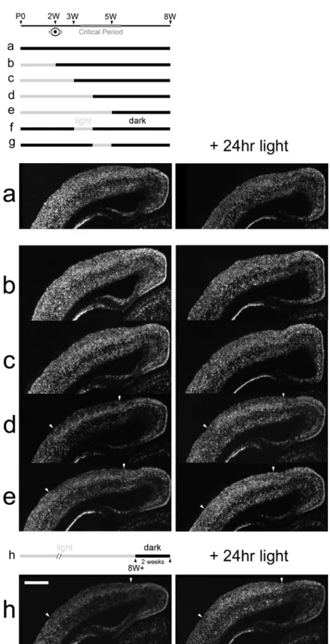
Previous studies suggest that early periods of visual experience are sufficient to trigger closure of the critical period despite later visual deprivation (Mower et al., 1983; Mower and Christen, 1985). To determine the window of early visual experience required for development of normal adult regulation of cpg15 expression in visual cortex, animals were raised in a normal 12/12 hour light/dark cycle to 2, 3, 4 or 5 weeks postnatal, then transferred to a dark environment until adulthood at 8 weeks. Animals were sacrificed without seeing any further light or following 24 hours of re-exposure to light, in order to assess their ability to regulate cpg15 expression. In normally raised adult rats cpg15 mRNA expression in visual cortex is down-regulated following a two-week period in the dark. After re-exposure to light for 24 hours, such dark-adapted rats will up-regulate cp15 levels (Nedivi et al., 1996) (Figs. 2-7h and 2-8h). In contrast, rats raised in
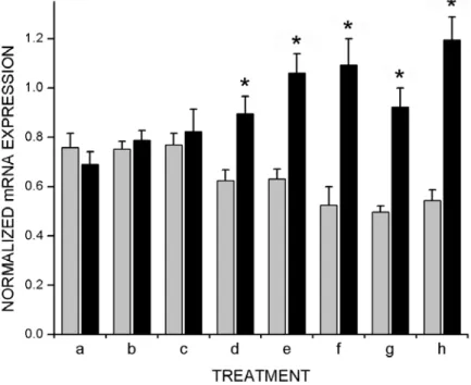
darkness to eight weeks show abnormally high levels of cpg15 expression in visual cortex and upon exposure to 24 hours of light, levels of expression are decreased (Figs. 2-4, 2-7a and 2-8a). These two extremes were compared with animals exposed to visual experience at restricted windows during development. Animals exposed to visual experience for two or three weeks prior to being placed in the dark to adulthood, displayed regulation of cpg15 expression essentially the same as in DR animals (Figs. 2-7b, 2-7c, and 2-8b, 2-8c). Animals reared with normal visual experience for the first four or five postnatal weeks and then placed in darkness until 8 weeks of age, regulate cpg15 expression in visual cortex similar to normal animals (Figs. 2-7d, 2-7e, and 2-8d, 2-8e). These results suggest that one week of visual experience at the outset of the critical period is sufficient to confer normal patterns of adult cpg15 regulation. To test this prediction, animals were raised to eight weeks in darkness except for a week of light either between weeks 3 and 4 or weeks 4 and 5. cpg15 expression in visual cortex was assayed at 8 weeks. cpg15 mRNA levels in animals allowed just one week of visual experience during the critical period are strikingly similar to those seen in normally raised animals and are dramatically induced upon re-exposure to light for 24 hours (Fig. 2-8f and 2-8g). Our results show that a restricted window of visual experience of one week during the critical period is sufficient to determine the adult patterns of cpg15 regulation in visual cortex.
Discussion
The experiments described here address the temporal and spatial expression patterns of cpg15 in the developing visual system of the normal rat and in response to visual manipulations. cpg15 expression was monitored in relation to the critical period for development of eye specific preference in visual cortex. We compared expression during normal development with that seen in response to dark rearing, monocular retinal action potential blockade or MD. The regulation of
cpg15 expression patterns suggests that it may serve as a molecular indicator of the potential for visual system plasticity.
Activity-independent and activity-dependent phases of cpg15 expression
We found that in the rat visual system the onset of cpg15 mRNA expression in visual cortex occurs at two weeks postnatal. cpg15 expression levels gradually rise and peak two weeks later, in the midst of the electrophysiologically mapped cortical critical period for shifts in eye preference. Following its peak expression, cpg15 mRNA levels in visual cortex decline to a lower basal level in adults, concomitant with critical period closure at approximately six weeks (Fagiolini et al., 1994; Gordon and Stryker, 1996). Although onset of cpg15 expression is coincident with eye opening, it is unaffected by dark rearing, blockade of retinally driven action potentials, or MD. This is consistent with previous studies of cpg15 expression in the cat visual system demonstrating that early cpg15 expression in visual cortex is activity-independent (Corriveau et al., 1999). The initial timing of cpg15 expression is therefore likely to be controlled by a developmentally regulated activity-independent mechanism. These findings are consistent with electrophysiological and optical imaging studies demonstrating that the basic structure of cortical maps is innate and develops in the absence of visual experience (Crair et al., 1998).
These studies also show that experience is necessary at a later stage of development for the refinement of ocular selectivity and maintaining responsiveness (Crair et al., 1998). We find that after the peak of the critical period, as levels of cpg15 in visual cortex begin to decline, a component of cpg15 expression dependent on retinally driven action potentials becomes evident and is progressively more pronounced with age. Activity-dependent regulation of cpg15 expression in the SC and LGN can be detected a week earlier than in visual cortex, perhaps
related to earlier maturation of these visual structures. Our results indicate that in all visual structures regulation of cpg15 expression is biphasic. Early cpg15 expression is independent of retinally driven action potentials. With maturation during the critical period, an activity-dependent component of cpg15 expression emerges, and the effect of retinal action potential blockade becomes progressively more pronounced with age. Activity-dependent regulation of cpg15 arises relatively late in development and may represent an adult feature of visual system plasticity.
Effect of visual experience on cpg15 expression
The aspects of cpg15 regulation that we found to be dependent on normal visual experience were its peak levels in visual cortex at P28 and its subsequent down-regulation following the closure of the critical period. Although cpg15 levels in visual cortex of DR rats are indistinguishable from those in control rats within the first 3 postnatal weeks, the expression at P28 is lower than controls. Rats DR beyond P28 maintain the same peak level of cpg15 expression through adulthood, levels that are significantly higher than in their control counterparts. There is a crossover of the cpg15 expression profiles in normal and DR rats, such that at the peak of the critical period expression is higher in normal animals, while after the critical period cpg15 is higher in DR animals. This crossover can also be seen in the developmental profiles of susceptibility to MD in normal and DR cats (Mower, 1991). In rodents, there have been no studies that examine the effects of dark rearing on susceptibility to MD during the height of the critical period. Studies at later ages show that similar to cats, DR rats also retain a prolonged capability to respond to MD, even at P90 (Guire et al., 1999).
In respect to dark rearing, we show here two additional cases in which cpg15 regulation closely parallels plasticity as measured by susceptibility to MD. Electrophysiological studies have demonstrated that a short exposure to light can trigger the end of the delayed plasticity that results from dark rearing (Mower et al., 1983; Philpot et al., 2001). Similarly, early visual experience in DR kittens attenuates the effects of later dark rearing so that there is no delayed plasticity (Cynader, 1983). These results show that the effect of dark rearing can be negated by a short period of light in DR adults, or with sufficient early visual experience. We find that a 24-hour exposure to light returns cpg15 levels in DR rats to those found in normally raised adults, and that one week of visual experience during the critical period is sufficient to confer normal adult patterns of cpg15 regulation. This suggests that visual experience is not persistently required during development for normal maturation of visual system function. Rather, exposure to patterned vision for at least a week during the critical period can irreversibly trigger the molecular machinery required for maturation, and will likely result in normal adult responses to visual manipulations. Since, in the absence of visual experience cpg15 expression is abnormally high in the adult, the molecular trigger for maturation may involve a general down-regulation of plasticity genes such as cpg15.
Differences in the effect of dark rearing on cpg15 from effects of activity blockade and MD The effect of binocular elimination of patterned vision on cpg15 expression during the critical period is profoundly different from the effect of retinal action potential blockade or MD. The effect of dark rearing is exclusive to visual cortex, and causes a prolonged up-regulation of cpg15 expression starting at the peak of the critical period and into adulthood. Retinal action potential blockade at the same developmental times causes a decrease in cpg15 expression in the
LGN and SC as well as visual cortex. The result, whereby the effect of dark rearing on cpg15 expression is restricted to visual cortex, corresponds with the observation that the delayed plasticity seen in DR cats is not manifested in the LGN (Mower et al., 1985; Mower and Christen, 1985).
During the critical period, down-regulation of cpg15 expression in visual cortex by MD is more severe than that caused by retinal activity blockade. This is consistent with electrophysiological studies showing that MD during the critical period produces a greater shift of ocular dominance in visual cortex than total blockade of retinal activity (Rittenhouse et al., 1999). A possible explanation is that during MD the residual activity from the retina actively depresses efficacy of synaptic connections driven by the deprived eye (Bear and Rittenhouse, 1999; Rittenhouse et al., 1999). Despite the fact that a monocularly deprived eye is generating some activity as opposed to total loss of activity generated by retinal action potential blockade, during the critical period cpg15 levels are lower in visual cortex of MD animals than in animals after retinal TTX blockade. This could reflect depression of cortical synaptic activity driven by the deprived eye.
Taken together, this indicates that regulation of cpg15 expression does not correspond directly to levels of activity, but rather, seems to reflect a propensity for functional plasticity.
Effect of dark rearing on expression of visually responsive genes
It has been proposed that mechanisms underlying adult plasticity during learning and memory, or long-term potentiation and long term depression, also play a key role in developmental plasticity (Kandel and O'Dell, 1992; Goodman and Shatz, 1993; Constantine-Paton and Cline, 1998; Nedivi, 1999). For this reason, many genes isolated or characterized on
the basis of their response to activity in the adult have been investigated in the context of developmental plasticity in the visual system. These include both regulatory genes that encode transcription factors as well as effector genes that can directly affect neuronal morphology and function (reviewed in (Nedivi, 1999)). Multiple genes show a transcriptional response to dark rearing, although the type of response varies. While GAP43, CaMKII and GAD all show the same increase in expression shown by cpg15 in response to dark rearing past the critical period (Neve and Bear, 1989), junB, zif/268, and BDNF show the opposite response and are down-regulated (Rosen et al., 1992; Lein and Shatz, 2000). In contrast to light-independent onset of cpg15 expression, dark rearing prevents the normal onset and transcriptional increase of Homer, zif/268, and BDNF in visual cortex (Worley et al., 1990; Brakeman et al., 1997; Capsoni et al., 1999; Lein and Shatz, 2000). Subsequent exposure to light causes their rapid induction (Worley et al., 1990; Brakeman et al., 1997; Capsoni et al., 1999; Lein and Shatz, 2000). The transcriptional regulation of this latter group provides an accurate ‘molecular readout’ of activity, while regulation of cpg15 together with GAP43, CaMKII and GAD corresponds more closely with the capacity for plasticity.
Summary
During Xenopus visual system development, CPG15 concurrently regulates multiple aspects of retinotectal circuit formation (Cantallops et al., 2000). CPG15 promotes tectal cell dendritic arbor growth, stabilizes retinal axon arbors, and promotes maturation of retinotectal synapses (Nedivi et al., 1998; Cantallops et al., 2000). Our finding that DR rats fail to down-regulate cpg15, raises the possibility that perhaps the residual plasticity measured electrophysiologically in these animals reflects an extended capacity for local synaptic
remodeling. The prolonged plasticity seen in DR animals may result from failure to down-regulate genes like cpg15 that could promote structural remodeling and synaptic maturation.
Methods
Animal manipulations and tissue isolation
All animal work was approved by the Massachusetts Institute of Technology Committee on Animal Care, and conforms to NIH guidelines for the use and care of vertebrate animals. Wistar-Kyoto rats (Taconic, Germantown, NY) were housed either in a room with a 12/12 hour light/dark cycle or in a room sealed from visible light. A 15-watt safelight shielded by a #2 Kodak dark-red filter (Kodak, Rochester, NY) was used for daily care and maintenance of animals housed in the darkroom (approximately 30 minutes per day). At various points during the experiment Polaroid photographic paper placed in the dark room was monitored for exposure. In cat, intermittent safelight exposure can be sufficient to prevent the apparent extension of the critical period by dark rearing. To test whether safelight exposure affected dark rearing of rats, a group of animals handled with an infrared viewing system (950 nm) was raised in the dark to 3, 4, 5, and 8 weeks (each group n=3). The levels of cpg15 expression in rats handled with intermittent safelight exposure were not significantly different from those handled under infrared conditions, and both were significantly different from their age-matched controls at 4 weeks and 8 weeks (data not shown). The difference in sensitivity to safelight exposure may reflect the relatively poor vision of albino rats as compared to cats.
Rats were reared under normal conditions (12/12-hour light/dark), DR from birth, or dark-adapted at different developmental time points. DR and age-matched control animals were sacrificed postnatally at progressive times, starting at postnatal day 10 (P10) and at 1 week
intervals from P14 (the approximate day of eye opening) to 8 weeks (P10, each group n=2; P14, P21, 5 weeks, 6 weeks, each group n=4; 4 weeks DR n=4, controls n=3; 7 weeks controls n=3; 8 weeks DR n=3, controls n=5). A parallel set of DR rats were removed from the darkroom at the same intervals, and exposed to light for 24 hours before sacrifice (n=3 for each time point). A group of DR 8 week old rats were exposed to light for 6H, 12H, 24H, 7 or 14 days (n=2-3 for each time point). Dark-adapted adult rats were placed in the dark for two weeks and sacrificed in the dark with a parallel set sacrificed following re-exposure to 24 hours of light (n=3 each group). Rats dark-adapted to adulthood were raised normally to 2, 3, 4, or 5 weeks then placed in the dark until 8 weeks and sacrificed in the dark or following re-exposure to light for 24 hours (n=3 per group per time point). Two additional groups of rats were allowed a 1 week window of visual experience (between weeks 3 and 4 or between weeks 4 and 5) during dark rearing. At 8 weeks, these animals were sacrificed in the dark (each group n=3) or following a 24 hour exposure to light (Lt3-4, n=3; Lt4-5, n=2).
Monocular blockades of retinal action potentials were done essentially as previously described (Prusky and Ramoa, 1999). A strip of Elvax containing either tetrodotoxin (TTX) or citrate buffer was surgically implanted into the vitreous of the left eye of each animal for 3 days of sustained release, starting at P11, P18, P25, P32, P39 and P53 (TTX, n=3; citrate, n=2). Briefly, 200 mg of washed Elvax beads (DuPont, Wilmington, DE) were dissolved in methylene chloride. The dissolved Elvax was mixed with 20 μl of 1% Fast Green in DMSO and either 20 μl of 0.3 M TTX in citrate buffer (CalBiochem, La Jolla, CA), or 20 μl of 18.6 mM citrate buffer
(Sigma, St. Louis, MO). The methylene chloride was slowly evaporated over the course of one day at -70°C and for five subsequent days at -20°C. The Elvax was then sectioned into 180-micron thick disks by cryostat and stored at -80°C. Before surgeries, the Elvax was washed in
70% ethanol for 30 minutes and twice in sterile PBS for 30 minutes. For younger animals, the Elvax was cut into approximately 2 x 0.75 mm strips. For older animals the Elvax strips were approximately 4 x 1 mm. The strips were carefully inserted into the vitreous following a small incision at the sclera’s edge. One eye in each animal was treated with either TTX or citrate Elvax while the other eye remained intact. Anesthesia was maintained by halothane/O2 via mask. Following Elvax implantation, the rats recovered from anesthesia under observation. The effectiveness of activity blockade was monitored daily by assaying for consensual pupil response to bright white light under light halothane/O2 anesthesia.
MD by eyelid suture was initiated at P18, P25, P32, P39 and P53 (for each time point, n=3). Under Ketamine/Xylazine (80/10 mg/kg) anesthesia, the area surrounding the left eye was cleaned with Betadine and isopropyl alcohol. The lid margins were trimmed and the eye flushed with sterile PBS. Two to three horizontal mattress sutures using 6.0 Ethilon (Johnson & Johnson, Somerville, NJ) closed the length of the apposed lids. Ophthalmic ointment (Fougera, Melville, NY) was applied and the animals were monitored for recovery. For the P14 time point (n=3), rather than suture the unopened eye, tissue adhesive (Vetbond; 3M, St. Paul, MN) was applied at P11 to prevent possible eye opening. Following three days of TTX blockade or MD, animals were sacrificed by guillotine decapitation, brains were removed immediately, trimmed and positioned for coronal sectioning before being frozen on powdered dry ice and stored at -80°C.
In situ hybridization
Ten micron coronal sections through anterior visual cortex were sectioned by cryostat, thaw mounted on Superfrost/plus microscope slides, dried, fixed in 4% paraformaldehyde, washed in PBS, dehydrated in ethanol, air-dried, then stored desiccated at -80°C. Before
hybridization, slides were pretreated (at room temperature, unless otherwise stated) with 0.2 M HCl (20 min.); DDW (5 min.); 2x standard saline citrate (30 min. at 70°C); DDW (5 min.). The
next prehybridization treatments, from pronase (Sigma, St. Louis, MO; Type XIV) to air-drying slides for 1 hour, were conducted as described (Hogan et al., 1994). RNA probes were synthesized with an RNA transcription kit (Stratagene, La Jolla, CA) and 35S-UTP (Amersham Pharmacia Biotech, Piscataway, NJ; 800 Ci/mmol), using linearized cpg15 cDNA as a template. Hybridizations were done as previously described (Nedivi et al., 1996). Posthybridization wash conditions: 3 hours at 50°C, in 50% formamide, 1x salt solution (Hogan et al., 1994), with 10 mM DTT; 15 min. at 37°C in TNE (10 mM Tris pH 7.5, 0.5 M NaCl, 1 mM EDTA); 30 min. at 37°C in TNE containing RNase A (Sigma, St. Louis, MO; 20 μg/ml); 30 min. at 37°C in TNE; and finally overnight at 50°C in 50% formamide, 1x salt solution. Slides were dehydrated with 0.3M NH4Ac in ethanol, air-dried, and processed for autoradiography as described (Hogan et al., 1994) using NTB-2 emulsion (Kodak, Rochester, NY) diluted 1:1 with 2% glycerol, and exposed for 3-5 days at 4°C.
Quantitative data analysis
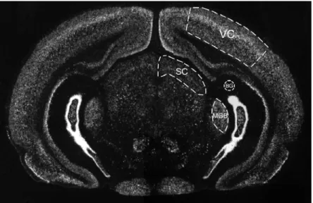
Darkfield images of 2-4 sections from each brain were acquired into Photoshop 5.0 with a Diagnostics Instruments Spot2 digital camera mounted on a Nikon Eclipse E600 using a 1x/0.04 Plan UW objective (MVI, Avon, MA). Images were saved as grayscale TIFFs and imported into NIH Image (version 1.62). Each section contained, in addition to visual cortex and SC, the medial geniculate body (MGB) of the thalamus, an auditory sensory area (Fig. 2-1). Areas were defined by Nissl staining of alternate sections. Mean pixel density measurements were taken from four areas on each section: the visual cortex, SC, background, and MGB. Pixel
density was measured on a 0 to 255 scale where 255 is white. The background served as a zero-labeling negative control, while the MGB served as a positive control with a high level of labeling that is unaffected by visual manipulations. Visual cortex and SC measurements were normalized on a scale of 0 to 1 interpolating between the background (0) and MGB (1) values. The background mean pixel density was first subtracted from the mean pixel densities in the visual cortex, SC, and MGB yielding the net mean pixel densities for each area. The net mean pixel density in the visual cortex or SC was then divided by the net mean pixel density from the MGB in the same section. Statistical significance was determined by unpaired Student’s t-test.
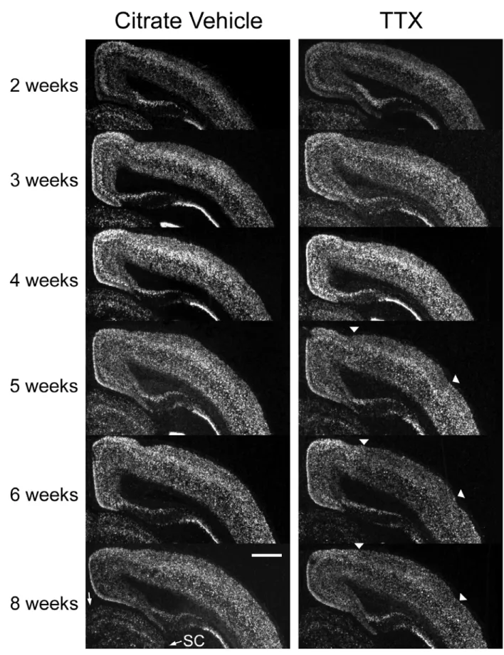
Figure 2-2. Developmental time course of cpg15 expression in the visual cortex of normal and DR rats.
Figure 2-3. Quantification of cpg15 expression in visual cortex of control, DR rats, rats after retinal activity blockade, or after MD.
Figure 2-5. cpg15 expression in visual cortex after 3 days of monocular TTX blockade initiated at different developmental times.
Figure 2-6. Quantification of cpg15 expression in superior colliculus after 3 days of monocular TTX blockade initiated at different developmental times.
Figure 2-8. One week of visual experience during the critical period is sufficient to confer normal adult patterns of cpg15 regulation.
Figure Legends
Figure 2-1. In situ hybridization on a coronal section through rat visual cortex using a cpg15 probe. Regions relevant for quantification are outlined. Mean pixel densities were measured in four areas on each section: the visual cortex (VC), superior colliculus (SC), medial geniculate body (MGB), and in an area of the section with zero labeling for background (BG). The background is used as the zero-point while the MGB served as a positive control with a high level of labeling that is unaffected by visual manipulations (see Methods).
Figure 2-2. Developmental time course of cpg15 expression in the visual cortex of normal and DR rats. Representative dark-field photomicrographs of in situ hybridizations for cpg15 mRNA. Coronal hemi sections through visual cortex of normal animals (left), and DR animals (right) are shown at the designated postnatal ages. The onset of cpg15 expression in neocortex for both normal and DR animals is coincident with eye opening at 2 weeks. Normally, cpg15 expression peaks at 4 weeks and then declines to a lower basal adult level. In DR animals, cpg15 levels remain elevated past 5 weeks. Arrowheads point to the earliest cpg15 expression at medial apex of neocortex. Some individual panels (6W, DR6W, 4W, DR4W, DR3W, and DR2W) were normalized with respect to MGB for assembly of the montage. Scale bar,1 mm.
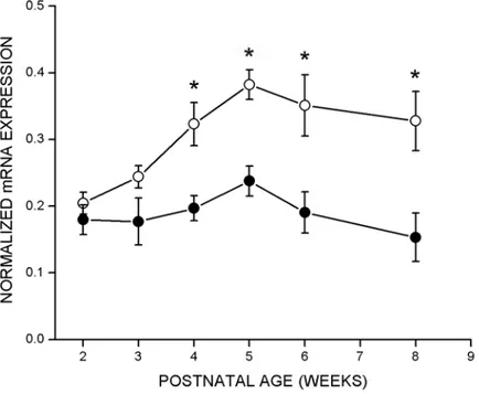
Figure 2-3. Quantification of cpg15 expression in visual cortex of control, DR rats, rats after retinal activity blockade, or after MD. In all cases, after background subtraction, the net average pixel value in visual cortex was normalized by the net average pixel value of the MGB (see methods for detail). a, Effect of dark rearing on cpg15 expression in visual cortex. Data from
four in situ experiments are shown, of which Figure 2-2 is representative. Filled circles represent DR animals, unfilled circles represent untreated control animals, and error bars represent SEM. Points marked with * are significantly different between normal and DR animals (at 4 weeks p = 0.001, at 8 weeks p = 0.016, unpaired Student’s t-test). b, Effect of 3 days monocular TTX blockade initiated at different developmental times on cpg15 expression in visual cortex. Data from three in situ experiments are shown, of which Figure 2-5 is representative. Filled circles represent visual cortex contralateral to TTX blockade, unfilled circles represent visual cortex of control animals implanted with a citrate control in the contralateral eye, and error bars represent SEM. Points marked with * are significantly different between visual cortex of control and TTX treated rats (at 5 weeks p = 0.0012, at 6 weeks p = 0.032, at 8 weeks p = 0.0008, unpaired Student’s t-test). c, Effect of 3 days MD by eyelid suture initiated at different developmental time points on cpg15 expression in visual cortex. Filled circles represent visual cortex contralateral to the sutured eye, unfilled circles represent visual cortex of untreated control animals, and error bars represent SEM. Points marked with * are significantly different between visual cortex of control and monocularly deprived rats (at 4 weeks p = 0.003, at 5 weeks p = 0.006, at 6 weeks p = 0.007, unpaired Student’s t-test).
Figure 2-4. Light triggers down-regulation of cpg15 expression in DR rats. Quantification of cpg15 expression in visual cortex of animals DR to 8 weeks and subsequently exposed to light (Lt) for designated times. Data from two in situ experiments are shown. In each section the net average pixel value in visual cortex is normalized by the net average pixel value of the MGB. Abnormally high levels of cpg15 expression in animals DR to 8 weeks decline to normal adult levels within 24 hours of exposure to light. Error bars represent SEM. * mark significant
differences compared to normal 8 week animals (DR8W p = 0.016, DR8W+6hl p = 0.009, DR8W+12hl p = 0.0086, unpaired Student’s t-test).
Figure 2-5. cpg15 expression in visual cortex after 3 days of monocular TTX blockade initiated at different developmental times. Dark-field photomicrographs of in situ hybridizations for cpg15 mRNA in coronal sections through the visual cortex contralateral to the treated eye of either control animals (left) implanted with the citrate vehicle or TTX-treated animals (right), at the designated ages. Starting at 5 weeks postnatal, retinal TTX blockade decreases levels of cpg15 expression in visual cortex (see cortical area between arrowheads). Starting at 4 weeks postnatal, retinal TTX blockade causes a down-regulation of cpg15 expression in the SC (see area between arrows marked SC). Some individual panels (C4W, TTX4W, TTX3W) in this figure were normalized with respect to MGB for assembly of the montage. Scale bar, 1 mm.
Figure 2-6. Quantification of cpg15 expression in superior colliculus after 3 days of monocular TTX blockade initiated at different developmental times. Data from three in situ experiments are shown, of which Figure 2-5 is representative. After background subtraction, the net average pixel value in superficial layers of the SC was normalized by the net average pixel value of the MGB. cpg15 expression in the SC is depressed by retinal TTX blockade soon after eye opening. Filled circles represent SC contralateral to TTX blockade, unfilled circles represent SC contralateral to citrate control, and error bars represent SEM. Points marked with * are significantly different between SC of control and TTX treated rats (at 4 weeks p = 0.026 at 5 weeks p = 0.0003 at 6 weeks p = 0.027, at 8 weeks p = 0.011, unpaired Student’s t-test).
Figure 2-7. Early visual experience confers normal adult patterns of cpg15 regulation. Representative dark-field photomicrographs of in situ hybridizations for cpg15 mRNA on coronal hemi sections through visual cortex. Animals were raised to different ages with normal visual experience, then transferred into a dark environment until 8 weeks (left), followed by a 24 hour re-exposure to light (right). Animals raised normally to 4 or 5 weeks of age (d, e), then DR to 8W, show the normal decline in cpg15 levels with maturation as well as the normal adult regulation (h) of cpg15 expression by light (arrowheads delineate visual cortex). Animals raised normally to 2 and 3 weeks of age (b, c), then DR to 8W, similarly to animals DR from birth (a), fail to down-regulate cpg15 as adults and do not re-induce cpg15 in response to light. Panels were not normalized for montage assembly. Scale bar, 1 mm.
Figure 2-8. One week of visual experience during the critical period is sufficient to confer normal adult patterns of cpg15 regulation. Quantification of cpg15 expression in visual cortex of rats with early visual experience. Measurements and calculations were done as described in Figure 2-3. The treatment legend is identical to Figure 2-7. A one week-window of light during the critical period, either between weeks 3 and 4 (f) or weeks 4 and 5 (g), confers normal adult patterns of cpg15 regulation. Grey bars represent cpg15 expression levels without re-exposure to light, black bars represent levels following 24 hours of light exposure, and error bars represent SEM. Points marked with * are significantly different between animals sacrificed in the dark or following 24 hours of light exposure (at e and h, p <0.0001; at f, p = 0.0049; at g, p = 0.0022; at d, p = 0.009, unpaired student’s t-test).
Chapter 3
Dynamic remodeling of dendritic arbors in GABAergic interneurons of adult
visual cortex
Abstract
Despite decades of evidence for functional plasticity in the adult brain, the role of structural plasticity in its manifestation remains unclear. To examine the extent of neuronal remodeling that occurs in the brain on a daily basis, we used a multi-photon based microscopy system for chronic in vivo imaging and reconstruction of entire neurons in the superficial layers of the rodent cerebral cortex. Here, we show the first unambiguous evidence of dendrite growth and remodeling in adult neurons. Over a period of months, neurons could be seen extending and retracting existing branches, and in rare cases adding new branch tips. Neurons exhibiting dynamic arbor rearrangements were GABA positive non-pyramidal interneurons, while pyramidal cells remained stable. These results are consistent with the idea that dendritic structural remodeling is a substrate for adult plasticity and suggest that circuit rearrangement in the adult cortex is restricted by cell type-specific rules.
Introduction
Hubel and Wiesel’s groundbreaking work in the 1960-1970’s defined a critical period in development when manipulating visual inputs causes dramatic functional and structural changes in layer 4 of primary visual cortex (Hubel and Wiesel, 1970; Hubel et al., 1977; LeVay et al., 1980). Since their finding that large scale rearrangement of thalamic afferents in visual cortex is restricted to a developmental critical period, the adult brain has been considered relatively
‘hard-sensory maps in the adult brain (reviewed (Buonomano and Merzenich, 1998)), even across long distances (Pons et al., 1991), was explained as unmasking of existing connections and was not considered to require outright growth (Ramachandran et al., 1992). Although there are indications that adult cortex is capable of anatomical change in response to peripheral manipulation, particularly in the superficial layers (Darian-Smith and Gilbert, 1994; Florence et al., 1998), the scale of change is small compared to the critical period, and is difficult to detect against the general variance in the size and shapes of cortical neurons. Moreover, such changes are seen only in response to external perturbation, leaving it unclear whether arbor remodeling normally occurs in the adult cortex on a daily basis, and to what extent.
With the advent of new technologies to time-lapse image neuronal morphology in vivo (Denk et al., 1990), the issue is now being revisited. Repeated in vivo imaging of apical dendrites extending from layer 5 pyramidal neurons into the superficial layers has been used to investigate dendritic spine dynamics in both somatosensory and visual cortex (Grutzendler et al., 2002; Trachtenberg et al., 2002; Holtmaat et al., 2005; Zuo et al., 2005a). Less attention has been paid to potential changes in the overall structure of dendritic arbors. In fact, it has been suggested that little if any structural plasticity occurs in the apical dendrites of layer 5 pyramidal neurons in the adult somatosensory cortex (Trachtenberg et al., 2002), or in the apical dendrites of mitral and tufted cells in the adult olfactory bulb (Mizrahi and Katz, 2003). No study has directly addressed the potential for structural dynamics in a cross section of neurons that reflects the diversity of neocortical cell types. More specifically, the non-pyramidal neurons of the neocortex have yet to be the focus of investigation by in vivo imaging studies, despite their important role in adult cortical plasticity and in reorganization of cortical maps (Jacobs and Donoghue, 1991; Jones, 1993).
Here, we investigate the dendritic arbor dynamics of pyramidal and non-pyramidal neurons in the superficial layers of the adult visual cortex in vivo. Our results show that while dendritic branches of layer 2/3 pyramidal cells remain stable, non-pyramidal interneurons in these layers are dynamic, exhibiting a range of structural changes on a weekly basis.
Results
Repeated in vivo imaging of cortical neurons
Studies suggest that intracortical connections in the supragranular layers of the neocortex, and in particular axonal sprouting, may be a locus of adult structural plasticity (Darian-Smith and Gilbert, 1994). To directly test the prediction that neurons in layer 2/3 are capable of structural change and to assess the extent of dendritic structural dynamics in the adult cortex, we chronically imaged neuronal morphology in the intact rodent brain. To allow long-term visualization of neuronal structure in vivo, cranial windows were bilaterally implanted over the visual cortices of thy1-GFP-S mice (Feng et al., 2000) between 4-6 weeks of age. These mice express green fluorescent protein (GFP) in a sparse pseudo-random subset of neocortical neurons. Imaging began at least 2 weeks after surgery to allow for recovery and optical clarification of the cranial windows. Brains were screened for optically accessible GFP positive neurons using wide-field fluorescence, and neuronal location was noted using local landmarks in the brain’s surface vasculature. Individual GFP labeled neurons in layers 2/3 of visual cortex of anesthetized adult mice were then time-lapse imaged using a custom made two-photon microscope (Kim et al., 1999). To include as many neuronal branch tips as possible within the imaging volume, nine slightly overlapping volumes were imaged in a 3 x 3 array through z-x-y translation of an automated motorized stage. Individual image planes were stitched together to create a montage of adjoining x-y sections for a given depth from the pial surface. In an attempt to provide a
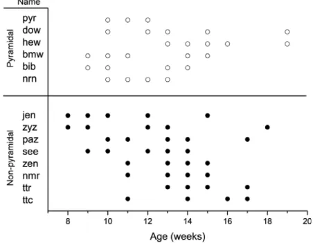
comprehensive view of adult structural plasticity, data collection was initially not restricted to any particular cell type. Six pyramidal cells and 8 non-pyramidal cells from 13 animals were time-lapsed imaged for 4 to 10 weeks (Fig. 3-1) and 4-dimensional morphometric analysis was carried out by quantitative comparison of dendritic branch tip length (BTL) as a function of time. Branch tips that were not imaged clearly for multiple imaging sessions or whose termination was unclear were excluded from analysis.
Pyramidal cells are stable over time
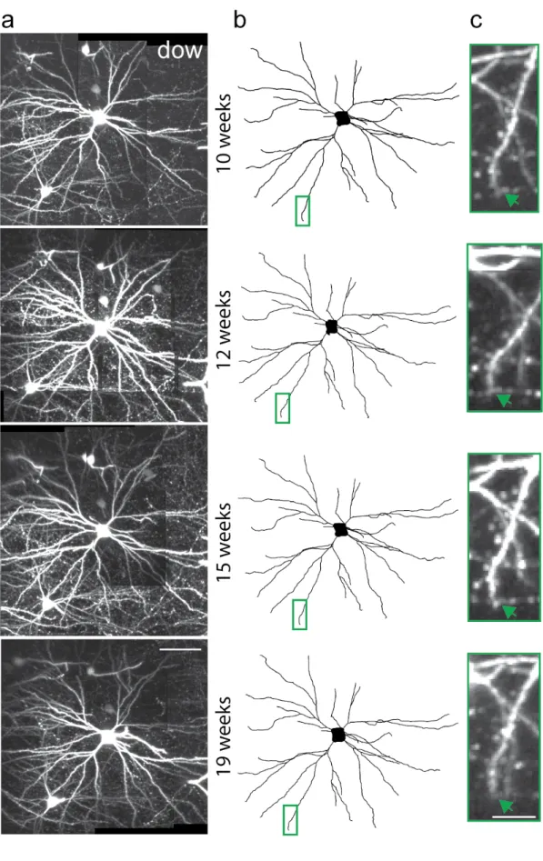
Imaged pyramidal cells exhibited typical small pyramidal morphologies with a spiny apical dendrite and a skirt of spiny basal dendrites emanating from the lower half of a pyramid-shaped cell body. An example is shown in Figure 3-2 and Supplementary Movie 3-1. The cell body of this pyramidal neuron, ‘dow’, was located 180 microns below the pial surface. It had a total of 57 dendritic branch tips on its apical dendrite and 5 primary basal dendrites. We monitored 28 of the 57 branch tips over 9 weeks. Examination of individual branch tips revealed no overt sign of structural change (Fig. 3-2a-c). Similar examination of all cells in the pyramidal population did not identify any change in apical and basal dendritic branches. These data suggest that under normal conditions the dendritic branches of layer 2/3 pyramidal neurons in visual cortex are relatively stable in the adult, and are consistent with previous studies reporting dendritic branch stability in other cortical areas (Trachtenberg et al., 2002; Mizrahi and Katz, 2003).
We next examined layer 2/3 non-pyramidal neurons. Figure 3-3 shows maximum-intensity z-projections (MZPs) of image planes close (± 15 microns) to the cell body of a non-pyramidal neuron ‘nmr’ with a bitufted dendritic morphology revealing its highly complex local arborization (Fig. 3-3a and Supplementary Movie 3-2). The cell body’s center of mass was 118 microns below the pial surface. This neuron had 4 primary dendrites with a total of 49 branch tips. 28 of the 49 branch tips were monitored for 4 weeks. Four of the 28 branch tips exhibited variations in length over 4 weeks. Two examples are shown where branch tips visibly elongated in the x-y plane (Fig. 3-3b-f). Branch tip #20 elongated by approximately 16 microns over 4 weeks in (Fig. 3-3b-c, f). Concurrently, branch tip #15 increased in length by approximately 10 microns (Fig. 3-3d-f). Both branch tips emanate from the same primary dendrite whose dendritic branch length accounts for 62% the total monitored dendritic length of the neuron. These results demonstrate that dendritic arbors of neurons within the adult neocortex are capable of growth.
A different non-pyramidal neuron, ’paz’, residing 78 microns below the pial surface, is shown in Figure 3-4. 2-D projections of the 3-D traces show a moderately branched interneuron with a bitufted dendritic morphology (Fig. 3-4a). This cell had 7 primary dendrites with 47 branch tips. 29 of 47 dendritic branch tips were monitored for 7 weeks. Virtually all the branches were stable, however two branches exhibited remodeling, one of which was so large that it exceeded the imaging volume. Time-lapse images revealed that within as little as 2 weeks, this branch tip more than doubled its length and exited the imaging volume (Fig. 3-4). Although at 13 weeks postnatal we were unable to follow the process to its termination, we measured a net extension of > 92 microns from the branch tip at 11 weeks to its location at the edge of the imaging volume two weeks later. The axon of this neuron projected from the cell body in the