Publisher’s version / Version de l'éditeur:
Glycobiology, 11, 11, pp. 957-967, 2001-11-01
READ THESE TERMS AND CONDITIONS CAREFULLY BEFORE USING THIS WEBSITE.
https://nrc-publications.canada.ca/eng/copyright
Vous avez des questions? Nous pouvons vous aider. Pour communiquer directement avec un auteur, consultez la première page de la revue dans laquelle son article a été publié afin de trouver ses coordonnées. Si vous n’arrivez pas à les repérer, communiquez avec nous à PublicationsArchive-ArchivesPublications@nrc-cnrc.gc.ca.
Questions? Contact the NRC Publications Archive team at
PublicationsArchive-ArchivesPublications@nrc-cnrc.gc.ca. If you wish to email the authors directly, please see the first page of the publication for their contact information.
Archives des publications du CNRC
This publication could be one of several versions: author’s original, accepted manuscript or the publisher’s version. / La version de cette publication peut être l’une des suivantes : la version prépublication de l’auteur, la version acceptée du manuscrit ou la version de l’éditeur.
For the publisher’s version, please access the DOI link below./ Pour consulter la version de l’éditeur, utilisez le lien DOI ci-dessous.
https://doi.org/10.1093/glycob/11.11.957
Access and use of this website and the material on it are subject to the Terms and Conditions set forth at
Genetic basis for expression of the major globotetraose containing
lipopolysaccharide from Haemophilus influenzae strain Rd (RM118)
Hood, Derek W.; Cox, Andrew D.; Wakarchuk, Warren W.; Schur, Melissa;
Schweda, Elke K. H.; Walsh, Shannon L.; Deadman, Mary E.; Martin, Adele;
Moxon, E. Richard; Richards, James C.
https://publications-cnrc.canada.ca/fra/droits
L’accès à ce site Web et l’utilisation de son contenu sont assujettis aux conditions présentées dans le site LISEZ CES CONDITIONS ATTENTIVEMENT AVANT D’UTILISER CE SITE WEB.
NRC Publications Record / Notice d'Archives des publications de CNRC:
https://nrc-publications.canada.ca/eng/view/object/?id=33ec888e-b4f7-43b3-ae36-fa88ebdce74e https://publications-cnrc.canada.ca/fra/voir/objet/?id=33ec888e-b4f7-43b3-ae36-fa88ebdce74e
Genetic basis for expression of the major globotetraose-containing lipopolysaccharide
from H. influenzae strain Rd (RM118)
Derek W. Hood1,2, Andrew D. Cox3, Warren W. Wakarchuk3,
Melissa Schur3, Elke K.H. Schweda4, Shannon L. Walsh2,
Mary E. Deadman2, Adele Martin3, E. Richard Moxon2,
and James C. Richards3
2Molecular Infectious Diseases Group, University of Oxford Department of Paediatrics, Weatherall Institute of Molecular Medicine, John Radcliffe Hospital, Headington, Oxford, OX3 9DS, UK; 3Institute for Biological Sciences, National Research Council of Canada, Ottawa, Ontario, K1A 0R6 Canada; and 4Clinical Research Centre, Karolinska Institutet and University College of South Stockholm, NOVUM, S-141 86 Huddinge, Sweden.
Received on April 19, 2001; revised on July 9, 2001; accepted on July 11, 2001
A genetic basis for the biosynthetic assembly of the globotetraose containing lipopolysaccharide (LPS) of
Haemo-philus influenzae strain RM118 (Rd) was determined by
structural analysis of LPS derived from mutant strains. We have previously shown that the parent strain RM118 elaborates a population of LPS molecules made up of a series of related glycoforms differing in the degree of oligosaccharide chain extension from the distal heptose residue of a conserved phosphorylated inner-core element, L-α-D-Hepp-(1 →2)-L-α-D-Hepp-(1→3)-[β-D-Glcp-(1→4)-]-L-α-D-Hepp-(1→5)-α-Kdo.
The fully extended LPS glycoform expresses the globotetraose structure, β-D-GalpNAc-(1→3)-α-D -Galp-(1→4)-β-D-Galp-(1→4)-β-D-Glcp. A fingerprinting strategy was employed to establish the structure of LPS from strains mutated in putative glycosyltransferase genes compared to the parent strain. This involved glycose and linkage analysis on intact LPS samples and analysis of O-deacylated LPS samples by electrospray ionization mass spectrometry and 1D 1H-nuclear magnetic resonance spectroscopy. Four
genes, lpsA, lic2A, lgtC, and lgtD, were required for sequential addition of the glycoses to the terminal inner-core heptose to give the globotetraose structure. lgtC and lgtD were shown to encode glycosyltransferases by enzymatic assays with synthetic acceptor molecules. This is the first genetic blueprint determined for H. influenzae LPS oligosaccharide biosynthesis, identifying genes involved in the addition of each glycose residue.
Key words: globotetraose/Haemophilus influenzae/ lipopolysaccharide
Introduction
Haemophilus influenzae is a bacterium that routinely colonizes the upper respiratory tract of humans. It is also the cause of
both upper and lower respiratory tract infections that result from contiguous spread. Occasionally, H. influenzae can cause systemic and life-threatening bacteraemic diseases, such as septicaemia and meningitis. Lipopolysaccharide (LPS) of
H. influenzae functions as a virulence determinant (Preston et al., 1996a). LPS results in major cytotoxic injury to host cells, is a target for host immune responses, and can influence each stage of the pathogenesis of H. influenzae infection (Moxon and Maskell, 1992). Changes in LPS expression can alter the viru-lence of this pathogen (Kimura and Hansen, 1986; Cope et al., 1990; Weiser et al., 1990b; Hood et al., 1996a). A feature of H.
influenzae LPS is that surface-exposed epitopes of the oligosac-charide are subject to high frequency on-off switching of expression (phase variation) (Weiser et al., 1990a; High et al., 1993; Jarosik and Hansen, 1994). This heterogeneity may be an advantage to the bacteria, allowing them to better confront different host compartments and microenvironments and to survive the host immune response (Weiser and Pan, 1998).
Determination of structure is crucial to understanding the biology of H. influenzae LPS and its role in bacterial virulence.
H. influenzae LPS comprises a variable oligosaccharide moiety and a membrane-anchoring lipid A component (Zamze and Moxon, 1987). LPS from a number of different strains have been shown to contain a common L-glycero-D -manno-heptose-containing inner-core trisaccharide unit attached to the lipid A moiety via a phosphorylated 2-keto-3-deoxyoctu-losonic acid (Kdo) residue (Phillips et al., 1992, 1993, 1996; Gibson et al., 1993; Schweda et al., 1993, 1995; Masoud et al., 1997; Risberg et al., 1997, 1999a,b; Rahman et al., 1999). Each of the Hep residues can provide a point for the addition of Hex residues, which in turn can lead to oligosaccharide chain extensions. The degree of substitution and chain extension from the triheptose unit varies within and between strains (Masoud et al., 1997; Risberg et al., 1999b). In addition, phos-phate-containing substituents that include free phosphate groups (P), phosphoethanolamine (PEtn), pyrophospho-ethanolamine (PPEtn), and phosphocholine (PCho) also contribute to the structural variability of these molecules. Recently we reported the structure of a globotetraose (β-D -GalpNAc-(1→3)-α-D-Galp-(1→4)-β-D-Galp-(1→4)-β-D-Glcp) containing LPS from H. influenzae strain RM118 (Risberg et
al., 1999b), the strain (Rd) for which the complete genome sequence has been determined (Fleischmann et al., 1995). For strain RM118, three major populations of LPS glycoforms were identified, all containing a PCho→6)-β-D-Glcp group off the Hep attached to the Kdo unit, but differing in the length of the oligosaccharide chains off the third Hep of the inner-core element. LPS glycoforms expressing a fully assembled globotetraose side chain and sequentially truncated glycoforms containing globoside (α-D-Galp-(1→4)-β-D-Galp-(1→4)-β-D-Glcp) and
lactose (β-D-Galp-(1→4)-β-D-Glcp) were characterized (Risberg et al., 1999b).
The availability of the complete genome sequence of
H. influenzae strain Rd facilitated a comprehensive study of LPS biosynthetic loci in the type b strains RM153 and RM7004. Many predicted gene functions were correlated with particular steps in the synthesis of the LPS in strain RM153 (Hood et al., 1996a). The LPS from strain RM118 has a significantly different structure to that of RM153, the pattern and degree of substitution of oligosaccharide chain extensions being entirely different. Thus it is not possible to assign the genetic basis for biosynthetic functions for RM118 LPS from the information currently available. In particular, the genetic basis for expression of the globotetraose structure and Hex addition to the first Hep has not previously been reported.
In this study we employ a structural fingerprinting strategy to determine and compare the structures of LPS obtained from a series of defined mutants in LPS biosynthetic genes in
H. influenzae strain RM118. We identify the glycosyltrans-ferases involved in the assembly of the globotetraose side chain and in the biosynthesis of the inner-core region of the LPS molecule. The transferase functions of gene products involved in sequential addition of α-1,4-linked Galp (LgtC) and β-1,3-linked GalpNAc (LgtD) to give the globoside and globotetraose structures, respectively, were unambiguously determined by enzymatic assays with synthetic acceptors.
Results
Construction and screening of mutant strains
A set of mutants was made (Table I) to investigate in detail the genetic basis of the biosynthesis of the oligosaccharide portion of LPS from H. influenzae strain RM118. The DNA constructs used to mutate the majority of these genes had been previously reported (Hood et al., 1996a). lic1 and lic2A are phase-variable LPS biosynthetic loci described previously (Weiser et al., 1989; High et al., 1993). In the course of this study there was no obvious candidate gene responsible for adding the Glc to the first Hep. Searching the Rd genome sequence database with the LgtF sequence from Neisseria gave a match (31% identify over 247 amino acids) to reading frame HI0653. lgtF was amplified by polymerase chain reaction (PCR) from chromosomal DNA of strain RM118, cloned then inactivated and used to transform H. influenzae. Genes responsible for the synthesis of Kdo (kdsA, kdsB) and the Kdo transferase (kdtA) have been identified from the genome sequence (Fleischmann
et al., 1995). Various attempts to construct strains mutated in the kdtA gene failed, similar to findings with type b strains (Hood et al., 1996a). This is assumed to be due to nonviability of this mutant. LPS isolated from RM118 and the isogenic mutant strains was analysed by tricine–sodium dodecyl sulfate–polyacrylamde gel electrophoresis (T–SDS–PAGE) (data not shown). Strains mutated in genes most likely encoding glycosyltransferases for RM118 oligosaccharide synthesis and that showed an altered pattern of LPS bands when compared to wild type (Figure 1), were selected for detailed structural analysis of their LPS as described below. A mutant in which the lic1 locus is inactivated was also investi-gated.
Structural characterization of LPS
Analysis of LPS from strain RM118 by T–SDS–PAGE (Figure 1) showed a heterogeneous pattern of bands corresponding in electrophoretic mobility to populations of low-molecular-mass LPS composed of lipid A and oligosaccharide components differing in the number of attached sugar residues. We have previously shown that strain RM118 grown under similar Table I. LPS-related genes investigated in strain RM118 in this study
HI numbers are the ORF designations given by the Institute for Genomic Research for the H. influenzae genome sequence database. The genes given in boldface type are those selected for detailed comparative analysis of the expressed LPS glycoforms.
Gene HI number Reference
kdtA 0652 Hood et al., 1996a
lgtC 0259 Hood et al., 1996a
lgtD 1578 Hood et al., 1996a
lpsA 0765 Hood et al., 1996a
orfZ 0260.1 Hood et al., 1996a
opsX 0261 Hood et al., 1996a
orfH 0523 Hood et al., 1996a
rfaF 1105 Hood et al., 1996a
lic2A 0550 High et al., 1993
lic1 1537–1540 Weiser et al., 1990a
lgtF 0653 This study
cld 0866 Hood et al., 1996a
galU 0812 Hood et al., 1996a
kfiC 0868 Hood et al., 1996a
lsg1 0867 Hood et al., 1996a
orfM 0260 Hood et al., 1996a
orfE 0869 Hood et al., 1996a
orfO 0870 Hood et al., 1996a
orfY 0871 Hood et al., 1996a
pgmB 0740 Hood et al., 1996a
pgmC 1337 Hood et al., 1996a
rfe 1716 Hood et al., 1996a
rfbP 0872 Hood et al., 1996a
rfbB 0873 Hood et al., 1996a
Fig. 1. The electrophoretic gel-migration patterns after T–SDS–PAGE of LPS
purified from RM118 wild type and strains mutated in putative
glycosyltransferase genes. RM118 corresponds to the wild-type strain, and the isogenic mutants are listed by the relevant LPS gene.
conditions expresses populations of LPS containing three to five glycose residues attached to a common inner-core element (Risberg et al., 1999b). LPS from strains with mutations in
lic1, lgtF, and lgtD showed similar complex banding patterns, and those from strains with mutations in lgtC, lic2A, lpsA,
orfH, rfaF, and opsX gave less complex patterns comprising bands having consecutively faster mobilities consistent with successive sugar deletions. Identification of the nature of sugar deletions in the LPS samples from mutant strains, grown in liquid culture, was achieved by comparative structural analysis. Structural fingerprinting was done using electrospray ionization mass spectrometry (ESI-MS) and 1D 1H–nuclear
magnetic resonance (NMR) analysis of O-deacylated LPS (LPS-OH) samples. In addition, glycose and linkage analyses were carried out on intact LPS samples. Such analyses established the key structural features of the altered LPS glycoforms. The ESI-MS data obtained in the negative ion mode is presented in Table II. The LPS-OH samples from the mutant strains gave data consistent with the presence of oligosaccharides linked
via Kdo-4-phosphate to a common O-deacylated lipid A moiety (lipid A-OH) differing in the number of Hep, Hex, and phosphate-containing substituents (Phillips et al., 1992, 1996; Schweda et al., 1993, 1995; Masoud et al., 1997; Risberg et al., 1997, 1999b). H. influenzae lipid A-OH is known (Masoud et al., 1997; Helander et al., 1988) to be composed of bisphos-phorylated β-1,6-linked glucosamine disaccharide substituted by 3-hydroxytetradecanoamide groups at C-2 and C-2′.
opsX mutant. Inactivation of opsX gave rise to deep-rough LPS, which was devoid of Hep or hexose residues, containing only a phosphorylated Kdo attached to lipid A (Table II). We have previously shown that mutation in the opsX gene of
H. influenzae type b strain RM153 results in truncation of the LPS between HepI and Kdo (Hood et al., 1996a). Tandem mass spectrometry (MS-MS) analysis of the LPS-OH sample by low-energy collisional activation of the doubly charged molecular ions (m/z 625.5 and 616.5) afforded in both cases a major fragment ion at m/z 952 (lipid A-OH) arising from cleavage of the Kdo-β-D-glucosamine bond (data not shown). The mass of this fragment ion is consistent with that expected for H. influenzae lipid A-OH (Helander et al., 1988). It seems likely that the anhydro-Kdo-P derivative (corresponding to m/z 616.5) is due to the Kdo molecule in this mutant only, not being substituted by the HepI residue at the 5-position. It is evident that RM118 and RM153 opsX mutants express LPS similar to that from the previously characterized Rdisn (I69) strain (Helander et al., 1988; Preston et al., 1996b). The I69 LPS phenotype arises from a mutation in the heptose biosynthetic gene
gmhA (Brook and Valvano, 1996), rendering the mutant strain incapable of adding heptose to LPS.
rfaF mutant. 1H-NMR analysis of LPS-OH from RM118rfaF
showed, in addition to the expected 1H resonance from the α-linked
glucosamine residue of lipid A, an anomeric proton resonance (∼5.19 ppm) in the low-field region from a single heptose unit. Sugar analysis confirmed the Hep residue to be L-glycero-D
-manno heptose. Correspondingly, the ESI-MS spectrum was dominated by a single abundant doubly charged ion at m/z 721.6 consistent with the structure Hep1-Kdo-lipid A-OH (Table II).
orfH mutant. Strain RM118orfH gave a mixture of LPS glyco-forms, each containing two Hep residues, as evidenced from the ESI-MS data (Table II). In addition to the major population of glycoforms containing an additional Hep residue, that is, Hep2•PEtn0-2•Kdo-lipid A-OH, compared to RM118rfaF LPS, were species containing a Hex-PCho unit. Sugar analysis indicated the presence of D-glucose and the PCho methyl protons gave an intense signal in the 1H-NMR at 3.24 ppm..
LPS from this strain reacted with TEPC-15, a PCho specific monoclonal antibody (MAb) (Weiser et al., 1997). Linkage analysis revealed the presence of terminal Hep, 3-substituted Hep and 3,4,-disubsituted Hep residues (Table III). This data is consistent with RM118orfH expressing the two major LPS glycoform structures 1 and 2 (see Scheme 1, PEtn shows partial substitution). The occurrence of two bands for the LPS of RM118orfH when analyszed by T–SDS–PAGE (Figure 1) is consistent with this conclusion.
lpsA mutant. ESI-MS analysis of LPS-OH from RM118lpsA indicated it to contain glycoforms having an additional Hep residue when compared to RM118orfH (Figure 2, Table II), the PCho containing Hex1 glycoform being the major LPS species. Linkage analysis was consistent with sequential addition of Hep to the terminal Hep in structure 2 (Table III). Correspondingly, the 1H-NMR spectrum of this LPS-OH
showed the characteristic pattern in the low-field region (5.0–6.0 ppm) for the LPS tri-Hep inner-core element (HepII, 5.76 ppm; HepI/HepIII, 5.16/5.15 ppm) of H. influenzae (Figure 3) (Risberg et al., 1999b). This data is consistent with the RM118lpsA derived LPS having the structure 3 (Figure 2, Table IV).
lic2A mutant. ESI-MS analysis of the LPS-OH samples from strain RM118lic2A revealed the presence of Hex2 glycoforms as the major LPS species (Table II). Analysis of the RM118lic2A LPS indicated the presence of D-glucose as the only neutral hexose, linkage analysis indicating it to be a terminal residue (Table III). A significant proportion of 2-linked Hep residues was also revealed by linkage analysis. It is note-worthy, that 2-subsituted Hep residues were not detected in the LPS sample from the lpsA mutant due to substitution of that residue by PEtn groups (cf. structure 3), which are not readily cleaved under the hydrolysis conditions employed in the linkage analysis procedure. In accord with these findings, it can be concluded that LPS from the lic2A mutant differs from that of the lpsA mutant in that it carries a glucose residue at the Scheme 1. Structures 1 and 2 of the LPS glycoform.
2-position of HepIII as shown in structure 4 (Table IV). The presence of an additional 1H-NMR signal at 4.65 ppm
indicated the terminal D-Glcp to have the β-configuration, the
upfield shifted value of the resonance for HepII (5.58 ppm) compared to that of the unsubstituted analogue (5.76 ppm),
being indicative of the 1,2-linkage to HepIII (structure 4) (Masoud et al., 1997; Schweda et al., 1993, 1995).
lgtC mutant. For the RM118lgtC mutant, ESI-MS analysis of the LPS-OH sample revealed the presence of Hex3 glycoforms. Table II. Negative ion ESI-MS data and proposed compositions of LPS-OH from H. influenzae strain RM118 mutants.
Average mass units were used for calculation of molecular weight based on proposed composition as follows: Hex, 162.15; HexNAc, 203.19; Hep, 192.17; Kdo-P, 300.16; PEtn, 123.05; PCho, 165.05. The average molecular mass of O-deacylated lipid A (lipid A-OH) is 953.03.
aData acquired by CE-ESI- MS on a crystal Model 310 CE instrument interfaced to an API 3000 triple quadrupole mass spectrometer (Perkin-Elmer/Sciex) fitted with a bare fused silica capillary column and using 30 mM morpholine-acetate (pH 9.0) containing 5% methanol as the separation buffer.
bMeasured from the respective molecular ions in the reconstructed spectrum. cThe major ion observed corresponded to the molecular ion –18 (loss of H
2O). Observed ions (m/z) Molecular mass (Da)
Strain (M-2H)2– (M-3H)3– Observed Calculated Relative intensityb Proposed composition
opsXa 616.5 — 1235.0 1235.2 80.0 Kdo-P (-H
2O)c, Lipid A-OH
625.5 — 1253.0 1253.2 20.0 Kdo-P, Lipid A-OH
rfaF 721.6 — 1445.2 1445.3 100.0 Hep, Kdo-P, Lipid A-OH
orfH 817.5 1637.5 1637.5 10.6 2Hep, Kdo-P, Lipid A-OH
879.2 — 1760.4 1760.6 42.4 2Hep, PEtn, Kdo-P, Lipid A-OH
940.5 — 1883.4 1883.6 15.2 2Hep, 2PEtn, Kdo-P, Lipid A-OH
1042.3 — 2086.6 2087.8 21.2 PCho, Hex, 2Hep, PEtn, Kdo-P, Lipid A-OH 1104.8 — 2211.6 2210.8 10.6 PCho, Hex, 2Hep, 2PEtn, Kdo-P, Lipid A-OH
lpsA 1056.3 — 2114.6 2114.9 10.3 Hex, 3Hep, PEtn, Kdo-P, Lipid A-OH
1139.0 759.0 2280.0 2279.9 69.0 PCho, Hex, 3Hep, PEtn, Kdo-P, Lipid A-OH 1200.4 800.0 2402.9 2403.0 20.7 PCho, Hex, 3Hep, 2PEtn, Kdo-P, Lipid A-OH
lic2A 1137.5 758.3 2277.5 2277.0 18.0 2Hex, 3Hep, PEtn, Kdo-P, Lipid A-OH
1220.0 813.3 2442.5 2442.1 51.0 PCho, 2Hex, 3Hep, PEtn, Kdo-P, Lipid A-O H 1281.9 854.3 2565.8 2565.1 31.0 PCho, 2Hex, 3Hep, 2PEtn, Kdo-P, Lipid A-OH
lgtC 1301.1 867.1 2603.8 2604.2 80.0 PCho, 3Hex, 3Hep, PEtn, Kdo-P, Lipid A-OH 1362.7 908.2 2727.5 2727.3 20.0 PCho, 3Hex, 3Hep, 2PEtn, Kdo-P, Lipid A-OH
lgtD 1300.6 867.1 2603.9 2604.2 62.0 PCho, 3Hex, 3Hep, PEtn, Kdo-P, Lipid A-OH
1362.0 2726.0 2727.3 5.0 PCho, 3Hex, 3Hep, 2PEtn, Kdo-P, Lipid A-OH
1381.6 921.2 2765.5 2766.4 28.0 PCho, 4Hex, 3Hep, PEtn, Kdo-P, Lipid A-OH 1443.3 962.1 2888.9 2889.4 5.0 PCho, 4Hex, 3Hep, 2PEtn, Kdo-P, LipidA-OH
lgtF 1137.4 757.8 2276.6 2277.0 16.0 2Hex, 3Hep, PEtn, Kdo-P, Lipid A-OH
— 798.9 2399.7 2400.0 5.0 2Hex, 3Hep, 2PEtn, Kdo-P, Lipid A-OH
1218.6 812.2 2439.4 2439.2 18.0 3Hex, 3Hep, PEtn, Kdo-P, Lipid A-OH 1280.0 853.2 2562.3 2562.2 7.0 3Hex, 3Hep, 2PEtn, Kdo-P, Lipid A-OH 1320.3 879.9 2642.8 2642.4 38.0 HexNAc, 3Hex, 3Hep, PEtn, Kdo-P, Lipid A-OH
— 920.8 2765.4 2765.4 16.0 HexNAc, 3Hex, 3Hep, 2PEtn, Kdo-P, Lipid A-OH
lic1 1056.2 — 2114.4 2114.9 12.8 Hex, 3Hep, PEtn, Kdo-P, Lipid A-OH
1117.4 744.9 2237.3 2237.9 7.2 Hex, 3Hep, 2PEtn, Kdo-P, Lipid A-OH
1218.3 812.1 2438.9 2439.2 20.0 3Hex, 3Hep, PEtn, Kdo-P, Lipid A-OH 1279.7 852.6 2561.1 2562.2 12.9 3Hex, 3Hep, 2PEtn, Kdo-P, Lipid A-OH
1299.2 865.7 2600.3 2601.3 9.0 4Hex, 3Hep, PEtn, Kdo-P, Lipid A-OH
1360.6 906.5 2722.9 2724.4 10.0 4Hex, 3Hep, 2PEtn, Kdo-P, Lipid A-OH 1401.1 933.7 2804.6 2804.5 12.9 HexNAc, 4Hex, 3Hep, PEtn, Kdo-P, Lipid A-OH 1462.5 974.6 2927.1 2927.6 15.1 HexNAc, 4Hex, 3Hep, 2PEtn, Kdo-P, Lipid A-OH
Sugar analysis indicated that LPS from the lgtC mutant contained D-galactose, which by linkage analysis was found to be present as a terminal residue (Table III). Linkage analysis also revealed 4-linked D-Glcp residues consistent with the major Hex3 glycoform (Table III) being substituted by a lactose moiety at HepIII (structure 5). The 1H-NMR spectrum
of the LPS-OH is identical to that previously reported by us (Risberg et al., 1999b) for the lactose-containing Hex3 LPS glycoform which is present in the parent strain. The lgtC gene was shown to encode a 1,4-α-galactosyltransferase by examination of the transferase activity of the recombinant enzyme (data not shown).
lgtD mutant. A mixture of Hex3 and Hex4 LPS glycoforms were elaborated by H. influenzae RM118lgtD (Table II). In accord, two bands were observed on T–SDS–PAGE analysis of the LPS, one corresponding in electrophoretic mobility to that from the lgtC mutant and a slower migrating band (Figure 1). LPS from this mutant strain contained terminal and 4-linked D-Galp residues (Table III). A comparison of the 1D Table III. Linkage analysis data for LPS derived from H. influenzae RM118 mutated in LPS biosynthesis genes
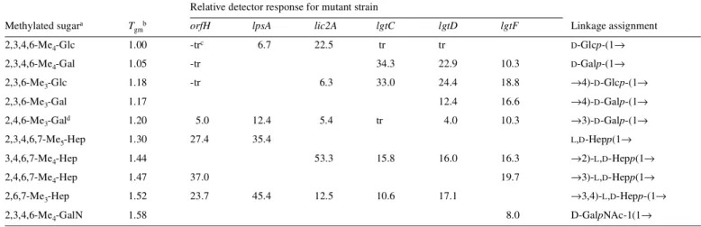
a2,3,4,6-Me
4-Glc represents 1,5-di-O-acetyl-2,3,4,6-tetra-O-methyl-D-glucitol-1-d1, etc. bRetention times (T
gm) are reported relative to 2,3,4,6-Me4-Glc, and values are not corrected for differences in detector response factors. cTrace amount detected.
dAll samples showed detectable levels of 2,4,6,-Me
3-Gal. Only the sample from mutant strain lgtF is expected to give rise to this methylated derivative (Risberg et al., 1999). We suspect a contaminating source contributes to this peak in all samples.
Relative detector response for mutant strain Methylated sugara T
gmb orfH lpsA lic2A lgtC lgtD lgtF Linkage assignment
2,3,4,6-Me4-Glc 1.00 -trc 6.7 22.5 tr tr D-Glcp-(1→
2,3,4,6-Me4-Gal 1.05 -tr 34.3 22.9 10.3 D-Galp-(1→
2,3,6-Me3-Glc 1.18 -tr 6.3 33.0 24.4 18.8 →4)-D-Glcp-(1→
2,3,6-Me3-Gal 1.17 12.4 16.6 →4)-D-Galp-(1→
2,4,6-Me3-Gald 1.20 5.0 12.4 5.4 tr 4.0 10.3 →3)-D-Galp-(1→
2,3,4,6,7-Me5-Hep 1.30 27.4 35.4 L,D-Hepp(1→
3,4,6,7-Me4-Hep 1.44 53.3 15.8 16.0 16.3 →2)-L,D-Hepp(1→
2,4,6,7-Me4-Hep 1.47 37.0 19.7 →3)-L,D-Hepp(1→
2,6,7-Me3-Hep 1.52 23.7 45.4 12.5 10.6 17.1 →3,4)-L,D-Hepp-(1→
2,3,4,6-Me4-GalN 1.58 8.0 D-GalpNAc-1(1→
Fig. 2. Negative-ion ESI-MS of the LPS-OH from the lpsA mutant of H. influenzae RM118 showing doubly and triply charged ions from the major Hex1 glycoform
(structure 3).
Fig. 3. 1H-NMR spectrum of the LPS-OH from the lpsA mutant of H. influenzae RM118 showing the α-anomeric proton region between 5.0 and 6.0 ppm. Anomeric resonances corresponding to the 3,4-disubstituted Hep (HepI), 6-PEtn substituted Hep (HepII), terminal Hep (HepIII), and phosphorylated α-GlcN in the lipid A region are indicated.
1H-NMR spectra with those of the parent strain and its lgtC
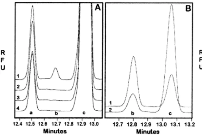
mutant, pointed to the presence of an α-D-Galp-(1→4)-β-D-Galp unit in the Hex4 glycoform, a signal at 5.01 p.p.m. being indicative of the terminal α-D-Galp residue (structure 6) (Table IV). The lgtD gene product was shown to have β-GalpNAc transferase activity with the synthetic acceptor FCHASE-pk, by a comparative assay of
the parent and the lgtD mutant strains (Figure 4).
lgtF mutant. Mutation of the lgtF gene in RM118 gave a strain from which the LPS neither reacted with MAb TEPC-15 (data not shown) nor showed the characteristic PCho methyl proton signal (3.24 ppm) in the 1H-NMR spectrum. Linkage analysis
indicated that the LPS lacked the terminal β-D-Glcp residue, containing only mono-3-substituted HepI residues (Table III). A similar distribution of glycoforms, as found in the parent strain LPS, differing in the length of the oligosaccharide chain from HepIII was observed for LPS-OH from the lgtF mutant in its ESI-MS (Table II). It is noteworthy that full extension of the globotetraose unit, β-D-GalpNAc-(1→3)-α-D -Galp-(1→4)-β-D-Galp-(1→4)-β-D-Glcp (Hex3.HexNAc) from HepIII can occur in the absence (Figure 5) or the presence (Table II, lic1) of the β-D-Glcp residue at HepI.
lic1 mutant. ESI-MS analysis of O-deacylated LPS from the
lic1 mutant gave a similar heterogeneous mixture of glyco-forms (Table II) as that observed in the parent strain but lacking PCho substituents. Examination of the 1H-NMR
spectrum of RM118lic1 LPS-OH revealed the absence of the characteristic PCho methyl proton signal at 3.24 ppm. Additionally, the LPS from this mutant did not react with MAb TEPC-15 (not shown).
Discussion
In a previous study, the complete genome sequence of H. influenzae strain Rd facilitated the identification and study of candidate LPS genes in a H. influenzae type b strain, RM153 (Hood et al., 1996a). Many probable gene functions were established relevant to LPS biosynthesis in strain RM153, but others remain unidentified. Knowledge of the detailed structure of the LPS from strain RM118 (Risberg et al., 1999b) has shown that the Hex extensions in the oligosaccharide portion are funda-mentally different to that of strain RM153. In this study we
investigate the genetic basis for the biosynthesis of the major LPS glycoforms expressed by RM118, the index sequenced strain.
In our investigation of the H. influenzae LPS inner core, attempts to remove Kdo, the first sugar added to the lipid A, have repeatedly failed presumably because Kdo is required to complete lipid A synthesis and is thus likely essential for cell viability. The Kdo transferase function of KdtA has been demonstrated by complementation experiments in Escherichia
coli (White and Raetz, 1998). The results from the present study would suggest that opsX, rfaF, and orfH are the genes encoding the enzymes that add the first, second, and third Hep, respectively, to the Kdo, to form the inner core of H. influenzae LPS (Figure 6). opsX, rfaF, and orfH have some homology to heptosyl transferases of other bacteria (Hood et al., 1996a). The data from RM118 mutants is consistent with that obtained in the type b strain RM153, where opsX, rfaF, and orfH were proposed as the genes encoding the HepI, HepII, and HepIII transferases, respectively (Hood et al., 1996a). This shows a conservation of the genetic basis for as well as the structure of the triheptosyl inner core of H. influenzae LPS.
Each heptose of the inner-core moiety of H. influenzae LPS provides a point for elongation by oligosaccharide chains (Masoud et al., 1997; Risberg et al., 1999b). Each of the genes
lpsA, lic2A, lgtC, lgtD, and lgtF are predicted to encode glyco-syltransferase enzymes involved in oligosaccharide elongation by homology comparisons with genes of similar function (Hood et al., 1996a; Campbell et al., 1997). The results of the present investigation would indicate that the lpsA gene product plays a role in controlling oligosaccharide chain extension from HepIII. A mutation in the lpsA gene affords a truncated LPS in which HepIII is devoid of oligosaccharide chain exten-sions. ESI-MS analysis of the RM118lpsA-derived LPS-OH indicated a PCho-containing Hex1 glycoform as the major LPS species (structure 2), confirming that HepI can be substituted in Table IV. Structure of the major LPS glycoforms of increasing
oligosaccharide chain length from HepIII in mutant stains of
H. influenzae RM118
Fig. 4. Capillary electrophoresis analysis of LgtD activity in H. influenzae strain
RM118. (A) Trace 1 is a complete reaction mixture using the 100,000 × g pellet of a sonicate as enzyme source; trace 2 is similar to the reaction mixture in 1, except it is missing UDP-GalNAc; trace 3 is a complete reaction mixture from the mutant RM118:lgtD; trace 4 is similar to trace 3 except it is missing UDP-GalNAc. The peak a is an impurity in the FCHASE-PK preparation, peak b is FCHASE-Globotetraose, peak c is FCHASE-PK. (B) The trace 1 is the purified product for thin-layer chromatography from a reaction as described for
A trace 1. Trace 2 is the same material as trace 1, but treated with
the absence of Hex extension from HepIII. The lic2A, lgtC, and
lgtD mutants, which contain a functional lpsA gene, are capable of adding a β-D-Glcp residue in a 1,2-linkage to initiate chain extension from HepIII (Table IV). LpsA has homology to the group of galactosyltransferases typified by Lic2A and LgtB of Haemophilus and Neisseria, respectively. In strain RM153, it was also found that LPS from a lpsA mutant lacked any extension from the third Hep (Hood et al., 1996a). However, in strain RM153, lpsA apparently plays a slightly different role being responsible for the addition of Gal as the sole extension from the third Hep (Hood et al., 1996a). Thus, LpsA is likely the transferase for the addition of the first glycose to HepIII in H. influenzae LPS biosynthesis.
The RM118lic2A mutant showed a PCho-containing Hex2 glycoform as a major LPS species (structure 4) and RM118lgtC, which contains a functional lic2A gene, elaborates LPS containing a lactose side chain at HepIII (structure 5). This is consistent with the involvement of the lic2A gene to add the
β-D-Galp unit in a 1,4 linkage to the terminal β-D-Glcp residue attached to HepIII. Lic2A homologues in type b strains have been shown to be involved in expression of the digalactoside-containing Pk epitope (α-D-Galp-[1→4]-β-D-Galp-[1→4]-β-D -Glcp). Homology comparisons with other databank sequences support the function of Lic2A as a β-galactosyltransferase; importantly, it has significant homology to the Neisseria LgtB and LgtE proteins, both of which are galactosyltransferases (Wakarchuk et al., 1996). Structural analysis of LPS from an RM118 strain mutated in lgtC confirmed the loss of α-D-Galp, supporting the α-galactosyltransferase function for this gene. Correspondingly the lgtD mutant and the parent strain, which contain a functional lgtC gene, are capable of adding a α-D-Galp in a 1,4 linkage to the terminal β-D-Galp of the lactose epitope (structure 6). The function of LgtC was confirmed by demon-strating α-galactosyltransferase activity with the recombinant protein and a synthetic FCHASE-Lac acceptor. In N. meningitidis, LgtC is also an α-galactosyltransferase (Gotschlich, 1994; Wakarchuk et al., 1998). It follows that the lgtC gene encodes the specific α-galactosyltransferase for the synthesis of the α-D-Galp-(1→4)-β-D-Galp of the RM118 Hex4 LPS glycoform (Figure 6). The parent strain RM118 that contains a functional
lgtD gene is capable (unlike the mutant) of elaborating the complete globotetraose unit, which is indicative of its role in adding the terminal β-D-GalpNAc. The H. influenzae lgtD gene is a homologue of two related Neisseria genes, lgtA and
lgtD, which add GlcpNAc and GalpNAc, respectively, to
N. gonorrhoeae LPS (Gotschlich, 1994). Enzyme assays with extracts of RM118 and the RM118lgtD mutant confirmed the β-D-GalpNAc transferase activity. The lgtD gene was found not to be present in the type b strains RM153 and RM7004 (called lgtA in Hood et al., 1996a). Correspondingly, the LPS elaborated by strain RM153 does not contain a GalpNAc moiety (Masoud et al., 1997).
It is noteworthy that, though the activity of glycosyltrans-ferases adding the distal residues of the globotetraose (lgtD) and globoside (lgtC) oligosaccharide side chains could be assayed with the appropriate synthetic acceptor, similar experi-ments to assay the activity of the transferases involved in synthesis of the lactose moiety (Lic2A and LpsA) were unsuccessful. It is likely that the latter two enzymes have more stringent specificities that require the acceptor sugar to be Fig. 5. Negative ion ESI-MS of the triply charged molecular ion region of the LPS-OH from the lgtF mutant of H. influenzae RM118. Peaks arising from the Hex 2
(β-D-Galp-(1→4)-β-D-Glcp), Hex 3 (α-D-Galp-(1→4)-β-D-Galp-(1→4)-β-D-Glcp), and Hex3•HexNAc (β-D-GalpNAc-(1→3)-α-D-Galp-(1→4)-β-D-Galp-(1→4)-β-D-Glcp) are indicated.
Fig. 6. A schematic representation of the structure of LPS from H. influenzae
strain RM118 based on the results of the analysis of Risberg et al., (1999b). The proposed site of action in LPS biosynthesis of loci characterized in this study are shown, linked by arrows to the relevant saccharide linkage. The phase variable loci are lic1, lic2A, and lgtC. Represented in the LPS structure: KDO, 2-keto-3-deoxyoctulosonic acid; Hep, L-glycero-D-manno-heptose; Glc, D-glucose; Gal, D-galactose; GalNAc, N-acetylgalactosamine; PEtn, phosphoethanolamine;
P, phosphate; PCho, phosphocholine. For the heptose residues, listed top to bottom are heptose I, heptose II, then heptose III.
linked to the inner-core Hep residues. Characterization of the initial set of genes and mutant strains available for study of RM118 LPS biosynthesis gave no obvious candidate respon-sible for addition of the β-D-Glcp unit to HepI. An lgtF homologue was identified in strain RM118 by searching the strain Rd genome sequence for matches to genes required for the addition of Hex sugars to Hep residues in the LPS of other organisms. These search sequences included the rfaK and lgtF genes of Neisseria (Kahler et al., 1996). Analysis of the LPS from strain RM118lgtF supported a role for LgtF in chain extension from HepI. The ESI-MS showed molecular ions corresponding to a mixture of glycoforms having chain extensions, including lactose and globotetraose, from HepIII of a triheptosyl inner-core unit that lacks PCho→6)-β-D-Glcp at HepI (Figure 3). Thus, the processes of chain extension from both HepI and HepIII appear to be largely independent in the LPS of strain RM118.
The heterogeneity observed in H. influenzae LPS structure may be due in part to intrinsic variation in the biosynthesis of such a complex structure, but the majority of variation observed is presumed to be due to specific LPS biosynthetic genes capable of variable expression (phase variation). This study has allowed us for the first time to confirm the genes involved in the synthesis of an important phase-variable epitope of H. influenzae LPS, the digalactoside. In strain RM118, Lic2A adds the proximal β-D-Galp and LgtC the terminal α-D-Galp to the digalactoside (-D-Galp-(1→4)-β-D -Galp) as part of the extension from HepIII, whereas the same epitope is expressed as the terminal extension from a diglucoside on the second Hep in the type b strain RM153 (Masoud et al., 1997). Both lic2A and lgtC are phase-variable genes (High et al., 1993; Hood et al., 1996b), making the expression of the epitope highly variable within and between organisms. The digalactoside epitope is expressed in the LPS of many related bacteria, including Neisseria (Virji et al., 1990). The epitope is potentially immunodominant, and its presence offers the potential for molecular mimicry of host structures and can influence the survival of Haemophilus within experimental systems (Hood et al., 1996b; Weiser and Pan, 1998).
In addition to the order and stereochemistry of the sugar residues, the location, type and frequency of substituents such as P, PEtn, and PCho can have a profound affect on LPS structure and biological function. The lic1 locus is essential for the phase-variable addition of PCho to the H. influenzae LPS molecule (Weiser et al., 1997; Lysenko et al., 2000). DNA sequence polymorphisms in lic1 direct the different acceptor specificity observed for PCho incorporation and influence the resistance of H. influenzae to innate humoral immunity (Weiser and Pan, 1998; Lysenko et al., 2000). The gene encoding a Kdo kinase, kdkA, responsible for phosphorylation of Kdo, has been identified (White et al., 1999). This gene has previously been investigated by us as orfZ and when mutated was shown to alter bacterial survival in an infant rat model of infection (Hood et al., 1996a). The only remaining substituents in the core oligosaccharide, whose genetic control remains unknown, therefore, are the PEtn residues that are attached to the 6-position of the HepII residue stoichiometrically and sometimes to the phosphate group on Kdo.
In summary, the genetic blueprint for synthesis of the major globotetraose-containing oligosaccharide of RM118 LPS has been elucidated. The type b strain RM153 and strain RM118
have a gene pool for LPS biosynthesis that is generally the same for the two strains but with some key differences related to their respective LPS structure and biology.
Materials and methods
Bacterial strains and culture conditions
The H. influenzae Rd strain was originally obtained from Alexander and Leidy by Herriot. It was given to H. O. Smith, who named the strain KW-20 and used it in the genome sequencing project (Fleischmann et al., 1995). This same strain obtained from the Smith laboratory has been used by us (RM118). The genotypes of mutants derived from this strain are listed in Table I. H. influenzae strains were grown at 37°C in brain heart infusion broth supplemented with hemin (10 µg/ml), nicotinamide adenine dinucleotide (2 µg/ml), and kanamycin (10 µg/ml) when appropriate.
E. coli strain DH5α was used to propagate cloned PCR products and gene constructs and was grown at 37°C in Luria-Bertani broth supplemented with ampicillin (100 µg/ml) or kanamycin (50 µg/ml) as required (Sambrook et al., 1989).
Identification of LPS-related genes from the H. influenzae
genome sequence
Putative LPS biosynthetic genes had been previously identified by an in silico search of the H. influenzae genome sequence with heterologous sequences of LPS biosynthetic genes from a wide range of organisms obtained from publicly available databases (Hood et al., 1996a). The RM118lgtF locus (HI0653) was identified by searching the Institute for Genomic Research H. influenzae strain Rd sequence database (www.tigr.org/tdb/CMR/ghi/htmls/SplashPage.html) for matches with the LgtF protein sequence from N. meningitidis (GenBank accession no. U58765).
Recombinant DNA methodology, cloning, and mutation
Restriction endonucleases and DNA-modifying enzymes were obtained from Boehringer Mannheim and used according to the manufacturer’s instructions. Plasmid DNA preparation, Southern blotting, and hybridization analysis were performed as described by Sambrook et al. (1989). Chromosomal DNA was prepared from Haemophilus by the method described elsewhere (High et al., 1993).
Apart from lgtF, putative H. influenzae LPS biosynthetic genes were cloned and mutated as previously reported (Hood
et al., 1996a). For the lgtF locus, oligonucleotide primers, lgtFa (5′-TGGTGGTGGGCAAGACGC-3′) and lgtFb (5′- AGCCTG-AATTCGACAGCC-3′), amplified a 1461-bp DNA fragment including HI0653 by PCR. PCR conditions were for 1-min periods of denaturation (94°C), annealing (50°C), and poly-merization (72°C) for 30 cycles. One microliter of PCR product was ligated with 50 ng of plasmid pT7Blue (Novagen) and transformed into E. coli strain DH5α. Recombinant plasmids were confirmed by restriction endonuclease digestion and sequencing from plasmid-specific primers (Hood et al., 1996a). The lgtF gene was inactivated by inserting a kana-mycin resistance cassette (released by digestion with EcoR1 from pUC4Kan, Pharmacia) into a MunI restriction site 257 bp inside the 5′ end of HI0653 to give plasmid pDQ1.
Construction of mutant strains
Two to three micrograms of linearized plasmid, containing mutated LPS biosynthetic genes, was used to transform
H. influenzae strain RM118 by the MIV procedure (Herriott
et al., 1970) and transformants were selected on kanamycin. To construct strain RM118lic1, RM118 was transformed with 5µg of sheared chromosomal DNA isolated from the corre-sponding RM153 mutant. Strain RM118lic2A was constructed by transformation of RM118 with 1 µg of a PCR product including inactivated lic2A and the adjacent gene ksgA amplified from strain RM153lic2A. PCR used the primers L2A (5′-CTCCATATTACATAAT-3′) and L2D (5′-AAACACT-TAGGCCATACG-3′) under conditions as described above. All transformants were recultured on appropriate brain heart infusion/antibiotic plates, then were confirmed as mutants by PCR amplification and/or Southern blotting/hybridization of digested chromosomal DNA.
Analysis of LPS by immunoblotting and electrophoresis
LPS isolated from wild type and mutants of H. influenzae strain RM118 was analyzed using LPS-specific monoclonal antibodies and by T–SDS–PAGE) as described previously (Hood et al., 1996a).
Structural fingerprinting of LPS
Cells from 10-L batch cultures (10 lots of 1 L) were harvested after overnight growth, LPS was extracted by the hot phenol-water method (Westphal and Jann, 1965), followed by ethanol precipitation as described by Thibault and Richards (2000). LPS was purified and O-deacylated as previously described (Holst et al., 1993). Sugars were identified by gas-liquid chromatography mass spectrometry (GLC-MS) as their alditol acetates as previously described (Masoud et al., 1997). Linkage analysis was accomplished following acetylation of the oligo-saccharides with acetic anhydride (0.5 ml) and 4-dimethyl-aminopyridine (0.5 mg) at room temperature for 24 h. Peracetylated material was then treated with methyl iodide in dimethylsulfoxide in the presence of lithium methylsulfinyl-methanide to afford the methylated oligosaccharides, which were recovered using a SepPak C18 cartridge and subjected to sugar analysis (Blakeney and Stone, 1985). The relative proportions of the various alditol acetates and partially methyl-ated alditol acetates obtained in sugar and methylation analyses were measured from the detector response of the GLC-MS and are uncorrected. GLC-MS was carried out with a Delsi Di200 chromatograph equipped with a NERMAG R10–10H quadrupole mass spectrometer or with a Varian Iontrap system using a DB-5 fused silica capillary column (25 m × 0.25 mm × 0.25 µm) and a temperature gradient of 160°C (1 min) → 250°C at 3°C/min.
ESI-MS was performed on a VG Quattro Mass Spectrometer (Micromass, Manchester, UK) in the negative-ion mode. Samples were dissolved in water and then mixed in a 1:1 ratio with 50% aqueous acetonitrile containing 1% acetic acid. Sample solutions were injected via a syringe pump into a running solvent of H2O:CH3CN (1:1) at a flow rate of 5 µl/min.
1D 1H-NMR spectra were recorded at 500 MHz for solutions
in deuterium oxide at 22°C, after several lyophyllizations with D2O, on a Bruker AMX 500 spectrometer. To enhance spectral resolution, perdeutero-EDTA (2 mM) and perdeutero-SDS
(10 mg/ml) were added to the D2O solutions (Risberg et al., 1997). Chemical shifts are referenced to the methyl proton resonance (δ; 2.225 ppm) of internal acetone.
Analysis of enzymatic activity from LgtC and LgtD
The enzyme encoded by lgtD was assayed with the synthetic acceptor FCHASE-Pk using capillary electrophoresis for
detection of the product (Wakarchuk et al., 1996). FCHASE-Pk
was synthesized from FCHASE-Lac using the N. meningitidis LgtC enzyme as previously described (Gotschlich, 1994; Wakarchuk et al., 1998). The reaction conditions were 0.5 mM acceptor, 1 mM UDP-GalNAc, 50 mM HEPES–NaOH (pH 7.0), 10 mM MgC12, 10 mM MnC12. Extracts from RM118 and RM118:lgtD were made by sonicating the cells, and then collecting the membrane fraction by centrifugation at 100,000 × g for 30 min. The product was isolated by thin-layer chromatography as previously described (Wakarchuk et al., 1996). Since the proportion of acceptor converted to product was small, some of the starting material was also isolated. The recovered mixture was divided into two parts and then treated with β-hexosaminidase under conditions recommended by the enzyme supplier (NEB).
Activity for LgtC was below the limits of detection in extracts of RM118, so the lgtC gene was cloned into an expression vector and activity was assayed in E. coli. The gene was amplified by PCR (as described above) using primers lgtCa (5′GGGGGGCATATGGGACGGACTGTCAGTCAGA-CAATG) and lgtCb (5′GGGGGGGTCGACTCATTAAT-TATCTTTTATTCTCTTTCTTAATC). The gene was then inserted into plasmid pCWori plus at the NdeI and SalI sites similar to what was described for lgtC from N. meningitidis (Gotschlich, 1994; Wakarchuk et al., 1998). Crude sonicated extracts of the recombinant clone were assayed with 1 mM FCHASE-Lac, 1 mM UDP-Gal, 10 mM MnC12, 5 mM
dithio-threitol, 50 mM HEPES, pH 7.5. The enzyme was shown to be unstable in E. coli and was assayed within a few hours of when the extracts were made. The product of the enzyme reaction was analyzed by specific glycosidase digestion, mass spectrometry, and cochromatography with authentic FCHASE-Pk.
Acknowledgments
We thank Don Krajcarski for assistance in ESI-MS analysis, Jianjun Li for CE-ESI-MS analysis, and Johan Sagemark and Anna-Karin Karlsson for methylation analysis. D.H., S.W., and M.D. were supported by a program grant from the Medical Research Council, UK.
Abbreviations
ESI-MS, electrospray ionization mass spectrometry; GLC-MS, gas-liquid chromatography mass spectrometry; Kdo, 2-keto-3-deoxyoctulosonic acid; LPS, lipopolysaccharide; LPS-OH,
O-deacylated lipopolysaccharide; MAb, monoclonal antibody; MS-MS, tandem mass spectrometry; NMR, nuclear magnetic resonance; P, free phosphate group; PCR, polymerase chain reaction PEtn, phosphoethanolamine; PPEtn, pyrophospho-ethanolamine; PCho, phosphocholine; T–SDS–PAGE, tricine– sodium dodecyl sulfate–polyacrylamide gel electrophoresis.
References
Blakeney, A.B. and Stone, B.A. (1985) Methylation of carbohydrates with lithium methylsulphinyl carbanion. Carbohydr. Res., 140, 319–324. Brook, J.S. and Valvano, M.A. (1996) Molecular cloning of the Haemophilus
influenzae gmhA (lpcA) gene encoding a phosphoheptose isomerase required for lipooligosaccharide biosynthesis. J. Bacteriol., 178, 3339–3341. Campbell, J.A., Davies, G.J., Bulone, V., and Henrissat, B. (1997) A classification
of nucleotide-diphospho-sugar glycosyltransferases based on amino acid sequence similarities. Biochem. J., 326, 929–942.
Cope, L.D., Yogev, R., Mertsola, J., Argyle, J.C., McCracken, G.H., and Hansen, E.J. (1990) Effect of mutation in lipopolysaccharide biosynthetic genes on virulence of Haemophilus influenzae type b. Infect. Immun., 58, 2343–2351.
Fleischmann, R.D., Adams, M.D., White, O., Clayton, R.A., Kirkness, E.F., Kerlavage, A.R., Butt, C.J., Tomb, J-F., Dougherty, B.A., Merrick, J.M., and others (1995) The genome of Haemophilus influenzae Rd. Science,
269, 496–512.
Gibson, B.W., Melaugh, W., Phillips, N.J., Apicella, M.A., Campagnari, A.A., and Griffiss, J.M. (1993) Investigation of the structural heterogeneity of lipooligosaccharides from pathogenic Haemophilus and Neisseria species and of R-type lipopolysaccharides from Salmonella typhimurium by electrospray mass spectrometry. J. Bacteriol., 175, 2702–2712.
Gotschlich, E.C. (1994) Genetic locus for the biosynthesis of the variable portion of Neisseria gonorrhoeae lipooligosaccharide. J. Exp. Med., 180, 2181–2190.
Helander, I.M., Lindner, B., Brade, H., Altmann, K., Lindberg, K.K., Rietschel, E.T., and Zahringer, U. (1988) Chemical structure of the lipo-polysaccharide of Haemophilus influenzae strain I-69 Rd–/b+: Description of a novel deep-rough chemotype. Eur. J. Biochem., 177, 483–492.
Herriott, R.M., Meyer, E.M., and Vogt, M.J. (1970) Defined non-growth media for stage II development of competence in Haemophilus influenzae.
J. Bacteriol., 101, 517–524.
High, N.J., Deadman, M.E., and Moxon, E.R. (1993) The role of the repetitive DNA motif (5′-CAAT-3′) in the variable expression of the Haemophilus
influenzae lipopolysaccharide epitope Galα(1-4)βGal. Mol. Microbiol., 9, 1275–1282.
Holst, O., Broer, W., Thomas-Oates, J.E., Mamat, U., and Brade, H. (1993) Structural analysis of two oligosaccharide bisphosphates isolated from the lipopolysaccharide of a recombinant strain of Escherichia coli F515 (Re chemotype) expressing the genus-specific epitope of Chlamydia lipo-polysaccharide. Eur. J. Biochem., 214, 703–710.
Hood, D.W., Deadman, M.E., Allen, T., Masoud, H., Martin, A., Brisson, J.R., Fleischmann, R., Venter, J.C., Richards, J.C., and Moxon, E.R. (1996a) Use of the complete genome sequence information of Haemophilus
influenzae strain Rd to investigate lipopolysaccharide biosynthesis.
Mol. Microbiol., 22, 951–965.
Hood, D.W., Deadman, M.E., Jennings, M.P., Bisceric, M., Fleischmann, R.D., Venter, J.C., and Moxon, E.R. (1996b) DNA repeats identify novel virulence genes in Haemophilus influenzae. Proc. Natl. Acad. Sci. USA,
93, 11121–11125.
Jarosik, G.P. and Hansen, E.J. (1994) Identification of a new locus involved in the expression of Haemophilus influenzae type b lipooligosaccharide.
Infect. Immun., 62, 4861–4867.
Kahler, C.M., Carlson, R.W., Rahman, M.M., Martin, L.E., and Stephens, D.S. (1996) Two glycosyltransferase genes, lgtF and rfaK, constitute the lipooligosaccharide ice (Inner Core Extension) biosynthesis operon of
Neisseria meningitidis. J. Bacteriol., 178, 6677–6684.
Kimura, A. and Hansen, E.J. (1986) Antigenic and phenotypic variants of
Haemophilus influenzae type b lipopolysaccharide and their relationship in virulence. Infect. Immun., 51, 60–79.
Lysenko, E., Richards, J.C., Cox, A.D., Stewart, A., Martin, A., Kapoor, M., and Weiser, J.N. (2000) The position of phosphorylcholine on the lipopoly-saccharide of Haemophilus influenzae affects binding and sensitivity to C-reactive protein-mediated killing. Mol. Microbiol., 35, 234–245. Masoud, H., Moxon, E.R., Martin, A., Krajcarski, D., and Richards, J.C.
(1997) Structure of the variable and conserved lipopolysaccharide oligosaccharide epitopes expressd by Haemophilus influenzae serotype b strain Eagan. Biochem., 36, 2091–2103.
Moxon, E.R. and Maskell, D.J. (1992) “Haemophilus influenzae lipopoly-saccharide: the biochemistry and biology of a virulence factor.” In Hormaeche, C.E., Penn, C.W., Smythe, C.J. (eds.), Molecular biology of
bacterial infection, current status and future perspectives. Society for
General Microbiology Symposium 49. Cambridge University Press, Cambridge, UK.
Phillips, N.J., Apicella, M.A., Griffiss, J.M., and Gibson, B.W. (1992) Structural characterization of the cell surface lipooligosaccharides from a non-typable strain of Haemophilus influenzae. Biochem., 31, 4515–4526. Phillips, N.J., Apicella, M.A., Griffiss, J.M., and Gibson, B.W. (1993)
Structural studies of the lipooligosaccharides from Haemophilus influenzae type b strain A2. Biochem., 32, 2003–2012.
Phillips, N.J., McLaughlin, R., Miller, T.J., Apicella, M.A., and Gibson, B.W. (1996) Characterization of two transposon mutants of Haemophilus
influenzae type b with altered lipooligosaccharide biosynthesis. Biochem.,
35, 5937–5947.
Preston, A., Mandrell, R.E., Gibson, B.W., and Apicella, M.A. (1996a) The lipopolysaccharides of pathogenic Gram-negative bacteria. Crit. Rev.
Microbiol., 22, 139–180.
Preston, A., Maskell, D., Johnson, A., and Moxon, E.R. (1996b) Altered lipopolysaccharide characteristic of the I69 phenotype in Haemophilus
influenzae results from mutation in a novel gene, isn. J. Bacteriol., 178, 396–402.
Rahman, M.M., Gu, X-X., Tsai, C-M., Kolli, V.S.K., and Carlson, R.W. (1999) The structural heterogeneity of the lipooligosaccharide (LOS) expressed by pathogenic non-typeable Haemophilus influenzae strain NTHi 9274. Glycobiology, 9, 1371–1380.
Risberg, A., Schweda, E.K.H., and Jansson, P.-E. (1997) Structural studies of the cell-envelope oligosaccharide from the lipopolysaccharide of Haemophilus
influenzae strain RM.118-28. Eur. J. Biochem., 243, 701–707.
Risberg, A., Alvelius, G., and Schweda, E.K.H. (1999a) Structural analysis of the lipopolysaccharide oligosaccharide epitopes expressed by Haemophilus
influenzae strain RM.118-26. Eur. J. Biochem., 265, 1067–1074. Risberg, A., Masoud, H., Martin, A., Richards, J.C., Moxon, E.R., and
Schweda, E.K.H. (1999b) Structural analysis of the lipopolysaccharide epitopes expressed by a capsular deficient strain of Haemophilus influenzae Rd. Eur. J. Biochem., 261, 171–180.
Sambrook, J., Fritsch, E. F., and Maniatis, T. (1989) Molecular cloning; a
laboratory manual, 2d ed. Cold Spring Harbor Laboratory, Cold Spring Harbor, NY.
Schweda, E.K.H., Hegedus, O.E., Borrelli, S., Lindberg, A.A., Weiser, J.W., Maskell, D.J., and Moxon, E.R. (1993) Structural studies of the saccharide portion of cell envelope lipopolysaccharide from Haemophilus influenzae strain AH1-3 (lic3+). Carbohydr. Res., 246, 319–330.
Schweda, E.K.H., Jansson, P.-E., Moxon, E.R., and Lindberg, A.A. (1995) Structural studies of the saccharide part of the cell envelope lipooligo-saccharide from Haemophilus influenzae strain galEgalK. Carbohydr.
Res., 272, 213–224.
Thibault, P. and Richards, J.C. (2000) Application of combined capillary electrophoresis electrospray mass spectrometry in the characterisation of short chain lipopolysaccharides: Haemophilus influenzae. In Holst, O. (ed.), Methods of molecular biology, vol. 45, Bacterial toxins: methods and
protocols. Humana Press, Totowa, NJ.
Virji, M., Weiser, J.N., Lindberg, A.A., and Moxon, E.R. (1990) Antigenic similarities in lipopolysaccharides of Haemophilus and Neisseria and expression of a digalactoside structure also present on human cells.
Microb. Pathogen., 9, 441–450.
Wakarchuk, W., Martin, A., Jennings, M.P., Moxon, E.R., and Richards, J.C. (1996) Functional relationships of the genetic locus encoding the glycosyl transferase enzymes involved in the expression of the lacto-N-neotetraose terminal lipopolysaccharide structure in Neisseria meningitidis. J. Biol.
Chem., 271, 19166–1917.
Wakarchuk, W.W., Cunningham, A.M., Watson, D.C., and Young, N.M. (1998) Role of paired basic residues in the expression of active recombinant galacto-syltransferases from the bacterial pathogen Neisseria meningitidis. Prot. Eng.,
11, 295–302.
Weiser, J.N. and Pan, N. (1998) Adaptation of Haemophilus influenzae to acquired and innate humoral immunity based on phase variation of lipo-polysaccharide. Mol. Microbiol., 30, 767–775.
Weiser, J.N., Love, J.M., and Moxon, E.R. (1989) The molecular mechanism of phase variation of Haemophilus influenzae lipopolysaccharide. Cell, 59, 657–665.
Weiser, J.N., Maskell, D.J., Butler, P.D., Lindberg, A.A., and Moxon, E.R. (1990a) Characterisation of repetitive sequences controlling phase variation of Haemophilus influenzae lipopolysaccharide. J. Bacteriol.,
Weiser, J.N., Williams, A., and Moxon, E.R. (1990b) Phase-variable lipo-polysaccharide structures enhance the invasive capacity of Haemophilus
influenzae. Infect. Immun., 58, 3455–3457.
Weiser, J.N., Shchepetov, M., and Chong, S.T.H. (1997) Decoration of lipopolysaccharide with phosphorylcholine: a phase variable characteristic of Haemophilus influenzae. Infect. Immun., 65, 943–950.
Westphal, O. and Jann, K. (1965) Bacterial lipopolysaccharides. Extraction with phenol-water and further applications of the procedure. Meth. Carbohydr.
Chem., 5, 83–91.
White, K.A. and Raetz, C.R.H. (1998) Complementation of a genomic disruption of the E.coli bifunctional KDO transferase by the H. influenzae mono-functional KDO transferase. FASEB J., 12, L44.
White, K.A., Lin, S., Cotter, R.J., and Raetz, C.R.H. (1999) A Haemophilus
influen-zae gene that encodes a membrane bound 3-deoxy-D-manno-octulosonic acid (Kdo) kinase: Possible involvement of KDO phosphorylation in bacterial virulence. J. Biol. Chem., 274, 31391–31400.
Zamze, S.E. and Moxon, E.R. (1987) Composition of the lipopolysaccharide from different capsular serotype strains of Haemophilus influenzae. J. Gen.