HAL Id: hal-01819569
https://hal-amu.archives-ouvertes.fr/hal-01819569
Submitted on 20 Jun 2018HAL is a multi-disciplinary open access archive for the deposit and dissemination of sci-entific research documents, whether they are pub-lished or not. The documents may come from teaching and research institutions in France or abroad, or from public or private research centers.
L’archive ouverte pluridisciplinaire HAL, est destinée au dépôt et à la diffusion de documents scientifiques de niveau recherche, publiés ou non, émanant des établissements d’enseignement et de recherche français ou étrangers, des laboratoires publics ou privés.
Galactolipase activity of Talaromyces thermophilus
lipase on galactolipid micelles, monomolecular films and
UV-absorbing surface-coated substrate
Inès Belhaj, Sawsan Amara, Goetz Parsiegla, Priscila Sutto-Ortiz, Moulay
Sahaka, Hafedh Belghith, Audric Rousset, Dominique Lafont, Frédéric
Carriere
To cite this version:
Inès Belhaj, Sawsan Amara, Goetz Parsiegla, Priscila Sutto-Ortiz, Moulay Sahaka, et al.. Galactoli-pase activity of Talaromyces thermophilus liGalactoli-pase on galactolipid micelles, monomolecular films and UV-absorbing surface-coated substrate. Biochimica et Biophysica Acta Molecular and Cell Biology of Lipids, Elsevier, 2018, 1863 (9), pp.1006-1015. �10.1016/j.bbalip.2018.05.016�. �hal-01819569�
PREPRINT - BBA Mol Cell Biol Lipids (2018) 1863(9):1006-1015
1
doi: 10.1016/j.bbalip.2018.05.016
2 3
Galactolipase activity of Talaromyces thermophilus lipase on galactolipid
4
micelles, monomolecular films and UV-absorbing surface-coated substrate
5 6
Inès Belhaja*, Sawsan Amarab,c, Goetz Parsieglab, Priscila Sutto-Ortizb, Moulay Sahakab, 7
Hafedh Belghitha, Audric Roussetd, Dominique Lafontd and Frédéric Carrièreb* 8
9
Running title: Galactolipase activity of Talaromyces thermophilus lipase 10
11
a
Laboratoire de Biotechnologie Moléculaire des Eucaryotes, Centre de Biotechnologies de 12
Sfax, Université de Sfax, BP «1177» 3018 Sfax, Tunisia 13
b
Aix-Marseille Université, CNRS, Bioénergétique et Ingénierie des Protéines UMR 7281, 31 14
Chemin Joseph Aiguier, 13402 Marseille Cedex 20, France 15
c
Lipolytech, Zone Luminy Biotech Entreprises Case 922, 163 avenue de Luminy, 13288 16
Marseille Cedex 09, France 17
d
Laboratoire de Chimie Organique II-Glycochimie, ICBMS UMR 5246, CNRS-Université 18
Claude Bernard Lyon 1, Université de Lyon, Bâtiment Curien, 43 Boulevard du 11 Novembre 19
1918, 69622 Villeurbanne Cedex, France 20
21
*
Correspondence to: Frédéric Carrière : carriere@imm.cnrs.fr 22
Inès Belhaj: ines.belhaj@cbs.rnrt.tn 23
Phone: +216 74874449; Fax: +216 74874449 24
Keywords: enzyme assay, galactolipid, galactolipase, in silico docking, lipase, monomolecular 25
films ; 26
Abbreviations: BHT, butylhydroxytoluene; β-CD, β-cyclodextrin; DGDG, 27
digalactosyldiacylglycerol ; αE-MGDG, 1,2-di-α-eleostearoyl-3-galactopyranosyl glycerol; 28
GPLRP2, guinea pig pancreatic lipase-related protein 2; MGDG, 29
monogalactosyldiacylglycerol; NaTDC, sodium taurodeoxycholate; PLRP2, pancreatic lipase-30
related protein 2; TC4, tributyrin ; TC8; trioctanoin, TLL, Thermomyces lanuginosus lipase ; 31
TTL, Talaromyces thermophilus lipase; 32
Abstract 33
Talaromyces thermophilus lipase (TTL) was found to hydrolyze monogalactosyl 34
diacylglycerol (MGDG) and digalactosyl diacylglycerol (DGDG) substrates presented in 35
various forms to the enzyme. Different assay techniques were used for each substrate: pHstat 36
with dioctanoyl galactolipid-bile salt mixed micelles, barostat with dilauroyl galactolipid 37
monomolecular films spread at the air-water interface, and UV absorption using a novel 38
MGDG substrate containing α-eleostearic acid as chromophore and coated on microtiter 39
plates. The kinetic properties of TTL were compared to those of the homologous lipase from 40
Thermomyces lanuginosus (TLL), guinea pig pancreatic lipase-related protein 2 and Fusarium 41
solani cutinase. TTL was found to be the most active galactolipase, with a higher activity on 42
micelles than on monomolecular films or surface-coated MGDG. Nevertheless, the UV 43
absorption assay with coated MGDG was highly sensitive and allowed measuring significant 44
activities with about ten ng of enzymes, against hundred ng to ten µ g with the pHstat. TTL 45
showed longer lag times than TLL for reaching steady state kinetics of hydrolysis with 46
monomolecular films or surface-coated MGDG. These findings and 3D-modelling of TTL 47
based on the known structure of TLL pointed out to two phenylalanine to leucine substitutions 48
in TTL, that could be responsible for its slower adsorption at lipid-water interface. TTL was 49
found to be more active on MGDG than on DGDG using both galactolipid-bile salt mixed 50
micelles and galactolipid monomolecular films. These later experiments suggest that the 51
second galactose on galactolipid polar head impairs the enzyme adsorption on its aggregated 52
substrate. 53
1. Introduction 54
Glycolipids are present in almost all biological membranes. Among them, 55
galactolipids are the most abundant membrane lipids in plants, especially in green tissues 56
where they generally represent about 75% of total membrane lipids [1]. They are especially 57
abundant in the photosynthetic membranes of the thylakoids in the chloroplast. These 58
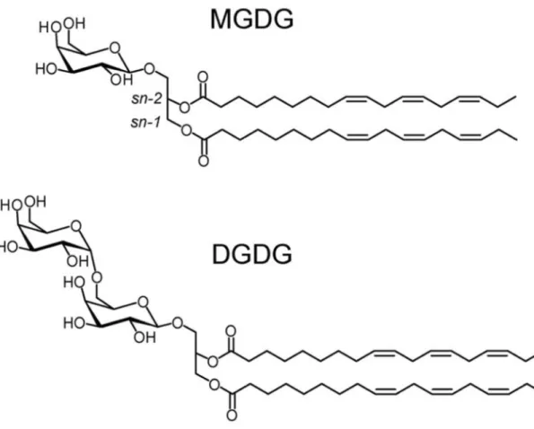
membranes contain mainly monogalactosyl diacylglycerols (MGDG) and digalactosyl 59
diacylglycerols (DGDG) (Fig.1), which represent 50% and 30% of total lipids, respectively 60
[2]. Plant galactolipids are characterized by long chain fatty acids, typically varying from C16 61
to C20 with one to three unsaturations [3] and they are particularly rich in polyunsaturated 62
fatty acids like C16:3 and C18:3 (ALA, α-linolenic acid) [4]. Although galactolipids are not 63
present at high levels in normal human diet compared to triglycerides, they may represent the 64
main source of the essential ALA in the absence of specific supplementation with ALA-rich 65
vegetable oils. Indeed, ALA can represent as much as 60 % w/w of the total fatty acid in 66
galactolipids from plant leaves, like in spinach [4], which is higher than what is found in 67
flaxseed oil [5]. The uptake of galactolipid fatty acids is made possible by the action of 68
galactolipases (1,2-Di-O-acyl-1-O-(β-D-galactosyl)-D-glycerol acylhydrolases; EC 3.1.1.26) 69
that hydrolyze the ester bonds at sn-1 and sn-2 positions of galactolipids. In humans and other 70
mammals, this function is mainly fulfilled by pancreatic lipase-related protein 2 (PLRP2) 71
produced by the exocrine pancreas and acting in the GI tract [6-8]. 72
Galactolipase activity is often displayed by lipases (triacylglycerol acylhydrolases 73
EC.3.1.1.3) with broad substrate specificity that can accept various acylglycerols as substrates 74
like phospholipids and galactolipids. Numerous enzymes with galactolipase activity have 75
been discovered in the plant kingdom [9-11] and more recently in microalgae [12]. Various 76
microbial lipases are also known to degrade galactolipids. For example, Dawson et al. found 77
that microbial lipases from rumen were able to decompose grass galactolipids in the sheep GI 78
tract [13]. Microbial lipases from Rhizopus arrhizus [14], Mucor javanicus [15] and 79
Rhizomucor miehei (Lipozyme TM) [16] have been shown to hydrolyze the ester bond of 80
galactolipids. More recently, synthetic medium chain MGDG and DGDG were used to 81
characterize the galactolipase activity of several well-known microbial lipases, such as the 82
lipases from Thermomyces lanuginosus (TLL), Rhizomucor miehei (RML), Rhizopus oryzae 83
(ROL), Candida antarctica A (CalA), and the cutinase from Fusarium solani (FsC) [17]. 84
Using these substrates and natural long chain galactolipids, a new galactolipase from 85
Fusarium solani was also identified [18]. 86
In contrast to the large biochemical and structural knowledge on lipases and 87
phospholipases, galactolipases remain poorly studied and characterized so far. This is mainly 88
due to the fact that galactolipid substrates are not commercially available at a large scale and 89
low cost. Natural galactolipids purified from plant green materials have been used for the 90
characterization of some galactolipases by thin layer chromatography analysis of lipolysis 91
products or free fatty acid titration [4, 18] but their limited availability does not allow 92
thorough kinetic studies. Nevertheless, medium galactolipid substrates have been synthesized 93
to develop continuous and sensitive assays using the pHstat [19] and the monomolecular film 94
[7, 20] techniques 95
Here we used these assays to search for a possible activity of Talaromyces 96
thermophilus lipase (TTL) on galactolipids. This enzyme was previously purified from a 97
newly isolated Talaromyces thermophilus fungal strain and was shown to have a high activity 98
towards short, medium and long chain triacylglycerols [21]. The use of wheat bran as carbon 99
source for cultivating T. thermophilus was found to be more effectively than olive oil to 100
induce the production of TTL [21], suggesting that other types of lipids present in wheat bran, 101
like galactolipids, could boost the level of TTL expression in this fungus. Since the expression 102
of microbial lipases is in general induced by the fatty acids released upon digestion of the 103
lipid used as the carbon source [22], we wondered whether TTL displays a galactolipase 104
activity, as previously identified with homologous enzymes of this fungal lipase family [17, 105
18]. 106
The galactolipase activity of TTL was investigated using medium chain galactolipids 107
presented in the form of mixed micelles with bile salts (pHstat assay; [19]) and 108
monomolecular films (Barostat assay; [7]). We also used the characterization of TTL for 109
developing a novel UV spectrophotometric galactolipase assay in microtiter plates using a 110
synthetic MGDG substrate containing α-eleostearic acid as chromophore. Various lipase 111
assays have been developed for high throughput screening using α-eleostearic acid esterified 112
into natural [23, 24] and synthetic triglycerides [25, 26] or phospholipids [27] but not 113
galactolipids so far. Using this combination of galactolipase assays, we showed that TLL is 114
active on both MGDG and DGDG and we compared its kinetic properties with those of other 115
galactolipases, the homologous fungal lipase from Thermomyces lanuginosus (TLL), guinea 116
pig pancreatic lipase-related protein 2 (GPLRP2) and cutinase from Fusarium solani (FsC). 117
118 119
2. Materials and methods 120
121
2.1. Enzymes 122
TTL was produced by a newly isolated thermo-tolerant fungal strain identified as 123
Talaromyces thermophilus Stolk and purified according to [21]. Recombinant GPLRP2 was 124
expressed in Aspergillus orizae and purified according to [28]. Cutinase from Fusarium solani 125
(FsC) was expressed in E.coli and purified as previously described [29]. Thermomyces 126
lanuginosus lipase (TLL; Lipolase™) was purchased from Sigma (St-Quentin-Fallavier, 127
France). The homogeneity of the various enzymes was routinely assessed by performing SDS-128
PAGE on 12% gels using Laemmli’s procedure [30]. The protein concentration was 129
determined with a good accuracy using the BCA kit (Pierce) and BSA as standard. 130
131
2.2. Potentiometric pHstat assay of galactolipase activity 132
Dioctanoyl galactolipid substrates, C8-MGDG (3-O-β-D-galactopyranosyl-1,2-di-O-133
octanoyl-sn-glycerol) and C8-DGDG (3-O-(6-O-α-D-galactopyranosyl-β-D -134
galactopyranosyl)-1,2-di-O-octanoyl-sn-glycerol), were synthesized as previously described 135
[4, 19]. The galactolipase activity was measured potentiometrically at 37 °C and at a constant 136
pH value of 8.0 by continuously measuring the release of free fatty acids from mechanically 137
stirred dispersions of galactolipids, using 0.1 N NaOH as titrant and a pHstat apparatus (718 138
STAT Titrino, Metrohm). To prepare the galactolipid dispersion, 25 mg of MGDG or C8-139
DGDG were mixed with 5 mL of 0.33 mM Tris-HCl buffer, containing 0.1 M NaCl and 140
various concentrations of sodium taurodeoxycholate (NaTDC) and then subjected to 141
ultrasonic treatment for 6-8 min in a water bath (HF-Frequency 35 kHz; SONOREX SUPER 142
compact ultrasonic bath model RK 31, BANDELIN electronic) [19]. One international 143
galactolipase unit (U) corresponds to the release of one µmol of fatty acid released per 144
minute. The specific activities were expressed in international units per milligram of enzyme 145
(U/mg). 146
147
2.3. Potentiometric pHstat assay of phospholipase and lipase activities 148
Phospholipase activities were measured potentiometrically at 37 °C and pH 8.0 by 149
automatically titrating the free fatty acids released from purified egg L-α-phosphatidylcholine 150
(Sigma) as substrate, as previously described [31]. Lipase activities were measured with 151
mechanically stirred triglyceride emulsions according to [21]. Specific activities were 152
expressed in international units (U) per milligram of enzyme. One U corresponds to 1 µmol of 153
fatty acid released per minute. 154
155
2.4. Monomolecular film experiments for measuring galactolipase activities 156
The galactolipase activity was measured using monomolecular films of 1,2-di-O-157
dodecanoyl-3-O-β-D-galactopyranosyl-sn-glycerol (C12-MGDG) and 1,2-di-O-dodecanoyl-158
3-O-(6-O-α-D-galactopyranosyl-β-D-galactopyranosyl)-sn-glycerol (C12-DGDG) as 159
substrates. C12-MGDG and C12-DGDG were synthesized as previously reported [7, 20]. All 160
experiments were performed using the KSV 5000 barostat equipment (KSV, Helsinki, 161
Finland) and a “zero order” Teflon trough [32]. The reaction compartment had a surface area 162
of 38.5 cm2 and a volume of 43 ml. The reservoir (24-cm long × 7.5-cm wide) had a surface 163
area of 156.5 cm2 and a volume of 203 mL. Lipid monomolecular films were formed at the 164
air/water interface by spreading the lipid solution (1 mg/mL C12-MGDG or C12-DGDG in 165
chloroform). The enzyme solution was injected into the subphase at a final concentration of 166
0.02 nM for rGPLRP2, 5nM for TLL and cutinase and 0.45 nM for TTL. The aqueous 167
subphase was composed of 10 mM Tris-HCl, 100 mM NaCl, 21 mM CaCl2, and 1 mM
168
EDTA, pH 8.0 and was prepared with double-distilled water. Residual surface-active 169
impurities were removed by simultaneous sweeping and suction of the surface before 170
spreading the lipid solution [32]. The reaction compartment was stirred with a 1-cm magnetic 171
bar rotating at 250 rpm. The reactions were performed at 25°C. Surface pressure was 172
measured using a Wilhelmy plate (perimeter, 3.94 cm) attached to an electromicro balance. 173
The trough was equipped with a mobile Teflon barrier to keep the surface pressure constant 174
during enzymatic hydrolysis of the substrate film and desorption of the soluble lipolysis 175
products (monododecanoyl-galactopyranosyl-glycerol, dodecanoic acid). Enzyme activity 176
was estimated from the surface of the trough covered by the mobile barrier and the known 177
molecular area of the substrate molecule. The molecular areas of the MGDG and C12-178
DGDG substrates were previously determined by performing compression isotherms [33]. 179
The galactolipase activity was expressed in moles of substrate hydrolyzed per surface unit 180
(cm2) per minute and referred to the overall molarity of the enzyme initially injected into the 181
aqueous subphase (mol.cm-2.min-1.M-1). 182
183
2.5. Synthesis of 1,2-Di-O-
α
-eleostearoyl-3-O-β
-D-galactopyranosyl-sn-glycerol (αE-MGDG) 184Dry dichloromethane was prepared by successive washing with water, drying with 185
calcium chloride, and distillation from calcium hydride. Thin layer chromatography (TLC) 186
was carried out on aluminium sheets coated with silica gel 60 F254 (Merck). TLC plates were
187
inspected by UV light (λ = 254 nm). Column chromatography was performed on Silica-gel 188
(Silicycle). 1H and 13C NMR spectra were recorded at 293 K using Bruker ALS300 or 189
DRX300 spectrometers. High resolution (HR-ESI-QToF) mass spectra were recorded using a 190
Bruker MicroToF-Q II XL spectrometer. 191 192 3-O-[2,3,4,6-Tetra-O-levulinoyl-β-D-galactopyranosyl]-1,2-O-isopropylidene-sn-glycerol (2): 193 3-O-[2,3,4,6-Tetra-O-acetyl-β-D-galactopyranosyl]-1,2-O-isopropylidene-sn-glycerol (1) 194
(2.60 g, 5.53 mmol) was added to dry methanol (50 mL) containing a chip of sodium and the 195
mixture was stirred for 2 h. Amberlite IR 120 [H+] resin was added to neutralize the solution 196
and after 2 min, the solution was filtrated and concentrated in vaccuo. The product was 197
coevaporated twice from toluene (2x15 mL), dissolved in ethyl acetate (50 mL) and levulinic 198
acid (3.92 g, 33.76 mmol), dicyclohexyldicarbodiimide (8.27 g, 40.00 mmol) and a catalytic 199
amount of 4-dimethylaminopyridine (50 mg) were successively added. The mixture was 200
stirred overnight, and after filtration, the solid was washed carefully with ethyl acetate. The 201
organic phase was concentrated to dryness, the crude product was purified by column 202
chromatography (4:1 ethyl acetate-petroleum ether to pure ethyl acetate) and the pure product 203
2 was recovered at 70% yield: 2.70 g, oily material, Rf 0.30 (5:1 ethyl acetate-petroleum
204
ether); [α]D +2.1 (c 1.0, CHCl3); 1H NMR (CDCl3):
δ
5.36 (dd, 1H, J3,4 3.4, J4,5 0.8 Hz, H-4),205
5.17 (dd, 1H, J1,2 7.9, J2,3 10.4 Hz, H-2), 5.01 (dd, 1H, H-3), 4.57 (d, 1H, H-1), 4.26 (dddd,
206
1H, H-2gly), 4.20 (dd, 1H, J5,6a 6.7, J6a,6b 11.2 Hz, H-6a), 4.10 (dd, 1H, J5,6b 6.4 Hz, H-6b),
207
4.04 (dd, 1H, J1agly,2gly 6.4, J1agly,1bgly 6.4 Hz, H-1agly), 3.92-3.86 (m, 2H, H-5, H-3agly), 3.83
208
(dd, 1H, J1bgly,2gly 6.2 Hz, H-1bgly), 3.62 (dd, 1H, J2gly,3bgly 6.2, J3agly,3bgly 10.5 Hz, H-3bgly),
209
2.80-2.40 (m, 16H, 4COCH2CH2COCH3), 2.19, 2.18, 2.17, 2.16 (4s, 12H, 4CH3CO), 1.41,
210 1.34 (2s, 6H, (CH3)2C); 13CNMR (CDCl3):
δ
206.52, 206.43, 206.16, 206.04 211 (4CH3CO),172.15, 171.90, 171.85, 171.41 (4OCOCH2), 109.26 (C(CH3)2), 101.23 (C-1), 212 74.25 (C-2gly), 70.81, 70.73 3, C-5), 69.38 3gly), 68.85 2), 67.24 4), 66.36 (C-2131gly), 61.38 (C-6), 37.84, 37.77, 37.76, 37.67 (CH2COCH3), 29.76, 29.67 (CH3COCH2), 27.79
214
(OCOCH2), 26.67, 25.20 ((CH3)2C).
215
HRMS calculated for C32H46NaO16 [M+Na]+ 709.2678; found 709.2651.
216 217
3-O-[2,3,4,6-Tetra-O-levulinoyl-β-D-galactopyranosyl]-sn-glycerol (3): a solution of 218
compound 2 (2.60 g, 1.46 mmol) in 70% aqueous acetic acid (20 mL) was stirred for 5 h at 219
60°C. After concentration, the residue was coevaporated from toluene (3x20 mL). The crude 220
product was purified by column chromatography (9:1 CHCl3-EtOH). Pure product 3 was
221
obtained in 90% yield: 2.20 g, oily material, Rf 0.46 (9:1 CHCl3-EtOH); [α]D-0.5 (c2.0,
222
CHCl3); 1H NMR (CDCl3):
δ
5.31 (bd, 1H, J3,4 3.4, J4,5 0.2 Hz, H-4), 5.12 (dd, 1 H, J1,2 7.8223
Hz, J2,3 10.5 Hz, H-2), 5.00 (dd, 1H, H-3), 4.49 (d, 1H, H-1), 4.16 (dd, 1H, J5,6a 7.1, J6a,6b
224
11.3, H-6a), 4.06 (dd, 1H, J5,6b 6.1 Hz, H-6b), 3.89 (bd, 1H, H-5), 3.88-3.55 (m, 5H, H-1agly,
225
H-1bgly, H-2gly, H-3agly, H-3bgly), 3.17-3.10 (m, 2H, 2OH), 2.85-2.35 (m, 16H,
226
4COCH2CH2COCH3), 2.19, 2.18, 2.17, 2.16 (4s, 12H, 4CH3CO); 13C NMR (CDCl3):
227
δ
207.76, 206.89, 206.69, 206.22 (CH3CO levulinoyl), 172.28, 171.99, 171.81, 171.75228
(OCOCH2 levulinoyl), 101.57 (C-1), 72.03 (C-3gly), 70.89 (C-5), 70.66 (C-3), 70.48, 69.02
229
(C-2, C-2gly), 67.35 (C-4), 63.35 (C-1gly), 61.61 (C-6), 37.87, 37.87, 37.80, 37.69
230
(CH3COCH2), 29.87, 29.85, 29.81, 29.72 (CH3CO), 27.82, 27.80 (OCOCH2).
231
HRMS calculated for C29H42NaO16 [M+Na]+ 669.2365; found 669.2344.
232 233
1,2-Di-O-α-eleostearoyl-3-O-[2,3,4,6-tetra-O-levulinoyl-β-D-galactopyranosyl]-sn-glycerol 234
(5): α-Eleostearic acid 4 (2.42 g, 8.42 mmol), dicyclohexyldicarbodiimide (3.58 g, 17.40 235
mmol) and a catalytic amount of 4-dimethylaminopyridine (50 mg) were successively added 236
under argon to a solution of product 3 (1.875 g, 2.90 mmol) in dichloromethane (40 mL). The 237
mixture was stirred overnight, and methanol (0.50 mL) was added. After 2 h, the solid was 238
removed by filtration and washed with dichloromethane. The combined organic phases were 239
concentrated to dryness and the crude product was purified by column chromatography (1:1 to 240
4:1 ethyl acetate-petroleum ether). A second column chromatography (9:1 CH2Cl2-EtOH) was
241
necessary to give the pure product 5, recovered in 73% yield: 2.47 g, oily material, Rf
0.20-242
0.25 (1:1 ethyl acetate-petroleum ether), 0.70 (9:1 CH2Cl2-EtOH); [α]D +3.7 (c 1.0, CHCl3);
243
1
H NMR (CDCl3):
δ
6.42-6.33 (m, 2H, 2H-11eleo), 6.20-6.13 (m, 2H, 2H-12eleo), 6.13-6.06 (m,2H, 2H-13eleo), 6.02-5.95 (m, 2H, 2H-10eleo), 5.75-5.65 (m, 2H, 2H-14eleo), 5.43-5.34 (m, 1H,
245
H-9eleo), 5.37 (bd, 1H, J3,4 3.4, J4,5 0.7 Hz, H-4), 5.20 (dddd, 1H, J1agly,2gly 3.0, J1bgly,2gly 6.2,
246
J2gly,3agly 4.9, J2gly,3bgly 5.9 Hz, H-2gly), 5.16 (dd, 1H, J1,2 7.9 Hz, J2,3 10.5 Hz, H-2), 5.02 (dd,
247
1H, H-3), 4.49 (d, 1H, H-1), 4.41 (dd, 1H, J1agly,1bgly 12.0 Hz, H-1agly), 4.20 (dd, 1H, J5,6a 6.6,
248
J6a,6b 11.1, H-6a), 4.14 (dd, 1H, H-1bgly), 4.10 (dd, 1H, J5,6b 6.6 Hz, H-6b), 3.95 (dd, 1H,
249
J3agly,3bgly 10.9 Hz, H-3agly), 3.89 (bdd, 1H, H-5), 3.69 (dd, 1H, H-3bgly), 2.85-2.45 (m, 16H,
250
8CH2 levulinoyl), 2.35-2.26 (m, 4H, 2COCH2 eleo), 2.19, 2.18, 2.17, 2.16 (4s, 12H, 4CH3CO
251
levulinoyl), 2.14-2.07 (m, 4H, 4H, 2CH2CH=), 1.65-1.55 (m, 4H, 2COCH2CH2 eleo),
1.40-252
1.25 (m, 24H, 12CH2 alkyl chains), 0.91 (t, 9H, J 6.5Hz, 2CH3CH2); 13C NMR (CDCl3):
δ
253
206.58, 206.49, 206.25, 206.09 (CH3CO levulinoyl), 173.36, 172.88 (COCH2 eleo), 172.26,
254
171.94, 171.94, 171.47 (OCOCH2 levulinoyl), 135.27 (C-14eleo), 132.94 (C-12eleo), 131.84
(C-255
9eleo), 130.63 (C-13eleo), 128.82 (C-10eleo), 126.01 (C-11eleo), 101.54 (C-1), 70.89 (C-5), 70.76
256
(C-3), 69.76 (C-2gly), 68.78 (C-2), 67.75 (C-3gly), 67.24 (C-4), 62.39 (C-1gly), 61.41 (C-6),
257
37.93, 37.87, 37.87, 37.76 (CH3COCH2), 34.27, 34.13 (COCH2 eleo), 32.57 (C-15 eleo),
258
31.53 (C-16 eleo), 29.82 (CH3CO), 29.73, 29.25, 29.18, 29.14, 29.11 (C-4, C-5, C-6, C-7),
259
27.88, 27.86 (OCOCH2 lev, C-8), 24.93 (C-3 eleo), 22.30 (C-17 eleo), 14.03 (C-18 eleo).
260
HRMS calculated for C65H98NaO10 [M+Na]+ 1189.6645; found 1189.6661.
261 262
1,2-Di-O-α-eleostearoyl-3-O-β-D-galactopyranosyl-sn-glycerol (6): a solution of hydrazine 263
hydrate (1.00 mL, 20.60 mmol) in 3:2 pyridine-acetic acid (20 mL) was added drop wise to a 264
solution of galactolipid 5 (1.167 g, 1.00 mmol) in pyridine. The mixture was stirred for 15 265
min, and poured in chloroform (150 mL). The organic phase was washed with water (75 mL) 266
and with saturated NaHCO3 solution (2x50 mL). The aqueous phases were extracted with
267
chloroform (6x40 mL) and the combined organic phases were dried (Na2SO4) and
268
concentrated. Pure product 6 was recovered in 60% yield after purification by column 269
chromatography (10:1 ethyl acetate-methanol): 0.465 g, oily material, Rf 0.70 (10:1 ethyl
270
acetate-methanol), 0.70 (9:1 CH2Cl2-EtOH); [α]D +3.8 (c 1.0, 4:1 CHCl3-MeOH); 1H NMR
271
(CDCl3):
δ
6.33-6.25 (m, 2H, 2H-11eleo), 6.12-6.05 (m, 2H, 2H-12eleo), 6.04-5.97 (m, 2H,2H-272
13eleo), 5.93-5.86 (m, 2H, 2H-10eleo), 5.66-5.57 (m, 2H, 2H-14eleo), 5.34-5.26 (m, 2H,
2H-273
9eleo), 5.19 (dddd, 1H, J1agly,2gly 3.2, J1bgly,2gly 6.5, J2gly,3agly 5.4, J2gly,3bgly 4.1 Hz, H-2gly), 4.28
274
(dd, 1 H, J1agly,1bgly 12.1 Hz, H-1agly), 4.14 (d, 1H, J1,2 7.1 Hz, H-1), 4.14 (dd, 1H, H-1bgly),
275
3.95 (dd, 1H, J3agly,3bgly 10.9 Hz, H-3agly), 3.81(bd, 1H, J3,4 3.4, J4,5 0.7 Hz, H-4), 3.76 (dd,
276
J5,6a 6.2, J6a,6b 11.9, H-6a), 3.67 (dd, 1H, J5,6b 5.2 Hz, H-6b), 3.63 (dd, 1H, H-3bgly), 3.47 (dd,
277
1H, J2,3 9.6 Hz, H-2), 3.44-3.40 (m, 2H, H-3, H-5), 2.26-2.20 (m, 4H, 2COCH2), 2.11-1.95
278
(m, 8H, 4CH2CH=), 1.57-1.47 (m, 4H, 2COCH2CH2 eleo), 1.32-1.17 (m, 24H, 12CH2 alkyl
279
chains), 0.81 (t, 6H, J 6.5Hz, 2CH3CH2); 13C NMR (CDCl3):
δ
173.98, 173.68 (COCH2 eleo),280
135.18 (C-14eleo), 132.85 (C-12eleo), 131.68 (C-9eleo), 130.53 (C-13eleo), 128.72 (C-10eleo),
281
125.88 (C-11eleo), 103.98 (C-1), 74.93 (C-3), 73.32 (C-5), 71.15 (C-2), 70.31 (C-2gly), 68.73
282
(C-4), 67.83 (C-3gly), 62.78 (C-1gly), 61.42 (C-6), 34.19, 34.05 (COCH2 eleo), 32.44 (C-15
283
eleo), 31.41 (C-16 eleo), 29.60, 29.12, 29.05, 29.01(C-4eleo, C-5eleo, C-6eleo, C-7eleo), 27.74
(C-284
8eleo), 24.79 (C-3 eleo), 22.17 (C-17eleo), 13.81 (C-18eleo).
285
HRMS calculated for C45H75O10 [M+H]+ 775.5353; found 775.5327.
286 287
2.6. Spectrophotometric assay of galactolipase activities using 1,2-Di-O-
α
-eleostearoyl-3-O-288β
-D-galactopyranosyl-sn-glycerol in microtiter plates 289Microtiter plates were coated with the UV-absorbing galactolipid substrate using an 290
αE-MGDG solution (0.5 mg mL-1) prepared in ethanol and containing 0.01% BHT as an 291
antioxidant. The wells of UV-transparent microtiter plates (Corning, Inc., Corning, NY, 292
catalog No. 3635) were filled with the substrate solution (100 µ L/well) and left to stand under 293
a fume hood until the solvent had completely evaporated (for around two hours). The wells 294
containing the coated galactolipids were washed three times with 0.2 mL of the assay buffer 295
(10 mM Tris-HCl buffer, pH 8.0, containing 150 mM NaCl, 6 mM CaCl 2, 1 mM EDTA, and
296
3 mg mL-1 β-cyclodextrin (β-CD)) and left to equilibrate at 37°C for at least 5 min with 200 297
µl of the assay buffer. The β-CD was used in the reaction buffer in order to solubilize the 298
long-chain of fatty acids released upon substrate hydrolysis. Assays were performed by 299
adding the lipase solutions (2–10 µl) into the wells, and the optical density (OD) at 272 nm 300
was recorded continuously at regular time intervals of 30 s for 15 min using a Powerwave TM 301
200 microtiter plate-scanning spectrophotometer (Bio-Tek Instruments Winooski, VT) 302
running using the KC4 software. OD measurements included pathlength correction and OD 303
values are given for an optical pathlength of 1cm. The steady-state rate of OD increase (Rss)
304
as well as the lag time (τ) required to reach the steady state were calculated by fitting the OD 305
variation with time to the following equation adapted from Verger et al. [34]: 306
307
OD272(t) – OD272(0) = Rss × t +
τ
.Rss(e-t/τ-1)308 309
where OD272(t) and OD272(0) are the optical densities recorded at 272 nm at reaction time t
310
(min) and zero (enzyme injection), respectively, Rss is the steady-state reaction rate (∆OD
311
min-1), and
τ
is the lag time (minutes). The specific activity of galactolipases was estimated 312from the steady-state reaction rate using an apparent molar extinction of 5320 M-1cm-1 for α-313
eleostearic acid [27] and was expressed as µmoles of fatty acid released per minute per mg of 314
enzyme, under the assay conditions. 315
316
2.7. Molecular modelling of Talaromyces thermophilus lipase 317
A 3D model of TTL was built based on its sequence homology with TLL (88% 318
sequence identity), and a known 3D structure of TLL with the lid in the open conformation 319
(PBD code 1GT6 [35]) using the swiss model server [36]. A dipolar vector for the constructed 320
model of TTL and the structure of TLL was calculated using partial Gasteiger charges 321
obtained with the Chimera program [39]. 322
323
2.8. Dynamic light scattering measurements 324
Dynamic light scattering (DLS) experiments on C8-MGDG and C8-DGDG 325
dispersions in 0.33mM Tris buffer, pH 8, 100mM NaCl and 5mM CaCl2 were carried out
326
using a Zetasizer Nano S (Malvern Instruments) at 37°C. Each measurement with mixtures of 327
galactolipids and bile salts was performed in triplicate and consisted in 10-15 runs of 10 328
seconds at a scattering angle of 173°. The determination of the hydrodynamic diameter (DH)
329
was based on the Einstein-Strokes relation to obtain the intensity averaged size distribution. A 330
viscosity of 0.6684 cP and a refractive index of 1.332 (at 37°C) were used for the dispersion 331
medium, while a value of 1.49 was used as an approximation of the refractive index for 332
micelles [40]. Changes in the viscosity and in the refractive index induced by the temperature 333
were taken into account by the software. Collected data were analyzed by applying a 334
customized method using 70 classes with a size-range analysis of 0.6 to 10000 nm. 335
3. Results and discussion 336
337
3.1. Galactolipase activity of TTL on C8-MGDG and C8-DGDG micelles 338
The galactolipase activity of TTL was first tested using synthetic medium chain (C8) 339
MGDG and DGDG mixed with bile salts (NaTDC) at molar ratios of 1.33 and 0.25, 340
respectively, to form mixed micelles. This presentation of substrate to the enzyme was 341
previously reported to be the most effective for various mammalian and microbial 342
galactolipases [4, 17, 19]. These micelles were not characterized, however, and dynamic light 343
scattering was used here to estimate their average particle size and distribution at 37°C and 344
pH 8. The hydrodynamic diameters (DH, z-average) of C8-MGDG (10 mM)-NaTDC (13 mM)
345
and C8-DGDG (10 mM)-NaTDC (2.5 mM) micelles were found to be 9.4 ± 0.3 and 24.5 ± 346
0.2 nm, respectively, with polydispersity index (PdI) of 0.303 and 0.178. These values were in 347
the same range as those measured with mixed micelles of phospholipids and bile salts at 348
similar concentrations [41, 42]. 349
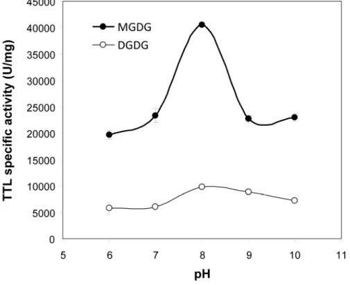
The optimum activity of TTL was found to occur at pH 8 on both MGDG and C8-350
DGDG (Figure 2). TTL specific activity on C8-MGDG was found to be 40,500 ± 125 U/mg, 351
compared to 4,658 ± 146 U/mg for Fusarium solani lipase [18], 5,420 ± 85 U/mg for 352
GPLRP2, 984 ± 62 U/mg for FsC and 450± 41U/mg for TLL [17]. Thus, to our knowledge, 353
TTL activity on C8-MGDG is the highest galactolipase activity measured. This activity is 7-354
fold higher than that of GPLRP2, the most active mammalian galactolipase characterized so 355
far. The maximum specific activity of TTL on C8-DGDG was also found to be the highest 356
galactolipase activity measured with DGDG (9,800 ±125 U/mg; Figure 2). 357
3.2. Galactolipase activity of TTL on C12-MGDG and C12-DGDG monomolecular films 359
The galactolipase activity of TTL was then tested on medium chain synthetic 360
galactolipids (C12-MGDG and C12-DGDG) that form stable monomolecular films at the air-361
water interface, while their lipolysis products are soluble in water [7, 17, 33, 43]. This allows 362
measuring galactolipase activities at various surface pressures using the barostat technique 363
[32, 34]. 364
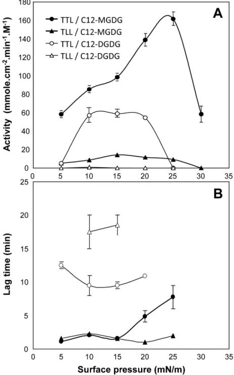
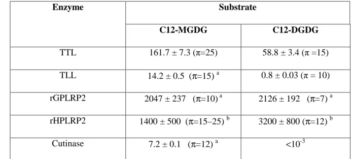
TTL activity on C12-MGDG showed a bell-shaped activity profile as a function of 365
surface pressure, with a maximum activity of 161.7 ± 7.3 mmol cm-2 min-1 M-1 at 25 mN/m 366
(Figure 3A). TTL was found to be 10 to 14-fold more active than the homologous fungal 367
lipase TLL, that showed its maximum activity (14.2 mmol cm-2 min-1 M-1) at a lower surface 368
pressure of 12 mN m-1 (Figure 3A). TTL was less active than rGPLRP2 but the optimum 369
activity of the latter (2047 ± 237 mmol cm-2 min-1 M-1; Table 1) was found at a much lower 370
surface pressure of 10 mN m-1 (Table 1). Remarkably, TTL was found to be able to 371
hydrolyze C12-MGDG monomolecular films at surface pressures up to 30 mN m-1. Most 372
galactolipases characterized so far are not active at such high surface pressures, except 373
recombinant human PLRP2 (Table 1; [33]). It confirms that lipases possessing a lid domain, 374
like TTL, TLL and rHPLRP2, are able to hydrolyze galactolipids at higher surface pressures 375
than those without a lid, like GPLRP2 and FsC [17]. 376
Similar features were observed with C12-DGDG but with optimum activities at lower 377
surface pressures (Figure 3A and Table 1). Remarkably, TLL only showed a very weak 378
activity of 0.8 ± 0.03 mmol cm-2 min-1 M-1 on C12-DGDG at 10 mN m-1 (Figure 3A). The 379
presence of a second galactose unit on the polar head of galactolipids has therefore a 380
significant effect on the penetration and activity of galactolipases as a function of surface 381
pressure, and this effect is particularly marked with TLL. This is also shown by the lag times 382
for measuring steady state kinetics of galactolipid hydrolysis using the barostat technique. 383
With C12-MGDG, the lag time values were low (1-2 min) below 15 mN m-1 and only 384
increased above and till 25 mN m-1 for TTL (Figure 3B). Lag times for both enzymes were 385
much higher (10-20 min) with C12-DGDG and this feature was already observed at low 386
surface pressure (Figure 3B). 387
The surface pressure and the size/steric hindrance of the hydrophilic polar head of 388
galactolipids are therefore two important parameters controlling the activity of galactolipases, 389
and this activity is favoured at higher surface pressure by the presence of a lid domain. 390
Enzymes without a lid, like GPLRP2 and cutinase show optimum activity at low surface 391
pressures and very long lag times (40-70 min) to reach steady state kinetics above 10 mN m-1 392
[17]. The presence of an amphiphilic lid probably favours the interaction of the lipase with the 393
galactolipid monolayer spread at the air-water interface. These results further underline the 394
crucial role of the lid in the interaction with the lipid substrate and the control of enzyme 395
activity [44, 45].. 396
397
3.3. Galactolipase activity of TTL on a surface-coated MGDG substrate containing
α
-398eleostearic acid 399
The conjugated triene present in α-eleostearic acid confers strong UV absorption 400
properties on both pure fatty acid and TAGs containing this fatty acid, as in tung oil in which 401
it represents around 70% of total fatty acids [23]. These UV absorption properties have been 402
used for developing lipase and phospholipase spectrophotometric assays in microtiter plates 403
using tung oil [24], synthetic TAG [25, 26] and phospholipid [27] containing α-eleostearic 404
acid as chromophore. Following a similar approach, we synthesized a 405
monogalactosyldiglyceride containing α-eleostearic acid (αE-MGDG) to establish a new UV 406
spectrophotometric assay of galactolipases in microtiter plates, in which the substrate is 407
coated on the well surface. 408
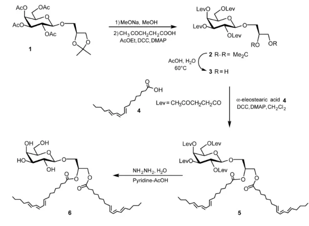
For the synthesis of αE-MGDG (compound 6 in Figure 4), we used 3-O-[2,3,4,6-409
Tetra-O-acetyl-β-D-galactopyranosyl]-1,2-O-isopropylidene-sn-glycerol (compound 1) that 410
was obtained in a previous study [7] and α-eleostearic acid (compound 4) prepared according 411
to Mendoza et al. [25] or O’Connor et al. [46]. Compound 1 was O-deacetylated under 412
Zemplén conditions (catalytic sodium methylate in methanol), the tetraol was esterified by 413
treatment with levulinic acid in ethyl acetate in the presence of dicyclohexylcabodimimide 414
(DCC) and 4-dimethylaminopyridine (DMAP), affording compound 2 in 70% yield. After 415
cleavage of the isopropylidene group under acidic medium (70% acetic acid, 60°C), the diol 3 416
was reacted with α-eleostearic acid 4 (DCC, DMAP, CH2Cl2) affording the product 5 in 73%
417
yield. Finally, the levulinoyl protecting groups were cleaved by hydrazine hydrate in a 3:2 418
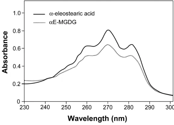
pyridine-acetic anhydride mixture yielding product 6 in 60% yields (Figure 4). 419
The UV absorption spectrum (230-300 nm) of an ethanolic solution of αE-MGDG 420
displayed three major peaks located at 260, 270 and 282 nm (Figure 5). This profile spectrum 421
is similar to that of pure eleostearic acid [24], pure tung oil triglycerides [23], synthetic α-422
eleostearic acid-containing triglycerides [25] and phosphatidylcholine [27]. In aqueous 423
buffer, the major absorption peak was shifted from 270 nm to 272 nm, as described earlier 424
[47]. 425
Assays of galactolipase activities were performed after coating UV-transparent 426
microtiter plates with αE-MGDG that was first added as a solution in ethanol before the 427
alcohol was evaporated. After coating the wells of microtiter plates, the absorbance at 272 nm 428
was recorded for 20 min in the presence of buffer without enzyme to determine background 429
absorbance. A constant baseline with optical density (OD) not exceeding 0.3 was recorded, 430
indicating that the substrate coating was not altered by the addition of buffer. TTL and other 431
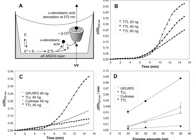
galactolipases were then tested, assuming that these enzymes will bind to the surface-coated 432
αE-MGDG substrate, will hydrolyze it and release α-eleostearic acid (Figure 6A). This long
chain fatty acid can be further solubilized by complex formation with β-CD present in the 434
buffer and its concentration can be measured continuously by monitoring UV absorbance at 435
272 nm (Figure 6A). 436
To validate the method, various amounts of substrate coated onto the plates (5, 10, 20 437
or 50 µ g per well) and enzymes (20, 40 and 80 ng of TTL, TLL, GPLRP2 or FsC) were 438
tested. The highest amount of substrate tested (50 µg αE-MGDG per well) was retained 439
because it formed a stable coating and allowed measuring steady-state enzyme kinetics for 440
longer period of time. Typical kinetics showing the increase in OD (∆OD = assay OD - initial 441
OD) at 272 nm during αE-MGDG hydrolysis by various amounts of TTL are shown in Figure 442
5B. Lag times of around 6 to 8 min were observed before recording linear OD variations with 443
time, but these variations were then proportional to TTL amounts in the 20 to 80 ng range 444
(Figure 6B). TTL was the most active enzyme according to ∆OD at 272 nm and compared to 445
TLL, GPLRP2 and FsC (Figure 6C). In all cases, steady state kinetics could be obtained after 446
various lag times (Table 2), with a good linearity of OD variations at 272 nm as a function of 447
enzyme amounts (20 to 80 ng; Figure 5D). 448
Based on a calibration with α-eleostearic acid, variations in OD at 272 nm could be 449
correlated with α-eleostearic acid concentration and further used for the estimation of enzyme 450
specific activities. The apparent molar extinction coefficient (εapp) of α-eleostearic acid has
451
been previously determined in microtiter plates by recording the absorbance at 272 nm of 452
various amounts of α-eleostearic acid dispersed in the buffer with β-CD at 37°C and it was 453
found to be 5320 M-1cm-1 [27]. Under these conditions, the increase with time of OD at 272 454
nm was converted into µmoles of α-eleostearic acid released per minand per mg of enzyme 455
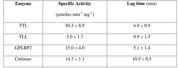
(Table 2). TTL was found to be the most active galactolipase on αE-MGDG under these 456
conditions with a specific activity of 50.3 ± 8.9 µmoles min-1 mg-1. TLL was 10-fold less 457
active while GPLRP2 and FsC were 3-fold less active. These enzymes also showed distinct 458
lag times with TLL reaching the most rapidly steady state kinetics although it was the enzyme 459
with the lowest specific activity (Table 2). These findings that differentiate TTL and TLL are 460
similar to those observed with MGDG monomolecular films at high surface pressures around 461
20 mN m-1 with similar ratio of enzyme activity (10 to 12) and lag times (5 to 8) between TTL 462
and TLL (Figure 3B). The presentation of the galactolipid substrate coated onto the microtiter 463
plate surface to the enzyme might therefore be similar to a substrate monolayer spread at the 464
air-water interface at 20 mN m-1. 465
466
3.4. TTL substrate specificity and structure-function relationships 467
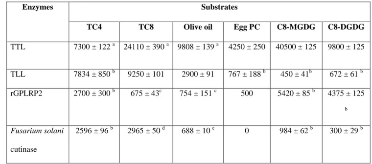
TTL possesses broad substrate specificity and was found to be active on triglycerides 468
with various acyl chain lengths, phospholipids and galactolipids (Table 3). It was however 469
more active on galactolipids than on triglycerides and phospholipids, with a galactolipase 470
activity on C8-MGDG micelles that is 1.68-fold, 2.4-fold and 9.5-fold higher than TTL 471
activities on trioctanoin, olive oil, and egg phosphatidylcholine, respectively. TTL substrate 472
preference was closer to that of GPLRP2 than to the closely related TLL (Table 3). Indeed, 473
TLL was 6 to 20-fold more active on triglycerides than on C8-MGDG, while its activity on 474
phospholipids was in the same order of magnitude as its activity on galactolipids (Table 3). 475
TTL was also found to be 4-fold more active on C8-MGDG than on C8-DGDG, while TLL is 476
slightly more active on C8-DGDG. TTL and TLL therefore display distinct substrate 477
preference while they share 89 % amino acid identities [48]. Although TTL was globally 478
more active on C8-MGDG than TLL at steady state, it showed longer lag times to reach the 479
steady state, particularly at high surface pressures, which suggests a slower 480
adsorption/penetration of TTL at the lipid-water interface compared to TLL. The presence of 481
an additional galactose on the galactolipid polar head led to a lower activity of TTL, but it is 482
unclear whether this results from a steric hindrance during the interfacial adsorption step or 483
within the active site. 484
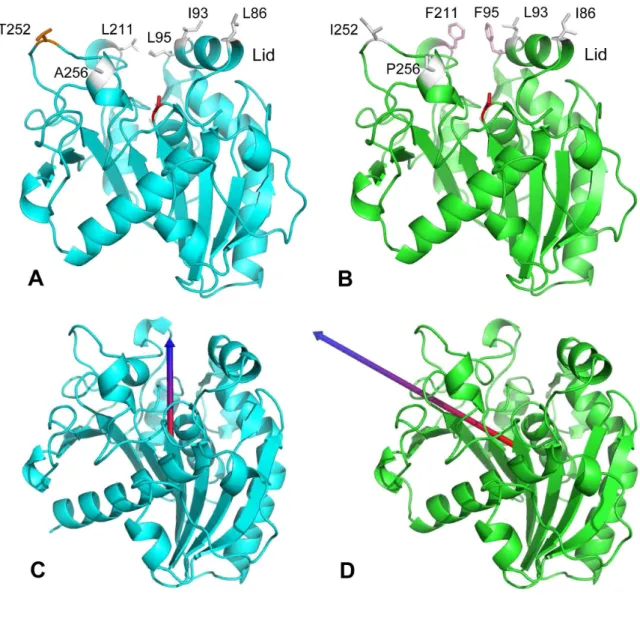
To gain more information on the structure-function relationships of TTL, we built a 485
3D model based on the known crystal structure of the homologous TLL with the lid in the 486
open conformation. The most remarkable difference between the two models was located in 487
the solvent exposed part of the hydrophobic substrate binding pocket hosting the acyl chains 488
which encompasses four amino acid substitutions in TTL vs. TLL (namely: Leu86Ile, 489
Leu93Ile, Phe95Leu, Phe211Leu) (Figure 7A-B). This region is also involved in the 490
interfacial recognition site (IRS) of the enzyme when the lid is in its open conformation [45], 491
as well as in the stabilization of the lid in its closed form. The replacement of two Phe by Leu 492
residues in TTL might explain the slower adsorption/penetration of TTL since aromatic 493
residues like Phe have strong contributions to protein transfer from water to water-lipid 494
interfaces [49]. The role of these residues in TTL will be investigated by site-directed 495
mutagenesis in future studies. 496
Since electrostatic interactions also play an important role in the interaction of 497
lipolytic enzymes with polar lipids, we calculated dipolar vectors for the TTL model and TLL 498
3D structure. Surprisingly the positive end of TTL dipolar vector was found to be oriented 499
from the bottom of the active site vertically to the surface (Figure 7C), while in TLL it was 500
oriented more horizontally along the active site (Figure 7D). This might favour a better 501
orientation of TTL towards polar or negatively charged lipid surfaces, and might explain the 502
5.5-fold higher activity of TTL on phospholipids compared to TLL (Table 3). 503
The comparison of TTL model with the crystal structure of TLL does not reveal 504
specific features that can explain the higher activity of TTL on galactolipids, nor the 505
preference for MGDG versus DGDG. The presence of a second galactose unit on DGDG 506
polar head may lead to steric hindrance within the enzyme active site, but monomolecular 507
film experiments rather suggest that the additional galactose unit impairs the enzyme 508
adsorption on its aggregated substrate (Figure 3). 509
510
4. Conclusions 511
Besides its activity on triglycerides [21] and phospholipids (this work), the TTL lipase 512
purified from the fungus Talaromyces thermophilus was found to hydrolyze a large variety of 513
synthetic galactolipid substrates presented in various forms to the enzyme (micelles, 514
monolayers, coating on solid surface). In all cases, it displays some of the highest lipolytic 515
activities recorded so far whatever the substrate. It appears to be more active on galactolipid 516
mixed micelles than on monomolecular films or surface-coated MGDG. Nevertheless, the 517
presence of a lid in TTL favours the hydrolysis of monomolecular films of galactolipids at 518
high surface pressure as observed with the homologous fungal lipase from Thermomyces 519
lanuginosus (TLL) belonging to the same gene family [48]. Differences in lag times for 520
reaching steady state kinetics of hydrolysis of galactolipid monomolecular films or surface-521
coated MGDG, and 3D modelling based on the known 3D structure of TLL, pointed out to 522
amino acid substitutions within the IRS of TTL that could be responsible for a slower 523
adsorption/penetration at lipid-water interface compared to TLL. 524
Finally, we have developed a fast, sensitive and continuous assay of galactolipases in 525
microtiter plates using a novel synthetic galactolipid substrate containing α-eleostearic acid 526
that allows a direct detection by UV absorption, of the fatty acids released upon lipolysis of 527
MGDG. Although the specific activities (U/mg) deduced from this assay (Table 2) are 2 to 3 528
order of magnitude lower than those estimated by the pHstat technique from C8-MGDG 529
micelle hydrolysis (Table 3), the UV detection of free fatty acids released from coated MGDG 530
is highly sensitive and allows measuring significant activities with about ten ng of enzymes, 531
against hundred ng to ten µg with the pHstat. The lower galactolipase activities measured 532
with the UV-spectrophotometric assay and substrate coated on microtiter plates are therefore 533
not an obstacle to the applicability of this novel assay. These lower activities are probably 534
linked to the mode of action of lipolytic enzymes that depends on the accessible surface 535
available for enzyme adsorption at the lipid-water interface, the first step in the overall 536
process of interfacial catalysis. With the pHstat method and the use of mixed micelles or fine 537
triglyceride emulsions as substrate, a very large accessible surface is created which ensures 538
maximum enzyme adsorption and thus maximum enzyme activity. With substrate coated onto 539
the wells of microtiter plates, as well as with monomolecular films, the accessible surface 540
available for enzyme adsorption is much reduced and one can expect a lower enzyme 541
turnover. 542
The novel spectrophotometric assay using surface-coated MGDG with UV-543
absorbing α-eleostearic acid allows the estimation of enzyme specific activities from steady 544
state kinetics. Moreover, the lag times for reaching these conditions give some idea about the 545
enzyme affinity for the lipid-water interface. Indeed, the differences observed between TTL 546
and TLL are in good agreement with independent experiments performed with galactolipid 547
monomolecular films. In addition to TTL characterization showing its potent galactolipase 548
activity, this novel assay will be an interesting tool for screening enzymes and mutant thereof 549
for their galactolipase activities. 550
551 552
Acknowledgements 553
We are grateful to Ali Gargouri (Centre de Biotechnologie de Sfax, Tunisia) for his 554
critical reading of the manuscript and constant support to this work, Rabaa Ben Ayed for her 555
technical assistance during the purification of TTL, Deborah Byrne for DLS measurements 556
(Institut de Microbiologie de la Méditerranée, Marseille, France) and Vanessa Point for her 557
technical assistance during monomolecular film experiments. This work received the financial 558
support of Agence Nationale de la Recherche in the framework of the GALACTOLIPASE 559
project (ANR-09-CP2D-06-01). This work also received financial support from the Ministry 560
of Higher Education and Scientific Research, Tunisia, granted to the Laboratoire de 561
Biotechnologie Moléculaire des Eucaryotes du Centre de Biotechnologie de Sfax. 562
563 564
Conflict of interest 565
The authors have declared no conflict of interest 566
References 567
568
[1] P. Dormann, C. Benning, Galactolipids rule in seed plants, Trends Plant Sci, 7 (2002) 112-569
118. 570
[2] G. Holzl, P. Dormann, Structure and function of glycoglycerolipids in plants and bacteria, 571
Prog Lipid Res, 46 (2007) 225-243. 572
[3] L.P. Christensen, Galactolipids as potential health promoting compounds in vegetable 573
foods, Recent Pat Food Nutr Agric, 1 (2009) 50-58. 574
[4] S. Amara, N. Barouh, J. Lecomte, D. Lafont, S. Robert, P. Villeneuve, A. De Caro, F. 575
Carriere, Lipolysis of natural long chain and synthetic medium chain galactolipids by 576
pancreatic lipase-related protein 2, Biochim Biophys Acta, 1801 (2010) 508-516. 577
[5] L. Couedelo, S. Amara, M. Lecomte, E. Meugnier, J. Monteil, L. Fonseca, G. Pineau, M. 578
Cansell, F. Carriere, M.C. Michalski, C. Vaysse, Impact of various emulsifiers on ALA 579
bioavailability and chylomicron synthesis through changes in gastrointestinal lipolysis, 580
Food Funct, 6 (2015) 1726-1735. 581
[6] L. Andersson, F. Carriere, M.E. Lowe, A. Nilsson, R. Verger, Pancreatic lipase-related 582
protein 2 but not classical pancreatic lipase hydrolyzes galactolipids, Biochim Biophys 583
Acta, 1302 (1996) 236-240. 584
[7] B. Sias, F. Ferrato, P. Grandval, D. Lafont, P. Boullanger, A. De Caro, B. Leboeuf, R. 585
Verger, F. Carriere, Human pancreatic lipase-related protein 2 is a galactolipase, 586
Biochemistry, 43 (2004) 10138-10148. 587
[8] J. De Caro, C. Eydoux, S. Cherif, R. Lebrun, Y. Gargouri, F. Carriere, A. De Caro, 588
Occurrence of pancreatic lipase-related protein-2 in various species and its relationship 589
with herbivore diet, Comp Biochem Physiol B Biochem Mol Biol, 150 (2008) 1-9. 590
[9] J.N. O'sullivan, N.W.M. Warwick, M.J. Dalling, A galactolipase activity associated with 591
the thylakoids of wheat leaves (Triticum aestivum L.). Journal of Plant Physiology, 131 592
(1987) 393-404. 593
[10] P.J. Helmsing, Purification and properties of galactolipase, Biochim. Biophys. Acta, 178 594
(1969) 519-533. 595
[11] T. Galliard, S. Dennis, Phospholipase, galactolipase and acyl transferase activities of a 596
lipolytic enzyme from potato., Phytochemistry, 13 (1974) 1731-1735. 597
[12] M. Terasaki, Y. Itabashi, Glycerolipid acyl hydrolase activity in the brown alga 598
Cladosiphon okamuranus TOKIDA, Biosci Biotechnol Biochem, 67 (2003) 1986-1989. 599
[13] R.M.C. Dawson, N. Hemington, G.P. Hazlewood, On the role of higher plant and 600
microbial lipases in the ruminal hydrolysis of grass lipids., Br J Nutr, 38 (1977) 225-232. 601
[14] W. Fischer, E. Heinz, M. Zeus, The suitability of lipase from Rhizopus arrhizus delemar 602
for analysis of fatty acid distribution in dihexosyl diglycerides, phospholipids and plant 603
sulfolipids., Hoppe-Seyler's Z. Physiol. Chem., 354 (1973) 1115-1123. 604
[15] T. Morimoto, A. Nagatsu, N. Murakami, J. Sakakibara, Chemoenzymatic synthesis of 1-605
O-acyl-3-0-(6'-0-acyl-b-D-galactopyranosyl )-sn-glycerol., Tetrahedron lett., 51 (1995) 606
6443-6450. 607
[16] P. Persson, I. Svensson, P. Adlercreutz, Enzymatic fatty acid exchange in 608
digalactosyldiacylglycerol., Chem Phys Lipids, 104 (2000) 13-21. 609
[17] A. Amara, D. Lafont, G. Parsiegla, V. Point, A. Chabannes, A. Rousset, F. Carrière, The 610
galactolipase activity of some microbial lipases and pancreatic enzymes., Eur. J. Lipid 611
Sci. Technol., 115 (2013) 442-451. 612
[18] R. Jallouli, H. Othman, S. Amara, G. Parsiegla, F. Carriere, N. Srairi-Abid, Y. Gargouri, 613
S. Bezzine, The galactolipase activity of Fusarium solani (phospho)lipase, Biochim 614
Biophys Acta, 1851 (2015) 282-289. 615
[19] S. Amara, D. Lafont, B. Fiorentino, P. Boullanger, F. Carriere, A. De Caro, Continuous 616
measurement of galactolipid hydrolysis by pancreatic lipolytic enzymes using the pH-stat 617
technique and a medium chain monogalactosyl diglyceride as substrate, Biochim Biophys 618
Acta, 1791 (2009) 983-990. 619
[20] D. Lafont, F. Carriere, F. Ferrato, P. Boullanger, Syntheses of an alpha-D-Gal-(1-->6)-620
beta-D-Gal diglyceride, as lipase substrate, Carbohydr Res, 341 (2006) 695-704. 621
[21] I. Belhaj-Ben Romdhane, A. Fendri, Y. Gargouri, A. Gargouri, H. Belghith, A novel 622
thermoactive and alkaline lipase from Talaromyces thermophilus fungus for use in 623
laundry detergents., Biochemical Engineering Journal, 53 (2010) 112-120. 624
[22] A. Najjar, S. Robert, C. Guerin, M. Violet-Asther, F. Carriere, Quantitative study of 625
lipase secretion, extracellular lipolysis, and lipid storage in the yeast Yarrowia lipolytica 626
grown in the presence of olive oil: analogies with lipolysis in humans, Appl. Microbiol. 627
Biotechnol., 89 (2011) 1947-1962. 628
[23] G. Pencreac'h, J. Graille, M. Pina, R. Verger, An ultraviolet spectrophotometric assay for 629
measuring lipase activity using long-chain triacylglycerols from Aleurites fordii seeds., 630
Anal. Biochem., 303 (2002) 17-24. 631
[24] C. Serveau-Avesque, R. Verger, J.A. Rodriguez, A. Abousalham, Development of a 632
high-throughput assay for measuring lipase activity using natural triacylglycerols coated 633
on microtiter plates, Analyst, 138 (2013) 5230-5238. 634
[25] L.D. Mendoza, J.A. Rodriguez, J. Leclaire, G. Buono, F. Fotiadu, F. Carriere, A. 635
Abousalham, An ultraviolet spectrophotometric assay for the screening of sn-2-specific 636
lipases using 1,3-O-dioleoyl-2-O-alpha-eleostearoyl-sn-glycerol as substrate, J Lipid Res, 637
53 (2012) 185-194. 638
[26] M. El Alaoui, L. Soulere, A. Noiriel, Y. Queneau, A. Abousalham, alpha-Eleostearic 639
acid-containing triglycerides for a continuous assay to determine lipase sn-1 and sn-3 640
regio-preference, Chem Phys Lipids, 206 (2017) 43-52. 641
[27] M. El Alaoui, A. Noiriel, L. Soulere, L. Grand, Y. Queneau, A. Abousalham, 642
Development of a high-throughput assay for measuring phospholipase A activity using 643
synthetic 1,2-alpha-eleostearoyl-sn-glycero-3-phosphocholine coated on microtiter plates, 644
Anal Chem, 86 (2014) 10576-10583. 645
[28] A. Hjorth, F. Carrière, C. Cudrey, H. Wöldike, E. Boel, D.M. Lawson, F. Ferrato, C. 646
Cambillau, G.G. Dodson, L. Thim, R. Verger, A structural domain (the lid) found in 647
pancreatic lipases is absent in the guinea pig (phospho)lipase, Biochemistry, 32 (1993) 648
4702-4707. 649
[29] S.B. Petersen, P.H. Jonson, P. Fojan, E.I. Petersen, M.T. Petersen, S. Hansen, R.J. Ishak, 650
E. Hough, Protein engineering the surface of enzymes., J Biotechnol 66 (1998) 11-26. 651
[30] U.K. Laemmli, Cleavage of structural proteins during the assembly of the head of 652
bacteriophage T4, Nature, 227 (1970) 680-685. 653
[31] S. Amara, V. Delorme, M. Record, F. Carriere, Inhibition of phospholipase A1, lipase 654
and galactolipase activities of pancreatic lipase-related protein 2 by methyl arachidonyl 655
fluorophosphonate (MAFP), Biochim Biophys Acta, 1821 (2012) 1379-1385. 656
[32] R. Verger, G.H. de Haas, Enzyme reactions in a membrane model. 1: A new technique to 657
study enzyme reactions in monolayers, Chem. Phys. Lipids, 10 (1973) 127-136. 658
[33] C. Eydoux, J. De Caro, F. Ferrato, P. Boullanger, D. Lafont, R. Laugier, F. Carriere, A. 659
De Caro, Further biochemical characterization of human pancreatic lipase-related protein 660
2 expressed in yeast cells, J Lipid Res, 48 (2007) 1539-1549. 661
[34] R. Verger, M.C.E. Mieras, G.H. de Haas, Action of phospholipase A at interfaces, J. 662
Biol. Chem., 248 (1973) 4023-4034. 663
[35] S. Yapoudjian, M.G. Ivanova, A.M. Brzozowski, S.A. Patkar, J. Vind, A. Svendsen, R. 664
Verger, Binding of Thermomyces (Humicola) lanuginosa lipase to the mixed micelles of 665
cis-parinaric acid/NaTDC, Eur J Biochem, 269 (2002) 1613-1621. 666
[36] M. Biasini, S. Bienert, A. Waterhouse, K. Arnold, G. Studer, T. Schmidt, F. Kiefer, T.G. 667
Cassarino, M. Bertoni, L. Bordoli, T. Schwede, SWISS-MODEL: modelling protein 668
tertiary and quaternary structure using evolutionary information., Nucleic Acid Res., 42 669
(2014) W252-W258. 670
[37] O. Trott, A.J. Olson, AutoDock Vina: improving the speed and accuracy of docking with 671
a new scoring function, efficient optimization, and multithreading, J Comput Chem, 31 672
(2010) 455-461. 673
[38] D. Seeliger, B.L. de Groot, Ligand docking and binding site analysis with PyMOL and 674
Autodock/Vina, J Comput Aided Mol Des, 24 (2010) 417-422. 675
[39] E.F. Pettersen, T.D. Goddard, C.C. Huang, G.S. Couch, D.M. Greenblatt, E.C. Meng, 676
T.E. Ferrin, UCSF Chimera--a visualization system for exploratory research and 677
analysis., J Comput Chem, 25 (2004) 1605-1612. 678
[40] M. Gagos, R. Koper, W.I. Gruszecki, Spectrophotometric analysis of organisation of 679
dipalmitoylphosphatidylcholine bilayers containing the polyene antibiotic amphotericin 680
B, Biochimica et Biophysica Acta-Biomembranes, 1511 (2001) 90-98. 681
[41] E. Mateos-Diaz, P. Sutto-Ortiz, M. Sahaka, D. Byrne, H. Gaussier, F. Carriere, IR 682
spectroscopy analysis of pancreatic lipase-related protein 2 interaction with 683
phospholipids: 2. Discriminative recognition of various micellar systems and 684
characterization of PLRP2-DPPC-bile salt complexes, Chem Phys Lipids, (2017). 685
[42] E. Mateos-Diaz, J.C. Bakala N'Goma, D. Byrne, S. Robert, F. Carriere, H. Gaussier, IR 686
spectroscopy analysis of pancreatic lipase-related protein 2 interaction with 687
phospholipids: 1. Discriminative recognition of mixed micelles versus liposomes, Chem 688
Phys Lipids, (2017). 689
[43] A. Roussel, Y. Yang, F. Ferrato, R. Verger, C. Cambillau, M. Lowe, Structure and 690
activity of rat pancreatic lipase-related protein 2, J Biol Chem, 273 (1998) 32121-32128. 691
[44] F. Carrière, K. Thirstrup, S. Hjorth, F. Ferrato, C. Withers-Martinez, C. Cambillau, E. 692
Boel, L. Thim, R. Verger, Pancreatic lipase stucture -function relationships by domain 693
exchange, Biochemistry, 36 (1997) 239-248. 694
[45] E. Mateos-Diaz, S. Amara, A. Roussel, S. Longhi, C. Cambillau, F. Carriere, Probing 695
Conformational Changes and Interfacial Recognition Site of Lipases With Surfactants 696
and Inhibitors, Methods Enzymol, 583 (2017) 279-307. 697
[46] R.T. O’Connor, D.C. Heinzelman, R.S. McKinney, F.C. Pack, The spectrophotometric 698
determination of alpha and beta isomers of eleostearic acid in tung oil., J. Am. Oil Chem. 699
Soc., 24 (1947) 212 - 216. 700
[47] C. Reichardt Solvatochromic dyes as solvent polarity indicators., Chemical Reviews, 94 701
(1994) 2319-2358. 702
[48] I. Belhaj-Ben Romdhane, F. Frikha, I. Maalej-Achouri, A. Gargouri, H. Belghith, Gene 703
cloning and molecular characterization of the Talaromyces thermophilus lipase catalyzed 704
efficient hydrolysis and synthesis of esters, Gene, 494 (2012) 112-118. 705
[49] W.C. Wimley, S.H. White, Experimentally determined hydrophobicity scale for proteins 706
at membrane interfaces, Nat Struct Biol, 3 (1996) 842-848. 707
[50] D.M. Lawson, A.M. Brzozowski, S. Rety, C. Verma, G.G. Dodson, Probing the nature of 708
substrate binding in Humicola lanuginosa lipase through X-Ray crystallography and 709
intuitive modelling, Protein Eng., 7 (1994) 543-550. 710
[51] K. Dridi, S. Amara, S. Bezzine, J.A. Rodriguez, F. Carriere, H. Gaussier, Partial deletion 711
of beta9 loop in pancreatic lipase-related protein 2 reduces enzyme activity with a larger 712
effect on long acyl chain substrates, Biochim Biophys Acta, 1831 (2013) 1293-1301. 713
[52] M. Schué, D. Maurin, R. Dhouib, J.C. Bakala N’Goma, V. Delorme, G. Lambeau, F. 714
Carrière, S. Canaan, Two secreted cutinase-like proteins from Mycobacterium 715
tuberculosis display very different lipolytic activities related to their physiological 716
function., FASEB J., 24 (2010) 1893-1903. 717
[53] A. Roussel, S. Amara, A. Nyyssola, E. Mateos-Diaz, S. Blangy, H. Kontkanen, A. 718
Westerholm-Parvinen, F. Carriere, C. Cambillau, A Cutinase from Trichoderma reesei 719
with a lid-covered active site and kinetic properties of true lipases, J Mol Biol, 426 (2014) 720
3757-3772. 721
Figure legends 723
724
Figure 1. Chemical structures of monogalactosyl diacylglycerol (MGDG; 1,2-diacyl-3-O-β-725
D-galactosyl-sn-glycerol) and digalactosyl diacylglycerol (DGDG; 1,2-diacyl-3-O-(6-O-α-D-726
galactosyl-β-D-galactosyl)-sn-glycerol). 727
Figure 2. pH-dependent galactolipase activity of TTL on synthetic medium chain MGDG and 729
DGDG as substrates. Activities were measured using the pHstat technique and substrate 730
micelles with a bile salt (NaTDC) to galactolipid molar ratio of 1.33. Values (U/mg) are 731
means ± SD (n= 3). 1 U = 1 µmole of free fatty acid released per min. 732
Figure 3. Variations with surface pressure in the activity of TTL and TLL on monomolecular 734
films of galactolipids. (A) Steady-state activities on C12-MGDG and C12-DGDG 735
monomolecular films; (B) Lag times observed in the course of C12-MGDG and C12-DGDG 736
monomolecular film hydrolysis by TTL and TLL. Global enzyme concentration was 0, 45 nM 737
for TTL and 5 nM for TLL. Values are means ± SD (n=3). 738
Figure 4. Synthesis scheme of 1,2-Di-O- α-eleostearoyl-3-O-β-D-galactopyranosyl-sn-740
glycerol (αE-MGDG). 741
Figure 5. UV absorption spectra of α-eleostearic acid (60 µg/mL) and αE-MGDG (60 µg/mL) 743
dissolved in ethanol containing BHT 0.001%. 744
Figure 6. UV-spectrophotometric assays of galactolipase activities using 1,2-Di-O- α-746
eleostearoyl-3-O-β-D-galactopyranosyl-sn-glycerol (αE-MGDG). (A) Schematic 747
representation of the assay showing the enzymatic hydrolysis of the αE-MGDG film coated 748
onto the wells of a microtiter plate, followed by the solubilization of free α-eleostearic acid 749
(FFA) in the bulk phase by formation of a complex with β-cyclodextrin (β-CD); E, lipase in 750
solution; E*, activated lipase at the interface; S, substrate. (B) Variations with time of optical 751
density at 272 nm (versus OD272 at time zero) using various amounts of TTL. (C) Variations
752
with time of optical density at 272 nm (versus OD272 at time zero) of various enzymes (40 ng
753
each). (D) Variations of optical density at 272 nm per min at steady state as a function of 754
enzyme amounts. Substrate coated onto the microtiter (50 µ g/well) was incubated with 20,40 755
and 80 ng of GPLRP2, TLL, cutinase or TTL, respectively. Enzymes were injected into the 756
well containing 200 µl of buffer. The increase in OD at 272 nm was recorded for 15 min. 757