HAL Id: hal-02512447
https://hal.univ-lorraine.fr/hal-02512447
Submitted on 9 Oct 2020
HAL is a multi-disciplinary open access
archive for the deposit and dissemination of
sci-entific research documents, whether they are
pub-lished or not. The documents may come from
teaching and research institutions in France or
abroad, or from public or private research centers.
L’archive ouverte pluridisciplinaire HAL, est
destinée au dépôt et à la diffusion de documents
scientifiques de niveau recherche, publiés ou non,
émanant des établissements d’enseignement et de
recherche français ou étrangers, des laboratoires
publics ou privés.
Accuracy of Several Lung Ultrasound Methods for the
Diagnosis of Acute Heart Failure in the ED: A
Multicenter Prospective Study
Aurélien Buessler, Tahar Chouihed, Kévin Duarte, Adrien Bassand, Matthieu
Huot-Marchand, Yannick Gottwalles, Alice Pénine, Elies André, Lionel Nace,
Déborah Jaeger, et al.
To cite this version:
Aurélien Buessler, Tahar Chouihed, Kévin Duarte, Adrien Bassand, Matthieu Huot-Marchand, et
al.. Accuracy of Several Lung Ultrasound Methods for the Diagnosis of Acute Heart Failure in the
ED: A Multicenter Prospective Study. Chest, American College of Chest Physicians, 2020, 157 (1),
pp.99-110. �10.1016/j.chest.2019.07.017�. �hal-02512447�
Accuracy of several lung ultrasound methods for the diagnosis of acute heart failure in
the emergency department: A multicenter prospective study
Aurélien Buessler*1 MD, Tahar Chouihed*1,3,9 MD, PhD, Kévin Duarte2,3 PhD, Adrien Bassand1 MD, Matthieu Huot-Marchand1 MD, Yannick Gottwalles4 MD, Alice Pénine5 MD, Elies André6 MD, Lionel Nace7, MD, Déborah Jaeger1 MD, Masatake Kobayashi8, MD, Stéfano Coiro10 MD, PhD, Patrick Rossignol3,9, MD, PhD, Nicolas Girerd3,9,11 MD, PhD.
1. Emergency Department, University Hospital of Nancy, France
2. Université de Lorraine, Institut Elie Cartan de Lorraine, Unité Mixte de Recherche 7502, Vandœuvre-lès-Nancy, France ; Centre National de la Recherche Scientifique, Institut Elie Cartan de Lorraine, Unité Mixte de Recherche 7502, Vandœuvre-lès-Nancy, France ; INRIA, Project-Team BIGS, Villers-lès-Nancy, France.
3. Université de Lorraine, Centre d’Investigations Cliniques Plurithématique 1433, Institut Lorrain du Cœur et des Vaisseaux, Vandoeuvre les Nancy France Groupe choc, INSERM U1116. Faculté de Médecine. 54500 Vandoeuvre les Nancy
4. Emergency Department, Colmar Hospital, Colmar, France
5. Emergency Department, Charleville-Maizières Hospital, Charleville-Maizières France 6. Emergency Department, Mercy Hospital, Metz, France
7. Intensive Care Unit, University Hospital of Nancy, France
8. Department of Cardiology, Tokyo Medical University, Tokyo, Japan.
9. F-CRIN INI-CRCT (Cardiovascular and Renal Clinical Trialists), Nancy, France 10. University of Perugia, School of Medicine, Perugia, Division of Cardiology
11. Département de Cardiologie, Institut Lorrain du Cœur et des Vaisseaux, CHRU Nancy, France
Acknowledgements and disclosures: PR, and NG are funded by a public grant overseen by the French National Research Agency (ANR) as part of the second “Investissements d’Avenir” program FIGHT-HF (reference: RHU-0004) and by the French PIA project “Lorraine Université d’Excellence”, reference ANR-15-IDEX-04-LUE. Prof. Rossignol has received board membership fees from Novartis, Relypsa, and Steathpeptides. Dr. Chouihed reports honoraria from Novartis. Prof Girerd reports honoraria from Novartis, Boehringer and Servier.
ABSTRACT Background
Early appropriate diagnosis of acute heart failure (AHF) is recommended by international guidelines. This study assessed the value of several lung ultrasound (LUS) strategies for identifying AHF in the emergency department (ED).
Methods
This prospective study, conducted in four EDs, included patients with diagnostic uncertainty based on initial clinical judgment. A clinical diagnosis score for AHF (Brest score) was quantified, followed by an extensive LUS examination performed according to the 4- (BLUE protocol), 6-, 8- and 28-point methods. The primary outcome was AHF discharge diagnosis adjudicated by 2 senior physicians blinded for LUS measurements. C-index was used to quantify discrimination.
Results
Among the 117 included patients, AHF (N=69) was identified in 27.4%, 56.2%, 54.8% and 76.7% of patients with the 4-point (2 bilateral positive points), 6-point, 8-point (≥1 bilateral positive point) and 28-point (B-line count ≥30) methods, respectively.
The C-index of the Brest score was 72.8 (65.3-80.3) whereas the C-index of the 4-, 6-, 8- and 28-point methods were respectively 63.7 (58.5-68.8), 72.4 (65.0-79.8), 74.0 (67.1-80.9) and 72.4 (63.9-80.9). The highest increase in C-index on top of the BREST score was observed with the 8-point method in the whole population (6.9 (1.6-12.2), p=0.010) and in the population with intermediate Brest Score, followed by the 6-point method.
Conclusions
In patients with diagnostic uncertainty, 6-point/8-point LUS method (using the 1 bilateral positive point threshold) improves AHF diagnosis accuracy on top of the BREST score.
INTRODUCTION
Dyspnea is one of the most frequent causes of admission in the emergency department (ED)1 and represents a significant diagnostic challenge for emergency physicians (EP). Acute heart failure (AHF) is one of the most common etiologies of acute dyspnea2. Guidelines recommend that diagnosis should be made as soon as possible in order to promptly begin appropriate early treatment3,4. Prognosis is related to initiation time of specific therapies5. In-hospital mortality is typically reported to be greater than 10%6 and has remained stable in the last 30 years.
Diagnostic approaches include clinical evaluation, chest X-ray, biological tests and specific biomarkers. Nevertheless, diagnosis remains difficult, especially in ED patients, many of whom feature atypical clinical presentation due to several prior comorbidities and mixed/concomitant etiologies of acute dyspnea2. Recently, Basset et al. developed the Brest score for the diagnosis of AHF in ED patients7. However, this score classified 50% of cases in the intermediate probability group7, hence supporting the importance of developing and promoting “new tools”8 which are complimentary to clinical scores in order to achieve quick diagnosis of AHF in patients admitted for acute dyspnea in the ED.
Ultrasound has gained widespread use in recent years and is now a highly valuable tool in the ED. Lung ultrasound (LUS) is a quick, reliable and easy-to-use exam that can improve the diagnostic accuracy for dyspneic patients9,10. Lichtenstein et al. furthermore highlighted its advantages in intensive care units for the evaluation of patients with respiratory distress (i.e. BLUE protocol)11. Several methods have been secondarily proposed to assess pulmonary congestion, using different analysis points, interpretation thresholds and various assessment conditions12–14. However, all of these studies focused on patients outside of the ED.
In light of the above, the present study aimed to evaluate and compare the diagnostic performance of currently available ultrasound protocols for pulmonary congestion assessment (i.e. the 4- (BLUE protocol), 6-, 8- and 28-point methods) in patients admitted for acute dyspnea in the ED. The study furthermore aimed to evaluate the diagnostic performance of these methods in patients with intermediate Brest scores (namely 4–8).
METHODS
Study protocol and design
This study is a part of the prospective PURPLE study (NCT 03194243, CNIL DR-2017-098). Patients admitted to the ED in four different hospitals including a university hospital over a 3-month period were included. All patients over 50 years old admitted for acute dyspnea for whom the treating physician had diagnostic uncertainty based on his/her initial clinical evaluation were included. Exclusion criteria consisted in traumatic dyspnea and systolic blood pressure < 70 mmHg.
For each patient, the Brest score was calculated7 and a standardized LUS was performed. All clinical and ultrasound analysis data were collected by the EPs and entered in the Clinical Research Form of the study.
Ultrasound methods
Ultrasounds were performed by ultrasound-certified EPs. Twenty-eight-point LUS were performed in all patients: for each point, a B-line grading from 0 to 10 was used. Using the data of this 28-point method, patients were able to be classified according to four published methods (Figure 1)11–13,15.
Four-point method (BLUE protocol)11
- Two scanning sites on each hemithorax:
o Second intercostal space, mid-clavicular line o Fourth intercostal space, anterior axillary line
- A positive point was defined as the presence of at least three B-lines.
- A positive exam was defined, according to the seminal publication16, by the presence of at least three B-lines on each scanning site.
Six-point method 12
- Three scanning sites on each hemithorax:
o Second intercostal space, mid-clavicular line o Fourth intercostal space, anterior axillary line o Fifth intercostal space, mid-axillary line
- A positive point was defined as the presence of at least three B-lines in a given scanning site.
- A positive exam was defined, according to the seminal publication12, by the presence of at least three B-lines on 2 scanning sites on each hemithorax.
Eight-point method13
- Four scanning sites on each hemithorax:
o Two anterior points, between the sternum and the anterior axillary line, comprised of 2 scanning sites
o Two lateral points between the anterior and the posterior axillary line, comprised of 2 scanning sites
- A positive point was defined as the presence of at least 3 B-lines in a given scanning site. Twenty-eight-point method15,17
- This exam was used both as a continuous count of overall B-lines as well as in the form of dichotomous variables (≥15 or ≥30).
A positive point was defined as the presence of at least three B-lines in a given scanning site. The exams were then categorized according to the presence and number of bilateral positive point. We considered two definitions of positive exams: a positive exam was either defined as at least one positive zone bilaterally (i.e. at least one on the right lung and at least one on the left lung) or as at least two positive zones bilaterally. The presence of two positive points on each hemithorax, irrespectively of their locations (p.e. positive points on the superior part of the right thorax and on the inferior part of the left thorax), qualified for being considered as having “≥2 bilateral positive points”.
Outcome
Diagnostic outcome was the final diagnosis at discharge collected from the patients’ medical records. The final diagnosis of the hospital stay was adjudicated by 2 senior physicians blinded (Emergency Physician and Cardiologist) to the LUS measurements.
Sample size
A random sample of 120 patients (60 with AHF and 60 without) was necessary, when the sample C-index was equal to 80%, to achieve a two-sided 95% confidence interval width of 16% (i.e. with a lower limit equal to 72% and a upper limit equal to 88%) by using the Hanley and McNeil's method. This setting also allows to have a confidence interval width of less than 0.18 for an C-index equal to 75% and a confidence interval width of 0.14 for an C-index of 0.85.
Statistical analysis
All analyses were performed using R software (the R foundation for Statistical Computation). The two-tailed significance level was set at p < 0.05.
Baseline characteristics are described as mean ± standard deviation or median (interquartile range) for continuous variables and frequency (percentage) for categorical variables. Comparison of baseline characteristics according to AHF and non-AHF groups were carried out using the non-parametric Wilcoxon test for continuous variables and chi-square or Fisher’s exact tests for categorical variables.
Associations between LUS measurements and AHF were assessed using logistic regression. Odds-ratios (ORs) with 95% confidence intervals (CIs) are reported. For certain variables, quasi-complete separation was detected. ORs with CIs were therefore estimated using a logistic regression model with Firth’s penalized likelihood. This method provides a solution to the phenomenon of monotone likelihood which causes parameter estimates of the usual logistic regression model to diverge, with infinite standard errors.
Individual performance of LUS measurements for diagnosing AHF was assessed by the calculation of C-index which is very similar to the area under the curve (AUC) of the receiver operating characteristic (ROC) used on univariable data. In addition, the increase in C-index was calculated to assess the additional value of LUS measurements on top of the Brest score for the diagnosis of AHF.
RESULTS
One hundred and seventeen patients were included, 62% of whom had a hospital discharge diagnosis of AHF (n=73) although only 54% (n=63) had an AHF diagnosis in the ED (3 patients with AHF at the ED had a non-AHF discharge diagnosis and 13 had an AHF diagnosis at discharge but not in the ED) (Table 1). The population was elderly (mean age =79.6 ± 11.8), mainly female (56%) and frequently had comorbidities. The majority of patients were hospitalized subsequent to ED admission (96%, n = 112), primarily in medical wards (n=68, 58%), while 25% (n=29) were admitted to intensive care units and only 13% (n=15) were admitted in a cardiology ward (Table 1). A majority of patients had an intermediate Brest score (64%, n=75) both in the AHF group (67%, n=49) and in the non-AHF group (59%, n=26)).
Diagnostic performances in the overall study population
In a first instance, the Brest score had a good diagnostic value when considered as a continuous variable index=81.8, 95% CI [74.2-89.4]), which subsequently decreased when using BREST score categories (C-index=72.8, 95% CI [65.3-80.3]).
Among the LUS methods, the 4-point method (2 bilateral positive points) had the lowest C-index (63.7(95% CI [58.5 - 68.8]) whereas the other methods had very similar C-index (6-point method for ≥ 1 bilateral positive point, 72.4 (95% CI [65.0 - 79.8]; 8-point method for ≥ 1 bilateral positive point, 74.0 (95% CI [67.1 - 80.9] and 28-point method for B-lines ≥30, 72.4 (95% CI [63.9-80.9]).
The 6-point method (≥ 1 bilateral positive point) had a specificity near 90% with a relatively low sensitivity (56.2%, 41.1-67.8%). The 8-point method (≥ 1 bilateral positive point) had a higher specificity (93.2%, 81.3-98.6%) and similar sensitivity (54.8%, 42.7-66.5%). In contrast, the 28-point method had high sensitivity (B-lines ≥15, 89.0 (79.5 - 95.1) and B-lines ≥ 30, 76.7 (65.4 - 85.8)) but low specificity (B-lines ≥15, 43.2 (28.3 - 59.0) and B-lines ≥ 30, 68.2 (52.4 - 81.4)).
For the 6-point and 8-point methods, the use of the ≥ 1 bilateral positive point threshold yielded higher C-index as well as a better sensitivity (13 and 6%) and moderately lower specificity (- 4 and - 11%).
Each method provided significant added value to the Brest score as assessed by changes in C-index. However, the highest increase in C-index were observed for the 6-point method (6.7 (95% CI [0.9-12.5], p = 0.024) and the 8-point method (6.9 (95% CI [1.6-12.2], p = 0.010) (Figure 2 and Table 3).
Diagnostic performances with intermediate Brest scores
In patients (n = 75) with intermediate Brest scores (4–8), the 4-point method (2 positive points bilaterally) had a C-index of 61.2 (95% CI [55.3 - 67.1]) and an added value to the Brest score of less than 5 as measured by an increase in C-index. In contrast, the 6-point and 8-point methods had a C-index greater than 70 when considering ≥1 positive point bilaterally (respectively 71.8 (95% CI [62.4 - 81.2]) and 72.7 (95% CI [63.9 - 81.5])).
Similarly, to the results in the overall population, the 6-point and 8-point methods (≥ 1 bilateral positive point) had a specificity near 90% and a sensitivity near 50%. For the 8-point method, the use of the ≥ 1 bilateral positive point threshold yielded a higher C-index as well as better sensitivity (14% increase) and moderately lower specificity (4% decrease).
A significant increase in C-index over the BREST score was only identified for the 8-point method (Increase in C-index = 10.7 (95% CI [1.7-19.7], p = 0.020). However, the increase in C-index with the 6-point method had a very similar point estimate (Increase in C-index = 8.9 (95% CI [-0.2-17.9], p = 0.054). Importantly, the 28-point method had a lower increase in C-index of 6.8 (95% CI [-2.6-16.1]) which was not statistically significant (p = 0.16) (Figure 2 and Table 3).
DISCUSSION
In the present study, the 6-point and 8-point methods were found to be the most relevant LUS methods for establishing AHF diagnosis in the ED. This result was furthermore confirmed among patients with intermediate Brest scores. In addition, all ultrasound methods (particularly the 6-point and 8-point methods) provided a diagnostic added value on top of the Brest score, both in the whole population (Increase in C-index 8-point method = 6.9, 95% CI [1.6-12.2], p=0.010) and in patients with intermediate Brest scores (Increase in C-index 8-point method = 10.7, 95% CI [1.7-19.7], p=0.020). The main results and techniques used as summarized in Figure 2.
Importantly, we identified a somewhat lower C-index for the diagnosis of AHF than that previously reported in a recent metanalysis18 in which AHF identified on LUS proved to be a diagnostic variable with discriminatory value (positive LR 7.4, 95% CI = 4.2 to 12.8; negative LR 0.16, 95% CI = 0.05 to 0.51) and for which the authors acknowledged the high statistical heterogeneity for these pooled estimates (I2 = 78% and I2 = 99%, respectively). However, contrary to the above studies, the present analysis was conducted in the specific setting of “real-life” patients admitted to the ED for whom the treating physician had diagnostic uncertainty based on his/her initial clinical evaluation.Our results can be synthesized as in figure 3.
Brest Score and AHF
Brest score is a clinical score recently developed for AHF diagnosis, with three probability categories: low, intermediate and high. Our study confirmed its good diagnostic capacity when considered as a continuous value analysis, although was decreased (C-index 72.8, 95% CI [65.3–80.3]) when dichotomized as risk categories. Indeed, the Brest score efficiently rules out AHF diagnosis for scores < 4 and affirms the diagnosis for scores > 9. However, for patients with an intermediate score (4–8), other complementary tools (biomarkers and/or LUS)9 seemingly appear necessary to improve diagnostic accuracy19.
Lung ultrasound methods using 6 scanning sites or more
LUS is recommended by international guidelines4. It is reliable, reproducible, quick and easy-to-use, which prompted its increasing use in patients with acute dyspnea. Its diagnostic performance has been reported to be excellent in a large meta-analysis (sensitivity: 94.1%, 95% CI [81.3–98.3], specificity: 92.4%, 95% CI [84.2–96.4] for AHF diagnosis)20. In addition, Zanobetti et al. reported that the diagnostic accuracy of LUS is better for AHF than for other etiologies of acute dyspnea21 and that 30 minutes of training is sufficient to provide a good expertise22,23. However, in these previous studies, a number of LUS methods were used, such that the indicated method in the aforementioned meta-analysis is unclear. Moreover, a head-to-head comparison of each
available method for AHF diagnosis was not conducted. In addition, previous studies typically did not specify if the clinical setting of the patients required the use of LUS. Indeed, it is likely that in patients with very unequivocal clinical pictures, the added value of LUS is moderate. Importantly, to the best of our knowledge, its added value on top of the Brest score, a recent and powerful clinical diagnostic tool, has not been previously assessed.
In the present study, the 6-point and 8-point methods were the most discriminative LUS tools for identifying AHF in elderly (mean age 79.6) patients in whom the ED physicians perceived diagnostic uncertainty. Importantly, in our study, uncertainty was purely physician-driven; This explains why only 2/3 of the population would qualify for uncertainty (i.e. intermediate risk of HF) using the Brest score. In this “real-life” clinical setting, the 6-point or 8-point method significantly increased the discrimination for AHF diagnosis on top of the Brest score (Table 3) along with an isolated C-index (i.e. not taking into account clinical features) greater than 70. In addition, the diagnostic performance of LUS was maintained in patients with intermediate BREST scores, which further strengthens the ability of LUS to correctly identify AHF in patients with the most clinical uncertainty.
While the present study reports less evocative C-index than in previous reports18,24, it should be emphasized that only patients with true diagnostic uncertainty were considered in the present analysis, which could have decreased the diagnostic performance of LUS. In this particular setting, an isolated C-index of 70% or greater together with a significant increase of 6 to 10% in C-index suggest a strong and clinically relevant improvement in diagnostic accuracy for AHF in actual clinical settings focusing on the most difficult cases. These results further confirm the strong diagnostic ability of LUS.
Lung ultrasound methods using 4 scanning sites
The BLUE protocol technique, developed in an intensive care unit by Lichtenstein et al, is the most widely used and taught LUS technique11. However, in the present study, the diagnostic ability of a 4-point LUS technique for diagnosing AHF was somewhat less than that of other methods which rely on a greater number of scanning points (6 to 28). The BLUE protocol, relying on 4 anterior scanning sites to identify AHF, may be less effective in ED patients due to the lower severity of patients with dyspnea (and subsequent pulmonary features/lesions) admitted in the ED comparatively to patients admitted in intensive care units.Patients admitted in the ED are likely to exhibit less extensive pulmonary abnormalities than patients admitted in the ICU and may therefore benefit from LUS techniques involving 6 scanning sites or more.
Perspectives
LUS is a new helpful tool in the ED as well as in the pre-hospital setting. While echocardiography can assess cardiac dysfunction and filling pressures, the latter requires trained practitioners and can be difficult to perform in the setting of acute dyspnea. Our results show that LUS using a 6- or 8-point method, as in other reports25, improves the diagnostic accuracy of AHF in the ED. Notwithstanding, while the specificity of LUS using either a 6-point or 8-point method herein was similar to other reports, the sensitivity documented in our study was only about 50%, which is much lower than the 90.5% (87.4- 93) reported by Pivetta et al.26 However, this previous study was performed by an ED group with extensive experience in LUS, which may have resulted in its higher diagnostic performance. In addition, the differences in diagnostic performance could also be partly related to the absence of identification of lung sliding and condensation in the present study. In addition, LUS
alone may not be sufficient to fully identify AHF in patients with high diagnostic uncertainty. Nazerian et al. showed a good diagnostic performance of a simplified echocardiography performed by emergency physicians for AHF diagnosis27. Other studies also suggest that using the size and collapsibility of the inferior vena cava, or other markers can improve diagnostic accuracy in dyspneic patients28,29,30. Furthermore, Laursen et al. showed that an algorithm using cardiac, vascular and lung ultrasound resulted in an improved early diagnostic accuracy31. Thus, the use of an ultrasound-based algorithm rather than a LUS-centered algorithm may be needed to further improve AHF diagnosis accuracy. Importantly, studies advocating a multimodal ultrasound approach for improving early diagnostic accuracy31 do not provide a precise algorithm. We believe that such an algorithm should be validated. It is the hope that the EMERALD-US study will be able to provide reliable evidence regarding an integrated ultrasound algorithm in the field of acute dyspnea admitted in the ED (NCT: NCT03691857).
LIMITATIONS
The present prospective multicenter study has certain limitations. First, various ultrasound devices were used as well as various patient positions32 (it is however likely that most patients were in semi-seated position) which could have resulted in some heterogeneity. However, given that LUS is likely to occupy an increasing place in emergency settings, including with various ultrasound devices, in various positions, pragmatic studies such as the present study more aptly reflect this intrinsic heterogeneity.
Uncertainty was an inclusion criteria but was purely physician-driven. This could have introduced some heterogeneity in the data as the perception of uncertain situations might vary across physicians.
The adjudicated diagnosis used for the current analysis was based on the hospitalization report extracted from the medical record. This diagnosis could have been influenced by the LUS results. However, the final diagnosis was adjudicated by 2 senior physicians blinded to the LUS measurements.
CONCLUSION
The present study suggests that LUS using the 8-point/6-point method improves AHF diagnosis on top of the BREST score, especially in patients with intermediate BREST scores. Validated algorithms centered not only on the positive diagnosis of AHF, but also on the competing diagnosis of dyspnea (e.g. pneumonia) using LUS, vascular ultrasound and simplified echocardiography could further improve LUS diagnostic accuracy in the ED.
BIBLIOGRAPHY
1.
Hunold KM, Caterino JM. High Diagnostic Uncertainty and Inaccuracy in Adult Emergency Department Patients with Dyspnea: A National Database Analysis. Acad Emerg Med Off J Soc Acad Emerg Med 2018;2. Ray P, Birolleau S, Lefort Y, et al. Acute respiratory failure in the elderly: etiology, emergency diagnosis and prognosis. Crit Care Lond Engl 2006;10(3):R82.
3. Ponikowski P, Voors AA, Anker SD, et al. 2016 ESC Guidelines for the diagnosis and treatment of acute and chronic heart failure: The Task Force for the diagnosis and treatment of acute and chronic heart failure of the European Society of Cardiology (ESC). Developed with the special contribution of the Heart Failure Association (HFA) of the ESC. Eur J Heart Fail 2016;18(8):891–975.
4. Mebazaa A, Yilmaz MB, Levy P, et al. Recommendations on pre-hospital & early hospital management of acute heart failure: a consensus paper from the Heart Failure Association of the European Society of Cardiology, the European Society of Emergency Medicine and the Society of Academic Emergency Medicine. Eur J Heart Fail 2015;17(6):544–558.
Hospitalized With Acute Heart Failure. J Am Coll Cardiol 2017;69(25):3042–3051.
6. Chouihed T, Buessler A, Bassand A, et al. Hyponatraemia, hyperglycaemia and worsening renal function at first blood sample on emergency department admission as predictors of in-hospital death in patients with dyspnoea with suspected acute heart failure: retrospective observational analysis of the PARADISE cohort. BMJ Open 2018;8(3):e019557.
7. Basset A, Nowak E, Castellant P, Gut-Gobert C, Le Gal G, L’Her E. Development of a clinical prediction score for congestive heart failure diagnosis in the emergency care setting: The Brest score. Am J Emerg Med 2016;
8. Girerd N, Seronde M-F, Coiro S, et al. Integrative Assessment of Congestion in Heart Failure Throughout the Patient Journey. JACC Heart Fail 2018;6(4):273–285.
9. Pirozzi C, Numis FG, Pagano A, Melillo P, Copetti R, Schiraldi F. Immediate versus delayed integrated point-of-care-ultrasonography to manage acute dyspnea in the emergency department. Crit Ultrasound J 2014;6(1):5.
10. Silva S, Biendel C, Ruiz J, et al. Usefulness of cardiothoracic chest ultrasound in the management of acute respiratory failure in critical care practice. Chest 2013;144(3):859–865.
11. Lichtenstein DA, Mezière GA. Relevance of lung ultrasound in the diagnosis of acute respiratory failure: the BLUE protocol. Chest 2008;134(1):117–125.
12. Pivetta E, Goffi A, Lupia E, et al. Lung Ultrasound-Implemented Diagnosis of Acute Decompensated Heart Failure in the ED: A SIMEU Multicenter Study. Chest 2015;148(1):202–210.
13. Volpicelli G, Mussa A, Garofalo G, et al. Bedside lung ultrasound in the assessment of alveolar-interstitial syndrome. Am J Emerg Med 2006;24(6):689–696.
14. Volpicelli G, Elbarbary M, Blaivas M, et al. International evidence-based recommendations for point-of-care lung ultrasound. Intensive Care Med 2012;38(4):577–591.
15. Frassi F, Gargani L, Tesorio P, Raciti M, Mottola G, Picano E. Prognostic Value of Extravascular Lung Water Assessed With Ultrasound Lung Comets by Chest Sonography in Patients With Dyspnea and/or Chest Pain. J Card Fail 2007;13(10):830–835.
16. Lichtenstein D, Goldstein I, Mourgeon E, Cluzel P, Grenier P, Rouby J-J. Comparative diagnostic performances of auscultation, chest radiography, and lung ultrasonography in acute respiratory distress syndrome. Anesthesiology 2004;100(1):9–15.
17. Jambrik Z, Monti S, Coppola V, et al. Usefulness of ultrasound lung comets as a nonradiologic sign of extravascular lung water. Am J Cardiol 2004;93(10):1265–1270.
18. Martindale JL, Wakai A, Collins SP, et al. Diagnosing Acute Heart Failure in the Emergency Department: A Systematic Review and Meta-analysis. Acad Emerg Med Off J Soc Acad Emerg Med 2016;23(3):223–242.
19. Chouihed T, Coiro S, Zannad F, Girerd N. Lung ultrasound: a diagnostic and prognostic tool at every step in the pathway of care for acute heart failure. Am J Emerg Med 2016;34(3):656–657.
20. Al Deeb M, Barbic S, Featherstone R, Dankoff J, Barbic D. Point-of-care ultrasonography for the diagnosis of acute cardiogenic pulmonary edema in patients presenting with acute dyspnea: a systematic review and meta-analysis. Acad Emerg Med Off J Soc Acad Emerg Med 2014;21(8):843–852.
21. Zanobetti M, Scorpiniti M, Gigli C, et al. Point-of-Care Ultrasonography for Evaluation of Acute Dyspnea in the ED. Chest 2017;151(6):1295–1301.
22. Chiem AT, Chan CH, Ander DS, Kobylivker AN, Manson WC. Comparison of expert and novice sonographers’ performance in focused lung ultrasonography in dyspnea (FLUID) to diagnose patients with acute heart failure syndrome. Acad Emerg Med Off J Soc Acad Emerg Med 2015;22(5):564–573.
23. Noble VE, Lamhaut L, Capp R, et al. Evaluation of a thoracic ultrasound training module for the detection of pneumothorax and pulmonary edema by prehospital physician care providers. BMC Med Educ 2009;9:3.
24. Wang Y, Shen Z, Lu X, Zhen Y, Li H. Sensitivity and specificity of ultrasound for the diagnosis of acute pulmonary edema: a systematic review and meta-analysis. Med Ultrason 2018;1(1):32.
25. Pivetta E, Goffi A, Nazerian P, et al. Lung ultrasound integrated with clinical assessment for the diagnosis of acute decompensated heart failure in the emergency department: a randomized controlled trial. Eur J Heart Fail 2019;
26. Pivetta E, Goffi A, Lupia E, et al. Lung Ultrasound-Implemented Diagnosis of Acute Decompensated Heart Failure in the ED: A SIMEU Multicenter Study. Chest 2015;148(1):202–210.
27. Nazerian P, Vanni S, Zanobetti M, et al. Diagnostic accuracy of emergency Doppler echocardiography for identification of acute left ventricular heart failure in patients with acute dyspnea: comparison with Boston criteria and N-terminal prohormone brain natriuretic peptide. Acad Emerg Med Off J Soc Acad Emerg Med 2010;17(1):18–26.
28. Bataille B, Riu B, Ferre F, et al. Integrated use of bedside lung ultrasound and echocardiography in acute respiratory failure: a prospective observational study in ICU. Chest 2014;146(6):1586–1593.
29. Laffin LJ, Patel AV, Saha N, et al. Focused cardiac ultrasound as a predictor of readmission in acute decompensated heart failure. Int J Cardiovasc Imaging 2018;
30. Öhman J, Harjola V-P, Karjalainen P, Lassus J. Rapid cardiothoracic ultrasound protocol for diagnosis of acute heart failure in the emergency department. Eur J Emerg Med Off J Eur Soc Emerg Med 2017;
31. Laursen CB, Sloth E, Lambrechtsen J, et al. Focused sonography of the heart, lungs, and deep veins identifies missed life-threatening conditions in admitted patients with acute respiratory symptoms. Chest 2013;144(6):1868–1875.
32. Frasure SE, Matilsky DK, Siadecki SD, Platz E, Saul T, Lewiss RE. Impact of patient positioning on lung ultrasound findings in acute heart failure. Eur Heart J Acute Cardiovasc Care 2015;4(4):326–332.
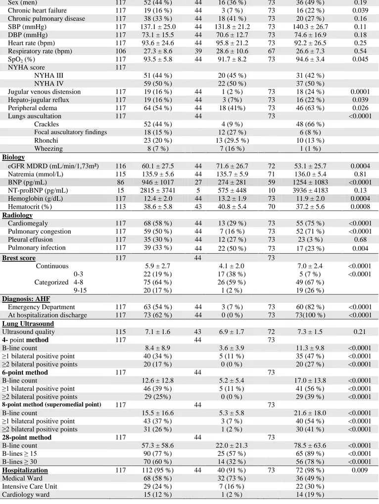
Population (n=117) No-AHF (n=44) AHF (n=73)
N Mean ± SD n (%) N Mean ± SD n (%) N Mean ± DS n (%) p Clinical Characteristics Age (years) 117 79.6 ± 11.8 44 77.0 ± 13.6 73 81.2 ± 10.3 0.088
Table 1: Characteristics of the study population
Sex (men) 117 52 (44 %) 44 16 (36 %) 73 36 (49 %) 0.19 Chronic heart failure 117 19 (16 %) 44 3 (7 %) 73 16 (22 %) 0.039 Chronic pulmonary disease 117 38 (33 %) 44 18 (41 %) 73 20 (27 %) 0.16 SBP (mmHg) 117 137.1 ± 25.0 44 131.8 ± 21.2 73 140.3 ± 26.7 0.11 DBP (mmHg) 117 73.1 ± 15.5 44 70.6 ± 12.7 73 74.6 ± 16.9 0.18 Heart rate (bpm) 117 93.6 ± 24.6 44 95.8 ± 21.2 73 92.2 ± 26.5 0.25 Respiratory rate (bpm) 106 27.3 ± 8.6 39 28.6 ± 10.6 67 26.6 ± 7.3 0.54 SpO2 (%) 117 93.5 ± 5.8 44 91.7 ± 8.2 73 94.6 ± 3.4 0.045 NYHA score 117 NYHA III 51 (44 %) 20 (45 %) 31 (42 %) NYHA IV 59 (50 %) 22 (50 %) 37 (50 %)
Jugular venous distension 117 19 (16 %) 44 1 (2 %) 73 18 (24 %) 0.0001 Hepato-jugular reflux 117 19 (16 %) 44 3 (7%) 73 16 (22 %) 0.039 Peripheral edema 117 64 (54 %) 44 18 (41%) 73 46 (63 %) 0.026
Lungs auscultation 117 44 73 <0.0001
Crackles 52 (44 %) 4 (9 %) 48 (66 %)
Focal auscultatory findings 18 (15 %) 12 (27 %) 6 (8 %)
Rhonchi 23 (20 %) 13 (29.5 %) 10 (13 %) Wheezing 8 (7 %) 7 (16 %) 1 (1 %) Biology eGFR MDRD (mL/min/1,73m²) 116 60.1 ± 27.5 44 71.6 ± 26.7 72 53.1 ± 25.7 0.0004 Natremia (mmol/L) 115 135.9 ± 5.6 44 135.7 ± 5.9 71 136.0 ± 5.4 0.81 BNP (pg/mL) 86 946 ± 1017 27 274 ± 281 59 1254 ± 1083 <0.0001 NT-proBNP (pg/mL) 15 2815 ± 3741 5 575 ± 448 10 3936 ± 4183 0.13 Hemoglobin (g/dL) 117 12.4 ± 2.0 44 13.2 ± 1.9 73 11.9 ± 2.0 0.0004 Hematocrit (%) 113 38.6 ± 5.8 43 40.8 ± 5.4 70 37.2 ± 5.6 0.0008 Radiology Cardiomegaly 117 68 (58 %) 44 13 (29 %) 73 55 (75 %) <0.0001 Pulmonary congestion 117 59 (50 %) 44 7 (16 %) 73 52 (71 %) <0.0001 Pleural effusion 117 35 (30 %) 44 12 (27 %) 73 23 (3 %) 0.68 Pulmonary infection 117 39 (33 %) 44 22 (50 %) 73 17 (23 %) 0.004 Brest score 117 44 73 Continuous 5.9 ± 2.7 4.1 ± 2.0 7.0 ± 2.4 <0.0001 Categorized 0-3 22 (19 %) 17 (38 %) 5 (7 %) <0.0001 4-8 75 (64 %) 26 (59 %) 49 (67 %) 9-15 20 (17 %) 1 (2 %) 19 (26 %) Diagnosis: AHF Emergency Department 117 63 (54 %) 44 3 (7 %) 73 60 (82 %) <0.0001 At hospitalization discharge 117 73 (62 %) 44 0 (0 %) 73 73(100 %) <0.0001 Lung Ultrasound Ultrasound quality 115 7.1 ± 1.6 43 6.9 ± 1.7 72 7.3 ± 1.5 0.21 4- point method 117 44 73 B-line count 8.4 ± 8.9 3.6 ± 3.9 11.3 ± 9.8 <0.0001
≥1 bilateral positive point 40 (34 %) 5 (11 %) 35 (47 %) <0.0001 ≥2 bilateral positive points 20 (17 %) 0 (0 %) 20 (27 %) <0.0001
6-point method 117 44 73
B-line count 12.6 ± 12.8 5.2 ± 5.4 17.0 ± 13.8 <0.0001
≥1 bilateral positive point 46 (39 %) 5 (11 %) 41 (56 %) <0.0001 ≥2 bilateral positive points 29 (25%) 0 (0 %) 29 (39 %) <0.0001
8-point method (superomedial point) 117 44 73
B-line count 15.5 ± 16.6 5.3 ± 5.8 21.6 ± 18.0 <0.0001
≥1 bilateral positive point 43 (37 %) 3 (7 %) 40 (54 %) <0.0001 ≥2 bilateral positive points 31 (26 %) 1 (2 %) 30 (41 %) <0.0001
28-point method 117 44 73 B-line count 57.3 ± 58.6 22.0 ± 21.3 78.5 ± 63.6 <0.0001 B-lines ≥ 15 90 (77 %) 25 (57 %) 65 (89 %) <0.0001 B-lines ≥ 30 70 (60 %) 14 (32 %) 56 (78 %) <0.0001 Hospitalization 117 112 (95 %) 44 40 (91 %) 73 72 (98 %) 0.009 Medical Ward 68 (58 %) 32 (73 %) 36 (49 %)
Intensive Care Unit 29 (24 %) 7 (16 %) 22 (30 %)
SBP/DPB: systolic/diastolic blood pressure; SpO2; blood oxygen saturation; NYHA: New York Heart
Association; eGFR: estimated glomerular filtration rate; MDRD: Modification of diet in renal disease; BNP: Brain natriuretic peptide; NT-proBNP: N-terminal pro b-type natriuretic peptide; AHF; acute heart failure
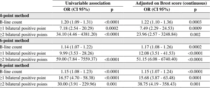
Univariable association Adjusted on Brest score (continuous)
OR (CI 95%) p OR (CI 95%) p
4-point method
B-line count 1.20 (1.09 - 1.31) <0.0001 1.22 (1.10 - 1.36) 0.0003 ≥1 bilateral positive point 7.18 (2.54 - 20.29) 0.0002 7.49 (2.29 - 24.53) 0.0009 ≥2 bilateral positive points 34.10 (4.46 - 4381.20) <0.0001 23.96 (2.57 - 3248.84) 0.002 6-point method
B-line count 1.14 (1.07 - 1.22) 1.17 (1.08 - 1.26) 0.0002 ≥1 bilateral positive point 9.99 (3.53 - 28.26) 12.08 (3.51 - 41.53) <0.0001 ≥2 bilateral positive points 59.00 (7.84 - 7559.37) <0.0001 51.15 (6.08 - 6740.40) <0.0001 8-point method
B-line count 1.15 (1.08 - 1.23) <0.0001 1.15 (1.07 - 1.24) <0.0001 ≥1 bilateral positive point 16.57 (4.70 - 58.38) <0.0001 15.68 (3.87 - 63.48) 0.0001 ≥2 bilateral positive points 30.00 (3.91 - 229.96) 0.001 38.75 (4.19 - 358.43) 0.001
Table 2: Association between the different lung ultrasound techniques and AHF diagnosis (in
univariable analysis and after adjustment on the Brest score)
Performance Diagnostic value of LUS techniques in addition to the Brest score C-index value of the
considered parameter (CI 95 %)
Specificity (CI 95%) Sensitivity (CI 95%)
C-index value of Brest Score and considered parameter
(CI 95 %)
P C-index increase in addition to the Brest score
(CI 95 %) p Overall Population Brest score Continuous 81.8 (74.2 - 89.4) Categories (0-3, 4-8, 9-15) 72.8 (65.3 - 80.3) 4- point method B-line count 76.7 (68.2 - 85.1) 88.1 (82.0 to 94.1) <0.0001 6.3 (1.0 to 11.6) 0.020
≥1 bilateral positive point 68.3 (60.8 - 75.8) 88.6 (75.4 - 96.2) 47.9 (36.1 - 60.0) 86.6 (80.1 to 93.1) <0.0001 4.8 (-0.1 to 9.6) 0.053 ≥2 bilateral positive points 63.7 (58.5 - 68.8) 100.0 (92.0 - 100.0) 27.4 (17.6 - 39.1) 85.3 (78.6 to 91.9) <0.0001 3.5 (0.4 to 6.5) 0.026 6-point method
B-line count 78.2 (70.1 - 86.4) 89.1 (83.3 to 94.8) <0.0001 7.3 (1.7 to 12.8) 0.010
≥1 bilateral positive point 72.4 (65.0 - 79.8) 88.6 (75.4 - 96.2) 56.2 (44.1 - 67.8) 88.5 (82.5 to 94.5) <0.0001 6.7 (0.9 to 12.5) 0.024 ≥2 bilateral positive points 69.9 (64.2 - 75.5) 100.0 (92.0 - 100.0) 39.7 (28.5 - 51.9) 88.4 (82.6 to 94.2) <0.0001 6.6 (2.3 to 10.8) 0.002 8-point method
B-line count 81.8 (74.3 - 89.3) 90.6 (85.2 to 96.0) <0.0001 8.8 (2.8 to 14.7) 0.004
≥1 bilateral positive point 74.0 (67.1 - 80.9) 93.2 (81.3 - 98.6) 54.8 (42.7 - 66.5) 88.7 (82.9 to 94.6) <0.0001 6.9 (1.6 to 12.2) 0.010 ≥2 bilateral positive points 69.4 (63.3 - 75.5) 97.7 (88.0 - 99.9) 41.1 (29.7 - 53.2) 88.7 (82.8 to 94.7) <0.0001 6.9 (1.7 to 12.1) 0.009
Patients with intermediate Brest score
Brest score
Continuous 71.7 (59.9 - 83.6) N/A
4-point method
B-line count 75.9 (65.0 - 86.8) 81.6 (71.7 to 91.5) <0.0001 9.9 (0.1 to 19.6) 0.047
≥1 bilateral positive point 68.7 (59.3 - 78.2) 88.5 (69.8 - 97.6) 49.0 (34.4 - 63.7) 78.5 (67.8 to 89.2) <0.0001 6.8 (-2.1 to 15.7) 0.13 ≥2 bilateral positive points 61.2 (55.3 - 67.1) 100.0 (86.8 - 100.0) 22.4 (11.8 - 36.6) 76.5 (65.9 to 87.1) <0.0001 4.8 (0.3 to 9.3) 0.037 6-point method
B-line count 78.4 (68.0 - 88.7) 83.4 (74.0 to 92.7) <0.0001 11.6 (1.9 to 21.4) 0.020
≥1 bilateral positive point 71.8 (62.4 - 81.2) 88.5 (69.8 - 97.6) 49.0 (34.4 - 63.7) 80.6 (70.4 to 90.8) <0.0001 8.9 (-0.2 to 17.9) 0.054 ≥2 bilateral positive points 69.4 (62.5 - 76.3) 100.0 (86.8 - 100.0) 22.4 (11.8 - 36.6) 81.4 (71.9 to 90.8) <0.0001 9.6 (3.1 to 16.1) 0.004 8-point method
B-line count 81.0 (71.2 - 90.8) 85.4 (76.4 to 94.3) <0.0001 13.6 (3.4 to 23.8) 0.009 ≥1 bilateral positive point 72.7 (63.9 - 81.5) 92.3 (74.9 - 99.1) 53.1 (38.3 - 67.5) 82.4 (72.6 to 92.2) <0.0001 10.7 (1.7 to 19.7) 0.020 ≥2 bilateral positive points 67.5 (59.6 - 75.3) 96.2 (80.4 - 99.9) 38.8 (25.2 - 53.8) 80.4 (70.3 to 90.4) <0.0001 8.6 (0.9 to 16.4) 0.029
Table 3. Diagnostic performance of the various lung ultrasound techniques in conjunction with the Brest score for pulmonary congestion
assessment
Supplementary table 1: Association between 28-point method and AHF diagnosis (in univariable analysis and after adjustment on the Brest score)
Univariable association Adjusted on Brest score (continuous)
OR (CI 95%) p OR (CI 95%) p
28-point method
B-line count 1.04 (1.02 - 1.06) <0.0001 1.04 (1.02 - 1.06) <0.0001 B-lines ≥ 15 6.17 (2.40 - 15.90) 0.0008 4.92 (1.68 - 14.44) 0.004 B-lines ≥ 30 7.06 (3.06 - 16.27) <0.0001 6.97 (2.58 - 18.79) 0.0001
Supplementary table 2: Performances for AHF with the 28-points method
Performances Increased level of evidence added by the LUS techniques
in addition to the Brest score (continuous)
C-index value of the
considered parameter (CI 95 %)
Specificity (CI 95%) Sensitivity (CI 95%)
C-index value of Brest Score and considered parameter
(CI 95 %)
P C-index increase in addition to the Brest score
(CI 95 %) p Overall Population 28-points method B-line count 82.1 (74.7 - 89.6) 90.5 (85.0 to 95.9) <0.0001 8.7 (2.6 to 14.7) 0.005 B-lines ≥15 66.1 (57.9 - 74.3) 43.2 (28.3 - 59.0) 89.0 (79.5 - 95.1) 85.0 (78.0 to 92.0) <0.0001 3.2 (-0.7 to 7.1) 0.11 B-lines ≥30 72.4 (63.9 - 80.9) 68.2 (52.4 - 81.4) 76.7 (65.4 - 85.8) 86.7 (80.1 to 93.2) <0.0001 4.8 (-0.6 to 10.3) 0.082
Patients with intermediate Brest score
28-points method
B-line count 80.1 (70.1 - 90.1) 84.3 (75.0 to 93.6) <0.0001 12.6 (2.5 to 22.6) 0.014
B-lines ≥15 64.0 (53.1 - 74.9) 42.3 (23.4 - 63.1) 85.7 (72.8 - 94.1) 78.3 (67.6 to 89.0) <0.0001 6.6 (-2.5 to 15.7) 0.16 B-lines ≥30 68.5 (57.2 - 79.8) 61.5 (40.6 - 79.8) 75.5 (61.1 - 86.7) 78.5 (67.4 to 89.6) <0.0001 6.8 (-2.6 to 16.1) 0.16