HAL Id: hal-00016689
https://hal.archives-ouvertes.fr/hal-00016689
Submitted on 31 May 2020
HAL is a multi-disciplinary open access
archive for the deposit and dissemination of
sci-entific research documents, whether they are
pub-lished or not. The documents may come from
teaching and research institutions in France or
abroad, or from public or private research centers.
L’archive ouverte pluridisciplinaire HAL, est
destinée au dépôt et à la diffusion de documents
scientifiques de niveau recherche, publiés ou non,
émanant des établissements d’enseignement et de
recherche français ou étrangers, des laboratoires
publics ou privés.
Identification of six novel allosteric effectors of
Arabidopsis thaliana Aspartate kinase-Homoserine
dehydrogenase. Physiological context sets the specificity
Gilles Curien, Stéphane Ravanel, Mylène Robert, Renaud Dumas
To cite this version:
Gilles Curien, Stéphane Ravanel, Mylène Robert, Renaud Dumas. Identification of six novel allosteric
effectors of Arabidopsis thaliana Aspartate kinase-Homoserine dehydrogenase. Physiological context
sets the specificity. Journal of Biological Chemistry, American Society for Biochemistry and Molecular
Biology, 2005, 280 (50), pp.41178-41183. �10.1074/jbc.M509324200�. �hal-00016689�
Identification of Six Novel Allosteric Effectors of
Arabidopsis thaliana Aspartate Kinase-Homoserine
Dehydrogenase Isoforms
PHYSIOLOGICAL CONTEXT SETS THE SPECIFICITY
*Received for publication, August 24, 2005, and in revised form, October 3, 2005 Published, JBC Papers in Press, October 10, 2005, DOI 10.1074/jbc.M509324200
Gilles Curien
1, Ste´phane Ravanel, Myle`ne Robert, and Renaud Dumas
From the Laboratoire de Physiologie Cellulaire Ve´ge´tale UMR5168, CEA-CNRS-INRA-Universite´ Joseph Fourier,
38054 Grenoble, France
The Arabidopsis genome contains two genes predicted to code for
bifunctional aspartate kinase-homoserine dehydrogenase enzymes (isoforms I and II). These two activities catalyze the first and the third steps toward the synthesis of the essential amino acids threo-nine, isoleucine, and methionine. We first characterized the kinetic and regulatory properties of the recombinant enzymes, showing that they mainly differ with respect to the inhibition of the homo-serine dehydrogenase activity by threonine. A systematic search for other allosteric effectors allowed us to identify an additional inhib-itor (leucine) and 5 activators (alanine, cysteine, isoleucine, serine, and valine) equally efficient on aspartate kinase I activity (4-fold activation). The six effectors of aspartate kinase I were all activators of aspartate kinase II activity (13-fold activation) and displayed a similar specificity for the enzyme. No synergy between different effectors could be observed. The activation, which resulted from a decrease in the Kmvalues for the substrates, was detected using low substrates concentrations. Amino acid quantification revealed that alanine and threonine were much more abundant than the other effectors in Arabidopsis leaf chloroplasts. In vitro kinetics in the presence of physiological concentrations of the seven allosteric effectors confirmed that aspartate kinase I and II activities were highly sensitive to changes in alanine and threonine concentrations. Thus, physiological context rather than enzyme structure sets the specificity of the allosteric control. Stimulation by alanine may play the role of a feed forward activation of the aspartate-derived amino acid pathway in plant.
The first and the third steps in the synthesis of the essential amino acids threonine (Thr) and methionine (Met) from aspartate (Asp) are catalyzed in bacteria and in plants by the bifunctional enzyme aspartate kinase-homoserine dehydrogenase (AK-HSDH)2 (E.C. 2.7.2.4-E.C. 1.1.1.3) (see Scheme 1). In Escherichia coli, two isoforms of AK-HSDH enzymes are found (thrA and metL gene products) (1). ThrA protein is inhibited by Thr on both activities. No allosteric effector could be iden-tified for the AK and the HSDH activities of the second isoform (MetL protein) (1). In plants, only one AK-HSDH isoform was identified after fractionation of crude extracts of soluble proteins (2, 3). However, the sequencing of the Arabidopsis genome revealed the existence of two
genes (At1g31230 and At4g19710) for AK-HSDH enzymes. The cDNA from one of these genes (At1g31230) was previously cloned (4) but the protein (AK-HSDH I) was not characterized. In a study from our group (5), the cDNA corresponding to the second AK-HSDH gene (At4g19710) was cloned and the protein (named AK-HSDH II) was overexpressed in E. coli, purified to homogeneity, and characterized.
To complete the analysis of Arabidopsis AK-HSDH enzymes the
Ara-bidopsis thalianaAK-HSDH I isoform was cloned and overexpressed in
E. coliand its kinetic and regulatory properties were determined. In the course of this work, a systematic survey of the effect of other amino acids led us to identify unexpected allosteric effectors of Arabidopsis Thr-sensitive AKs. The key point of this discovery was the measurement of AK activities in the presence of low (i.e. physiological) concentrations of the substrates ATP and Asp.
MATERIALS AND METHODS
Construction of pET23AK-HSDH I Plasmid
The plasmid coding for AK-HSDH I devoid of its predicted transit peptide was constructed by PCR using the same strategy as for AK-HSDH II (5). The initiating Met in recombinant AK-AK-HSDH I was aligned with the position of the initiating Met in the recombinant AK-HSDH II (5). As part of the insertion of a NcoI restriction site, Gly62and
Ser63were replaced by Met62and Ala63in the recombinant protein.
Sequencing of the cDNA was carried out and was in agreement with the predicted cDNA sequence of At1g31230.
Protein Expression and Purification
Transformed E. coli BL21pLys(S) cells were grown at 37 °C in 800 ml of LB medium with 100g/ml carbenicillin and 34 g/ml chlor-amphenicol. Isopropyl 1-thio--D-galactopyranoside was added to a final concentration of 0.4 mMwhen bacterial growth was equivalent
to an A600of 0.8. Cells were further grown for 15 h at 28 °C and
collected by centrifugation. The preparation of the crude extract and the first two purification steps (anion exchange chromatography and gel filtration) of AK-HSDH I were as for AK-HSDH II (5). AK-HSDH I eluted from the DEAE column at a higher KCl concentration (300 mM) than AK-HSDH II (250 mM). After elution from the gel filtra-tion column, the protein was concentrated on a Centricon device (50 K) and loaded on a Green Sepharose column equilibrated with 25 mM
Hepes, pH 7.5, 10% (v/v) glycerol, and 1 mMThr. AK-HSDH I protein was excluded. Pure protein was quickly frozen in N2and stored at
⫺80 °C for several months without any loss of activity. Recombinant AK-HSDH II protein was overexpressed in E. coli and purified as previously reported (5).
*The costs of publication of this article were defrayed in part by the payment of page charges. This article must therefore be hereby marked “advertisement” in accordance with 18 U.S.C. Section 1734 solely to indicate this fact.
1To whom correspondence should be addressed. Tel.: 78-23-64; Fax:
33-4-38-78-50-91; E-mail: gcurien@cea.fr.
2The abbreviations used are: AK, aspartate kinase; ASADH, aspartate semialdehyde
dehydrogenase; HSDH, homoserine dehydrogenase.
at INRA Institut National de la Recherche Agronomique, on November 8, 2010
www.jbc.org
Initial Velocity Measurements
All experiments were carried out in the spectrophotometer in a quartz cuvette thermostated at 30 °C.
AK Assay—AK activity was measured with a coupled assay using aspartate semialdehyde dehydrogenase (ASADH) previously purified to homogeneity in the laboratory (6). The amount of ASADH was adjusted so that the flux through the enzymatic chain was independent on the ASADH concentration (typically 50 nMAK and 0.8MASADH). Activ-ity was measured in 50 mMHepes, pH 8.0, 150 mMKCl, 20 mMMgCl2,
200MNADPH in the presence of fixed or variable amounts of ATP and Asp (see figure legends). Reaction was initiated by addition of the enzyme. In this coupled assay 2 mol of NADPH are consumed per mol of Asp phosphorylated (1 mol consumed by ASADH and 1 mol by HSDH). NADPH oxidation rate at steady-state was divided by two to obtain actual AK rates.
HSDH Assay—HSDH substrate, aspartate semialdehyde, was pre-pared as previously described (7). Activity measurements were carried out in 50 mMHepes, pH 8.0, 150 mMKCl, 100Maspartate semialde-hyde, and 200MNADPH. Reaction was initiated with the enzyme (final concentration 20 nM).
Free Amino Acid Analysis in Purified Arabidopsis Chloroplasts
A. thaliana(ecotype Columbia) plants were grown for 3 weeks on soil in a growth chamber (22 °C, 60% air humidity, light intensity of 150E m⫺2s⫺1, 16 h light/8 h dark) before rosette leaves were harvested. Chlo-roplasts were purified on Percoll gradients as described in Ref. 8. Free amino acids were prepared from intact chloroplasts by three consecu-tive extractions in aqueous ethanol solutions buffered with Hepes-KOH, pH 7.5, as described in Ref. 9. Between the extraction steps, the samples were centrifuged for 10 min at 16,000⫻ g and the supernatants collected. The combined ethanol/water extracts were used for amino acid derivatization using o-phthaldialdehyde. The derivatized amino acids were separated by reverse phase high pressure liquid chromatog-raphy using a Hypersil C18 column (150⫻ 4.6 mm inner diameter; 3 m) at 37 °C and 0.8 ml/min using a 65-min gradient of methanol in a sodium acetate buffer. Derivatized amino acids were detected using a SFM25 fluorimeter (Kontron) by excitation at 340 nm and emission at 455 nm. Free amino acid contents were normalized to chlorophyll
con-centration (10). Three extractions were performed for two independent purifications of chloroplasts to ensure reproducibility.
RESULTS
The cDNA corresponding to the At1g31230 gene from A. thaliana and predicted to code for a Thr-sensitive AK-HSDH was the first to be cloned (4), but the recombinant protein (named AK-HSDH I) was not characterized. Following the publication of the Arabidopsis genome, a second gene (At4g19710) predicted to code for a Thr-sensitive AK-HSDH was identified. In a previous study (5) from our group the corre-sponding recombinant protein (named AK-HSDH II) was overex-pressed in E. coli, purified to homogeneity, and characterized. In the present work, to determine the kinetic and regulatory properties of AK-HSDH I, the cDNA sequence corresponding to the predicted mature form of the enzyme (devoid of its predicted chloroplastic transit peptide) was amplified by PCR and cloned into a pET23d(⫹) expression vector. The AK-HSDH I recombinant protein was expressed in E. coli as a soluble form but at a lower level than observed for AK-HSDH II (5). Pure protein could be obtained after three purification steps (see “Mate-rials and Methods”).
Comparison of AK-HSDH Isoform Kinetic Properties—AK I and AK II catalytic constants were similar (v/[E]0⫽ 5 ⫾ 0.5 s⫺1for AK I and v/[E]0
⫽ 6.2 ⫾ 0.5 s⫺1for AK II). Compared with AK II, AK I displayed a higher
Kmfor aspartate and a lower Kmfor ATP, see TABLE ONE.
HSDH activities were measured in the physiological direction (pro-duction of homoserine from aspartate semialdehyde). The Kmfor
aspar-tate semialdehyde proved to be very similar for both isoforms (290⫾ 17 and 311⫾ 6Mfor HSDH I and HSDH II, respectively). Catalytic
constants were also similar (245⫾ 19 and 196 ⫾ 21 s⫺1for HSDH I and HSDH II, respectively).
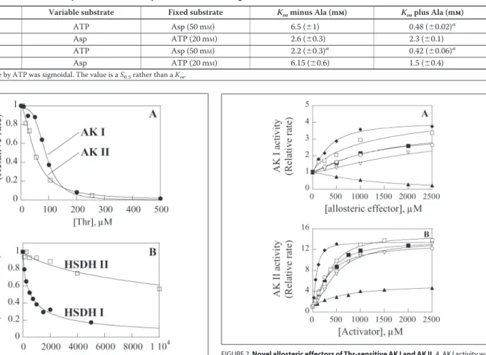
Inhibition of AK Activities by Thr at Low Substrate Concentrations— The inhibition of AK II by Thr was previously measured in the presence of 50 mMAsp and 20 mMATP (7). In the present work, to describe and compare the inhibition by Thr under more physiological conditions, we used low substrate concentrations, representative of the chloroplastic environment, i.e. 1 mMAsp and 2 mMATP (11). Fig. 1A shows that AK I, which was predicted to be a Thr-sensitive AK, was indeed inhibited by Thr. The inhibition curve was sigmoidal. The K0.5value for Thr was
about twice as high as that of AK II (91⫾ 3 and 49 ⫾ 3Mfor AK I and AK II, respectively). Also the cooperativity of the inhibition was much higher for AK I (Hill number, nH⫽ 3.7 ⫾ 0.5) than for AK II (nH⫽ 1.7 ⫾
0.15).
It is worth noting that the K0.5value of AK II for Thr was 10-fold lower
than previously reported for this enzyme when activity was measured in the presence of saturating concentrations of substrates (7). A lower K0.5
value for Thr in the presence of low substrate concentrations was expected as the inhibition of the AK activity by Thr is competitive with respect to the substrates (7).
Inhibition of HSDH Activities by Thr—Thr inhibited HSDH I activity in a hyperbolic manner (Fig. 1B). Inhibition was partial at saturation. In the presence of 100Maspartate semialdehyde, a K0.5value for Thr of
407⫾ 56Mwas calculated for HSDH I. In the same experimental conditions, HSDH II displayed a K0.5value for Thr about 1 order of
magnitude higher than HSDH I (K0.5⫽ 8500 ⫾ 1800M). At saturation
of Thr HSDH I retained 14% of activity, whereas HSDH II retained 25% of activity.
Identification of New Allosteric Effectors of AK I—As the present work was the first characterization of AK I we checked the effect of various other amino acids on its activity, in the absence of Thr and in the pres-ence of physiological concentrations of substrates (1 mMAsp, 2 mM
SCHEME 1. Aspartate pathway of amino acid biosynthesis. Each arrow represents an enzyme-catalyzed conversion. Dotted lines represent several steps.
Arabidopsis Aspartate Kinase-Homoserine Dehydrogenase
at INRA Institut National de la Recherche Agronomique, on November 8, 2010
www.jbc.org
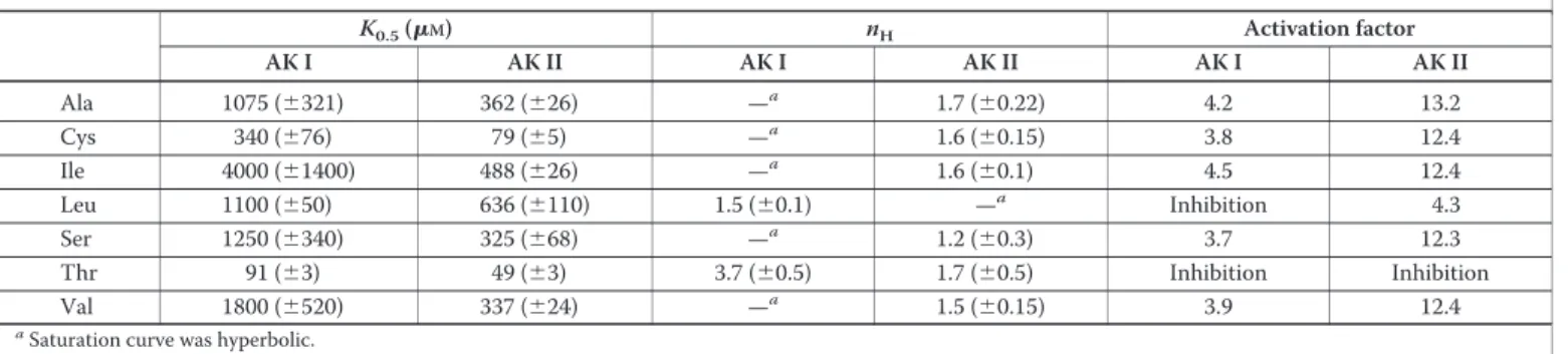
ATP). We initially tested amino acids produced in the Asp-derived amino acid pathway (Lys, Met, Leu, Val, Ile), or connected to this path-way (Cys, the precursor of Met). Interestingly, Cys, Ile, and Val stimu-lated the enzyme activity about 3-fold at 2.5 mM. On the other hand, Leu inhibited AK I. These results engaged us to test other amino acids exter-nal to the pathway but of similar size (Gly, Ser, and Ala). Surprisingly, Ser and Ala (2.5 mM) also stimulated AK I activity about 3-fold. Response curves are displayed in Fig. 2A. The activation was hyperbolic or slightly sigmoidal (for Cys). Cys was the strongest activator. Activa-tion by cysteine did not reflect a general sulfhydryl effect, because dithi-othreitol did not activate AK at 1 mM. Parameters of the activation by
Ala, Cys, Ile, Ser, Val, and of the inhibition by Leu are displayed in TABLE TWO. To test for synergistic effects between different activa-tors the different couples were tested at concentrations representing one-fourth the K0.5value calculated from the response curves in Fig. 2A.
For each couple tested, activation was additive, indicating the absence of synergy between two different activators. In the same manner no syn-ergy could be detected between the inhibitors Leu and Thr (not shown).
Among the 6 novel effectors of AK I, tested at 2.5 mM, only Cys had an effect on HSDH I activity. Cys inhibition was hyperbolic and partial at saturation (not shown). Fourteen percent activity was retained at satu-ration with Cys and a K0.5value for Cys of 290⫾ 45Mwas calculated.
Thus, Cys was a more potent inhibitor of HSDH I activity than Thr (K0.5⫽ 405M, Fig. 1B). The cysteine effect on HSDH activity did not
reflect a general sulfhydryl effect as dithiothreitol was not inhibitory at 1 mM. No synergy between Cys and Thr could be detected.
Re-examination of AK-HSDH II Regulatory Properties—We checked the effect of the allosteric effectors identified for AK I on the AK II activity, in the presence of physiological concentrations of ATP and Asp.
Interestingly, the effect on the AK II activity was even more pro-nounced than for AK I and qualitatively different. A 13-fold stimulation of AK II activity was observed with Ala, Cys, Ile, Ser, and Val each at 2.5 mM. Leu, an inhibitor of AK I, was stimulatory with AK II although to a lower extent than observed for the other activators (4-fold activation at 2.5 mM). The response curves for the strongest activators of AK II dis-played sigmoidal shapes (Fig. 2B). The saturation curve by Cys (Fig. 2B) had the lowest K0.5value (80⫾ 5M). K0.5values for Val, Ile, Ala, and Ser
TABLE ONE
Kmof AK I and AK II for ATP and Asp in the absence and presence of a saturating concentration of Ala
Enzyme Variable substrate Fixed substrate Kmminus Ala (mMM) Kmplus Ala (mMM)
AK I ATP Asp (50 mM) 6.5 (⫾1) 0.48 (⫾0.02)a
Asp ATP (20 mM) 2.6 (⫾0.3) 2.3 (⫾0.1)
AKII ATP Asp (50 mM) 2.2 (⫾0.3)a 0.42 (⫾0.06)a
Asp ATP (20 mM) 6.15 (⫾0.6) 1.5 (⫾0.4)
aSaturation curve by ATP was sigmoidal. The value is a S
0.5rather than a Km.
FIGURE 1. Inhibition of the two bifunctional AK-HSDH isoenzymes of Arabidopsis by Thr. A, AK activities were measured as indicated under “Materials and Methods” in the presence of 1 mMaspartate, 2 mMATP, and variable amounts of Thr. Curves are the best fit obtained by non-linear regression analysis of the experimental data to a Hill equation. Values for AK I are K0.5⫽ 90.6 (⫾0.3)M, nH⫽ 3.7 (⫾0.5). Values for AK II are K0.5⫽ 49 (⫾3)
M, nH⫽ 1.7 (⫾0.15). Uninhibited activities were normalized to 1. Actual rates (v/[E]0)
were 0.23⫾ 0.01 and 0.31 ⫾ 0.01 s⫺1for AK I and AK II, respectively. B, HSDH activities
were measured as indicated under “Materials and Methods.” Curves are the best fit obtained by non-linear regression analysis of the experimental data to a hyperbolic equation with a constant term at saturation. K0.5values for threonine were 407 (⫾56) and
8500 (⫾1800)Mfor HSDH I and II, respectively. Residual activity was 14 and 25% for HSDH I and II, respectively. Uninhibited activities were normalized to 1.
FIGURE 2. Novel allosteric effectors of Thr-sensitive AK I and AK II. A, AK I activity was measured in the absence of threonine at low substrate concentrations (2 mMATP, 1 mM
Asp) in the presence of variable amounts of Ala (䡺), Cys (⽧), Ile (ƒ), Leu (Œ), Ser (E), and Val (f). Curves are the best fit obtained by non-linear regression analysis of the experi-mental data to hyperbolic equations except for Leu where a Hill equation was used. Parameters of the activation are summarized in TABLE TWO. Activities in the absence of effectors were normalized to 1. B, AK II activity was measured in the absence of threonine at low substrate concentrations (2 mMATP, 1 mMAsp). Curves are the best fit obtained by non-linear regression analysis of the experimental data to a hyperbolic equation for Leu (Œ) or Hill equations for Cys (⽧), Ala (䡺), Ile (ƒ), Ser (E), and Val (f). Parameters of the activation are summarized in TABLE TWO. Activities in the absence of effectors were normalized to 1.
at INRA Institut National de la Recherche Agronomique, on November 8, 2010
www.jbc.org
were similar and about 4-fold higher (350M) than for Cys (see TABLE TWO). The saturation curve for Leu (Fig. 2B) displayed a higher K0.5
value (635⫾ 100M) and was hyperbolic. As observed for AK I no synergy could be detected between different activators of AK II (not shown).
At 2.5 mM, the 6 activators of AK II were without any detectable effect
on HSDH II activity in the presence or the absence of Thr, with the exception of Cys (not shown). Inhibition of HSDH II by Cys was hyper-bolic and partial at saturation. The enzyme retained 15% of activity at saturation with Cys and a K0.5value of 715⫾ 171Mwas calculated.
Thus, as observed for HSDH I, Cys was a more potent inhibitor of HSDH II than Thr. HSDH II K0.5value for Cys was higher than that of
HSDH I. No synergy between Thr and Cys for the inhibition of HSDH II could be observed (not shown).
Activation Results from a Decrease in the Kmfor the Substrates—AK I
and AK II kinetic parameters in the absence and presence of a saturating concentration of Ala (10 mM) were compared. No catalytic effect could be detected (not shown). However, as indicated in TABLE ONE, in the presence of 10 mMAla, AK I and AK II displayed much lower K0.5values
for ATP than in the absence of Ala (13- and 5-fold decrease, respec-tively). In the presence of Ala, AK II Kmfor Asp was reduced 4-fold,
whereas AK I displayed a Kmfor Asp virtually unchanged. The minor
effect of Ala on AK I apparent affinity for Asp explains why AK I is less efficiently activated than AK II at physiological concentrations of Asp and ATP (see Fig. 2). Both isoforms displayed similar kinetic parameters with respect to ATP and Asp in the presence of 10 mMAla. The activa-tion thus results from an increase in the apparent affinity for the sub-strates, and thus can only be detected at low substrate concentrations.
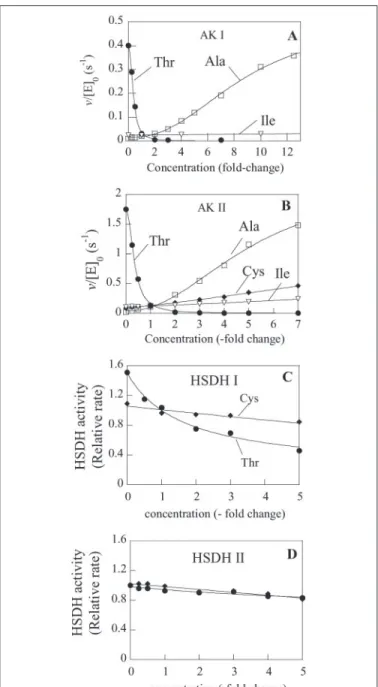
Enzyme Response in the Presence of Physiological Concentrations of Effectors—From a physiological point of view, the existence of several allosteric activators for a single enzyme, with some amino acids pro-duced downstream in the pathway (Leu, Val, and Ile) and others exter-nal to the pathway (Cys, Ser, and Ala) was perplexing. To examine this further in a quantitative manner, the abundance of the different amino acids was measured in the stroma of chloroplasts isolated from A.
thali-analeaves. Results presented in TABLE THREE indicated that Ala and Thr were from 5- to 20-fold more abundant than the other amino acids. Cys, the strongest activator of AK I and AK II and the best inhibitor of HSDH I and II was the lowest abundant of the 7 effectors. In the absence of observed synergy between different amino acids, these results sug-gested that the broad specificity for the control of AK activity observed when amino acids were tested one by one (Fig. 2) might be restricted to the two highly abundant amino acids Ala and Thr. We thus designed experiments in which AK I and AK II activities were measured in the presence of the 7 allosteric effectors at their chloroplastic concentra-tions. From a physiological point of view it was more pertinent to con-sider relative changes of concentration rather than absolute changes.
Thus, the concentration of each of the allosteric effectors of the AK activities was varied in turn, from zero to several times their chloroplas-tic concentration, in the presence of the other effectors at their chloro-plastic concentration. Fig. 3, A and B, show that, when the modification of amino acid abundance was expressed in terms of fold-changes, AK I and AK II activities were efficiently inhibited by Thr and activated by Ala only. Manyfold changes in Cys, Ile, Leu, Ser, or Val concentrations had only minor quantitative effects on AK I and AK II activities (Fig. 3). Results in Fig. 3, A and B, also show that Ala and Thr are able to displace each other. Moreover, the inhibition by Thr and the activation by Ala were observed for rather similar ranges of concentrations of effectors for AK I and AK II. Indeed, half-inhibition by Thr was observed at a concentration of Thr of 104Mand 114Mfor AK I and AK II,
respec-tively. Also, half-maximum activation was observed for 2250 and 3400 MAla for AK I and AK II, respectively.
Similar experiments were carried out to examine the sensitivity of HSDH activities to Cys and Thr. As expected from the low abundance of Cys, HSDH activities are only feebly affected by manyfold changes of Cys concentrations around its physiological value. HSDH I was much more sensitive to Thr than HSDH II (compare Fig. 3, C and D), which virtually escaped control by Thr at physiological concentration.
DISCUSSION
In the present work, the previously uncharacterized AK-HSDH iso-form I from A. thaliana was expressed as a recombinant protein in
E. coli, purified, and its kinetic and regulatory properties were deter-mined. We first showed that the AK and HSDH activities of this isoform were inhibited by Thr. HSDH I activity was inhibited by physiological concentrations of Thr, in marked difference with HSDH II, which was virtually unaffected in these conditions. A systematic examination of the
TABLE THREE
Concentrations of the effectors of AK-HSDH in the stroma
The amino acids Ala, Ile, Leu, Ser, Thr, and Val were quantified in chloroplasts isolated from 21-day-old Arabidopsis leaves.
Amino acid Chlorophyll Concentration in the stromaa
nmol/mg M Ala 26.96 (⫾1.9) 408 Cys 15b Ile 3.19 (⫾0.79) 48 Leu 3.57 (⫾0.53) 54 Thr 20.90 (⫾2.23) 317 Ser 4.05 (⫾0.72) 61 Val 6.36 (⫾1.09) 96 a66l of stroma/mg of chlorophyll (17).
bCys was assumed to be homogeneously distributed in the cell and we used the same
value as in Ref. 18.
TABLE TWO
Parameters of the allosteric activation and inhibition of AK I and AK II
K0.5(M) nH Activation factor AK I AK II AK I AK II AK I AK II Ala 1075 (⫾321) 362 (⫾26) —a 1.7 (⫾0.22) 4.2 13.2 Cys 340 (⫾76) 79 (⫾5) —a 1.6 (⫾0.15) 3.8 12.4 Ile 4000 (⫾1400) 488 (⫾26) —a 1.6 (⫾0.1) 4.5 12.4 Leu 1100 (⫾50) 636 (⫾110) 1.5 (⫾0.1) —a Inhibition 4.3 Ser 1250 (⫾340) 325 (⫾68) —a 1.2 (⫾0.3) 3.7 12.3 Thr 91 (⫾3) 49 (⫾3) 3.7 (⫾0.5) 1.7 (⫾0.5) Inhibition Inhibition Val 1800 (⫾520) 337 (⫾24) —a 1.5 (⫾0.15) 3.9 12.4 a
Saturation curve was hyperbolic.
Arabidopsis Aspartate Kinase-Homoserine Dehydrogenase
at INRA Institut National de la Recherche Agronomique, on November 8, 2010
www.jbc.org
effect of other amino acids leads us to the discovery of new allosteric activators and inhibitors of Arabidopsis Thr-sensitive AK activities. When tested in isolation and in the absence of Thr, amino acids Ala, Cys, Ile, Ser, and Val had very similar stimulating effects on a given AK
activity. The extent of stimulation and the apparent affinity were higher for AK II than for AK I. As the activation resulted from a decrease in the apparent Kmfor the substrates ATP and Asp it could only be detected
when activity measurements were carried out in the presence of low concentrations of substrates. In the first paper describing a Thr-sensi-tive AK from plant, Aarnes and Rognes (2), working on a partially puri-fied enzyme fraction from pea leaf observed a 2-fold stimulation of the AK activity by 8 mMAla, Val, or Ile (2). Little attention was given to this result in later reviews about the regulation of the Asp-derived amino acid synthesis, probably because the activation factor was low and observed at a high concentration of activators. We are now able to explain why the activation factor was so low. Indeed, in their assay the authors used high concentrations of Asp (12 mM) and ATP (8 mM). Under these conditions the activating effect was partly masked. Together, the observation of Aarnes and Rognes (2) on the pea enzyme and our results on the recombinant A. thaliana enzymes suggest that the activation of Thr-sensitive AK may be a general feature in plants. In this respect plant AK-HSDH enzymes clearly differ from their bacterial equivalent. Indeed, Ser was shown to be an inhibitor of the E. coli Thr-sensitive AK activity (K0.5⫽ 25 mM(12)), rather than an activator, as
observed here for the A. thaliana enzymes.
The identification of novel ligands for Arabidopsis AK-HSDH enzymes raised the question of their binding mode on the enzyme. AK-HSDH enzymes are predicted to be formed of three domains, the AK and HSDH domains, separated by a regulatory domain (7). We previously showed that the AK-HSDH II regulatory domain contains two non-equivalent threonine binding sites. Amino acid sequence anal-ysis and secondary structure prediction allowed us to identify an amino acid in each subdomain (Gln443and Gln524) potentially involved in the
binding of Thr, the only known effector at this time. Steady-state kinetic analyses of Q443A and Q524A mutants showed that the AK activity of the Q443A mutant was not any more inhibited by Thr, whereas the AK activity of the Q524A mutant was inhibited as observed for the wild type (7). Preliminary kinetic measurements (not shown) indicated that the allosteric stimulation by Ala was strongly reduced for both mutants of the AK-HSDH II regulatory domain. Thus both regulatory subdomains are involved in the allosteric activation. The two regulatory subdomains of AK-HSDH are repeated conserved motifs that belong to the ACT domain family (16). These domains are found associated with many allosteric enzymes involved in the amino acid metabolism. In this respect, the present results illustrate the remarkable plasticity of the ACT domains in their binding properties and their ability to control enzyme activities by activation and inhibition.
The broad specificity observed for the control of the Arabidopsis AK-HSDH activities was surprising. We first checked whether some interactions could be synergistic (as observed for the Lys-sensitive plant AK that is synergistically inhibited by Lys and S-adenosylmethionine, (13)). As we could not detect any synergy between different effectors, one predicted that the relative abundance of the effectors determines their contributions to the control of the enzyme activity. We thus quan-tified the concentrations of the different allosteric effectors in
Arabi-dopsischloroplasts. The inhibitor Thr and the activator Ala proved to be much more abundant than the other allosteric effectors suggesting that Thr and Ala were the physiological effectors of the enzymes. In vitro kinetics in the simultaneous presence of the seven allosteric effectors confirmed this prediction. These experiments also showed that AK I and AK II were modulated by similar ranges of Thr or Ala concentra-tions. This was a priori non-obvious at the examination of the results in Fig. 2. Indeed, one may conclude from these results that AK I and AK II strongly differ with respect to their sensitivity to Ala. Indeed, AK I
FIGURE 3. AK and HSDH activities in vitro in a physiological context. AK and HSDH activities were measured in the simultaneous presence of Ala, Cys, Ile, Leu, Ser, Val, and Thr at their physiological concentrations (see TABLE THREE). Each amino acid concentra-tion was then varied in turn around this value. Concentraconcentra-tions are expressed as fold-changes of the physiological concentrations. A, AK I. Inhibition curve by Thr (●) and activation curve by Ala (䡺) are the best fit obtained by non-linear regression analysis of the experimental data to Hill equations. Values for the inhibition by Thr are K0.5⫽ 114 ⫾
3Mand nH⫽ 2.4 (⫾0.1). Values for the activation by Ala are K0.5⫽ 3400 ⫾ 200Mand
nH⫽ 2.4 (⫾0.2). Activity was virtually unchanged when the concentrations of the other
effectors were changed (the response to Ile (ƒ) only is displayed). B, AK II. Inhibition curve by Thr (●) and activation curve by Ala (䡺) are the best fit obtained by non-linear regres-sion analysis of the experimental data to Hill equations. The data points for Val and Ser were similar to those obtained for Cys (⽧) and Ile (ƒ), respectively, and are not displayed for clarity reasons. Values for the inhibition by Thr are K0.5⫽ 104 ⫾ 2.5Mand nH⫽ 2.14
(⫾0.1). Values for the activation by Ala are K0.5⫽ 2250 (⫾ 320)Mand nH⫽ 2.15 (⫾0.3).
C, HSDH I. Curves are the best fit obtained by non-linear regression analysis of the
exper-imental data to hyperbolic equations with a constant term. K0.5value for the inhibition
by Thr corresponds to a concentration of Thr of 490⫾ 50M. D, HSDH II inhibition by Thr or Cys.
at INRA Institut National de la Recherche Agronomique, on November 8, 2010
www.jbc.org
displayed much higher K0.5values for the activators Ala than AK II.
Also, the activation factor was lower than for AK II. However, results in Fig. 3 show that these quantitative differences observed when amino acids were tested in isolation were strongly attenuated when activities were measured in the simultaneous presence of the inhibitor Thr and the activator Ala. As AK I displayed a lower apparent affinity than AK II for both the activator and the inhibitor, the ratio of concentration rather than the absolute concentrations of Ala and Thr determined the output. It is interesting to observe from results in Fig. 3 that the other activa-tors do contribute altogether to the activation of the enzyme. For exam-ple, in the presence of a physiological concentration of Thr (300M),
the six allosteric activators together stimulate 25-fold the AK II activity. Sixty percent of this activation are because of the abundant amino acid Ala. Nevertheless, the other activators contribute to the remaining 40%. Whether this non-negligible contribution constitutes a noise in the reg-ulatory processes that cannot be avoided or whether it has important physiological function remains to be determined.
An interesting conclusion from this work is that the cellular context rather than the enzyme structure determines the specificity of the allos-teric controls of Arabidopsis AK-HSDH enzyme activities. It is worth noting that the present conclusions are valid for Arabidopsis leaf chlo-roplasts. The conclusions might be different in a different tissue, under different environmental or developmental conditions leading to changes in the relative abundance of the seven allosteric effectors.
AK I, AK II, and HSDH I activities are inhibited by physiological concentrations of Thr. However, one can conclude from our results that HSDH activities are not stringent control points by Thr. Indeed, in a reaction media that mimics the metabolic composition of the leaf chlo-roplast, half-inhibition of HSDH I occurs for a concentration of Thr 6-fold higher than that required for half-inhibition of AK I or AK II (600
versus100M, compare Fig. 3, A and C). Also, molecular analyses using a GUS reporter-gene strategy suggested that the promoters of AK-HSDH I and AK-HSDH II were simultaneously active in different tissues of the plant (15) and especially in the leaf. If AK-HSDH I and AK-HSDH II enzymes are indeed simultaneously expressed in the same compartment, and HSDH II is fully active at a physiological concentra-tion of Thr, then HSDH activities are definitely not stringent control points by Thr. Note also that, although the inhibition of HSDH activities by Cys is more efficient than by Thr in vitro, the low abundance of this metabolite in vivo excludes a sensitive control of HSDH activities by Cys.
Functional redundancy is frequently invoked to justify the existence of isoforms. The present analysis shows that the two enzymes are not exactly similar. First, one observed that AK I was about 4-fold less active than AK II when activity was measured in the presence of the seven allosteric effectors at their physiological concentration (see Fig. 3, A and
B). Also, as shown in these figures, the response curve for Ala is shifted
to slightly higher concentrations of Ala for AK I compared with AK II. Finally, the two isoforms strongly differ with respect to the control of the HSDH activity by Thr. Whether these differences have important phys-iological implications remains to be determined. Other levels of con-trols, which would affect the two isoforms differentially are not excluded.
From a functional point of view, the existence of activators and inhib-itors of an enzyme activity presents two advantages. First, it increases the sensitivity to small changes in the activator or the inhibitor concen-trations. Second, as Ala not only displaces Thr but also activates the enzyme by itself, the existence of an activator and an inhibitor increases the range of enzyme activity for which control can be exercised. Addi-tional function, if any, of the activation of plant AKs by Ala, remains obscure. In plants, after incorporation into glutamate, nitrogen is quickly distributed to other amino acids, much of it going directly to Ala and Asp via transamination of pyruvate and oxaloacetate, respectively (14). Ala is thus directly linked to the carbon and the nitrogen metabo-lisms of the cell. In this respect, Ala may be a feed-forward activator of the aspartate-derived amino acid pathway. Activation of AKs by Ala might couple the supply of carbon and nitrogen to the demand for Asp-derived amino acids synthesis. This is new information that might prove useful for understanding of the aspartate-derived amino acid metabolism in vivo and the improvement of crop quality.
Acknowledgments—We thank Dr. Claude Alban, Prof. Roland Douce, and Dr. Michel Matringe for critical reading of the manuscript.
REFERENCES
1. Umbarger, H. E. (1978) Annu. Rev. Biochem. 47, 533– 606 2. Aarnes, H., and Rognes, S. E. (1974) Phytochemistry 13, 2717–2724
3. Azevedo, R. A., Arruda, P., Turner, W. L., and Lea, P. J. (1997) Phytochemistry 46, 395– 419
4. Ghislain, M., Frankard, V., Vandenbossche, D., Matthews, B. F., and Jacobs, M. (1994)
Plant Mol. Biol. 24,835– 851
5. Paris, S., Wessel, P. M., and Dumas, R. (2002) Protein Expression Purif. 24, 105–110 6. Paris, S., Wessel, P. M., and Dumas, R. (2002) Protein Expression Purif. 24, 99 –104 7. Paris, S., Viemon, C., Curien, G., and Dumas, R. (2003) J. Biol. Chem. 278, 5361–5366 8. Block, M. A., Tewari, A. K., Albrieux, C., Marechal, E., and Joyard, J. (2002) Eur.
J. Biochem. 269,240 –248
9. Kreft, O., Hoefgen, R., and Hesse, H. (2003) Plant Physiol. 131, 1843–1854 10. Lichtenthaler, H. K. (1987) Methods Enzymol. 148, 350 –382
11. Krause, G. H., and Heber, U. (1976) in The Intact Chloroplast (Barber, J., ed) pp. 174 –175, Elsevier/North-Holland Biomedical Press, Amsterdam, The Netherlands 12. Costrejean, J. M., and Truffa-Bachi, P. (1977) J. Biol. Chem. 252, 5332–5336 13. Rognes, S. E., Lea, P. J., and Miflin, B. J. (1980) Nature 287, 357–359
14. Ireland, R. (1997) in Plant Metabolism (Dennis, D. T., Turpin, D. H., Lefebvre, D. D., and Layzell, D. B., ed) 2nd Ed., pp. 483, Addison Wesley Longman, Essex 15. Rognes, S. E., Dewaele, E., Aas, S. F., Jacobs, M., and Frankard, V. (2003) Plant Mol.
Biol. 51,281–294
16. Chipman, D. M., and Shaanan, B. (2001) Curr. Opin. Struct. Biol. 11, 694 –700 17. Winter, H., Robinson, D. G., and Heldt, H. W. (1994) Planta (Basel) 193, 530 –535 18. Curien, G., Ravanel, S., and Dumas, R. (2003) Eur. J. Biochem. 270, 4615– 4627
Arabidopsis Aspartate Kinase-Homoserine Dehydrogenase
at INRA Institut National de la Recherche Agronomique, on November 8, 2010
www.jbc.org