Anatomic bases of medical,
and surgical techniques
radiological
Surgical o0
Radiologic
Anatomy
Journal of Clinical Anatomy © Springer-Verlag 1999
The medial malleolar network: a constant vascular base of the distally
based saphenous neurocutaneous island flap
F.T. Ballmer 1, R. Hertel 1, H.P. Noetzli 1 and A.C. Masquetet 2
I Department of Orthopaedic Surgery, University of Berne, Inselspital, CH-3010 Berne, Switzerland
2 Service de Chirurgie Orthop6dique et R6paratrice, H6pital Avicenne, 125, route de Stalingrad, F-93000 Bobigny, France
Summary: Based on 30 fresh cadaver dissections a detailed anatomic study of the medial malleolar network is presen- ted with particular attention to the ana- stomoses between the latter mad the vas- cular axis that follows the saphenous nerve. The media] malleolar network is formed by the anterior medial malleolar artery, branches from the media] tarsal arteries, the posterior medial malleolar artery and branches f r o m the medial plantar artery. A distinct anterior medial matleolar artery and posterior medial malleolar artery could be identified in 80 and 20%, r e s p e c t i v e l y , as well as constant additional small branches ari- sing from the anterior tibial or posterior tibial artery. A constant anastomosis was found between the arcade formed by the media1 tarsal arteries and the medial plantar a. in 60%, mad the medial branch of the medial plantar artery in 40%, res- pectively. This anastomosis always gave rise to branches to the medial malleolar network. In the perimalleolar area and with regard to the great saphenous v. a larger anterior and a smaller posterior branch of the saphenous nerve was found in 100 and 90%, respectively. In all dis- sections, for both branches of the saphe-
Correspondence to: F.T. Ballmer
nous nerve two to four small, but distinct anastomoses between the medial malleo- lar network and the perineural vascular axis were identified. These constant ana- stomoses represent a new and reliable v a s c u l a r b a s e for the d i s t a l l y - b a s e d saphenous neurocutaneous island flap. Thus, the pivotal point of the flap can be chosen in the area of the medial malleo- lus without respecting the most distal septocutaneous anastomosis between the perineural vascular axis and the posterior tibial artery. Additionally, an illustrative clinical case is presented.
Le r6seau mall6olaire m6dial, origine constante du lambeau en ilot neuro-cutan6 saph~ne ~ p6dieule distal R~sum~ : A partir de la dissection de 30 pibces cadavrriques fra~ches, nous avons rralis6 une 6tude anatomique drtaillre du rrseau malldotaire mddial, particulibre- ment orientre sur les anastomoses entre ce dernier et l'axe vasculaire qui suit le n e f f saph~ne. Le r r s e a u mall6olaire mrdiat est form6 par l'art~e mallrolaire m6diale ant6rieure, les branches des attires tarsales mrdiales, l'art~re mallro- laire mrdiale postrrieure et des branches de l'artbre plantaire mddiale. Une artbre mall6olaire mrdiale antrrieure et une attire mallrolaire m6diale post6rieure distinctes ont pu ~tre idenfifires dans res- pectivement 80 et 20 % des cas, en plus
de l'apport constant rralis6 par de fines branches igsues des attires tibiales ant& rieure et postrrieure. Nous avons trouv6 une anastomose constante entre l'arcade formge par les artbres tarsales mrdiales et l'art~re plantaire mrdiale dans 60 % des cas, la branche mrdiale de l'art~re plan- taire mrdiale darts 40 % des cas. Cette anastomose donnait toujours naissance des branches alimentant le rrseau mall6o- laire mddial. Dans la rrgion prri-mallro- laire et en regard de la veine grande saph~ne, nous avons trouv6 respective- ment dans 100 et 90 % des cas deux branches de division du nerf saph~ne, une branche antrrieure plus volumineuse et une branche postdrieure plus petite. Sur toutes les dissections nous avons idenfiflr, pour chacune des branches de division du neff saph~ne, deux ~ qnatre anastomoses distinctes entre le rrseau mallrolaire mrdial et l ' a x e vasculaire prri-neural. Ces anastomoses constantes reprrsentent une origine vasculaire fiable nouvellement identifire pour la rralisa- tion d'un lambeau en tlot neuro-vasculai- re saph~ne ~t prdicule distal. Le point de rotation de ce lambeau peut atre choisi au niveau de la rrgion mallrolaire mrdiale, sans respecter les anastomoses septo- cutanres les plus distales entre l'axe vas- culaire prri-neural et l'art~re tibiale pos- trrieure. Un cas clinique illustrant ce lambeau est prrsentr.
298 F.T. Ballmer, et al.: The medial malleolar network
Key words: Medial matleolar network - - Saphenous n e r v e - Neurocutaneous flap - - Island flap - - Lower limb
At the turn of the century Qutnu and Lejars d e m o n s t r a t e d that c u t a n e o u s nerves are accompanied by a vascular axis, and in 1936, Salmon mentioned neurocutaneous arteries without giving details [26, 30]. In the late seventies, the saphenous n. was believed to be the best suited free vascularized nerve graft based on studies on the vascularization of seve- ral nerves [7, 33]. But although the inti- mate relationship between the sensory nerves and the skin had been known for a long time, only recently has the concept of neurocutaneous island flaps for cove- rage of soft-tissue defects been developed and cfinlcally applied [5, 21]. The initial anatomic studies on neurocutaneous flaps showed that the vascular axis that follows a superficial sensory nerve in its suprafas- cial course ensures the vascularization of the nerves and gives off several cuta- neous branches to supply the skin. The vascular n e t w o r k of the nerve itself, which can be either a true artery or an interlacing network, is fed by anasto- m o s e s with s e p t o c u t a n e o u s v e s s e l s issuing from a deep main vessel or direct cutaneous arteries [21]. For the vascular axis of the saphenous n. two to seven sep- tocut~eous anastomoses with the poste- rior tibial artery along its course in the leg have been identified. The most distal ana- stomosis is located 3 to 5 cm proximal to the medial matleolus. Thus, this area cor- responds to the most distal pivotal point of the original distally based saphenous neurocutaneous island flap relying on a septocutaneous vessel [8, 21]. As the saphenous n. passes anterior to the medial malleolus to the dorsum of the foot, it was hypothesised that reliable anasto- moses between the medial malleolar net- work and the vascular axis of the saphe- nous n. m a y exist. Consequently, the pivotal point of a distally based saphe- nous neurocutaneous island flap could be chosen in the area of the media] maUeo- lus without identifying or respecting the most distal septocutaneous anastomosis between the vascular axis of the saphe- nous n. and the posterior tibial a. Thus, the d i s s e c t i o n w o u l d be fairly e a s y
consisting of elevation of a broad subcu- taneous fascial pedicle, which includes the branches of the saphenous n. with the perineural vascular axes and the saphe- nous v. Additionally, the more distally located pivotal point would lengthen the pedicle of the flap. Therefore, the purpose of the study was to examine in detail the anatomy of the medial malleolar vascular network with particular attention to the anastomoses of the latter with the vascu- lar axis that follows the saphenous n. Material and methods
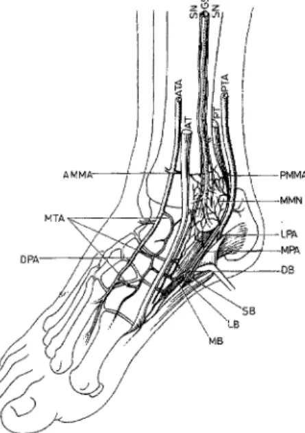
The anatomic study was performed on 30 fresh cadaver lower limbs of both sexes. The mean age at death ranged from the fifth to the ninth decade. Both legs were injected with coloured neoprene latex via the femoral artery. The injection was continued until the capillary beds of the toes were seen to fill. Limbs lacking in good peripheral staining were excluded. The cadavers were subsequently refrige- rated, and dissections under loupe magni- fication were carried out after 2 days. With the cadaver in the supine posi- tion, an anterior incision was made from the middle third of the leg to the metatar- sal region and the skin was carefully dis- sected off. While respecting the saphe- nous n., including its vascular axis and the great saphenous v., the fascia and the retinacula were opened and removed as well as tendons and muscles if required for better exposure. During the subse- quent careful dissection the anatomical structures Iisted below were identified and analysed. In particular the medial m a l l e o l a r n e t w o r k ( M M N ) , and the blood supply to the vascular axis of the saphenous n. were studied. The acro- nyms of the anatomic structures descri- bed below and shown in Fig. 1 are listed at the table 1.
The following data were recorded: a) the presence or absence of the anterior tibial a. (ATA) and dorsalis pedis a. (DPA), respectively, and their location with respect to the transverse bimalleolar axis;
b) the presence or absence of the anterior medial maUeolar a. (AMMA); if present, its location with respect to the ankle joint-line, as well as the number of additional smaller vessels present;
c) the presence or absence of the medial tarsal a. (MTA); if present, the number of vessels as well as the presence or absence of anastomosis of the MTA with the MMN and the medial plantar a. (MPA);
d) the presence or absence of the posterior tibial a. (PTA);
e) the presence or absence of the posterior medial malleolar a. (PMMA); if present, its location with respect to the ankle joint-line, as well as the number of additional smaller vessels present;
f) the diameter at its origin of the DPA (at the level of the ankle joint-line), AMMA, MTA, PTA, PMMA;
g) the presence or absence of the saphenous n. (SN) at the level of the ankle joint-line; if present, the number of branches and location with respect to the great saphenous v. (GSV);
h) the presence or absence of anasto- mosis between the MMN and the vascu- lar axis of the SN.
The anatomic study is supplemented by an illustrative clinical case.
Results
Anterior tibial a. (ATA) and dorsaIis pedis a. (DPA) (Figs. 1, 2)
The DPA was the continuation of the A T A in 27 o f the 30 d i s s e c t i o n s (90%). In 2/30 s p e c i m e n s (7%) the DPA and the perforating branch of the peroneaI a. were of a similar calibre, and in 1/30 limbs (3%) only a narrow DPA was found. The mean diameter of dhe DPA at the level of the ankle joint interline was 2.1 m m (range, 0.8 to 3.5 ram). In 11/30 dissections (37%) the distal ATA was located laterally to the middle of the transverse bimalleo- lar axis (Fig. 2).
Anterior medial malleolar a. (AMMA )
(Figs. 1, 2)
In 24/30 specimens (80%) a distinct branch was identified as the AMMA, which travelled medially and branched while joining the MMN. The mean dia- meter of this vessel at its origin was 0.5 m m (range, 0.3 to 0.7 ram). The arte- ry was located between approximately the ankle joint interline and up to 4 cm
Fig, 1. Schematic drawing of the anatomy of the medial malleolar area, especially of the vascular branches forming the medial maUeolar network. The vascular axes of the two branches o f the saphenous nn. and their anastomoses with the medial malleolar network are not shown (AT,
tibialis anterior m.; PT, tibialis posterior m.; other abbreviations are listed in table 1)
Table 1. Acronyms of the anatomic structures in alphabetical order ~ig. 1, 5c, 6a,b)
AMMA, anterior medial maUeolar artery; ATA, ante- rior tibial artery; DB, deep branch of the MPA;
DPA, dorsalis perils artery; GSV, great saphenous v.; LB, lateral branch of the DB; MB, medial branch of the DB; MMN, medial malleolar network; MPA,
medial plantar artery; MTA, medial larsal artery;
PMMA, posterior medial malleolar artery; PTA,
posterior tibial artery; SN, saphenous n.; SCA, sep- tocutaneous anastomosis; SB, superficial branch of the MPA
proximally. Besides this main vessel, up to three small branches arose from the A T A travelling m e d i a l l y to join the MMN.
Medial tarsal aa. (MTA) (Figs. 1, 2)
From its medial side, the DPA gave two to four branches: the average diameter at their origin measured 0.4 m m (range, 0.2 to 0.7 ram). Those branches given off at the level of the cuneiform bones and the navicular were the most prominent. The branches divided to form a vascular arca-
de with each other always anastomosing with the MMN. Additionally, in all dis- sections this arcade anastomosed with the MPA or the medial branch of the MPA (see also section MPA).
Posterior tibial a. (PTA) (Figs. 1, 3)
The PTA was present in all limbs. The mean diameter of the PTA 4 cm proxi- mal to the tip of the medial malleolus was 2.7 mm (range, 1.2 to 3.9 mm). In its retromalleolar course the PTA gave off a variable number of small branches to the MMN. Deep to the flexor retinaculum the PTA divided into the medial and late- ral plantar aa.
Posterior medial malIeolar a. ( PMMA )
(Figs. 1, 3)
In only 6/30 dissections (20%) could a dominant vessel with a mean diameter of 0.4 m m be identified as the PMMA, Totally, the PTA gave off 2 to 4 small branches (mean diameter 0.25 mm) one to four cm proximal to the tip of the medial malleolus. The branches travel- led around the medial malleolus anasto- m o s i n g with each other to j o i n the MMN.
Medial plantar a. (MPA) (Figs. 1, 3)
The MPA travelled in the medial com- partment of the sole of the foot covered by abductor hallucis; it was present in all limbs. It divided into two branches, superficial (SB) and deep (DB). The deep branch in turn divided into two fur- flier branches: a lateral branch (LB) and a medial branch (MB) which crossed the terminal part of the tibialis posterior ten- don to run distally along the medial mar- gin of the foot. This pattern was seen in 25/30 dissections (83%). In 5/30 limbs (17%) the medial branch of the deep branch of the MPA arose directly from the MPA.
In 12/30 specimens (40%) an anasto- mosis with a mean diameter of 0.5 m m (range, 0.3 to 0.7 ram) was found bet- ween the medial branch of the MPA and the vascular arcade formed by the MTA. In 18/30 specimens (60%), the anasto- m o s i n g v e s s e l with the M T A arose directly from the MPA (Fig. 3).
In all dissections the medial malleotar network r e c e i v e d branches f r o m the MPA and the anastomosis between the MPA and MTA.
Saphenous n. (SN) (Figs. 1, 4)
In the area of the medial malleolus, ante- rior to the great saphenous v. (GSV), a large branch of the saphenous n. could be identified in all dissections, passing dis- tally to reach the medial side of the dor- sum of the foot. In all but three of the 30 dissections (90%) a smaller branch could also be found, which is closer and poste- rior to the GSV. This posterior branch terminated at about the level of the ankle. All dissected nerves were well vasculari- zed by a rich vascular axis that showed good filling (Fig. 4). The two branches of the SN together with the GSV were inti- mately related to the MMN. In all dissec- tions for both branches of the saphenous n. two to four small, but distinct anasto- moses between the MMN and the per- ineural vascular axis were identified. In its course from the ankle joint-line to the ctmeonavicular articulation the anterior branch of the SN variably received addi- tional small branches from the arcade formed by the MTA.
Clinical application
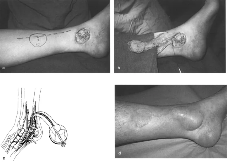
A 50-year old patient presented with a chronic ulcer in the area of the medial malleolus, whereby the anterior margin of the defect is located posteriorly to the course of the GSV as well as the anterior branch of the SN. The anterior feeding vessels of the medial malleolar network, i.e. the anterior medial malleolar a. and the medial tarsal aa. are preserved. The course of the great saphenous v., and consequently of the branches of the saphenous n. is marked over the lower medial aspect of the leg. The design of the flap is outlined on this course accor- ding to the size of the defect and the required length of the pedMe (Fig. 5a). An island flap of 6 x 5 cm is elevated and includes the fascia. The subcuta- neous fascial pedicle is elevated after performing two skin flaps by subdermal dissection. The pedicle should have a width o f 2 to 3 c m to include the branches of the SN with the perineural
300 F.T. Ballmer, et al.: The medial malleolar network
Fig. 2. Dorsal a~le area of left foot (bottom, dis- tal leg; top, mid-foot; on the right, medially). Anterior tibial a. (black arrow), anterior medial malleolar a. (long arrowhead), dorsalis pedis a.
(hollow arrow), medial tarsal aa. (short arrow- heads) forming an arcade as well as an anastomo- sis (blue arrow) with the medial plantar a. Medial malleolar area (curved arrow) with stained vascu- lar axis of the anterior branch of the saphenous n.
(yellow rubber strips)
Fig. 3. View of right medial malleolar area (on the right, distal leg; top left, mid-foot; bottom left, heel). Medial malteolar network (curved arrow) recieving branches from the posterior tibial a. (black arrow),
medial plantar a. (hollow arrow), anastomosis (star) between the medial plantar and medial tarsal aa. Great saphenous v. (dot). Note distinct anastomosis (short arrowhead) between the vascular axes of the saphenous n. branches (long arrowheads) as well as the medial malleolar network
vascular axes a n d the G S V . The septocu- t a n e o u s a n a s t o m o s e s to t h e p o s t e r i o r tibial a. are n o t respected o n the posterior m a r g i n o f the pedicle. T h e perfusion o f the pedicle relies on a retrograde flow in the perineural vascular axis, which is fed b y the anterior anastomoses o f the medial malteolar network. Therefore, n o dissec- tion is carried out in the medial premal- leolar area to avoid any surgical trauma. At the proximal e n d o f the flap the v. is ligated a n d cut, a n d the n. branches are severed (Fig. 5b and c). T h e donor site of the skin paddle a n d the pedicle are cove- red with split-thickness skin grafts. T h e postoperative course was uneventful. T h e final result is excellent (Fig. 5d).
Discussion
In various standard textbooks of a n a t o m y the m e d i a l m a l l e o l a r n e t w o r k a n d t h e saphenous n. are described, h o w e v e r nei- ther anatomic details n o r the presence or a b s e n c e o f v a s c u l a r a n a s t o m o s e s bet-
Fig. 4. View of right medial malleolar area (on the right, distal leg; on the left, mid-foot; bottom left, heel). Vascular anastomoses (arrowheads) between saphenous n. branches with stained vascular axes (yel- low rubber bands) and the medial malleolar network. Great saphenous v. (blue rubber band)
w e e n the two structures are g i v e n [12, 25, 32, 34, 36]. A possible e x p l a n a t i o n for that deficit m i g h t have b e e n absence o f clinical relevance in the past. H o w e - ver, with the recent d e v e l o p m e n t o f the c o n c e p t o f n e u r o c u t a n e o u s i s l a n d flaps our attention has b e e n d r a w n to this area
a n d has stimulated us to perform a detai- led anatomic study.
S o m e f i n d i n g s o f the p r e s e n t s t u d y need further discussion. In the dissected limbs, the incidence of the dorsal pedis a. as the continuation o f the anterior tibial a., as well as its location with respect to
Fig. 5a-d. a Chronic ulcer in the medial malleolar area. Outline of a 6 x 5 cm island flap and of the course of the great saphenous v. and of the anterior saphenous n., which are located anterior to the ulcer. The anterior feeding vessels of the medial malleolar network, i.e. the anterior medial malleolar a. and the medial tarsal aa. are preserved; b Note the width of the subcutaneous fascial pedicle, which includes the branches of the saphenous n. with the perineural vascular axes and the saphenous v. Deliberately, no dissection is carried out in the medial premalleolar area to avoid may surgical trauma to the anterior feeding vessels of the medial malleoiar network; e Schematic drawing of an elevated distally based saphenous neurocutaneous island flap relying on the medial malleolar network. The vascular axes of the two saphenous nerve branches and their anastomoses with the medial malleolar network are not shown (AT, tibialis anterior m.; PT, tibialis posterior m.; other abbreviations are listed in table 1 ); d Final result after uneventful postoperative course
the t r a n s v e r s e b i m a l l e o l a r axis, are in agreement with the literature [15, 22, 37]. In none of our specimens was the dorsal p e d i s a. t o t a l l y a b s e n t , in c o n t r a s t to s o m e p u b l i s h e d d a t a i n d i c a t i n g t h e "absence" of the dorsal pedis a. in 2.25% and 14.2% o f specimens [10, 15, 22, 23, 27, 28, 31]. However, a close analysis o f the literature reveals that in those series with the highest incidences of "absence" not o n l y a b s e n t but also small arteries were excluded [15, 27, 37]. Even a total absence of the dorsal pedis a. would not preclude a g o o d perfusion o f the medial malleolar network as it is constantly sup- plied from the posterior tibial and medial plantar aa. In our dissections the origin o f the dorsalis pedis a. was never exclusive-
ly from the perforating branch of the per- oneal a. as d e s c r i b e d in up to 7.7% o f specimens in the literature [1, 15, 24]. However, w e found in 7% the dorsalis pedis a. and the perforating branch of the p e r o n e a l a. to b e o f a b o u t the s a m e calibre. Pachnik et al described this pat- tern in 2% [24].
There is some difficulty in deciding which vessel should be called the ante- rior m e d i a l m a l l e o l a r a. In this respect w e a g r e e w i t h H u b e r [15]. H o w e v e r , our study r e v e a l e d in 80% o f cases a distinct branch as well as several smal- ler branches. Our findings are consis- t e n t w i t h o t h e r i n v e s t i g a t i o n s w h o observed malleolar branches originating from the level o f the ankle joint-line to
about 5 c m proximal to it [6, 11, 12, 17, 19, 34].
In our specimens a constant anasto- mosis between the first medial tarsal aa. and the llledial m a l t e o l a r n e t w o r k was identified. While Wildenauer also regu- larly found this connection, Bailleul et al. described it in only 82% [2, 35].
The anastomosis between the medial tarsal and the medial plantar aa. has been described by several authors [3, 20, 29, 31, 35]. A n extensor d i g i t o r u m b r e v i s flap was designed based on this constant- ly p r e s e n t v e s s e l [3]. O u r d i s s e c t i o n s r e v e a l e d that the a n a s t o m o s i s arose in 60% directly from the medial plantar a. and in 40% from the m e d i a l branch o f the medial plantar a.
302 F.T. Ballmer, et al.: The medial malleolar network The posterior tibial a. was substantial
and always present in our specimens. This is in agreement with the literature, which indicates an absent posterior tibial pulse in less than t% [28]. The posterior medial malleolar a. is described as a single vessel [12, 31]. In our specimens a distinct posterior medial malleolar a. could be defined in only 20%, but as well as this main vessel there were always present several small branches to the medial malleolar network issuing from the posterior tibial a.
The pattern of division of the medial plantar a. does not vary significantly from other reports [20, 29, 31].
The constant pattern of two main branches of the saphenous n. at the level of the ankle is confirmed [12, 14]. In all our specimens a substantial number of small vascular anastomoses could be identified between the branches of the saphenous n. and the medial malleolar network. To the best of our knowledge, these have not been previously described in the literature. These constant and reliable anastomoses represent a new vascular base for the distally based saphenous neurocutaneous island flap. The originally designed flap is depen- dent on septocutaneous anastomoses with the posterior tibia] a. (Fig. 6a). The most distal of such perforating branches is located 3 to 5 cm proximal to the medial malleolus, and is considered to be the most distal pivotal point of a distally based saphenous neurocutaneous island flap 0~ig. 6b) [8, 21]. However, the use of the vascular base of the medial mal- leolar network facilitates dissection as identification and preservation of the most distal septocutaneous anastomosis is avoided. The dissection consists of a safe elevation of a broad subcutaneous fascial pedicte, which includes the branches of the SN with the perineural vascular axes and the GSV. To avoid sur- gical trauma to the feeding medial mal- leolar network, any dissection in the mal- leolar area should be avoided. Additio- nally, the more distally located pivotal point appreciably lengthens the pedicle of the flap (Fig. 5c, 6b).
The aforementioned statements may widen the spectrum of indications for this flap which already has some prominent advantages, e.g. simple and rapid eleva-
- - - 5 C A
/
Fig. 6a, b. a Drawing of the saphenous v. and three septocutaneous anastomoses between the saphenous nn. and the posterior tibiat a. [21]. The perineural vascular axes are not shown (AT, tibialis anterior m,; other abbreviations are listed in table 1); b Schematic drawing of an elevated original distally based saphe- nous neurocutaneous island flap relying on the most distal septocuta_neous anastomosis between the saphe- nous nn. and the posterior tibial a, In comparison with Fig. 5c the pivotal point is appreciably more proxi- maUy located, thus shortening the pedicle of the flap (AT, tibialis anterior m.; other abbreviations are listed in table 1)
tion, reliability and versatility, no sacrifi- ce of a major artery, and one-stage proce- dure requiring no microsurgical tech- nique [8, 9, 13, 16]. While the sacrifice of the saphenous n. has not been reported to be a problem in patients, the division of the great saphenous v. may be hazar- dous in case of deep venous system pathology, which should be ruled out preoperatively [8].
Due to the high number of newly d e s i g n e d skin flaps in the last few decades, some concerns regarding the correct nomenclature and classification have been raised [18]. Although the term "neurocutaneous" is widely used in the literature, "neuroskin" or "neurovascu- lar" could also be used for the same
concept of flap [4, 5, 8, 9, 21]. In an effort to standardize the classification of the skin flaps, Le Nen et al. suggested that for the flap described above the term "fascio-cutaneous flap with saphenous neuro-vascular pedicle based distally" would be appropriate [18].
Conclusion
The medial malleolar network can be considered as a new and reliable vascular base for the distally based saphenous neurocutaneous island flap due to the constant vascular anastomoses between the SN and the medial malleolar net- work. Therefore, the pivotal point can be chosen in the area of the medial malleo-
lus without identifying or respecting the most distal septocutaneous anastomosis between the perineural vascular axis and the posterior tibial a. [8, 21]. Additional- ly, the more distally located pivotal point lengthens the pedicle of the flap.
Acb~owtedgemems. This work was supported by a
grant from the Swiss Orthopaedic Society (SGO), by the Department of Orthopaedic Surgery, Insets- pital, University of Berne (Switzerland) and by the Laboratory of Anatomy, Ren6 Descartes Universi- ty, Paris (France).
References
1. Adachi B (1928) Das Arteriensystem der Japa- her. Mamzen Co., Kyoto und Tokyo
2. Bailleul JP, Olivez PR, Mestdagh H, Vilette B, Depreux R (1984) Anatomie descriptive et topographique de l'artSre dorsale du pied (p6dieuse). Bull Assoc Anat (Nancy) 68:15-25 3. Bakhach J, Demiri E, Chahidi N, Baudet J (1998) Extensor digitorum brevis muscle flap: new refinements. Plast Reconstr Surg 102:
103-110
4. Bertelli JA (1995) Neurocutaneous flaps. Plast Reconstr Surg 95:1133-1134
5. Bertelli JA, Khoury Z (1992) Neurocutaneous island flaps in the hand: anatomical basis and preliminary results. Br J Plast Surg 45: 586- 590
6. Beveridge J, Masquelet AC, Romana MC, Vinh TS (1988) Anatomic basis of a fascio- cutaneous ftap supplied by the perforating branch of the peroneal artery. Surg Radiol Anat 10:195-199
7. Breidenbach WC, Terzis JK (t986) The Nood supply of vascularized nerve grafts. J Reconstr Microsurg 3:43~55
8. Cavadas PC (1997) Reversed saphenous nen- rocutaneous island flap: clinical experience. Plast Reconsu- Surg 99:1940-1946
9. Chang S-M (1996) The pedicle of neurocuta- neous island flaps. Plast Reconstr Surg 98: 374-376
10. Chavatzas D (1974) Revison of the incidence of congenital absence of dorsalis pedis artery by an ultrasonic technique. Anat Rec 178: 289- 290
11. Gahhos FN, Jaquith M, Hidalgo R (1989) The extended digitorum brevis muscle flap. Ann P1ast Surg 23:255-262
12. Gray H (1985) Anatomy of the human body. Thirthieth American Edition ed. Lea & FeN- get, Philadelphia
13. Hasegawa M, Torii S, Katoh H, Es~d S (1994) The distally based superficial sural artery flap. Plast Reconstr Surg 93:1012-1020
14. Horwitz MT (t938) Normal anatomy and variations of the peripheral nerves of the leg and foot. Application in operations for vascular diseases: study of one hundred specimens. Arch Surg 36:626-636
15. Huber JF (1941) The arterial network sup- pIying the dorsum of the foot. Anat Rec 80: 373-39I
16. Jeng S-F, Wei F-C (1997) Distally based stu-al island flap for foot and ankle reconstruction. Plast Reconstr Surg 99:744-750
17. Le Nen D, Beal D, Person H, Lefevre C, Sdn6- cail B (1994) Anatomical basis of a fascio- cutaneous pedicle flap based on the infero-late- ral collateral artery of the leg. Surg Radiol Anat 16:3-9
18. Le Nell D, Hu WG (1998) Classification des lambeanx cutan6s. Ma~trise Orthop6dique 76: 18-26
19. Masquelet AC, Beveridge J, Romana C, Ger- ber C (1988) The lateral supramalleolar flap. Plast Reconstr Surg 81:74-81
20. Masquelet AC, Romana MC (1990) The medialis perils flap: a new fasciocutaneous flap. Plast Reconstr Surg 85:765-772 2t. Masquelet AC, Romana MC, Wolf G (1992)
Skin island flaps supplied by [he vascular axis of the sensitive superficial nerves: anatomic study and clinical experience in the leg. Plast Reconstr Surg 89:1115-1121
22. Massin P, Romana C, Masquelet AC (1988) Anatomic basis of a pedicled extensor digito- rum brevis muscle flap. Surg Radiol Anat 10: 267-272
23. Morrison H (1933) A study of the dorsalis pedis and posterior tibial pulses in one thou- sand individuals without symptoms of ch'cula-
tory affections of the extremities. N Engl J Med 208:438-440
24. Pachnik RL, Howard K (1977) Variations of origins of the dorsalis pedis artery. J Am Podiatr Med Assoc 67:550-552
25. Pemkopf E (1980) Atlas der topographischen nnd angewandten Anatomie des Menschen. 2. Auflage ed. Urban & Schwarzenberg, Mfin- chen Wien Baltimore
26. Qu6nu J, Lejars F (1892) Etude anatomique sur les vaisseaux sanguins des nerfs. Arch Neurol (Paris) 23:1-35
27. Reich RS (1934) The pulses of the foot, Their value in the diagnosis of peripheral circulatory disease. Ann Surg 99:613-622
28. Robertson GSM, Ristic CD, Bniten BR (1990) ]he incidence of congenital absent foot pulses. Ann R Coil Surg Eng172:99-100
29. Romana MC, Masquelet AC (1989) Vasculari- zation of the inner border of the foot: surgical applications (24.06.88). Surg Radiot Anat 11: 177-178
30. Salmon M (1936) Les art~res de la peau: Etude anatomique et chirurgicale. Masson, Paris 31. Sarrafian SK (1993) Anatomy of the foot and ankle. Descriptive, topographic, functional. Second edition ed. J. B. Lippincott Company, Philadelphia
32. Spalteholz-Spanner (1961) Handaflas der Ana- tomie des Menschen. 16. Auflage ed. Schelte- ma & Holkema N.V., Amsterdam
33. Taylor GI, Ham FJ (1976) The free vascularized nerve graft. Plast Reconstr Surg 57:4t3-425 34. Testut L, Latarjet A (1948) Trait6 d'anatoirfie humaine, neuvi~me 6dition ed. G. Doin & Cie, Paris
35. Wildenauer E (1950) Die Blutversorgung des Talus. Z Anat Entwicklung 1 1 5 : 3 2 - 3 6 36. Wotf-Heidegger G (1962) Atlas of systematic human anatomy. Second edition ed. S. Karger, Basle (Switzerland) New York
37. Yamada T, Gloviczki P, Bower TC, Naessens JM (1993) Variations of the arterial anatomy of the foot. Am J Surg 166:130-135
Received January 13, 1999 / Accepted in final form May 4, 1999