HAL Id: hal-02944087
https://hal.archives-ouvertes.fr/hal-02944087
Submitted on 21 Sep 2020HAL is a multi-disciplinary open access archive for the deposit and dissemination of sci-entific research documents, whether they are pub-lished or not. The documents may come from teaching and research institutions in France or abroad, or from public or private research centers.
L’archive ouverte pluridisciplinaire HAL, est destinée au dépôt et à la diffusion de documents scientifiques de niveau recherche, publiés ou non, émanant des établissements d’enseignement et de recherche français ou étrangers, des laboratoires publics ou privés.
Using Native Chemical Ligation for Site-specific
Synthesis of Hetero- bis-lanthanide Peptide Conjugates:
Application to Ratiometric Visible or Near-infrared
Detection of Zn 2+
Céline Cepeda, Laurent Raibaut, Guillaume Fremy, Svetlana Eliseeva,
Anthony Romieu, Jacques Pécaut, Didier Boturyn, Stéphane Petoud, Olivier
Sénèque
To cite this version:
Céline Cepeda, Laurent Raibaut, Guillaume Fremy, Svetlana Eliseeva, Anthony Romieu, et al.. Using Native Chemical Ligation for Site-specific Synthesis of Hetero- bis-lanthanide Peptide Conjugates: Application to Ratiometric Visible or Near-infrared Detection of Zn 2+. Chemistry - A European Journal, Wiley-VCH Verlag, 2020, 26 (59). �hal-02944087�
Using Native Chemical Ligation for Site-specific Synthesis of
Hetero-bis-lanthanide Peptide Conjugates: Application to Ratiometric Visible
or Near-infrared Detection of Zn
2+Céline Cepeda,ab Laurent Raibaut,a Guillaume Fremy,ab Svetlana V. Eliseeva,c Anthony Romieu,d
Jacques Pécaut,e Didier Boturyn,b Stéphane Petoud,*c and Olivier Sénèque*a
aUniv. Grenoble Alpes, CNRS, CEA, IRIG, LCBM (UMR 5249), F-38000 Grenoble, France. b Univ. Grenoble Alpes, CNRS, DCM (UMR 5250) F-38000 Grenoble, France.
cCentre de Biophysique Moléculaire, UPR CNRS 4301, Université d’Orléans, rue Charles Sadron, F-45071 Orléans, France.
d ICMUB, UMR 6302, CNRS, Univ. Bourgogne Franche-Comté, 9 Avenue Alain Savary, 21000 Dijon, France.
eUniv. Grenoble Alpes, CEA, CNRS, IRIG, SyMMES, F-38000 Grenoble, France.
Email: olivier.seneque@cea.fr and stephane.petoud@inserm.fr
Abstract
The interest for ratiometric luminescent probes that detect and quantify a specific analyte is growing. Due to their special luminescence properties, lanthanide(III) cations offer attractive opportunities for the design of dual-color ratiometric probes. Here, we describe the design principle of hetero-bis-lanthanide peptide conjugates using native chemical ligation for a perfect control of the localization of each lanthanide cation within the molecule. Two zinc-responsive probes, r-LZF1Tb|Cs124|Eu and r-LZF1Eu|Cs124|Tb are described on the
basis of a zinc finger peptide and two DOTA complexes of terbium and europium. Both display dual-color ratiometric emission responding to the presence of Zn2+. Using a screening approach, anthracene was identified for the sensitization of the luminescence of two near infrared-emitting lanthanides, Yb3+ and Nd3+. Thus, two novel zinc-responsive hetero-bis-lanthanide probes, r-LZF3Yb|Anthra|Nd and r-LZF3Nd|Anthra|Yb were assembled,
the former offering a neat ratiometric response to Zn2+ with emission in the near-infrared around 1000 nm, which is unprecedented.
Introduction
Fluorescence – or more generally luminescence – is a highly sensitive, inexpensive and easy-to-use analytical technique that finds a large scope of applications in various scientific and medical fields, including analytical chemistry, biological research,[1] medical diagnosis[2] and image-guided surgery.[3,4] Responsive luminescent probes capable of detecting a specific molecular species in a complex medium are in high demand today and find many applications, for instance, in biology to obtain a better knowledge of molecular processes at play in living organisms[5–10] or to detect relevant disease biomarkers.[11] Luminescent probes most often offer an intensity-based response: the interaction (reversible binding or chemical reaction) with the analyte turns the emission ON or OFF with a uniform evolution over the whole emission spectrum. Such probes are the easiest to design for a chemist and quantification of the analyte is possible provided that the concentration of the probe is known. However, in a complex biological medium such as a living cell or whole organism, the spreading of the probe may not be uniform and its concentration may vary depending on its location, thereby precluding quantification of the analyte. It is possible to overcome this issue by designing ratiometric probes that feature dual-wavelength excitation or emission. The ratio of intensities observed at two different wavelengths (excitation or emission) provides a reliable signal read-out that is independent of the probe concentration. Most common ratiometric probes display a dual-wavelength emission due to a spectral shift caused by an interaction with the analyte.[12] For such probes, the emission ratio measured at two wavelengths is directly related to the analyte concentration.
Complexes of trivalent lanthanide cations (Ln3+) have fantastic luminescent properties. They offer an attractive alternative to the classical organic dyes, especially for biological application.[13–17] They possess (i) atom-like narrow emission bands at fixed wavelength that are independent of the experimental conditions and characteristic of the nature of the Ln3+, (ii) a large energy difference between the excitation and emission wavelengths, (iii) long luminescence lifetimes (micro to millisecond range) allowing suppression of background fluorescence by time-resolved detection and (iv) an excellent photostability. Since most 4f-4f transitions are forbidden by Laporte rules,[18] an efficient strategy for the sensitization of lanthanide cations to generate their luminescence is based on the excitation of a proximal highly-absorbing organic chromophores (antenna) that can transfer the resulting energy to the Ln3+ in order to populate its emissive excited state. This process has been termed the “antenna effect”. Several responsive lanthanide-based luminescent probes have been described to date but only few of them are ratiometric.[14,17] Actually, lanthanides offer two options to elaborate ratiometric probes. First, the relative intensity of each Ln3+ emission band and the shape of these bands depend on the Ln3+ coordination environment. Hence, ratiometric responsive sensing can be obtained in the case of coordinatively unsaturated luminescent Ln3+ complexes that allow the analyte to replace one or two coordinated water molecules in the Ln3+ coordination sphere.[19–23] This alters the geometry and symmetry around the Ln3+ affecting the intensity ratios between the different f-f transitions and their Stark splitting. In this respect, Eu3+ is the most interesting Ln3+ due to the high sensitivity of its emission bands to changes in coordination environment. Emission of other Ln3+ like Tb3+, for example, is less affected and changes are subtler.[21] The second approach for the design of Ln-based ratiometric probes takes advantage of their narrow emission bands with fixed emission wavelengths and uses two different Ln3+ whose emissions vary in different ways upon interaction of the probe with the analyte. The simplest approach in this respect is to exploit a pair of responsive and non-responsive Ln3+ complexes with distinct emission profiles. In such a mix, the non-responsive complex serves as a reference for the calibration of the non-responsive probe. The Ln3+ chelating ligands may be different for the two complexes[24] but, nevertheless, for biological imaging applications in living cells, it is recommended to use the same ligand in order to favour the same biodistribution of the responsive and non-responsive complexes in order to allow an accurate calibration .[25–29] This approach requires a common
antenna that is able to sensitize both Ln3+. To date, the Tb3+/Eu3+ pair has been widely used not only because of the availability of numerous antennas that can sensitize both cations but also because they exhibit easily detectable visible emission with long luminescence lifetimes within the millisecond range compared to microseconds for other Ln3+. Tb3+ and Eu3+ emission spectra overlap in the 560-670 nm spectral range but distinct and specific emission bands can be monitored at 490 and 545 nm for Tb3+ and 700 nm for Eu3+ (Figure 1). The proportion of Tb3+ and Eu3+ complexes in the global mixture can be adjusted to compensate for the differences in sensitization and emission efficiencies in order to provide emissions intensities in the same dynamic range for both complexes.
Figure 1: Examples of emission spectra of Tb3+, Eu3+, Yb3+ and Nd3+ with DOTA-monoamide ligands obtained from compounds LZF1Cs124|Tb, LZF1Cs124|Eu and LZF1NBD|Nd for Tb3+, Eu3+ and Nd3+,[30] respectively, and compound LZF3Anthra|Yb for Yb3+ (this work).
Nevertheless, even if based on the same ligand, the use of a mix of two distinct Ln3+ complexes do not fully ensure that the ratio of the two complexes is kept constant in cells as subtle changes in structure can affect biodistribution of molecules (possible discrepancy might arise from difference in Ln3+ ionic radius or hydration). Therefore, designing ratiometric probes with two distinct Ln3+ within the same molecules appears an interesting strategy to overcome this problem. For such a strategy we can envisage either a single antenna adapted both Ln3+ or a pair of antennas, each sensitizing one of the Ln3+. The main challenges in this strategy is to selectively introduce each of the two Ln3+ in a specific site of the molecule and to avoid scrambling. Such regiospecific hetero-bis-lanthanide complexes remain scarce. An example of hetero-bis-lanthanide complex with site-specific metalation was reported by Sames et al. with a DOTA/DTPA bis-chelate (DOTA = 1,4,7,10-tetraaza-cyclododecane-1,4,7,10-tetraacetic acid, DTPA = diethylene-triaminepentaacetic acid).[31] The kinetic inertness of the DOTA macrocycle compared to DTPA was exploited to specifically metalate this molecule with a Tb3+ and Eu3+ pair at chosen chelating sites (in this case, Tb3+ in DOTA and Eu3+ in DTPA). The emission spectrum of this molecule is dependent on the solvent polarity. Faulkner et al. used the four-component Ugi reaction to covalently assemble two DOTA-Tb3+ and DOTA-Eu3+ complexes and a naphthalene antenna thereby providing an O2-responsive ratiometric probe featuring a Tb3+/Eu3+ pair at controlled positions.[32] Interestingly, the Tb/Eu probe and the inverted Eu/Tb counterpart do not behave the same toward O2. In this article, we describe a novel strategy relying on native chemical ligation[33,34] (NCL) to assemble hetero-bis-lanthanide ratiometric responsive probes based on a peptide scaffold. Indeed, by assembling two peptide segments, each containing a distinct DOTA-Ln3+ complex, we were able to get two ratiometric probes that respond to Zn2+, a cation of interest in living systems.[30,35–40] The first one features the classical visible-emitting Tb3+/Eu3+ pair while the second one features a NIR-emitting Yb3+/Nd3+ pair, which is unprecedented. 500 600 700 1000 1100 λ / nm No rm a liz e d in te n sit y Tb Eu Yb Nd
Results and discussion
Design of hetero-bis-lanthanide peptide Zn2+-responsive probes
During the past years, we have described several responsive luminescent probes combining luminescent lanthanides, for their emission properties, and peptides, for their highly specific recognition properties.[30,40–43] Especially, we have shown that a classical bba zinc finger peptide with an appended DOTA-Ln3+ complex and a suitable antenna can operate as a turn-ON intensiometric luminescent probe for the detection of Zn2+.[30] Zinc fingers are small protein domains that bind a Zn2+ ion thanks to a set of four cysteine and histidine residues. Among them, classical bba zinc fingers possess a (Tyr/Phe)-Xaa-Cys-Xaa2/4-Cys-Xaa3-Phe-Xaa5-Leu-Xaa2-His-Xaa3-His consensus sequence, where Xaa can be any amino acid. The peptide is folded into a double-stranded b-sheet and an a-helix when bound to zinc but it is unfolded in its Zn2+-free form. To take advantage of this conformational change, we have grafted a DOTA-Ln3+ complex and a sensitizing antenna onto the peptide in remote positions in the sequence that come in close proximity when the peptide folds upon Zn2+-binding. Hence, the efficiency of the antenna effect increases upon Zn2+ binding, thereby increasing the Ln3+ emission intensity. Our system is quite versatile by its design, allowing to change the nature of the Ln3+ and the corresponding antenna to control both the emission and excitation wavelengths. The first family of luminescent zinc finger-based probes, named LZF1antenna|Ln, encompass visible-emitting
probes were obtained with Tb3+ or Eu3+ as a lanthanide and a NIR-emitting probe was obtained with Nd3+.[30,40] Depending on the LZF1antenna|Ln probe, the Ln3+ emission intensity is increased 8 to 30 times in response to Zn2+. In order to improve the properties of these probes, we thought to design ratiometric probes instead of intensiometric ones by adding a second lanthanide emitter. Our hypothesis was to keep the previous design with antenna and Ln3+ complex (Ln1 in Figure 2A) at the same positions in the sequence and to choose a second Ln3+ (Ln2 in Figure 2A) that can be sensitized by the same antenna. We decided to graft the second Ln3+ complex (Ln2) at the N-terminus, in close proximity to the antenna. In this case, the distance between the antenna and Ln2 is short enough to ensure a proper sensitization and to ensure that it is unaffected by Zn2+ -induced folding of the probe. Therefore, we were expecting the emission of Ln1 to increase upon Zn2+ binding but the emission of Ln2 to be almost constant for self-calibration purpose. Our first hetero-bis-lanthanide probe was based on the Tb3+/Eu3+ pair and an amide derivative of Carbostyril 124 (Cs124, 7-amino-4-methyl-2(1H)-quinolinone) was selected as an antenna because it is known to readily sensitize both of these Ln3+.[30,44]
Figure 2: (A) Principle of dual-color ratiometric Zn2+-sensing by r-LZF peptides (ET = energy transfer). (B) Amino acid sequences of r-LZF peptide probes with Zn2+-binding cysteines and histidines underlined.
Design of hetero-bis-lanthanide peptide Zn2+-responsive probes
As mentioned in the introduction, the main issue in the synthesis of hetero-bis-lanthanide compounds lies in the accurate control of the location of the two Ln3+ within the molecule. It would have seemed possible to graft the DOTA-Ln3+ complexes by chemoselective (click) reactions such as copper-catalysed alkyne-azide cycloaddition (CuAAC) or thiol-maleimide coupling. However, CuAAC is not suitable for peptides with many cysteine or histidine residues such as zinc fingers because of their high affinity for copper(I). Thiol-maleimide ligation would be difficult to control because of the presence of Zn-binding cysteines in the peptide. Zinc finger peptide comprises several cysteines that can be used to chemically synthetize the zinc finger protein by NCL.[45,46] Compared to other chemoselective ligations, NCL allows the assembly of two peptide segments by establishing a native amide bound between them, ensuring that neither the nature of the final zinc finger protein nor its folding properties are impaired. Additionally, this strategy allows using in both segments the thermodynamically and kinetically DOTA-Ln3+ complex, which is an asset in view of acidic HPLC conditions required for the purification of peptides. Thus, our strategy was to prepare two peptide segments, each of them comprising a DOTA-Ln3+ complex, but with a distinct Ln3+, and to assemble them by NCL[33,34] to get a peptide with Tb3+ and Eu3+ at chosen pre-defined positions (Figure 3). The amidated C-terminal cysteinyl segment contains a DOTA-Ln3+ complex whereas the N-terminal segment with a SEA latent thioester (SEA = bis(2-sulfanylethyl)amido) contains the amidated Cs124 antenna and the second DOTA-Ln3+. The C-terminal segment was synthesized on Rink Amide resin using an Alloc-protected diaminopropionic acid (Dap) residue, which can be selectively deprotected by Pd0, to selectively incorporate the DOTA ligand using
DOTA-tris(tBu)ester as a reagent (see Supporting Information for details of the synthesis). After resin cleavage and
removal of protecting groups, this segment was metalated with either Tb3+ or Eu3+. The N-terminal segment was synthesized on SEA-PS resin using the Fmoc-Glu(Cs124)-OH building block to incorporate the antenna during peptide elongation and the N-terminus was capped by coupling DOTA-tris(tBu)ester. After resin cleavage and removal of protecting groups, this segment was metalated with either Tb3+ or Eu3+ as well.
A
B
high-efficiency ET Zn2+ Cys Cys His His Cys Cys His His Antenna low-efficiency ET weak Ln1 emission strong Ln1 emission excitation excitation Antenna strong Ln2 emission strong Ln2 emission high-efficiency ET high-efficiency ET n = 1: Dap(DOTA-Ln) HN N N N N O O -O O -O -O O N H O Ln3+ n n = 4: Lys(DOTA-Ln)
Figure 3: Synthesis of r-LZF probes by native chemical ligation.
As a next step, in order to check that the scrambling of Ln3+ cannot occur, the Tb-loaded N-terminal and Eu-loaded C-terminal segments were mixed in an ammonium acetate buffer at neutral pH. After 3 days on stirring, no trace of Eu-loaded N-terminal segment or Tb-loaded C-terminal segment could be detected by ESI-MS (electrospray ionization mass spectrometry). This indicates that no exchange of Ln3+ occurs between the two DOTA units (Figure S8 of Supporting Information). Thus, the Tb-loaded N-terminal and Eu-loaded C-terminal segments were readily assembled by SEA ligation conducted in phosphate buffer in the presence of MPAA (4-mercaptophenylacetic acid) and TCEP (tris(carboxyethyl)-phosphine) to trigger the NCL. This synthesis afforded peptide r-LZF1Tb|Cs124|Eu (Figure 2B). Similarly, because the respective positions of Tb3+ and Eu3+ might be important, peptide r-LZF1Eu|Cs124|Tb (Figure 2B), with inverted Tb3+ and Eu3+ sites, was obtained from the Eu-loaded N-terminal and Tb-loaded C-terminal segments. Both peptides were obtained as pure products and were identified by ESI-MS analysis (Figure S7 of Supporting Information).
Characterization of ratiometric probes emitting in the visible
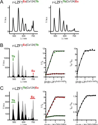
The luminescence properties of r-LZF1Eu|Cs124|Tb and r-LZF1Tb|Cs124|Eu were investigated in HEPES
buffer at pH 7.5. When the antenna is excited at 330 nm, the characteristic emissions of the antenna (Figures S9 and S10 of Supporting Information) as well as Tb3+ and Eu3+ are observed for both peptides. Excitation spectra of Tb3+ (lem = 545 nm) and Eu3+ (lem = 700 nm) confirm that both lanthanides are sensitized by the amide derivative of Cs124. The normalized time-resolved emission spectra (100 µs delay) of the Zn-free peptides are displayed in Figure 4A. In both cases, the terbium emission dominates the europium one, which is not surprising since this antenna is known to sensitize Tb3+ more efficiently than Eu3+.[25] Comparatively, the Eu3+ emission is far less intense and the Tb/Eu emission ratio is larger for r-LZF1Tb|Cs124|Eu vs
r-LZF1Eu|Cs124|Tb (Table 1). In agreement with our design, locating the Eu3+ at the N-terminus, closer to the antenna improves Eu3+ sensitization efficiency and corresponding emission intensity. When r-LZF1Eu|Cs124|Tb
is titrated by Zn2+, Tb3+ emission increases linearly with the amount of Zn2+ up to 1.0 eq. and plateaus above.
Antenna N O S S HN N N N N O O -O O -O -O O Ln1
SEA native chemical ligation MPAA, TCEPphosphate buffer, pH 6-7, 37°C
H2N HS O GKP-Z-ECTKSA GKKFTK-X-EELQKHAKTHTG n N N N N O O -O -O O O -O Ln2 r-LZF1Ln2|Cs124|Ln1 GKP-Z-ECTKSACGKKFTK-X-EELQKHAKTHTG N N N N O O -O -O O O -O Ln2 HN N N N N O O -O O -O -O O Ln1 O NH HN O GKP-Z-ECTKSACGKKFTK-X-EELQKHAKTHTG N N N N O O -O -O O O -O Ln2 HN N N N N O O -O O -O -O O Ln1 4 r-LZF3Ln2|Anthra|Ln1 Z = Glu(Cs124) X = Dap(DOTA-Ln1) Z = Ala(9-Anthryl) X = Lys(DOTA-Ln1)
Zn2+-binding causes an 11-fold increase of Tb3+ emission intensity, similar to the previously described intensiometric probe LZF1Cs124|Tb (13-fold increase).[30] Comparatively, the increase in Eu3+ emission intensity is less affected, i.e. only ca. 30 % increase, and the Eu3+ signal remains very weak in comparison to the one of Tb3+ (Figure 4B and Figure S9 of Supporting Information). Therefore, the evolution of Tb3+ and Eu3+ emissions upon Zn2+ addition differs markedly making r-LZF1Eu|Cs124|Tb a ratiometric probe. Nevertheless, Eu3+ emission is too weak and hardly detectable in comparison to Tb3+, complicating practical use of this probe.
Figure 4: Time-gated luminescence spectroscopy ( ex = 330 nm, delay time = 100 µs, gate time = 2 ms) of visible-emitting ratiometric probes. (A) Normalized emission spectra of r-LZF1Eu|Cs124|Tb (left) and
r-LZF1Tb|Cs124|Eu (right) in their Zn-free forms. (B) Emission spectra of r-LZF1Eu|Cs124|Tb in its Zn-free (black)
and Zn-bound (grey) forms (left) and monitoring of Tb3+ (490 nm, green) and Eu3+ (702 nm, red) channels during a Zn2+ titration of r-LZF1Eu|Cs124|Tb (middle) and evolution of the emission ratio of the Tb3+ channel over the Eu3+ channel (right). (C) Emission spectra of r-LZF1Tb|Cs124|Eu in its Zn-free (black) and Zn-bound
(grey) forms (left) and monitoring of integrated Tb3+ (470-510 nm, green) and Eu3+ (680-720 nm, red) channels during a Zn2+ titration of r-LZF1Eu|Cs124|Tb (middle) and evolution of the emission ratio of the Eu3+ channel over the Tb3+ channel (right). Solutions were prepared with 10 µM r-LZF probe in aerated HEPES buffer (10 mM, pH 7.5) containing TCEP (250 µM).
The overall behaviour of the other peptide LZF1Tb|Cs124|Eu is quite similar (Figure 4C and Figure S10
of Supporting Information) with a linear increase of both Tb3+ and Eu3+ emissions up to 1.0 eq. of Zn2+ but with an 8-fold increase of the Eu3+ emission and a much smaller (1.4-fold) increase of the Tb3+ signal providing a ratiometric response upon binding of one Zn2+ ion. However, compared to r-LZF1Eu|Cs124|Tb, r-LZF1Tb|Cs124|Eu
displays Tb3+ and Eu3+ emissions of comparable intensities allowing practical ratiometric detection of Zn2+ as confirmed by the evolution of the emission ratio of Eu3+ channel around 700 nm over Tb3+ channel around 490 nm (Figure 4C). For both peptides, Tb3+ and Eu3+ luminescence lifetimes for Zn-free probes were ca. 1.93 ±
500 600 700 0 1 λ / nm I / a .u . 500 600 700 0 1 λ / nm I / a .u . Eu 500 600 700 0 500 1000 1500 λ / nm I / a .u . Tb 0 1 2 0 2 4 6 8 10 12 [Zn]/[r-LZF1Eu|Cs124|Tb] I / I0 500 600 700 0 100 200 300 400 λ / nm I / a .u . Tb Eu 0 1 2 0 2 4 6 8 10 [Zn]/[r-LZF1Tb|Cs124|Eu] I / I 0 0 1 2 0.0 0.5 1.0 1.5 [Zn]/[r-LZF1Tb|Cs124|Eu] IEu / ITb A B r-LZF1Eu|Cs124|Tb r-LZF1Tb|Cs124|Eu C r-LZF1Eu|Cs124|Tb r-LZF1Tb|Cs124|Eu 0 1 2 0 2 4 6 8 10 [Zn]/[r-LZF1Eu|Cs124|Tb] ITb / IEu
0.03 ms and 0.65 ± 0.02 ms, respectively, and they remain unchanged for the Zn-bound peptide (Table 1 and Table S2 of Supporting Information). These luminescence lifetime values are typical of DOTA-monoamide Tb3+ and Eu3+ complexes in related mono-lanthanide peptide conjugates.[30,32,40] Complementary luminescence lifetimes measurements on r-LZF1Tb|Cs124|Eu conducted in deuterated water shows that one water molecule is
bound to Tb3+ or Eu3+ for both Zn-free and Zn-bound forms (Figure 17 of Supporting Information). Altogether, this result indicates that the ratiometric behaviour of r-LZF1Eu|Cs124|Tb and r-LZF1Tb|Cs124|Eu is caused only by
the Zn2+-induced peptide folding that shortens the distance between the antenna and the Ln3+ and that it is not due to a change in the Ln3+ hydration number or to a Tb3+-to-Eu3+ energy transfer. Quantum yields of Ln3+ luminescence upon excitation of the Cs124 antenna (Table 1) are rather modest but similar to those of the related probe LZF1Cs124|Tb.[30] Finally, the dissociation constant (Kd) of the Zn·r-LZF1Tb|Cs124|Eu-complex was
measured to be 10-9.2 by competitive Zn2+ titration using EGTA (ethylene glycol-bis(
-aminoethylether)-N,N,N’,N’-tetraacetic acid) as a competitor (Figure S20 and Table S3 of Supporting Information). This value
falls within the suitable range for detection of circulating Zn2+ in cells, where [Zn2+] lies between 10-11 and 10 -8 M.[37,47] Hence, the design of r-LZF1Tb|Cs124|Eu and its spectral behaviour in response to Zn2+ demonstrate that NCL gives access to hetero-bis-lanthanide peptide conjugates that can behave as ratiometric luminescent sensors. The comparison between r-LZF1Eu|Cs124|Tb and r-LZF1Tb|Cs124|Eu clearly indicates the importance of
the location of each lanthanide relative to the antenna to adapt their emission range and to optimize the probe response.
Table 1: Summary of r-LZF1Eu|Cs124|Tb and r-LZF1Tb|Cs124|Eu probes characterizations: dissociation constants,
terbium and europium luminescence enhancements, ratios of terbium over europium emission, quantum yields (FLn) and Ln3+ luminescence lifetimes.
r-LZF1Eu|Cs124|Tb r-LZF1Tb|Cs124|Eu log Kd n.d.a -9.2 ± 0.2 IZn/I0 Tb: 11 Eu: 1.3 Tb: 1.4 Eu: 8 Tb/Eu ratiob Zn-free Zn-bound 2.2 17.8 8.1 1.4 FLn / %c Zn-free Zn-bound 0.13 1.6 0.24 0.60 lifetimes / ms Zn-free Zn-bound Tb: 1.96 Tb: 1.92 Eu: 0.64 Eu: 0.66 Tb: 1.93 Tb: 1.91 Eu:0.66 Eu:0.67
a Not determined. b The Tb/Eu ratio was obtained by deconvolution of the emission spectra using spectra of related Tb- and Eu-DOTA-monoamide compounds, i.e. LZF1Cs124|Tb and LZF1Cs124|Eu.[30]c lex = 330 nm.
Toward NIR-emissive probes: screening of antennas for the sensitization of Nd3+ and Yb3+ luminescence
Besides the Tb3+/Eu3+ pair, other pairs of lanthanides could be used to design ratiometric probes. Of special interest are NIR-emitting lanthanides, neodymium and ytterbium, which emit around 1000 nm (Figure 1). Therefore, our next challenge was the unprecedented design of a ratiometric probe based on these two lanthanides for ratiometric NIR detection. We have previously described that an amino-derivative of 7-nitro-2,1,3-benzoxadiazole (NBD) can sensitize Nd3+ luminescence and that LZF1NBD|Nd featuring a Dap(NBD)
amino acid and a DOTA-Nd3+ complex is a NIR-emitting intensiometric luminescent probe for the detection of Zn2+.[30] Unfortunately, NBD is unable to sensitize Yb3+. Therefore, we looked for antennas that could
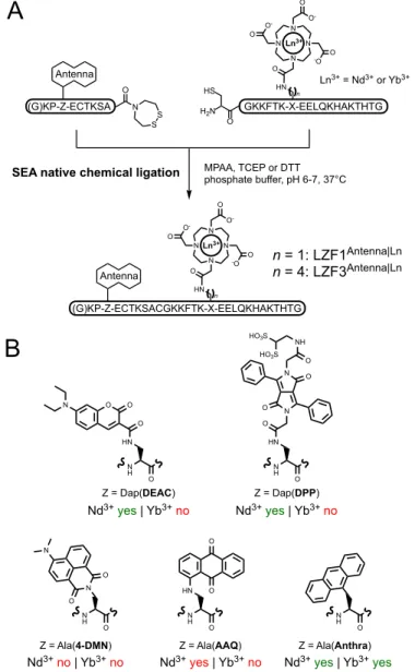
sensitize both Nd3+ and Yb3+. For this purpose, and to limit the synthetic effort, we decided to use once again NCL to perform this screening. We synthesized a series of N-terminal segments bearing a candidate antenna chromophore and two C-terminal segments bearing either a DOTA-Yb3+ or a DOTA-Nd3+ complex (Figure 5A). Note that C-terminal segment with both Dap or Lys anchorage for the DOTA chelator were used in this screening process, giving LZF1 or LZF3 probes, respectively. Indeed, we switched from Dap to Lys residue because the C-terminal segment with Lys anchorage was obtained in better yield. This structural variation has a minor impact on the probe properties as evidenced by comparison of LZF3Cs124|Ln probes (Ln = Tb or Eu)
with the previously described LZF1Cs124|Ln probes[30] (Figure S18 and S19 of Supporting Information). For the implementation of this screening, we assessed the potential of five antennas for the sensitization of NIR-emitting Ln3+ through various chromophores absorbing in the UV or the visible ranges (Figure 5B): 4-(dimethylamino)-1,8-naphthalimide (4-DMN, lmax = 447 nm), 1-amino-anthraquinone (AAQ, lmax = 505 nm), 7-(diethylamino)-coumarin (DEAC, lmax = 433 nm), a water-soluble derivative of 3,6-diphenyl-diketopyrrolopyrrole[48] (DPP, lmax = 455 nm) and anthracene (Anthra, lmax = 369 nm). The experimental procedures for the synthesis of the various LZF1Antenna|Ln or LZF3Antenna|Ln (Ln = Nd and Yb) peptides are
described in the Supporting Information.
Figure 5: Screening of antennas for Nd3+ and Yb3+ sensitization by native chemical ligation. (A) Reaction scheme for the 2-segment peptide assembly by NCL. (B) Structures of antenna amino acids explored in this screening and summary of their ability to sensitize Nd3+ and Yb3+ luminescence.
Antenna N O S S HN N N N N O O -O O -O -O O Ln3+
SEA native chemical ligation MPAA, TCEP or DTTphosphate buffer, pH 6-7, 37°C
H2N HS O (G)KP-Z-ECTKSA GKKFTK-X-EELQKHAKTHTG n (G)KP-Z-ECTKSACGKKFTK-X-EELQKHAKTHTG HN N N N N O O -O O -O -O O Ln3+ Antenna n N H O HN O N N O O O NH HO3S HO3S N H O N O O N N H O HN O O N H O HN N H O Z = Ala(4-DMN) Z = Ala(AAQ) Z = Dap(DEAC) Z = Ala(Anthra) Z = Dap(DPP) n = 1: LZF1Antenna|Ln n = 4: LZF3Antenna|Ln Ln3+ = Nd3+ or Yb3+ Nd3+yes | Yb3+no Nd3+yes | Yb3+no
Nd3+no | Yb3+no Nd3+yes | Yb3+no Nd3+yes | Yb3+yes O O O N
A
B
Ln3+ complexes with a coordinated amido-derivative of anthraquinone have already been described to sensitize both Nd3+ and Yb3+ luminescence in deuterated water[49] and a remote 1,8-diamino-anthraquinone antenna has been shown to sensitize both lanthanides in methanol.[50] However, no sensitized NIR lanthanide luminescence could be detected for LZF1AAQ|Nd or LZF1AAQ|Yb with their amino-anthraquinone chromophore
in buffered water (HEPES, pH 7.5). 4-Amino-1,8-naphthalimide has already been used by one of us as a sensitizing antenna for Nd3+ and Yb3+ in dendrimeric systems[51] but unfortunately, here again, neither Nd3+ nor Yb3+ luminescence could be observed for LFZ34DMN|Ln upon excitation of the chromophore. In contrast,
LZF1DPP|Ln (Figure S11 of Supporting Information) and LFZ3DEAC|Ln (Figure S12 of Supporting Information)
displayed sensitized Nd3+ emission but not the one arising from Yb3+. Upon Zn2+ binding, the Nd3+ emission is increased ca. 3-fold for LZF1DPP|Nd and 7-fold for LFZ3DEAC|Nd, showing that these two peptides can operate
as intensiometric probes for Zn2+ detection in the NIR. The fifth antenna to be assessed was anthracene, which has already been reported to successfully sensitize Nd3+, Yb3+ and Er3+ NIR luminescence in dichloromethane solution or in the solid state.[52,53] Indeed, with LZF3Anthra|Nd and LZF3Anthra|Yb, anthracene was able to sensitize
both lanthanides upon excitation in the 330-400 nm spectral range and both peptides show a weak lanthanide luminescence increase upon zinc binding, i.e. 1.3 and 1.5-fold for Nd3+ and Yb3+, respectively (Figure 6 and Figure S13 and S14 of Supporting Information). The quantum yields of Ln3+ luminescence upon antenna (DPP, DEAC or Anthra) excitation (Table S1 of Supporting Information) are in the same order of magnitude as the one of the previously described LZF1NBD|Nd probe, i.e. 10-4 – 6´10-3 %. From this screening study, it appears that anthracene is a suitable antenna for both Nd3+ and Yb3+ in our peptide-based system. Thus, it appeared to be a promising candidate for the design of a dual-wavelength NIR-emitting ratiometric probes. Therefore, the hetero-bis-lanthanide peptides r-LZF3Yb|Anthra|Nd and r-LZF3Nd|Anthra|Yb (Figure 2B) were assembled by NCL
of a N-terminal segment bearing a DOTA-Ln3+ complex and the anthracene antenna and a C-terminal segment bearing the second DOTA-Ln3+ complex, as described in Figure 3.
Figure 6: (A) Absorption spectrum of the anthracene antenna. (B,C) Excitation (lem = 990 nm for Yb3+ and 1060 nm for Nd3+) and emission (lex = 370 nm) spectra of (B) LZF3Athra|Yb and (C) LZF3Athra|Nd in their
Zn-free (black) and Zn-bound (grey) forms. Solutions were prepared with 50 µM probe in degassed HEPES buffer (10 mM, pH 7.5). 300 350 400 0 5000 10000 λ / nm ε / M -1 c m -1 950 1000 1050 1100 1150 0 5000 10000 λ / nm I / a .u . 300 350 400 λ / nm I / a .u . 300 350 400 λ / nm I / a .u . 1000 1050 1100 1150 1200 0 2000 4000 6000 λ / nm I / a .u .
B
C
LZF3Anthra|Yb LZF3Anthra|NdA
Characterization of ratiometric probes emitting in the NIR
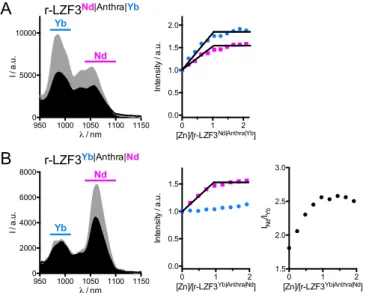
The luminescence properties of r-LZF3Nd|Anthra|Yb and r-LZF3Yb|Anthra|Nd were investigated in degassed
HEPES buffer at pH 7.5. Upon excitation of the anthracene chromophore at 370 nm, both r-LZF3Nd|Anthra|Yb
and r-LZF3Yb|Anthra|Nd display the characteristic emission signals of Yb3+ and Nd3+ (Figure 7) with similar intensities (see Nd/Yb ratio in Table 2). Addition of Zn2+ up to 1.0 eq. leads to an overall emission increase (Figure 7 and Figure S15 and S16 of Supporting Information). In the case of r-LZF3Nd|Anthra|Yb, emission of
both Yb3+ and Nd3+ increase in a similar extent, i.e. 1.8 and 1.5-fold for Yb3+ and Nd3+, respectively, making ratiometric detection difficult (Figure 7A). Conversely, the other peptide, r-LZF3Yb|Anthra|Nd, displays an
increase of the Nd3+ emission only, while the one of Yb3+ remains constant, making the ratiometric detection obvious as seen in Figure 7B. The quantum yields of Ln3+ emission of these two ratiometric probes (Table 2) are comparable to those of LZF3Antra|Nd and LZF3Antra|Yb (Table S1 of Supporting Information). The
dissociation constant (Kd) of the Zn·r-LZF3Yb|Anthra|Nd complex was determined to be 10-10.8 by competitive Zn2+ titration using EGTA (Figure S20 of Supporting Information), in line with the lower Kd for probes containing a Lys(DOTA-Ln) residue with respect to Dap(DOTA-Ln), as observed for LZF1Cs124|Ln vs
LZF3Cs124|Ln probes (Figure S20 and Table S3 of Supporting Information). To the best of our knowledge,
r-LZF3Yb|Anthra|Nd constitute the first proof-of-principle responsive and lanthanide-based ratiometric probe with
dual-wavelength emission in the NIR.
Figure 7: Luminescence spectroscopy (lex = 370 nm) of NIR-emitting ratiometric probes. (A) Emission spectra of r-LZF3Nd|Athra|Yb in its Zn-free (black) and Zn-bound (grey) forms (left) and monitoring of Yb3+ (970-1010 nm, blue) and Nd3+ (1040-1100 nm, pink) channels during a Zn2+ titration of r-LZF3Nd|Anhra|Yb
(right). (B) Emission spectra of r-LZF3Yb|Anthra|Nd in its Zn-free (black) and Zn-bound (grey) forms (left) and
monitoring of integrated Yb3+ (970-1010 nm, blue) and Nd3+ (1040-1100 nm, pink) channels during a Zn2+ titration of r-LZF3Yb|Anhtra|Nd (middle) and evolution of the emission ratio of the Nd3+ channel over the Yb3+ channel. Solutions were prepared with 25 µM probe in degassed HEPES buffer (10 mM, pH 7.5).
950 1000 1050 1100 1150 0 5000 10000 λ / nm I / a .u . Yb Nd 0 1 2 0.0 0.5 1.0 1.5 2.0 [Zn]/[r-LZF3Nd|Anthra|Yb] In te n si ty / a .u . 950 1000 1050 1100 1150 0 2000 4000 6000 8000 λ / nm I / a .u . Yb Nd 0 1 2 0.0 0.5 1.0 1.5 [Zn]/[r-LZF3Yb|Anthra|Nd] In te n si ty / a .u . 0 1 2 1.5 2.0 2.5 3.0 [Zn]/[r-LZF3Yb|Anthra|Nd] INd /IYb A B r-LZF3Nd|Anthra|Yb r-LZF3Yb|Anthra|Nd
Table 2: Summary of r-LZF3Nd|Anthra|Yb and r-LZF3Yb|Anthra|Nd probes characterizations: dissociation constant,
ytterbium and neodymium luminescence enhancement, ratio of neodymium over ytterbium emission in the spectra. r-LZF3Nd|Anthra|Yb r-LZF1Yb|Anthra|Nd log Kd n.d.a -10.8 ± 0.2 IZn/I0 Yb: 1.8 Nd: 1.5 Yb: 1.0 Nd: 1.5 Nd/Yb ratiob Zn-free Zn-bound 0.44 0.33 1.6 2.6 FLn / %c Zn-free Zn-bound 4.4´10-3 6.6´10-3 3.9´10-3 4.7´10-3
a Not determined. b The Nd/Yb ratio was obtained by deconvolution of the emission spectra using spectra of the related Yb- and Nd-DOTA-monoamide compounds LZF3Anthra|Yb and LZF3Anthra|Nd. c
l
ex
= 370 nm.
Conclusions
In summary, we have demonstrated that hetero-bis-lanthanide peptide conjugates site-specifically labeled with two distinct Ln3+ within the same molecule framework can be easily prepared by native chemical ligation of two peptide segments, each bearing a distinct kinetically and thermodynamically-stable DOTA-Ln3+ complex. As a proof-of-concept of this strategy, we have created several zinc-responsive probes with dual-wavelength ratiometric emission in the visible or in the NIR. Among them, r-LZF1Tb|Cs124|Eu and
r-LZF3Yb|Anthra|Nd display ratiometric detection properties in response to the presence of Zn2+ with visible (480-710 nm) and NIR (950-1150 nm) emission signals, respectively. These two probes clearly demonstrate that NCL is an attractive and efficient approach to properly design ratiometric luminescent lanthanide-based probes.
Acknowledgment
Authors acknowledge the Agence Nationale de la Recherche (12-BS07-0012 and
ANR-13-BSV5-009), La Ligue Contre le Cancer, Le Cancéropôle Grand Ouest, La Region Centre and the
Labex ARCANE and CBH-EUR-GS (ANR-17-EURE-0003) for financial support. S.P.
acknowledges support from the Institut National de la Santé et de la Recherche Médicale (INSERM).
A.R. thank the French "Investissements d’Avenir" program, project ISITE-BFC (contract
ANR-15-IDEX-0003), for its financial support for research in the field of DPP-based fluorophores for
biosensing/bioimaging.
References
[1] E. Haustein, P. Schwille, HFSP J. 2007, 1, 169, DOI: 10.2976/1.2778852.
[2] H. Kobayashi, M. Ogawa, R. Alford, P. L. Choyke, Y. Urano, Chem. Rev. 2010, 110, 2620, DOI: 10.1021/cr900263j.
[3] T. Nagaya, Y. A. Nakamura, P. L. Choyke, H. Kobayashi, Front. Oncol. 2017, 7, 314, DOI: 10.3389/fonc.2017.00314.
[4] C. H. F. Wenk, F. Ponce, S. Guillermet, C. Tenaud, D. Boturyn, P. Dumy, D. Watrelot-Virieux, C. Carozzo, V. Josserand, J.-L. Coll, Cancer Lett. 2013, 334, 188, DOI: 10.1016/j.canlet.2012.10.041. [5] T. Ueno, T. Nagano, Nat. Methods 2011, 8, 642, DOI: 10.1038/nmeth.1663.
[6] J. Zhou, H. Ma, Chem. Sci. 2016, 7, 6309, DOI: 10.1039/C6SC02500E.
[7] H. Zhu, J. Fan, J. Du, X. Peng, Acc. Chem. Res. 2016, 49, 2115, DOI: 10.1021/acs.accounts.6b00292. [8] A. Kaur, J. L. Kolanowski, E. J. New, Angew. Chem., Int. Ed. 2016, 55, 1602, DOI:
10.1002/anie.201506353.
[9] D. Andina, J.-C. Leroux, P. Luciani, Chem.-Eur. J. 2017, 23, 13549, DOI: 10.1002/chem.201702458. [10] J. Zhang, X. Chai, X.-P. He, H.-J. Kim, J. Yoon, H. Tian, Chem. Soc. Rev. 2019, 48, 683, DOI:
10.1039/C7CS00907K.
[11] O. Tagit, N. Hildebrandt, ACS Sens. 2017, 2, 31, DOI: 10.1021/acssensors.6b00625.
[12] M. H. Lee, J. S. Kim, J. L. Sessler, Chem. Soc. Rev. 2015, 44, 4185, DOI: 10.1039/C4CS00280F. [13] C. P. Montgomery, B. S. Murray, E. J. New, R. Pal, D. Parker, Accounts Chem. Res. 2009, 42, 925,
DOI: 10.1021/ar800174z.
[14] M. C. Heffern, L. M. Matosziuk, T. J. Meade, Chem. Rev. 2014, 114, 4496, DOI: 10.1021/cr400477t. [15] M. Sy, A. Nonat, N. Hildebrandt, L. J. Charbonnière, Chem. Commun. 2016, 52, 5080, DOI:
10.1039/C6CC00922K.
[16] J.-C. G. Bünzli, J. Lumin. 2016, 170, 866, DOI: 10.1016/j.jlumin.2015.07.033.
[17] K. Y. Zhang, Q. Yu, H. Wei, S. Liu, Q. Zhao, W. Huang, Chem. Rev. 2018, 118, 1770, DOI: 10.1021/acs.chemrev.7b00425.
[18] J.-C. G. Bünzli, S. V. Eliseeva, in ‘Lanthanide Luminescence’, Eds. P. Hänninen, H. Härmä, Springer Berlin Heidelberg, 2011, pp. 1.
[19] Y. Bretonniere, M. J. Cann, D. Parker, R. Slater, Org. Biomol. Chem. 2004, 2, 1624, DOI: 10.1039/B400734B.
[20] R. Pal, D. Parker, Chem. Commun. 2007, 474, DOI: 10.1039/b616665b.
[21] D. G. Smith, B. K. McMahon, R. Pal, D. Parker, Chem. Commun. 2012, 48, 8520, DOI: 10.1039/c2cc34267g.
[22] L. B. Jennings, S. Shuvaev, M. A. Fox, R. Pal, D. Parker, Dalton Trans. 2018, 47, 16145, DOI: 10.1039/C8DT03823F.
[23] S. J. Butler, Chem. Commun. 2015, 51, 10879, DOI: 10.1039/C5CC03428K.
[24] E. A. Weitz, J. Y. Chang, A. H. Rosenfield, E. A. Morrow, V. C. Pierre, Chem. Sci. 2013, 4, 4052, DOI: 10.1039/C3SC51583D.
[25] M. S. Tremblay, M. Halim, D. Sames, J. Am. Chem. Soc. 2007, 129, 7570, DOI: 10.1021/ja070867y. [26] G.-L. Law, R. Pal, L. O. Palsson, D. Parker, K.-L. Wong, Chem. Commun. 2009, 7321, DOI:
10.1039/b920222f.
[27] D. G. Smith, R. Pal, D. Parker, Chem.-Eur. J. 2012, 18, 11604, DOI: 10.1002/chem.201201738. [28] C. Song, Z. Ye, G. Wang, J. Yuan, Y. Guan, Chem.-Eur. J. 2010, 16, 6464, DOI:
10.1002/chem.201000528.
[29] Z. Dai, L. Tian, B. Song, Z. Ye, X. Liu, J. Yuan, Anal. Chem. 2014, 86, 11883, DOI: 10.1021/ac503611f.
[30] M. Isaac, L. Raibaut, C. Cepeda, A. Roux, D. Boturyn, S. V. Eliseeva, S. Petoud, O. Sénèque,
Chem.-Eur. J. 2017, 23, 10992, DOI: 10.1002/chem.201703089.
[31] M. S. Tremblay, D. Sames, Chem. Commun. 2006, 4116, DOI: 10.1039/b607949k.
[32] T. J. Sørensen, A. M. Kenwright, S. Faulkner, Chem. Sci. 2015, 6, 2054, DOI: 10.1039/C4SC03827D. [33] P. E. Dawson, T. W. Muir, I. Clarklewis, S. B. H. Kent, Science 1994, 266, 776, DOI:
10.1126/science.7973629.
[34] V. Agouridas, O. El Mahdi, V. Diemer, M. Cargoët, J.-C. M. Monbaliu, O. Melnyk, Chem. Rev. 2019,
119, 7328, DOI: 10.1021/acs.chemrev.8b00712.
[35] W. Maret, Met. Ions Life Sci. 2013, 12, 479, DOI: 10.1007/978-94-007-5561-1_14.
[36] K. P. Carter, A. M. Young, A. E. Palmer, Chem. Rev. 2014, 114, 4564, DOI: 10.1021/cr400546e. [37] A. M. Hessels, M. Merkx, Metallomics 2015, 7, 258, DOI: 10.1039/C4MT00179F.
[38] Y. Chen, Y. Bai, Z. Han, W. He, Z. Guo, Chem. Soc. Rev. 2015, 44, 4517, DOI: 10.1039/C5CS00005J. [39] S.-H. Park, N. Kwon, J.-H. Lee, J. Yoon, I. Shin, Chem. Soc. Rev. 2020, 49, 143, DOI:
10.1039/C9CS00243J.
[40] M. Isaac, A. Pallier, F. Szeremeta, P.-A. Bayle, L. Barantin, C. S. Bonnet, O. Sénèque, Chem. Commun. 2018, 54, 7350, DOI: 10.1039/C8CC04366C.
[41] M. Isaac, S. A. Denisov, A. Roux, D. Imbert, G. Jonusauskas, N. D. McClenaghan, O. Sénèque, Angew.
Chem., Int. Ed. 2015, 54, 11453, DOI: 10.1002/anie.201505733.
[42] A. Roux, M. Isaac, V. Chabert, S. A. Denisov, N. D. McClenaghan, O. Sénèque, Org. Biomol. Chem. 2018, 16, 5626, DOI: 10.1039/C8OB01044G.
Chem. Sci. 2017, 8, 1658, DOI: 10.1039/C6SC04086A.
[44] M. Li, P. R. Selvin, J. Am. Chem. Soc. 1995, 117, 8132, DOI: 10.1021/ja00136a010. [45] G. S. Beligere, P. E. Dawson, Peptide Sci. 1999, 51, 363.
[46] S. Futaki, K. Tatsuto, Y. Shiraishi, Y. Sugiura, Peptide Sci. 2004, 76, 98, DOI: 10.1002/bip.10562. [47] W. Maret, Metallomics 2015, 7, 202, DOI: 10.1039/C4MT00230J.
[48] E. Heyer, P. Lory, J. Leprince, M. Moreau, A. Romieu, M. Guardigli, A. Roda, R. Ziessel, Angew.
Chem., Int. Ed. 2015, 54, 2995, DOI: 10.1002/anie.201411274.
[49] J. E. Jones, S. J. A. Pope, Dalton Trans. 2009, 8421, DOI: 10.1039/b913902h.
[50] O. J. Stacey, B. D. Ward, A. J. Amoroso, S. J. A. Pope, Dalton Trans. 2016, 45, 6674, DOI: 10.1039/C5DT04351D.
[51] A. Foucault-Collet, Ph.D. Thesis Université d’Orléans, Orléans, 2013.
[52] T. Lazarides, M. A. H. Alamiry, H. Adams, S. J. A. Pope, S. Faulkner, J. A. Weinstein, M. D. Ward,
Dalton Trans. 2007, 1484, DOI: 10.1039/b700714k.
[53] B. Casanovas, S. Speed, O. Maury, M. S. E. Fallah, M. Font-Bardía, R. Vicente, Eur. J. Inorg. Chem. 2018, 2018, 3859, DOI: 10.1002/ejic.201800624.
Using Native Chemical Ligation for Site-specific Synthesis of
Hetero-bis-lanthanide Peptide Conjugates: Application to Ratiometric Visible
or Near-infrared Detection of Zn
2+Céline Cepeda,ab Laurent Raibaut,a Guillaume Fremy,ab Svetlana V. Eliseeva,c Anthony Romieu,d
Jacques Pécaut,e Didier Boturyn,b Stéphane Petoud,*c and Olivier Sénèque*a
aUniv. Grenoble Alpes, CNRS, CEA, IRIG, LCBM (UMR 5249), F-38000 Grenoble, France. b Univ. Grenoble Alpes, CNRS, DCM (UMR 5250) F-38000 Grenoble, France.
cCentre de Biophysique Moléculaire, UPR CNRS 4301, Université d’Orléans, rue Charles Sadron, F-45071 Orléans, France.
d ICMUB, UMR 6302, CNRS, Univ. Bourgogne Franche-Comté, 9 Avenue Alain Savary, 21000 Dijon, France.
eUniv. Grenoble Alpes, CEA, CNRS, IRIG, SyMMES, F-38000 Grenoble, France.
Email: olivier.seneque@cea.fr and stephane.petoud@inserm.fr
Supporting Information
Content
Abbreviations ………2
Materials and methods ………. 2
Amino acid synthesis ………3
Peptide sequences ………. 4
Peptide synthesis ………...………6
ESI-MS monitoring of the absence of Ln3+ scrambling ………..14
Luminescence spectroscopy ………14
Influence of Dap vs Lys anchorage for DOTA (LZF1 vs LZF3) ………20
Determination of Zn2+ binding constants ………21
Abbreviations
AAQ: 1-aminoanthraquinone; AcOH: acetic acid; AcOEt: ethyl acetate; Alloc: allyloxycarbonyl; Boc: tert-butyloxycarbonyl; Cs124: carbostyril 124; Dap: diaminopropionic acid; DCM: dicholoromethane; DEAC: 7-dimethylamino-coumarin-3-carboxy; diamide: N,N,N′,N′-tetramethylazodicarboxamide; DIEA: N,N-diisopropyl-ethylamine; DOTA: 1,4,7,10-tetraazacyclododecane-1,4,7,10-tetraacetic acid; DMF: N,N-dimethylformamide; 4-DMN:
N,N’-dimethyl-1,8-naphthalimido; DMSO: dimethylsulfoxide; DTT: dithiothreitol; EDTA, ethylenediamine-tetraacetic
acid; EGTA: ethylene glycol-bis(2-aminoethylether)-N,N,N',N'-tetraacetic acid; ESI: electrospray ionization; Et2O:
diethylether; Fmoc: 9-fluorenyl-methoxycarbonyl; ; Fmoc-OSu: Fmoc hydroxysuccinimide ester; HATU: [(dimethylamino)-1H-1,2,3-triazolo[4,5-b]-pyridin-1-ylmethylene]-methylmethanaminium hexafluoro-phosphate N-oxide; HEDTA: N-hydroxyethyl-ethylenediamine-triacetic acid; HPLC: high performance liquid chromatography; LRMS: low resolution mass spectrometry; MeCN: acetonitrile; MPAA: 4-mercaptophenylacetic acid; NH4OAc:
ammonium acetate; Pd(PPh3)4: tetrakis(triphenylphosphine)palladium(0); PyBOP:
(benzotriazol-1-yloxy)tripyrrolidinophosphonium-hexafluoro-phosphate; SEA: bis(2-sulfanyl-ethyl)amino; SEA-PS: (bis(2-sulfanyl-ethyl)-amino)-2-chlorotrityl-polystyrene; TCEP: tris(2-carboxyethyl)phosphine; TFA: trifluoroacetic acid; TIS: triisopropylsilane; tBu: tert-butyl; Trt: trityl; UV-Vis: ultraviolet-visible.
Materials and methods
Reagents and solvents: N-α-Fmoc-protected amino acids for peptide synthesis, PyBOP and HATU coupling reagents
were obtained from Novabiochem or Iris Biotech. Fmoc-L-Glu(Cs124)-OH and Fmoc-L-Ala(4-DMN)-OH and disulfonated 3,6-diphenyl-DPP dye (abbreviated as DPP-OH in this article) were synthetized according to literature procedures.[1–3] Fmoc-3-(9-anthryl)-L-Ala-OH was purchased from Santa Cruz Biotechnology. SEA-PS and NovaPEG
Rink Amide resin were purchased from X’prochem and Novabiochem, respectively. DOTA-tris(tBu)ester was purchased from CheMatech. Other reagents for peptide synthesis, solvents, buffers and metal salts were purchased from Sigma-Aldrich. All buffer or metal solutions for spectroscopic measurements were prepared with ultrapure water produced by a Millipore Milli-Q® purification system (purified to 18.2 MW.cm). The concentration of the Zn2+ solution was determined
by colorimetric EDTA titrations.[4] Buffer solutions were treated with Chelex 100 resin (Bio-Rad) to remove trace metal
ions.
Characterization and purification: Analytical HPLC separations were performed on an Agilent Infinity 1260 system
using Merck Chromolith RP-18e (100 mm ´ 4.6 mm) columns at a flow rate of 2 mL/min. Preparative HPLC separations were performed on a VWR LaPrepS system using Waters XBridge Peptide BEH130 C18 (5 µm, 150 mm ´ 19 mm) or Waters XBridge Peptide BEH130 C18 (5 µm, 150 mm ´ 10 mm) columns at flow rates of 14 or 6 mL/min, respectively. Mobile phase consisted in a gradient of solvent A (0.1% TFA in H2O) and B (0.1% TFA in MeCN/H2O 9:1). For analytical
separations, Method A consisted in 5% B during 1 min followed by a 5 to 50 % B linear gradient in 14 min at 2 mL/min and Method B consisted in 5% B during 1 min followed by a 5 to 100 % B gradient in 14 min at 2 mL/min. Eluate was monitored by electronic absorption at 214, 280 and 331 nm. ESI-MS analyses were performed on a Thermo LXQ spectrometer. UV-Vis absorption spectra were recorded on a Perkin-Elmer Lambda 35 spectrophotometer equipped with a thermo-regulated cell holder.
Amino acid synthesis
N-a-Boc-3-(1-anthraquinone-amino)-L-Ala-OH: To a solution of Boc-Dap-OH (500 mg, 2.45 mmol) and DIEA (1.9
mL, 11 mmol) in H2O/DMF 1:1 (36 mL) was added 1-chloroanthraquinone (297 mg, 1.2 mmol) and CuSO4 (586 mg,
3.7 mmol). The reaction mixture was stirred for 24 h on reflux. The solvents were removed under reduced pressure and the oily residue was dissolved in DCM, washed twice with 0.01 M aq. HCl, then twice with 1 mM aq. NaOH. DCM was added to the basic aqueous phases and 0.1 M aq. HCl was added to adjust the pH of the aqueous layer to 3. The organic layer was separated, dried over anhydrous Na2SO4 and evaporated under reduced pressure to give the desired product as
a red solid (27 mg, 0.07 mmol, 5.6 %). 1H NMR (200 MHz, 298 K, CDCl
3): d = 9.89 (s, 1H), 8.14 (m, 2H), 7.66 (t, 2H),
7.56-7.44 (m, 2H), 7.16 (d, J = 7.5 Hz, 1H), 5.60 (d, J = 7.6 Hz, 1H), 4.66 (m, 1H), 3.83 (br, 2H), 1.45 (s, 9H) ppm; HPLC (anal.): tR = 9.8 min (method B); LRMS (ESI+): monoisotopic m/z = 411.0 (+) / calculated monoisotopic m/z = 411.16
[M+H]+ for M = C22H22N2O6.
N-a-Fmoc-3-(1-anthraquinone-amino)-L-Ala-OH: Boc-3-(1-anthraquinone-amino)-L-Ala-OH (58 mg, 140 µmol)
was dissolved in DCM/TFA 1:1 (10 mL) and stirred at room temperature for 2 h. The solvents were removed under reduced pressure. The residue was triturated in Et2O and the supernatant was removed (three times). The solid was dried
under vacuum. It was then dissolved in H2O (3 mL) with K2CO3 (29 mg, 275 µmol). Then a solution of Fmoc-OSu (70
mg, 185 µmol) in 1,4-dioxane (6 mL) was added. After 30 minutes on stirring, H2O (25 mL) was added and the aqueous
solution was washed twice with Et2O (150 mL). The aqueous layer was acidified to pH 3 with 2 M aq. HCl and extracted
with AcOEt. The organic layer was dried over anhydrous Na2SO4 and evaporated under reduced pressure. The residue
was sequentially triturated in toluene, Et2O and acetone to give the desired N-Fmoc amino acid as a red solid (78 mg, 146
µmol, 78 %). 1H NMR (400 MHz, 298 K, acetone-d6): d = 10.01 (br t, 1H), 8.17 (m, 2H), 7.86-7.74 (m, 4H), 7.73-7.61
(m, 3H), 7.55 (d, J = 6.9 Hz, 1H), 7.46-731 (m, 3H), 7.26 (t, J = 6.9 Hz, 2H), 7.10 (d, J = 7.0 Hz, 1H), 4.67 (m, 1H), 4.45-4.17 (m, 3H), 4.05 (m, 1H), 3.87 (m, 1H) ppm; 13C NMR (100 MHz, 298 K, acetone-d6): d = 185.6, 183.8, 172.0, 157.2,
157.1, 152.4, 145.1, 145.0, 142.1, 136.4, 135.7, 135.6, 134.1, 133.8, 128.5, 127.9, 127.5, 127.2, 126.2, 126.1, 120.7, 118.9, 116.5, 114.3, 67.5, 54.4, 48.0, 44.5 ppm; HPLC (anal.): tR = 10.4 min (method B); LRMS (ESI–): monoisotopic
m/z = 531.1 (–) / calculated monoisotopic m/z = 531.16 [M–H]– for M = C
Peptide sequences
1aCs124:[5] Ac-Gly-Lys-Pro-Glu(Cs124)-Glu-Cys(StBu)-Thr-Lys-Ser-Ala-SEAoff 1aDEAC: Ac-Lys-Pro-Dap(DEAC)-Glu-Cys(StBu)-Thr-Lys-Ser-Ala-SEAoff 1aDPP: Ac-Lys-Pro-Dap(DPP)-Glu-Cys(StBu)-Thr-Lys-Ser-Ala-SEAoff 1aDMN: Ac-Lys-Pro-Ala(4-DMN)-Glu-Cys(StBu)-Thr-Lys-Ser-Ala-SEAoff 1aAAQ: Ac-Lys-Pro-Ala(AAQ)-Glu-Cys(StBu)-Thr-Lys-Ser-Ala-SEAoff
1aAnthra: Ac-Gly-Lys-Pro-Ala(9-anthryl)-Glu-Cys(StBu)-Thr-Lys-Ser-Ala-SEAoff 1bCs124: DOTA-Gly-Lys-Pro-Glu(Cs124)-Glu-Cys(StBu)-Thr-Lys-Ser-Ala-SEAoff 1bTb|Cs124: DOTA[Tb]-Gly-Lys-Pro-Glu(Cs124)-Glu-Cys(StBu)-Thr-Lys-Ser-Ala-SEAoff 1bEu|Cs124: DOTA[Eu]-Gly-Lys-Pro-Glu(Cs124)-Glu-Cys(StBu)-Thr-Lys-Ser-Ala-SEAoff 1bAnthra: DOTA-Gly-Lys-Pro-Ala(9-anthryl)-Glu-Cys(StBu)-Thr-Lys-Ser-Ala-SEAoff 1bNd|Anthra: DOTA[Nd]-Gly-Lys-Pro-Ala(9-anthryl)-Glu-Cys(StBu)-Thr-Lys-Ser-Ala-SEAoff 1bYb|Anthra: DOTA[Yb]-Gly-Lys-Pro-Ala(9-anthryl)-Glu-Cys(StBu)-Thr-Lys-Ser-Ala-SEAoff
2a:[5] Cys-Gly-Lys-Lys-Phe-Thr-Lys-Dap(DOTA)-Glu-Glu-Leu-Gln-Lys-His-Ala-Lys-Thr-His-Thr-Gly-NH2 2aTb:[5] Cys-Gly-Lys-Lys-Phe-Thr-Lys-Dap(DOTA[Tb])-Glu-Glu-Leu-Gln-Lys-His-Ala-Lys-Thr-His-Thr-Gly-NH 2 2aEu:[5] Cys-Gly-Lys-Lys-Phe-Thr-Lys-Dap(DOTA[Eu])-Glu-Glu-Leu-Gln-Lys-His-Ala-Lys-Thr-His-Thr-Gly-NH2 2aNd:[5] Cys-Gly-Lys-Lys-Phe-Thr-Lys-Dap(DOTA[Nd])-Glu-Glu-Leu-Gln-Lys-His-Ala-Lys-Thr-His-Thr-Gly-NH 2 2aYb: Cys-Gly-Lys-Lys-Phe-Thr-Lys-Dap(DOTA[Yb])-Glu-Glu-Leu-Gln-Lys-His-Ala-Lys-Thr-His-Thr-Gly-NH2 2b: Cys-Gly-Lys-Lys-Phe-Thr-Lys-Lys(DOTA)-Glu-Glu-Leu-Gln-Lys-His-Ala-Lys-Thr-His-Thr-Gly-NH2 2bTb: Cys-Gly-Lys-Lys-Phe-Thr-Lys-Lys(DOTA[Tb])-Glu-Glu-Leu-Gln-Lys-His-Ala-Lys-Thr-His-Thr-Gly-NH2 2bEu: Cys-Gly-Lys-Lys-Phe-Thr-Lys-Lys(DOTA[Eu])-Glu-Glu-Leu-Gln-Lys-His-Ala-Lys-Thr-His-Thr-Gly-NH 2 2bNd: Cys-Gly-Lys-Lys-Phe-Thr-Lys-Lys(DOTA[Nd])-Glu-Glu-Leu-Gln-Lys-His-Ala-Lys-Thr-His-Thr-Gly-NH2 2bYb: Cys-Gly-Lys-Lys-Phe-Thr-Lys-Lys(DOTA[Yb])-Glu-Glu-Leu-Gln-Lys-His-Ala-Lys-Thr-His-Thr-Gly-NH 2 LZF3Cs124|Tb: Ac-Gly-Lys-Pro-Glu(Cs124)-Glu-Cys-Thr-Lys-Ser-Ala-Cys-Gly-Lys-Lys-Phe-Thr-Lys-Lys(DOTA[Tb])-Glu-Glu-Leu-Gln-Lys-His-Ala-Lys-Thr-His-Thr-Gly-NH2 LZF3Cs124|Eu: Ac-Gly-Lys-Pro-Glu(Cs124)-Glu-Cys-Thr-Lys-Ser-Ala-Cys-Gly-Lys-Lys-Phe-Thr-Lys-Lys(DOTA[Eu])-Glu-Glu-Leu-Gln-Lys-His-Ala-Lys-Thr-His-Thr-Gly-NH2 LZF1AAQ|Nd: Ac-Lys-Pro-Ala(AAQ)-Glu-Cys-Thr-Lys-Ser-Ala-Cys-Gly-Lys-Lys-Phe-Thr-Lys-Dap(DOTA[Nd])-Glu-Glu-Leu-Gln-Lys-His-Ala-Lys-Thr-His-Thr-Gly-NH2 LZF1AAQ|Yb: Ac-Lys-Pro-Ala(AAQ)-Glu-Cys-Thr-Lys-Ser-Ala-Cys-Gly-Lys-Lys-Phe-Thr-Lys-Dap(DOTA[Yb])-Glu-Glu-Leu-Gln-Lys-His-Ala-Lys-Thr-His-Thr-Gly-NH2 LZF1DPP|Nd: Ac-Lys-Pro-Dap(DPP)-Glu-Cys-Thr-Lys-Ser-Ala-Cys-Gly-Lys-Lys-Phe-Thr-Lys-Dap(DOTA[Nd])-Glu-Glu-Leu-Gln-Lys-His-Ala-Lys-Thr-His-Thr-Gly-NH2 LZF1DPP|Yb: Ac-Lys-Pro-Dap(DPP)-Glu-Cys-Thr-Lys-Ser-Ala-Cys-Gly-Lys-Lys-Phe-Thr-Lys-Dap(DOTA[Yb])-Glu-Glu-Leu-Gln-Lys-His-Ala-Lys-Thr-His-Thr-Gly-NH2 LZF3DEAC|Nd: Ac-Lys-Pro-Dap(DEAC)-Glu-Cys-Thr-Lys-Ser-Ala-Cys-Gly-Lys-Lys-Phe-Thr-Lys-Lys(DOTA[Nd])-Glu-Glu-Leu-Gln-Lys-His-Ala-Lys-Thr-His-Thr-Gly-NH2 LZF3DEAC|Yb: Ac-Lys-Pro-Dap(DEAC)-Glu-Cys-Thr-Lys-Ser-Ala-Cys-Gly-Lys-Lys-Phe-Thr-Lys-Lys(DOTA[Yb])-Glu-Glu-Leu-Gln-Lys-His-Ala-Lys-Thr-His-Thr-Gly-NH2 LZF3DMN|Nd: Ac-Lys-Pro-Ala(4-DMN)-Glu-Cys-Thr-Lys-Ser-Ala-Cys-Gly-Lys-Lys-Phe-Thr-Lys-Lys(DOTA[Nd])-Glu-Glu-Leu-Gln-Lys-His-Ala-Lys-Thr-His-Thr-Gly-NH2 LZF3DMN|Yb: Ac-Lys-Pro-Ala(4-DMN)-Glu-Cys-Thr-Lys-Ser-Ala-Cys-Gly-Lys-Lys-Phe-Thr-Lys-Lys(DOTA[Yb])-Glu-Glu-Leu-Gln-Lys-His-Ala-Lys-Thr-His-Thr-Gly-NH2
LZF3Anthra|Nd: Ac-Gly-Lys-Pro-Ala(9-anthryl)-Glu-Cys-Thr-Lys-Ser-Ala-Cys-Gly-Lys-Lys-Phe-Thr-Lys-Lys(DOTA[Nd])-Glu-Glu-Leu-Gln-Lys-His-Ala-Lys-Thr-His-Thr-Gly-NH2 LZF3Anthra|Yb: Ac-Gly-Lys-Pro-Ala(9-anthryl)-Glu-Cys-Thr-Lys-Ser-Ala-Cys-Gly-Lys-Lys-Phe-Thr-Lys-Lys(DOTA[Yb])-Glu-Glu-Leu-Gln-Lys-His-Ala-Lys-Thr-His-Thr-Gly-NH2 r-LZF1Eu|Cs124|Tb: DOTA[Eu]-Gly-Lys-Pro-Glu(Cs124)-Glu-Cys-Thr-Lys-Ser-Ala-Cys-Gly-Lys-Lys-Phe-Thr-Lys-Dap(DOTA[Tb])-Glu-Glu-Leu-Gln-Lys-His-Ala-Lys-Thr-His-Thr-Gly-NH2 r-LZF1Tb|Cs124|Eu: DOTA[Tb]-Gly-Lys-Pro-Glu(Cs124)-Glu-Cys-Thr-Lys-Ser-Ala-Cys-Gly-Lys-Lys-Phe-Thr-Lys-Dap(DOTA[Eu])-Glu-Glu-Leu-Gln-Lys-His-Ala-Lys-Thr-His-Thr-Gly-NH2 r-LZF3Yb|Anthra|Nd: DOTA[Yb]-Gly-Lys-Pro-Ala(9-anthryl)-Glu-Cys-Thr-Lys-Ser-Ala-Cys-Gly-Lys-Lys-Phe-Thr-Lys-Lys(DOTA[Nd])-Glu-Glu-Leu-Gln-Lys-His-Ala-Lys-Thr-His-Thr-Gly-NH2 r-LZF3Nd|Anthra|Yb: DOTA[Nd]-Gly-Lys-Pro-Ala(9-anthryl)-Glu-Cys-Thr-Lys-Ser-Ala-Cys-Gly-Lys-Lys-Phe-Thr-Lys-Lys(DOTA[Yb])-Glu-Glu-Leu-Gln-Lys-His-Ala-Lys-Thr-His-Thr-Gly-NH2
Peptide synthesis
Peptide elongation: Peptide elongation was performed using standard SPPS procedure using Fmoc/tBu chemistry either
manually or on an automated peptide synthesizer (CEM Liberty1 Microwave Peptide Synthesizer). Double couplings (30 min) were performed using 4-fold molar excess of Fmoc-L-amino acid, 4-fold molar excess of PyBOP and 8-fold molar excess of DIEA at room temperature. A capping step was performed after each coupling with Ac2O/DIEA in DMF (5
min). Fmoc removal was performed using 20% piperidine in DMF (2×10 min).
SEAoff N-terminal segments 1:
Figure S1. Synthetic pathway for the preparation of 1aCs124 and 1bLn|Cs124 (Ln = Tb or Eu). * denotes standard side chain
protecting group (Boc for Lys; tBu for Glu, Thr and Ser).
1aCs124: The synthesis was performed as described previously.[5]
1bCs124: Peptide elongation was performed as described above on SEA-PS resin (0.025 mmol, 0.16 mmol/g) after
attachment of the first amino acid by double manual coupling (30 min) using 10-fold excess of Fmoc-Ala-OH, 9.5-fold excess of HATU and 10-fold excess of DIEA in DMF with pre-activation (5 min) followed by acetylation using Ac2O/DIEA/DCM (2:1:17 v/v/v, 10 mL, 2×5 min).[6] Non-standard Fmoc-L-Cys(StBu)-OH amino acid was used to
introduce the cysteine residue. Fmoc-L-Glu(Cs124)-OH[1] was used to introduce the antenna. DOTA-tris(tBu)ester (3 eq.)
was coupled overnight to the N-terminus using PyBOP (3 eq.) activation with DIEA (6 eq.) in DMF (5 mL). Removal of acid-labile side chain protecting groups and cleavage of the peptidyl resin was performed with TFA/H2O/TIS/thioanisole
(92.5:2.5:2.5:2.5 v/v/v/v, 10 mL) during 2 h. The peptide was then precipitated in ice-cold Et2O/heptane (1:1 v/v, 100
mL), dissolved in H2O, and lyophilized. The peptide was dissolved in H2O/AcOH and treated with a solution of I2 (200
mM in DMSO) to oxidize the C-terminal SEAon group into SEAoff group.[6,7] After 30 s, DTT (65 mM in H2O, 500 µL)
was added to quench the excess of iodine. The oxidized peptide was immediately purified by HPLC to give fragment 1b
N S S Trt Trt Fmoc/tBu SPPS Fmoc-Ala G K P E C T K S A N StBu S O HN HN O E Ac G K P E C T K S A N S S StBu S O HN HN O E 1) Ac2O, pyridine, DMF 2) TFA/TIS/H2O/thioanisole 3) I2, DMSO, H2O/AcOH S S Trt Trt N N N N O OH O HO O OH O G K P E C T K S A N S S StBu S O HN HN O E N N N N O O – O –O O O– O G K P E C T K S A N S S StBu S O HN HN O E
1) DOTA-tris(tBu)ester, PyBOP, DIEA, DMF 2) TFA/TIS/H2O/thioanisole 3) I2, DMSO, H2O/AcOH LnCl3, H2O pH 6.2 Ln3+ 1bLn|Cs124 Ln = Tb or Eu 1bCs124 1aCs124 * * * * *
as a powder (10 mg, 21 % yield for the 1bCs124×(TFA)2 salt). HPLC (anal.): tR = 10.3 min (method A); LRMS (ESI+):
monoisotopic m/z = 1797.7 (+), 898.9 (2+), 599.8 (3+) / calculated monoisotopic m/z = 1796.78 [M+H]+, 898.90
[M+2H]2+, 599.60 [M+3H]3+ for M = C76H121N19O23S4.
Formation of Ln3+ complexes 1bLn|Cs124 (Ln = Tb, Eu): Compound 1bCs124 (2.5 µmol, 5 mg) was dissolved in H2O and
the pH was adjusted to 6.2 using NaOH. Then, the lanthanide salt LnCl3 (10 µmol) was added. The solution was stirred
overnight under argon (after 1h, the pH was controlled and adjusted to 6.2 if needed). TCEP (35 µmol, 10 mg) was added prior to removal of excess Ln3+ by HPLC purification. The Ln-loaded peptide was obtained as a white powder after
freeze-drying (85-90% yield).
1bTb|Cs124: HPLC (anal.): t
R = 9.8 min (method A); LRMS (ESI+): average m/z = 1953.5 (+), 977.3 (2+), 651.9 (3+) /
calculated av. m/z = 1954.06 [M+H]+, 977.54 [M+2H]2+, 652.03 [M+3H]3+ for M = C76H118N19O23S4Tb). 1bEu|Cs124: HPLC (anal.): t
R = 9.8 min (method A); LRMS (ESI+): average m/z = 1946.5 (+), 974.1 (2+), 649.8 (3+) /
calculated av. m/z = 1947.10 [M+H]+, 974.06 [M+2H]2+, 649.71 [M+3H]3+ for M = C76H118N19O23S4Eu).
Figure S2. Synthetic pathway for the preparation of 1aDEAC and 1aDPP. * denotes standard side chain protecting group
(Boc for Lys; tBu for Glu, Thr and Ser).
1aDEAC: Peptide elongation was performed as described above on SEA-PS resin (0.1 mmol, 0.16 mmol/g) after attachment
of the first amino acid by double manual coupling (30 min) using 10-fold excess of Fmoc-L-Ala-OH, 9.5-fold excess of HATU and 10-fold excess of DIEA in DMF with pre-activation (5 min) followed by acetylation using Ac2O/DIEA/DCM
(2:1:17 v/v/v, 10 mL, 2×5 min).[6] Fmoc-L-Dap(Alloc)-OH and Fmoc-L-Cys(StBu)-OH amino acids were used to
introduce the diaminopropionic acid and cysteine residues, respectively. After acetylation of the N-terminus, removal of
N S S Trt Trt Fmoc/tBu SPPS Fmoc-Ala StBu S 1aDPP TFA/TIS/H2O/thioanisole 1) Pd(PPh3)4,PhSiH3, DCM
2) DPP-OH, PyBOP, DIEA, DMF
Ac K P Dap C T K S A StBu S HN Alloc N S S Trt Trt Trt Trt StBu S * * * * * * * * HN O N N O O NH O HO3S SO3H HN O N N O O NH O HO3S SO3H OH O N N O O HN O HO3S HO3S DPP-OH = E Ac K P Dap C T K S A N S S E Ac K P Dap C T K S A N SH SH E StBu S 1aDEAC 1) TFA/TIS/H2O/thioanisole 2) diamide, Pi 100mM pH 7.5 Trt Trt StBu S * * * * Ac K P Dap C T K S A N S S E Ac K P Dap C T K S AN S S E HN O O O N HN O O O N 1) Pd(PPh3)4,PhSiH3, DCM
2) DEAC-OH, PyBOP, DIEA, DMF
DEAC-OH =
OH O O O N