HAL Id: hal-02164409
https://hal.archives-ouvertes.fr/hal-02164409
Submitted on 25 Jun 2019
HAL is a multi-disciplinary open access
archive for the deposit and dissemination of
sci-entific research documents, whether they are
pub-lished or not. The documents may come from
teaching and research institutions in France or
abroad, or from public or private research centers.
L’archive ouverte pluridisciplinaire HAL, est
destinée au dépôt et à la diffusion de documents
scientifiques de niveau recherche, publiés ou non,
émanant des établissements d’enseignement et de
recherche français ou étrangers, des laboratoires
publics ou privés.
nanoparticles into membranes of well-defined
poly(ethylene oxide)-block-poly(ϵ-caprolactone)
nanoscale magnetovesicles as ultrasensitive MRI probes
of membrane bio-degradation
Adeline Hannecart, Dimitri Stanicki, Luce Vander Elst, Robert N Muller,
Annie Brûlet, Olivier Sandre, Christophe Schatz, Sébastien Lecommandoux,
Sophie Laurent
To cite this version:
Adeline Hannecart, Dimitri Stanicki, Luce Vander Elst, Robert N Muller, Annie Brûlet, et al..
Em-bedding of superparamagnetic iron oxide nanoparticles into membranes of well-defined poly(ethylene
oxide)-block-poly(ϵ-caprolactone) nanoscale magnetovesicles as ultrasensitive MRI probes of
mem-brane bio-degradation. Journal of materials chemistry B, Royal Society of Chemistry, 2019, 7 (30),
pp.4692-4705. �10.1039/C9TB00909D�. �hal-02164409�
RTICL
ARTICLE
Received 7th May 2019, Accepted 18th June 2019 DOI: 10.1039/C9TB00909D
www.rsc.org/
Embedding of superparamagnetic iron oxide nanoparticles into
membranes of well-defined poly(ethylene
oxide)-block-poly(ε-caprolactone) nanoscale magnetovesicles as ultrasensitive MRI
probes of membrane bio-degradation
Adeline Hannecart,
aDimitri Stanicki,
aLuce Vander Elst,
aRobert N. Muller,
a,bAnnie Brûlet
c, Olivier
Sandre,
dChristophe Schatz,
dSébastien Lecommandoux,
dand Sophie Laurent
*a,bThe present study reports the preparation of poly(ethylene oxide)-block-poly(ε-caprolactone) (PEO-b-PCL) polymer vesicles
via a nanoprecipitation method and the loading of two different size hydrophobically coated ultrasmall superparamagnetic
iron oxide (USPIO) nanoparticles (magnetic core size of 4.2 nm and 7.6 nm) into the membrane of these nanovesicles, whose thickness was measured precisely by small angle neutron scattering (SANS). Spherical nano-assemblies with a high USPIO payload and a diameter close to 150 nm were obtained as confirmed by dynamic light scattering (DLS), transmission electron microscopy (TEM) and cryo-TEM. Vesicular structure of these hybrid nano-assemblies was confirmed by multi-angle light scattering (MALS) measurements. Their magnetic properties were evaluated by T1 and T2 measurements (20 and 60 MHz) and by nuclear magnetic relaxation dispersion (NMRD) profiles. The size of USPIO entrapped in the membranes of PEO-b-PCL vesicles has a strong impact on their magnetic properties. It affects both their longitudinal and their transverse relaxivities and thus their magnetic resonance imaging (MRI) sensitivity. Acid-catalyzed hydrolysis of PCL membrane also influences their relaxivities as shown by measurements carried out at pH 7 vs. pH 5. This property was used to monitor the membrane hydrolytic degradation in vitro, as a proof of concept of potential monitoring of a drug delivery by nanomedicines
in vivo and non-invasively, by MRI.
Introduction
Magnetic resonance imaging (MRI) is currently a leading imaging modality for soft tissues and organs owing to its high spatial resolution, unlimited tissue penetration and non-invasive nature. However, MRI suffers from a low sensitivity and requires in many cases the administration of contrast agents. In this context, the development of MRI contrast agents with great stability and high efficiency is required. The efficiency of a MRI contrast agent is quantified by its longitudinal (r1) and
transverse (r2) relaxivities, defined as the increases of the
proton nuclear relaxation rates 1/T1 and 1/T2 of the solvent
brought by one millimole of magnetically active compound
(such as iron or gadolinium) per litre (s-1 mM-1). Paramagnetic
compounds are used as positive or T1-contrast agents because
they predominantly reduce the longitudinal relaxation time of protons (T1) and increase the signal where they are present. On
the contrary, ultrasmall superparamagnetic iron oxide nanoparticles (USPIO) are effective MRI contrast agents because of their very large magnetic moment. They predominantly reduce the transverse relaxation time of water protons (T2) and thus decrease the MRI signal intensity.
Consequently they are mainly used as negative or T2-contrast
agents [1-3].
Superparamagnetic iron oxide nanoparticles (SPION) can be produced by different synthetic methods carried out either in
a.Department of General, Organic and Biomedical Chemistry, NMR and Molecular Imaging Laboratory, University of Mons, 19 avenue Maistriau B-7000 Mons, Belgium b.Center for Microscopy and Molecular Imaging, 8 rue Adrienne Bolland, B-6041 Charleroi, Belgium
c.Laboratoire Léon Brillouin, CNRS, CEA, Univ. Paris-Saclay, UMR12, F-91191 Gif sur Yvette, France
d.Laboratoire de Chimie des Polymères Organiques, Univ. Bordeaux, CNRS, Bordeaux INP, LCPO, UMR 5629, F-33607 Pessac, France
Electronic Supplementary Information (ESI) available: Fig. S1: (a) 500 MHz 1H NMR spectrum of PEO
45-b-PCL111 copolymer in CDCl3; (b) chromatogram of PEO45-b-PCL111
obtained by GPC; Fig. S2: Plot of the I336/I331 ratio against the logarithm of concentration of PEO45-b-PCL111 suspensions in the presence of pyrene at fixed concentration of
0.6 µM; Fig. S3: TEM image of PEO45-b-PCL111 polymersomes negatively stained with uranyl acetate (scale bar = 200 nm); Fig. S4: NMRD profiles of the longitudinal relaxivity
vs. proton Larmor resonance frequency at 37°C for (a) 7.6 nm USPIO and (b) 4.2 nm USPIO; Fig. S5: Size and morphology of PEO45-b-PCL111 vesicles loaded at 32% FWR with
iron oxide nanoparticles of 4.2 nm diameter; Fig. S6: Multi-angle DLS plots: Variations of decay rate versus squared scattering vector q2 measured by multi-angle DLS; Fig.
S7: Multi-angle SLS plots: Guinier (a,b) and Berry (c,d) plots on nanoprecipitated objects made by co-assembly of PEO45-b-PCL111 with 7.6 nm USPIOs at 16% FWR (a,c) or
with 4.2 nm USPIOs at 32% FWR (b,d); Fig. S8: NMRD profiles of the longitudinal relaxivity vs. proton Larmor resonance frequency at 37°C for PEO45-b-PCL111 vesicles loaded
at 16% FWR with 7.6 nm USPIOs (circles) and at 32% FWR of 4.2 nm USPIOs (squares); Table. S1: Parameters extracted from the fitted NMRD profiles (DNMRD and MSNMRD) of
USPIOs loaded in PEO45-b-PCL111 vesicles (in water) and individually dispersed USPIOs (in THF); Fig. S9: Intensity-weighted size distribution measured by DLS of PEO45
-b-PCL111 vesicles measured after synthesis and after magnetic chromatography; Fig. S10: TEM image of PEO45-b-PCL111 vesicles loaded at 16% FWR with iron oxide
nanoparticles of 7.6 nm diameter after magnetic chromatography; Table. S2: Longitudinal (r1) and transverse (r2) relaxivities (in water and at 37°C) and resultant r2/r1 ratios
of USPIO loaded in PEO45-b-PCL111 vesicles before and after magnetic chromatography; Fig. S11: TEM images (a and b) and intensity-weighted size distributions (c and d) of
PEO45-b-PCL111 vesicles loaded at 16% FWR with 7.6 nm iron oxide nanoparticles: a) and c) sample left 7 days at 37°C and at pH 7 and b) and d) sample left 7 days at 37°C
and at pH 5; Fig. S12: Longitudinal relaxivities (a), transverse relaxivities (b) and transverse to longitudinal relaxivitiy ratios (c) at 20 MHz for PEO45-b-PCL111 vesicles loaded
at 16% FWR with 7.6 nm USPIOs at pH 7 (red markers) and pH 5 (blue markers) as function of time. See DOI: 10.1039/x0xx00000x
View Article Online
ARTICLE
aqueous or organic media. A commonly used method in aqueous media is the iron salts alkaline coprecipitation but it needs precise adjustment of the experimental conditions and often results into a quite broad particle size distribution. A remarkable non-aqueous process for producing highly magnetized, well crystallized and monodisperse (in size and shape) nanoparticles is the thermal decomposition method [4, 5]. It involves the decomposition of iron complexes in the presence of surfactants (oleic acid, oleylamine,…) in an organic solvent of high boiling temperature (>200°C). However, nanoparticles obtained by this way exhibit a hydrophobic layer and require surface modification to be transferred into aqueous media [6, 7]. In the recent years, several strategies have been developed to modify the nanoparticle surface like ligand addition or ligand exchange [8].
Another interesting way to obtain stable aqueous dispersions from iron oxide nanoparticles synthesized by the thermal decomposition method is their encapsulation via the self-assembly of amphiphilic block copolymers [9]. This approach offers the opportunity to encapsulate several USPIO inside a single nanoplatform, resulting in a significative increase of their MRI sensitivity [10-15]. Moreover, nanocarriers designed by the self-assembly of amphiphilic block copolymers can entrap both imaging and therapeutic agents, extending their applications to the theranostic field. Multifunctional nanocarriers for biomedical applications can therefore be assembled in this way [10, 15, 16]. As shown by Lecommandoux
et al. [17], two main morphologies can be produced by the
self-assembly of amphililic copolymers with USPIO: magnetomicelles and magnetovesicles (i.e. magnetic polymersomes). While micelles are generally formed by the self-assembly of amphiphilic copolymers presenting a large hydrophilic part, vesicles are formed with block copolymers presenting a large hydrophobic part [18]. A parameter used to predict the morphology of the self-assembled system is the volume fraction of the hydrophilic block (f). As a rule of thumb, for PEO-based block copolymers, copolymers with fEO greater
than 0.45 will usually induce the formation spherical micelle, while those with 0.40 > fEO > 0.20 will progressively form
worm-like micelles and then vesicles for the lowest hydrophilic fractions [19, 20]. Although the hydrophilic block fraction is an important parameter, the final morphology and size also depend on the crystallinity of the copolymer, and the experimental conditions used during the self-assembly process (temperature, addition of solvent into water or the reverse, mixing rate etc…).
Polymersomes are attractive nanocarriers because they can load hydrophilic as well as hydrophobic compounds in their aqueous lumen and membrane, respectively [21-26]. In contrast to their lipid counterparts (liposomes), they possess thicker membranes which make them more robust and facilitate the incorporation of hydrophobic nanoparticles [27]. Indeed, the inclusion of hydrophobic nanoparticles into liposome membranes can be difficult because these nanoparticles are usually larger than the hydrophobic bilayer which is typically 3-4 nm thick [28].
Several studies have reported the formation of poly(ethylene oxide)-block-poly(ε-caprolactone) (PEO-b-PCL) based vesicles, which are predominantly formed when fEO is
between 12 and 28 % [29-38]. Among the different PEO-b-PCL formulations screened, compositions close to PEO45-b-PCL105
(where 45 and 105 refer to the respective average degree of polymerization of each monomer unit) have been widely reported to form vesicles [29, 30, 32, 34, 35]. PEO-b-PCL polymersomes possess a high potential for biomedical applications. PEO block is biocompatible and its presence on the surface of nanoparticles reduces their non-specific interactions with plasma proteins which decrease their opsonization and increases their blood plasma circulation time. On the one hand, polymer vesicles made from PEO-based copolymers have been shown to remain intact and circulate for more than one day in rats [35]. On the other hand, PCL block is biodegradable and the hydrolysis of its ester linkages is accelerated in an acidic medium providing a controlled release of drugs at acidic pH [29, 35]. Until now, studies on PEO-b-PCL vesicles mainly focused on their preparation and characterization methods and/or on the loading and release of drugs. However, to the best of our knowledge, no work has be done on the encapsulation of hydrophobically coated USPIOs into these nano-assemblies produced from PEO-b-PCL copolymers with long PCL blocks.
In the present work, USPIOs possessing two different sizes (DTEM = 4.2 nm and DTEM = 7.6 nm) were synthesized by the
thermal decomposition method and their encapsulation into the membranes of PEO45-b-PCL111 polymer nanovesicles is
reported. Magnetopolymersomes obtained in this study are characterized by very large r2/r1 ratios (at 20 and 60 MHz),
which makes them highly promising canditates as T2-contrast
agents for MRI. The effect of the size of the membrane-embedded iron oxide nanoparticles on r1 and r2 relaxivities was
studied. Furthermore, we demonstrated for the first time in
vitro that PCL hydrolysis in magnetopolymersome membranes
significantly affects their magnetic relaxation properties, illustrating their potential to monitor PCL membrane degradation in vivo, which opens very promising possibilities for image-guided anti-cancer nanotherapies.
Experimental
Materials.
Iron(III) acetylacetonate Fe(acac)3 (97%), oleic acid (90%),
oleylamine (70%), benzylether (99%), tetrahydrofuran (> 99%), pyrene (> 99%) and iron standard for ICP (1000 ppm) were purchased from Sigma-Aldrich (Bornem, Belgium) and used as received. 1,2-hexadecanediol (> 98%) and oleyl alcohol (> 60%) was provided by TCI (Zwijndrecht, Belgium). Ethanol (100%) was purchased from Chem-Lab (Zedelgem, Belgium).
Synthesis of iron oxide nanoparticles.
Iron oxide nanoparticles were synthesized by the thermal decomposition method with the use of iron (III) acetylacetonate (Fe(acac)3) as the organometallic precursor. Two different
organic solvents were used in order to get USPIO with two
View Article Online
10.1039/C9TB00909D
different sizes, namely dibenzyl ether and a mixture of oleyl alcohol/dibenzyl ether (50 :50).
Synthesis of 7.6 nm iron oxide nanoparticles. Briefly, 1,
2-hexadecanediol (10 mmol, 2.58 g), oleic acid (2 mmol, 636 µL) and oleylamine (2 mmol, 658 µL) were added to dibenzyl ether (10 mL) in a three-neck round-bottom flask under magnetic stirring and a nitrogen atmosphere. This mixture was heated to 300°C before the rapid injection of an iron precursor solution: Fe(acac)3 in dibenzyl ether (706 mg in 10 mL, 2 mmol). The
reaction mixture was kept under heating at 300°C under magnetic stirring and nitrogen atmosphere for 30 minutes. After cooling down to room temperature, 40 mL of ethanol was added to the mixture and the black precipitate was isolated by magnetic decantation. The precipitate was then washed three times with ethanol. Finally, nanoparticles were dispersed in 40 mL of tetrahydrofuran (THF) and the solution was centrifuged (16000 g, 30 minutes) to remove any undispersed material.
Synthesis of 4.2 nm iron oxide nanoparticles. 4.2 nm iron oxide
nanoparticles were synthesized by using a mixture of oleyl alcohol/dibenzyl ether (50:50) instead of dibenzyl ether as reaction solvent. 1, 2-hexadecanediol (10 mmol, 2.58 g) and oleylamine (2 mmol, 658 µL) were added to oleyl alcohol (10 mL) in a three-neck round-bottom flask under magnetic stirring and a nitrogen atmosphere. This mixture was heated to 300°C before the rapid injection of the iron precursor solution (2 mmol, 706 mg of Fe(acac)3 dissolved in dibenzyl ether (10 mL)).
The reaction mixture was kept under heating at 300°C under magnetic stirring and a nitrogen atmosphere for 30 minutes. The black-coloured mixture was then cooled down to room temperature. Following the workup procedures described in the synthesis of 7.6 nm nanoparticles, a black-brown THF dispersion of 4.2 nm Fe3O4 nanoparticles was produced.
Self-assembly of PEO45-b-PCL111 copolymer.
PEO45-b-PCL111 was synthesized by ring opening polymerization
(ROP) of ɛ-caprolactone (CL) monomer in the presence of methyl-PEO-OH macroinitiator [40]. Nanoprecipitation of PEO45-b-PCL111 copolymer was carried out by dissolving the
polymer (10 mg) in THF (500 µL). Deionized water (9 mL) was then added instantaneously to the THF solution under magnetic stirring at room temperature. The mixture was kept under stirring for 15 minutes. THF was removed from the aqueous solution by dialysis against deionized water (MWCO: 12000-14000 gmol-1). Samples were finally sized down by extrusions
repeated an odd number of times at 60°C through polycarbonate (PC) filters of diameter from 1 µm down to 100 nm using a thermally controlled steel cylinder connected to pressurised nitrogen gas.
Self-assembly of PEO45-b-PCL111 with iron oxide nanoparticles.
USPIO-embedding polymersomes were prepared by co-assembly using the same nanoprecipitation method. Hydrophobically coated iron oxide nanoparticles at a concentration in THF (500 µL) in a predetermined feed weight ratio (FWR) with polymer were mixed with PEO45-b-PCL111
copolymer (10 mg). Deionized water (9 mL) was then added
under magnetic stirring at room temperature. The mixture was kept under stirring for 15 minutes. Subsequently, THF was eliminated from the suspension by dialysis against deionized water (MWCO: 12000-14000 gmol-1). The suspension was
finally gently centrifuged to remove any agglomerates and then downsized by an odd number of extrusion steps at 60°C through PC membranes using a thermally controlled steel cylinder connected to pressurized nitrogen gas.
Evidence of the PEO45-b-PCL111 acidolysis
Magnetovesicle suspensions (encapsulating 7.6 nm USPIOs at 16% FWR) were left at 37°C at pH 5.2 (Sorensen buffer) or at pH 7.4 (PBS buffer) for a period of one week. Relaxation times were determined at regular intervals on aliquots.
Similarly, empty-vesicle suspensions were left at 37°C at pH 5.2 (Sorensen buffer) or at pH 7.4 (PBS buffer) for a period of one week. Aliquots taken at different times (24, 72, 144, 168, 208 hours) were lyophilized and the resulting residues were purified by dialysis against deionized water (MWCO: 100-500 gmol-1)
before lyophilisation and GPC analysis in THF.
Magnetic chromatography.
Magnetovesicle suspensions were deposited onto a magnetic separation column (LS column™, Miltenyi Biotec, Leiden, The Netherlands) placed in a magnet. The retained vesicles were washed with deionized water and then retrieved after removal of the magnet and elution with deionized water.
Critical aggregation concentration (CAC) determination.
The CAC of the copolymer in aqueous medium was determined by a fluorescence technique using pyrene as a nonpolar probe. Pyrene solution in acetone (4.8 × 10⁻⁵ M, 50 µL) was added to 4
mL of aqueous polymer solutions at different concentrations. The obtained samples were stirred for 48 h at room temperature to allow evaporation of acetone. The resulting samples possess a constant pyrene concentration of 0.6 µM. Fluorescence spectra were recorded on a spectrofluorometer (LS 55, PerkinElmer, Waltham (MA), USA) at a scanning rate of 50 nm/min at room temperature; the slit widths were set at 2.5 nm. Excitation spectra of pyrene were recorded in the range 300-360 nm with the emission wavelength set at 371 nm. The CAC was estimated as the cross point of straight lines obtained at low and high concentration regions by plotting the intensity ratio I336/I331 against the logarithmic concentration of the
copolymer [41].
Instrumentation.
Dynamic light scattering (DLS) measurements were conducted at 20°C on a Zetasizer NanoZS ZEN 3600 instrument (Malvern, UK) to measure the hydrodynamic diameters (DH) and
polydispersity index (PDIDLS). Reported DH and PDIDLS values
were measured in triplicate from the 2nd order cumulant fit of the correlograms obtained from the backscattered light intensity at 173° scattering angle.
Multi-angle DLS and static light scattering (SLS) studies were performed with an ALV/CGS-3 compact laser goniometer equipped with a 632.8 nm HeNe laser (4 mW) and an ALV-5000/EPP multi tau digital correlator. Measurements were
ARTICLE
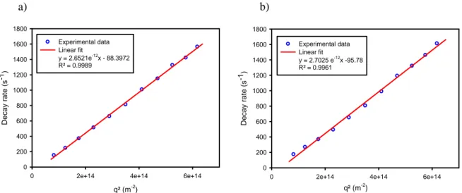
carried out over an angle range from 40° to 140° in 10° stepwise increments. A cylindrical glass cell containing 1 mL of the sample was immersed in a filtered toluene bath, whose background signal was measured at all angles for Rayleigh ratio determination. Refractive index increment (dn/dc) was determined with a gel permeation chromatography (GPC) system equipped with a refractometer detector. Suspensions of copolymer co-assembled with IONPs at constant FWR and different total concentrations c were analysed and a linear fit yielded the value of dn/dc. For multi-angle DLS experiments, the CONTIN fit introduced by Provencher [42] was applied on the autocorrelograms to obtain the decomposition into decay rate modes i without any assumption on their number. Then i
values were plotted versus the square of the light scattering vector q=4nsin(/2)/ where n is the solvent refraction index,
the wavelength and the scattering angle in order to get the corresponding translational diffusion constants, converted into hydrodynamic diameters using Stokes-Einstein’s relationship.
Transmission electron microscopy (TEM) images were recorded on a Microscope Fei Tecnai 10 operating at an accelerating voltage of 80 kV (Oregon, USA). For USPIO samples, a drop of diluted suspension ([Fe] ≈ 0.5 mM) in THF was placed on a carbon-coated copper grid (300 mesh). Images were analysed using automated particle counting of ImageJ software after thresholding and watershed filtering, enabling to perform a statistical treatment of the diameters. Based on a minimum of 1000 nanoparticles per batch, the polydispersity index (PDITEM)
was calculated using the following equations, by analogy with dispersity (Ð) defined for polymer chains, but subtracting 1 to obtain a PDI below 1 (like in DLS):
n w TEM
D
D
PDI
– 1 where
i i i i i nn
D
n
D
and
i i i i i i w D n D n D 3 4 (1)and are the number-average and the weight-average
n
D Dw
diameters, respectively, and ni the number of nanoparticles of
diameter Di.
For empty vesicles (pure PEO-b-PCL nanoprecipitated copolymer), negatively stained TEM images were acquired by placing successively a drop of diluted suspension (2 minutes), a drop of phosphate-buffered saline (pH 7.4) (2 minutes), a drop of DI water (2 minutes) and a drop of uranyl acetate 2% (w/v) solution (10 minutes) on a carbon-coated copper grid (300 mesh). Surplus of solutions were removed before each grid transfer. TEM images of vesicles loaded with USPIOs were obtained on grids prepared by placing a drop of diluted suspension ([Fe] ≈ 0.1 mM) on a Formvar™-coated copper grid (300 mesh).
Cryo-TEM images were recorded at the Institut de Physique et Chimie des Matériaux of Strasbourg (IPCMS, Pr. O. Ersen and D. Ihiawakrim) on a Microscope JEOL 2100 F operating at an accelerating voltage of 200 kV. A drop of diluted suspension was
placed on a carbon-coated copper grid (300 mesh), which was beforehand treated with an ELMO glow discharge unit (Cordouan Technologies, Pessac, France) to render the surface hydrophilic. The drop size was reduced with a filter paper until having a thin film. The grid was then plunged into liquid nitrogen and subsequently in liquid ethane. Maintained at a temperature near liquid nitrogen temperature (-190°C), the grid was transferred to a cryo-holder which was introduced into the high-vacuum column of the microscope.
Small angle neutron scattering (SANS) measurements were performed on the PACE spectrometer of Laboratoire Leon Brillouin (CEA-Saclay, France). Three configurations were used by varying the sample to detector distance and the neutrons wavelength () in order to cover a large q range: 3.18 10-3 – 3.70
10-1 A-1. The samples were prepared in pure heavy water (pure
copolymer) or in a mixture of 20% v/v D2O and H2O, matching
the theoretical neutron scattering length density (SLD) of the PCL blocks for those loaded with iron oxide NPs, in quartz cuvettes of respective thicknesses 2 mm or 1 mm. The scattering curves were divided by the transmission factor and by the thickness after subtracting the signal of an empty cuvette. Correction of the efficacy of the detector was performed by normalizing the curves by the signal of a 1 mm cuvette filled with light water to obtain the scattering intensity,
I(q), in absolute value (cm-1) [43].
Dosage of iron was performed by Inductively Coupled Plasma-Atomic Emission Spectroscopy (ICP-AES) on a Jobin Yvon JY70C instrument (Longjumeau, France). The total amount of iron was determined after microwave digestion (Milestone MLS-1200 MEGA, Gemini B.V., Apeldoorn, the Netherlands) in a mixture of nitric acid (600 µL, 65 %) and hydrogen peroxide (300 µL, 35%). An iron standard solution for ICP (1000 mgmL-1 of iron
in nitric acid) was used to obtain a calibration curve (linear correlation coefficient = 0.99).
Molecular weight distribution of the copolymers was measured by gel permeation chromatography (GPC) analysis (Agilent 1200 apparatus) equipped with a differential refractive index detector. Sample solutions (1 mg/mL in THF, 2% NEt3)
were injected with a 1 mLmin-1 flow rate at 35°C, in a
pre-column PLgel 10 µm (50 7.5 mm) and in two gradient pre-columns PLgel 10 µm mixed-B (300 7,5mm) (Laboratory of Polymeric and Composite Materials, Pr. P. Dubois, UMonS). Additional GPC analyses of PCL block hydrolysis as a function of time and pH were performed with THF as eluent on an UltiMate 3000 system from Thermo Fisher Scientific equipped with a diode array detector (DAD), a multi-angle light scattering (MALS) detector and a differential refractive index (dRI) detector from Wyatt Technology Corp. (Toulouse, France). Polymers were separated on three G2000, G3000 and G4000 TOSOH HXL gel columns (300 7.8 mm) (size exclusion limits from 1000 to 400 000 gmol-1) held at 40°C and under a THF flowrate of 1 mL/min.
EasiVial™ kit of Polystyrene from Agilent was used as the Mn
calibration standard (from 162 to 364 000 gmol-1).
Nuclear magnetic resonance (NMR) spectrum was recorded on a Bruker Avance 500 MHz spectrometer (Karlsruhe, Germany). DOSY-NMR measurements were conducted using a
View Article Online
10.1039/C9TB00909D
ARTICLE
STE-LED (stimulated echo-longitudinal eddy current delay) standard sequence with a diffusion time (Δ) of 250 ms, a gradient pulse length (δ) of 1 ms and 16 gradients ranging from 2% to 95% of the maximum gradient (53 G/cm).
Nuclear magnetic relaxation dispersion (NMRD) profiles reporting the longitudinal relaxation rates of water protons (R1)
over a magnetic field range from 0.25 mT to 0.94 T (0.015 - 40 MHz proton resonance frequency) were recorded on a Fast Field Cycling (FFC) relaxometer (Stelar, Mede, Italy). The apparent saturation magnetization (MSNMRD), the Néel
relaxation time (τN) and size of superparamagnetic crystal
(DNMRD) were determined by fitting these NMRD curves
numerically with the MINUIT minimisation program within the frame of the Outer Sphere relaxation model and a standard phenomenological approximation [40]. Additional longitudinal (T1) and transverse (T2) relaxation times were measured at 37°C
on Bruker Minispec mq20 and mq60 relaxometers working respectively at 20 MHz (0.47 T) and 60 MHz (1.41 T). The diamagnetic contributions of THF to the relaxation rates at 37°C are R1 = 0.2759 s-1 and R2 = 0.2984 s-1 at 20 MHz and R1 = 0.2227
s-1 and R2= 1.4837 s-1 at 60 MHz.
Results and discussion
Characterization and self-assembly of PEO45-b-PCL111 copolymer.
A copolymer of PEO-b-PCL was synthesized by ring opening polymerization of ɛ-CL monomer initiated by PEO chains with
Mn of 2000 gmol-1 possessing two different end-groups,
respectively –CH3 and –OH. The chemical structure of
PEO-b-PCL copolymer was assessed by 1H NMR spectroscopy. The 1H
NMR spectrum shows typical peaks of PEO-b-PCL copolymers, whose integral intensities were used to determine the composition of the PEO-b-PCL copolymer (Fig. S1a in supporting information). The final block copolymer composition is PEO45
-b-PCL111, where 45 and 111 are the respective average degrees of
polymerization (DP) of each monomer unit. This composition is close to the target hydrophilic ratio (14% weight fraction) of PEO45-b-PCL105, which was chosen owing to its tendency to form
a vesicular morphology [29, 30, 32, 34, 35, 37]. The copolymer is characterized by a relatively wide molecular weight dispersity (Ð=Mw/Mn = 1.35) as indicated by GPC (Fig. S1b in supporting
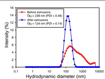
information). In this work, we opted for a fast, efficient and easy to implement method for the formation of the nano-assemblies, i.e. by the solvent shift method, also called nanoprecipitation. Sequential extrusions at 60°C (i.e. above the melting curve of PCL) through 1 µm, 0.8 µm, 0.4 µm and 0.2 µm pore size filters were necessary to narrow the size distribution (Fig. 1). TEM experiments (Fig. S3 in supporting information) confirm the formation of 60 nm diameter spherical nanostructures, suggesting thus the formation of the expected vesicles.
Hydrodynamic diameter (nm)
0.1 1 10 100 1000 10000In
te
n
si
ty
(%)
0 2 4 6 8 10 12 14 16 Before extrusions DH = 238 nm (PDI = 0.48) After extrusions DH = 124 nm (PDI = 0.14)Figure 1. Intensity-weighted size distributions of PEO45-b-PCL111 suspensions
obtained by the nanoprecipitation method before (empty circles) and after (filled circles) extrusions through 1 µm to 0.2 µm pore size filters.
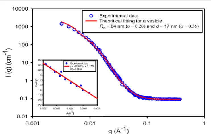
SANS experiments were performed on PEO45-b-PCL111
self-assemblies in order to confirm their vesicular structures and estimate their membrane thicknesses. SANS curves were fitted with the vesicle (shell) form factor thanks to SASView software. The values obtained from the fitting are Rin = 84 nm and d = 17
nm, where Rin corresponds to the inner aqueous compartment
radius and d to the membrane thickness (the radius of the vesicle Rtot, is given by: Rtot = Rin + d). One can note that radii and
thickness values calculated by fitting the SANS curves exhibit broad dispersity parameters (no unit),e as samples analysed by SANS were not extruded to avoid sample loss on the filters as SANS analysis requires concentrated enough samples, in this case 6.2 mgmL-1). The I(q) curves computed using a hollow
spherical shell model are well superimposed on experimental data, supporting the assumption that nano-vesicles are formed through the self-assembly of PEO45-b-PCL111 copolymers (Fig. 2).
In the intermediate range of scattering vectors, the neutron scattering intensities vary as q-2, this scaling law being
characteristic of vesicular structures that exhibit locally planar morphology. The Kratky-Porod plot (ln [I(q)q²] versus q²) enables accessing the membrane thickness (d) with more robustness than a fit thanks to the following asymptotic law [45]:
(2) ln
[
𝐼(𝑞)𝑞2]
≅(―𝑞²𝑑²12 )
Ln [I(q)q²] plotted versus q² in figure 2 enables deducing a membrane thickness of 21 nm from the slope. Noteworthy, a membrane thickness of 22.5 nm was reported for PEO45
-b-PCL105 vesicles in the literature (measured by cryo-TEM analysis)
[29], in good agreement with the value determined by SANS in the present study. The critical aggregation concentration (CAC) of PEO45-b-PCL111 copolymer in aqueous medium was
determined by the fluorescence technique using pyrene as a probe sensitive to a nonpolar environment (at fixed concentration of 0.6 µM). As the block copolymer concentration increases, a shift of the band from 331 to 336 nm was observed: then by plotting the I336/I331 ratio (from the excitation spectra)
ARTICLE
DOI: 10.1039/C9TB00909D against the logarithm of the concentration (Fig. S2 in supportinginformation) a break is observed corresponding to the value of log (CAC). This low CAC value of 1.4 µgmL-1 indicates a good
stability of PEO45-b-PCL111 vesicles upon dilution.
q (A-1) 0.001 0.01 0.1 1 I ( q ) ( cm -1) 0.01 0.1 1 10 100 1000 10000 Experimental data Theoritical fitting for a vesicle
Rin = 84 nm ( and d = 17 nm ( q²(A-2) 0.0002 0.0003 0.0004 0.0005 0.0006 ln (Iq²) -2.2 -2.0 -1.8 -1.6 -1.4 -1.2 -1.0 -0.8 -0.6 -0.4 Experimental data y = -3529.73 x + 0. 1776 R² = 0.9686
Figure 2. SANS curves of PEO45-b-PCL111 vesicles: experimental data are plotted as open
circles while solid lines represent the simulated intensity considering the form factor of hollow spheres with log-normal distributions on both Rin and d, whose standard values
() are indicated. The insets shows the representation of ln[I(q)q²] versus q² calculated from SANS measurements in the Kratky-Porod regime giving a membrane thickness of 21 nm.
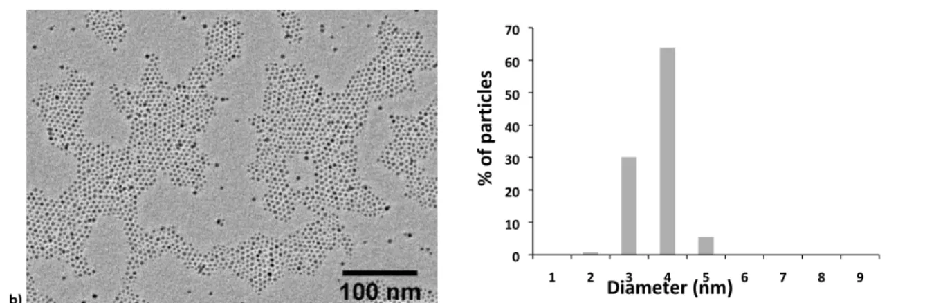
Characterization of iron oxide nanoparticles.
Iron oxide nanoparticles having two different sizes (4.2 nm and 7.6 nm) were synthesised by the thermal decomposition method. Narrow size distributions typical of this synthesis procedure were obtained. TEM images of USPIO were used to determine the size distribution of the magnetic cores (Fig. 3). The average diameters (DTEM) and the polydispersity index
(PDITEM) extracted from a statistical treatment of sizes
measured on TEM images as well as hydrodynamic diameters (DH) and PDI measured by DLS (PDIDLS) using the 2nd order
Cumulant fit of correlograms are reported in Table 1. NMRD profiles which display the evolution of proton longitudinal relaxivity r1 as a function of the applied field (or the
corresponding proton resonance frequency) were recorded on these two samples (Fig. S4 in supporting information). These profiles were fitted using the usual Outer Sphere theory modelling the acceleration of proton relaxation induced by superparamagnetic NPs [44] applied to suspensions in THF, for which a molecular diffusion coefficient of 3.27 10-9 cm2s-1 at
37°C has been measured independently by diffusion-ordered NMR spectroscopy (DOSY). Table 1 shows the values of the relaxometric Outer Sphere diameter (DNMRD), which represents
the shortest distance between the centre of the magnetic core of the USPIOs and the THF molecules, together with their apparent saturation magnetization (MsNMRD) values that are
almost identical ( 50 Am2kg-1) for both samples.
Table 1. Physicochemical characteristics of the USPIOs synthesised by the thermal decomposition method: diameter measured by TEM, DLS, NMRD, saturation magnetization
(MsNMRD) and Néel relaxation time (τN) extracted from the fits of NMRD profiles.
Sample DTEM (nm) PDITEM D
H(nm) PDIDLS DNMRD (nm) MsNMRD (Am²kg-1) τN (ns) 7.6 nm USPIO 7.6 0.14 11.8 0.05 8.5 49.4 2.24 4.2 nm USPIO 4.2 0.08 8.4 0.03 5.6 49.7 2.69 a) 1 2 3 4 5 6 7 8 9 10 11 12 13 14 15 0 10 20 30 40
Diameter (nm)
% of pa
rti
cl
es
DOI: 10.1039/C9TB00909D
ARTICLE
b) 1 2 3 4 5 6 7 8 9 0 10 20 30 40 50 60 70Diameter (nm)
% of pa
rti
cl
es
Figure 3. TEM images and histogram of diameters of USPIO synthesized by the thermal decomposition method: (a) USPIO with DTEM = 7.6 nm (7.6 ± 1.6 nm, PDITEM = 0.14) and (b)
USPIO with DTEM = 4.2 nm (4.2 ± 0.5 nm, PDITEM = 0.08). PDI values are defined by Eq. (1).
Longitudinal (r1) and transverse (r2) relaxivities were measured
in THF (37°C) at 20 and 60 MHz. Iron oxide nanoparticles with larger diameters possess higher longitudinal and transverse relaxivities and higher transverse to longitudinal relaxivity ratios, while staying at low values (r2/r1 below 3) typical for
small and well-dispersed (un-aggregated) USPIOs (Table 2).
Table 2. USPIO’s longitudinal and transverse relaxivities
measured at 20 and 60 MHz (in THF and 37°C) and resultant r2/r1
ratios. Sample Frequency (MHz) r1 (s-1 mM-1) r2 (s-1mM-1) r2/r1 7.6 nm USPIO 20 60 21.9 12.8 36.6 37.7 1.7 2.9 4.2 nm USPIO 20 60 8.6 7.8 12.1 17.9 1.4 2.3 Formulation and characterization of USPIO loaded in PEO45-b-PCL111 polymersomes
Nanoprecipitation of PEO45-b-PCL111 co-assembled with iron
oxide nanoparticles was performed by varying the initial USPIO concentration at constant copolymer concentration, the ratio of their weight concentrations in THF being defined as the “feed weight ratio” (FWR). After adjustment, the best results were obtained by mixing 10 mg of the block copolymer with 500 μL of THF containing USPIOs at concentrations close to [Fe]0 ~ 40
mM for 7.6 nm USPIO and [Fe]0 ~ 80 mM for 4.2 nm USPIO. The
corresponding FWR values are respectively 16% and 32%, within the same range as in the study by Arosio et al. [46] with another amphiphilic copolymer also forming vesicles. Above these threshold values, no more USPIOs could be entrapped in the vesicle membranes and large aggregates of USPIOs were observed by mere visual inspection. The TEM image of the sample obtained by nanoprecipitation of PEO45-b-PCL111 loaded
at 16% FWR with 7.6 nm USPIO shows spherical structures with a high USPIO content as well as some filamentous fragments connected to some of the spheres certainly formed during the drying step of the TEM grid preparation (Fig. 4a). To confirm that these fragments arise from the destruction of spherical structures during the drying step, cryo-TEM imaging allowing the visualization of colloids in the frozen-hydrated state was
performed. As expected, only spherical structures were observed (Fig. 4b). However, it can be noted that these spherical structures are arranged into chain-like aggregates, which could be an artefact due to their accumulation within the wedge of the ultrathin frozen film near a border of the TEM grid (being sterically excluded from the centre of the film because its thickness becomes smaller than vesicles diameter). The absence of clusters in the liquid suspensions is confirmed by dynamic light scattering measurements showing a monomodal size distribution with a mean hydrodynamic diameter close to 130 nm (DH = 129 nm, PDIDLS = 0.13) (Fig. 4c), characteristic of
well-defined and individually dispersed objects.
a) b) c) Hydrodynamic diameter (nm) 1 10 100 1000 10000 In te n s it y ( % ) 0 2 4 6 8 10 12 14
Figure 4. Size and morphology of PEO45-b-PCL111 vesicles loaded at FWR=16% with USPIO
nanoparticles of 7.6 nm diameter. (a) Low magnification TEM image showing intact spherical structures as well as some fragments (scale bar = 500 nm). (b) Cryo-TEM image showing only spherical structures (scale bar = 100 nm). (c) Intensity-weighted size distribution measured by DLS (DH = 129 nm, PDIDLS = 0.13).
ARTICLE
DOI: 10.1039/C9TB00909D Nanoprecipitation of PEO45-b-PCL111 loaded at 32% FWRwith 4.2 nm USPIO gave similar results. Spherical structures exhibiting a high USPIO content were observed on the TEM images (Fig. S4a in supporting information). A mean hydrodynamic diameter of 146 nm (PDIDLS = 0.10) was measured
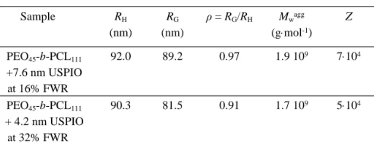
by DLS (the intensity-weighted size distribution is given in Fig. S4b). Concerning vesicle stability on time, the size distribution of magnetovesicles did not change significantly after storage for one month at 4°C. Multi-angle dynamic (DLS) and static (SLS) light scattering measurements were conducted on nanoprecipitated samples obtained by co-assembly of PEO45
-b-PCL111 with USPIOs of both sizes. Mean values of the radius of
gyration (RG), aggregate weight-averaged molar mass (Mwagg)
and aggregation number, i.e. the number of macromolecules per aggregates (Z), were determined from SLS measurements upon drawing Guinier and Berry plots (at several weight concentrations and over a scattering angular range of 40-140°). These plots are provided in Supporting Information (Fig. S5 and Fig. S6). The determination of the refractive index increment dn/dc as measured using a RI detector for GPC was required to determine the molar masses of the self-assembled nanostructures. Resulting dn/dc values were 0.102 Lg-1 for PEO45-b-PCL111 loaded at 16% FWR
with 7.6 nm USPIOs and 0.147 Lg-1 for PEO45-b-PCL111 loaded at 32%
FWR with 4.2 nm USPIOs. Such increase of the RI increment when inorganic NPs like USPIOs (whose RI is 2.3) are embedded in the objects was predictable. As the mean value hydrodynamic radius can depend on the scattering angle used for the measurement (as ascribed to size dispersity), multi-angle DLS measurements were performed over the same scattering angle range to measure the angle-average hydrodynamic radius (RH). From these measurements,
one can calculate the ratio ρ = RG/RH, which enables to estimate the
morphology of the self-assembly nanostructures: vesicles are characterized by ρ values close to 1 while ρ values around √(3⁄5)≈0.775 are expected for spherical micelles [47, 48]. In the present study, the ρ values calculated from the experimental multi-angle DLS and SLS results are in good agreement with vesicular structures (Table 3). Although they appear a bit higher, the Z values 5-7104 calculated for the aggregation numbers of copolymers
(after subtracting the fraction of mass of iron oxide in the objects) are also in agreement with values Z 3104 reported by Adams et al.
[26] for polymersomes made of PEO45-b-PCL101. In conclusion,
nano-objets produced by the self-assembly of PEO45-b-PCL111
with hydrophobically coated USPIOs are vesicles possessing a diameter close to 160-180 nm. These nanoprecipitated objects remain of vesicular morphology, despite their high USPIO payload (16 or 32% FWR) that can fit within the thick hydrophobic membranes.
Table 3. Structural characteristics of PEO45-b-PCL111 magnetovesicles measured by
multi-angle DLS and SLS measurements: Hydrodynamic radius RH measured by decay rate plots
vs. square of scattering vector (Fig. S5a,b), radius of gyration RG measured from Guinier
plots (Fig. S6a,b), molar mass of scatterers Mwagg and aggregation number
Z=Mwagg/(1+FWR)/Mwpolymer of the constituting macromolecules deduced from Berry
plots (Fig. S6c,d). Sample RH (nm) RG (nm) ρ = RG/RH Mwagg (gmol-1) Z PEO45-b-PCL111 +7.6 nm USPIO at 16% FWR 92.0 89.2 0.97 1.9 109 7104 PEO45-b-PCL111 + 4.2 nm USPIO at 32% FWR 90.3 81.5 0.91 1.7 109 5104
Relaxometric properties of USPIOs loaded in PEO45-b-PCL111 vesicles.
An MRI contrast agent is a magnetic compound that alters the signal intensity of specific parts on MR images by shortening longitudinal relaxation time (T1) and transverse relaxation time
(T2) of water protons in the biological tissues. The efficiency of
an MRI contrast agent is quantified in vitro by its longitudinal and transverse relaxivities (r1 and r2), which reflect the ability of
the contrast agent to decrease T1 and T2 relaxation times of the
water protons, respectively. In particular, T2-weighted contrast
agents decrease MRI signal intensity and thus darken regions where they accumulate (while those where they are absent, like a liver tumour, appear brighter). The most effective T2-weighted
contrast agents are those characterized by the highest r2/r1
ratio. USPIOs can also be used as T1-contrast agents, but only as
long as their r2/r1 ratio remains at a value lower than 3, typically
[49]. Relaxometry studies were performed on magnetic polymersomes loaded with two different size and loading contents of USPIOs, namely at 32% FWR with those of 4.2 nm diameter and 16 % FWR for the 7.6 nm diameter ones. Relaxation rates 1/T1 (s-1) and 1/T2 (s-1) were measured at 20
and 60 MHz (37°C) at different iron concentrations (mM). The plots of the relaxation rates versus equivalent iron concentration are given in Fig. 5. Longitudinal and transverse relaxivities deduced from the slopes are reported in Table 4. Encapsulation of USPIOs into the hydrophobic membranes of PEO45-b-PCL111 polymersomes leads to a significant decrease of r1 (due to a reduced accessibility of water protons to USPIO
surface), and an increase of r2 (due to USPIO clustering into the
vesicle) compared to USPIOs individually dispersed in the solvent (see r1 and r2 values in THF in Table 2). In addition, it is
well-known in the literature of MRI contrast agents [50] that r1
decreases as a function of the applied magnetic field while r2
reaches a plateau value. Consequently, higher transverse to longitudinal relaxivity ratios (r2/r1) were obtained at 60 MHz
DOI: 10.1039/C9TB00909D
ARTICLE
Table 4. Longitudinal (r1) and transverse (r2) relaxivities (in water and at 37°C) andresulting r2/r1 ratios of USPIO loaded in PEO45-b-PCL111 vesicle membranes.
Sample Frequency (MHz) r1 (s-1mM-1) r2 (s-1mM-1) r2/r1 PEO45-b-PCL111 + 7.6 nm USPIO at 16% FWR 20 60 3.3 1.3 187 197 57 154 PEO45-b-PCL111 + 4.2 nm USPIO at 32% FWR 20 60 0.6 0.3 109 110 171 389
As already reported in the literature for vesicles made from other copolymers, higher transverse relaxivities were obtained for polymersomes loaded with larger USPIOs [14, 46]. In this work, polymer vesicles loaded at 16% FWR with 7.6 nm USPIO have a transverse relaxivity of 197 s-1mM-1 (60 MHz) while
those loaded at 32% FWR with 4.2 nm USPIO have a smaller transverse relaxivity of 110 s-1mM-1 (60 MHz). Interestingly,
polymersomes loaded with 4.2 nm exhibit significantly lower longitudinal relaxivities: r1 = 0.3 s-1mM-1 at 60 MHz compared
to r1 = 1.3 s-1mM-1 for vesicles loaded with 7.6 nm USPIOs.
This can be explained by their total embedding within the hydrophobic PCL blocks of the membranes (of thickness 21 nm according to SANS, much larger than 4.2 nm), thereby avoiding
any direct contact of the water molecules with the iron oxide surface (thus preventing the contribution of the so-called “Inner Sphere” mechanism [50] to proton relaxations). Therefore even higher transverse to longitudinal relaxivity ratios (r2/r1) were
obtained for vesicles loaded with smaller USPIOs compared to the larger ones, thereby demonstrating their high potential as
T2-contrast agents despite their lower transverse relaxivity,
however that is analogous to the one of commercial SPIO MRI contrast agents.
NMRD profiles were recorded on polymersomes loaded with USPIOs possessing a diameter of 4.2 nm at 32% FWR and 7.6 nm at 16% FWR (Fig. S7 in supporting information). One can clearly note that longitudinal relaxivities of polymer vesicles loaded with the smallest USPIO (4.2 nm USPIO) are lower than those of vesicles loaded with largest USPIO (7.6 nm USPIO) over the whole range of measured frequencies. Compared to the NMRD profiles of individual USPIOs in THF (Fig. S4), a shift of the maxima towards lower Larmor frequencies were observed, and became more pronounced when the diameter of incorporated USPIOs was larger. A similar shift was already reported for
PGA-b-PTMC vesicles incorporating USPIOs of different sizes [46] and
ascribed to their embedding in the membranes, making them behave as aggregates of larger size than individually dispersed USPIOs. a) b) 0 30 60 90 120 150 0 0.2 0.4 0.6 0.8 R 2 20MHz R 2 60MHz 1 /T 2 (s -1 ) Iron concentration mM 0 0.5 1 1.5 2 2.5 3 0 0.2 0.4 0.6 0.8 R 1 20MHz R 1 60MHz 1 /T1 (s -1 ) Iron concentration mM c) d) 0 20 40 60 80 100 120 140 0 0.2 0.4 0.6 0.8 1 1.2 R 2 20MHz R 2 60MHz 1 /T 2 (s -1 ) Iron concentration mM 0 0.2 0.4 0.6 0.8 1 0 0.2 0.4 0.6 0.8 1 1.2 R 1 20MHz R1 60MHz 1 /T1 (s -1 ) Iron concentration mM
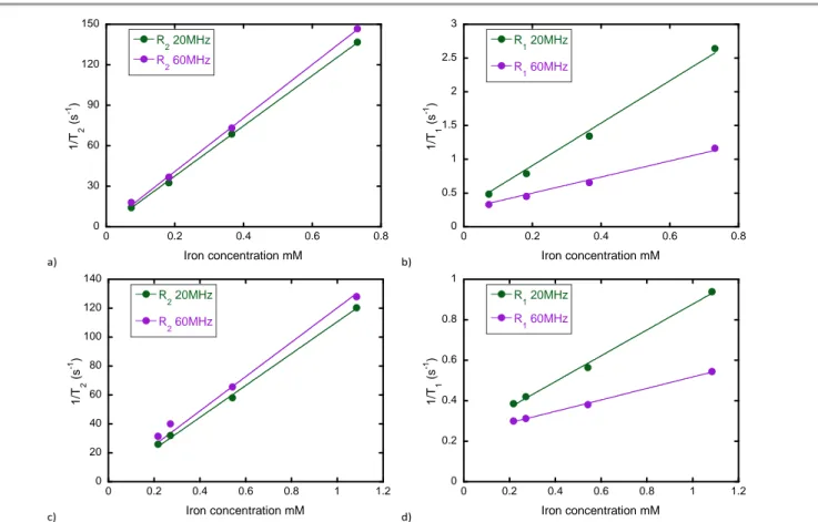
Figure 5. Relaxations rates as a function of iron concentration (mM) for PEO45-b-PCL111 vesicles loaded with iron oxides nanoparticles: (a) 1/T2 (s-1) for vesicles loaded at 16% FWR
with 7.6 nm USPIOs: slopes and intercepts are r2=186 s-1mM-1 and R20=0.506 s-1 at 20MHz, r2=198 s-1mM-1 and R20=1.54 s-1 at 60MHz. (b) 1/T1 (s-1) for vesicles loaded with 7.6 nm
USPIOs at 16% FWR: slopes and intercepts are r1=3.14 s-1mM-1 and R10=0.28 s-1 at 20MHz, r1=1.19 s-1mM-1 and R10=0.26 s-1 at 60MHz. (c) 1/T2 (s-1) for vesicles loaded with 4.2 nm
USPIOs at 32% FWR: slopes and intercepts are r2=110 s-1mM-1 and R20=0.506 s-1 at 20MHz, r2=119 s-1mM-1 and R20=1.54 s-1 at 60MHz. (d) 1/T1 (s-1) for vesicles loaded with 4.2 nm
RTICL
ARTICLE
In addition, the inflection points observed at low fields (below 1 MHz) on the NMRD profiles of individual USPIOs, usually ascribed to Néel relaxation of the moments, are absent on the NMRD profiles of the magnetic vesicles. These were fitted using the usual “Outer Sphere” model for superparamagnetic MRI contrast agents. Parameters extracted from these fittings (DNMRD and MSNMRD) are reported in Table S1 (supporting
information). Compared to individually dispersed USPIOs, an increase of DNMRD and a decrease of MSNMRD were observed.
These results are in agreement with the presence of a hydrophobic polymer layer surrounding the nanoparticles, preventing water molecules to approach surface of the magnetic centre (thus avoiding any Inner Sphere contribution to proton relaxation). The decreased magnetization values can be easily understood by the dilution of the magnetic iron oxide within the non-magnetic phase. It is also very interesting to note that the values of “Outer Sphere” diameters DNMRD deduced
from the fits are nearly equal to the hydrophobic membrane thickness measured by SANS (20.8 nm for 7.6 nm USPIOs at 16% FWR and 18.2 nm for 4.2 nm ones at 32% FWR). This parameter deduced by relaxometric experiments corresponds to a characteristic distance on which the presence of water molecules is forbidden, thus it is equal to the membrane hydrophobic thickness as from SANS and TEM.
In addition to their use as MRI contrast agents, magneto-vesicles described in the present study can also be used for magnetic separation, as they are retained by a LS column™ placed in an electromagnet, which confirms their high USPIO content. A study reported that magneto-liposomes containing a low amount of ferrofluid are not retained on the same magnetic column [51]. Size distributions (Fig. S8), TEM images (Fig. S9) and proton NMR relaxivities (Table S2 in supporting information) before and after magnetic chromatography are similar, which suggests that the samples are not affected by the magnetic chromatography and can therefore be concentrated by this method. Magnetophoretic separation can also be used to purify the sample and change the external aqueous medium, after loading with a hydrosoluble drug such as doxorubicin for example [52].
Degradation of USPIO loaded in PEO45-b-PCL111 vesicles in acidic aqueous conditions.
The degradation of PEO45-b-PCL111 based
magneto-polymersomes (encapsulating 7.6 nm USPIOs at 16% FWR) was studied in weakly acidic conditions, mimicking biologically relevant conditions as tumour environment or intra-lysosomal location after cell uptake. It is indeed known that hydrolysis of polyesters shortens hydrophobic blocks and consequently increases the hydrophilic mass fraction of block copolymer chains. Such degraded chains with comparatively short hydrophobic blocks will perturb local bilayer curvature and induce the growth of pores in polymersome membranes [29, 53-55]. Poration of polyester membranes at acidic pH is particularly interesting for drug delivery purpose as it can induce drug release from polymersomes uptaken by biological cells through an endocytosis pathway that end up in acidic (pH around 5) lysosomal compartments [29, 54].
To evaluate if the overall structure is affected or not by the acidic pH, GPC, DLS and TEM measurements performed at pH 7 were compared to the results obtained at weakly acidic pH (pH
5) after 7 days. The TEM images of the sample left 7 days at pH 7 and 37°C are similar to those of the sample left at pH 5 and 37°C. Spherical nanostructures with a high USPIO content as well as some filamentous fragments were observed (Fig. S10a and Fig. S10b in supporting information). DLS measurements also show similar size distributions (sample left at pH 7 and at pH 5) with mean hydrodynamic diameters in the range between 130 and 150 nm and PDIDLS values smaller than 0.17 (Fig. S10c
and Fig. S10d in supporting information). This suggests that spherical structures loaded with iron oxide nanoparticles were still present in the suspensions even after one week (168 hours) at 37°C and pH 5. a) Time (days) 0 2 4 6 8 r 1 6 0 M H z (s -1 m M -1) 1.0 1.1 1.2 1.3 1.4 1.5 1.6 pH 7 pH 5 b) Time (days) 0 2 4 6 8 r2 6 0 M H z (s -1 m M -1) 80 100 120 140 160 180 200 220 pH 7 pH 5 c) Time (days) 0 2 4 6 8 r 2 /r1 60 M H z 40 60 80 100 120 140 160 180 pH 7 pH 5
View Article Online
DOI: 10.1039/C9TB00909D
ARTICLE
Figure 6. Longitudinal relaxivities (a), transverse relaxivities (b) and transverse tolongitudinal relaxivity ratios (c) at 60 MHz for PEO45-b-PCL111 vesicles loaded at 16% FWR with 7.6 nm USPIOs at pH 7 (red filled markers) and pH 5 (blue empty markers) as a function of time.
In parallel, the relaxivities of polymer vesicles loaded at 16% FWR with 7.6 nm USPIO were measured over time at two different pH (pH 7 and pH 5). Figure 6 shows results obtained at 60 MHz. Similar trends were observed at 20 MHz as shown in supporting information (Fig. S11). At pH 7 and at 37°C, longitudinal and transverse relaxivities did not vary significantly during the studied period (7 days). On the contrary, at pH 5, the longitudinal relaxivity increases abruptly and the transverse relaxivity decreases more continuously over time, which results in an sigmoid decrease of the transverse to longitudinal relaxivity ratio (r2/r1), with a jump occurring after 2 days of
sample acidolysis. These relaxivity variations can be interpreted by the formation of hydrophilic pores in the PCL membranes occurring as the hydrophobic PCL block is progressively hydrolysed [25, 50, 51]. Water molecules could pass through PCL membrane leading to an increase of r1 (due to an increased
accessibility of water protons directly to the USPIO surface) and a decrease of r2 (due to a reduced USPIO clustering effect). Such
polymer degradation has been further highlighted by GPC for empty vesicles (Fig. 7), for which we could notice the appearance of a second peak at higher retention time corresponding to a Mn 2600 gmol-1, and attributable to PEG
chains liberated in the medium, presumably still joined to a much degraded PCL block.
Figure 7. GPC traces in THF (RI detector) of the copolymer dissolved in two different
buffers (pH7.4 and pH5.2) after dialysis, freeze-drying and recovery in THF at different times. The main peak around an elution volume of 20 mL correspond to following molar masses expressed relatively to the PS standard curve: Mn=12000 g·mol-1 and Mw= 15700
g·mol-1 at t=0;, M
n=11700 g·mol-1 and Mw= 15800 g·mol-1 at t=24h (pH5; flashy green
curve); Mn=12300 g·mol-1 and Mw= 16200 g·mol-1 at t=72h (pH5; blue curve); Mn=12600
g·mol-1 and Mw= 16500 g·mol-1 at t=144h (pH5; pink curve); Mn=12400 g·mol-1 and Mw=
17000 g·mol-1 at t=168h (pH5; black curve); M
n=12800 g·mol-1 and Mw= 16600 g·mol-1 at
t=208h (pH5; dark green curve); Mn=12100 g·mol-1 and Mw= 16000 g·mol-1 at t=208h
(pH7). After 72h at pH5, a second peak appeared around 24 mL, corresponding to
Mn=2600 g·mol-1 and Mw= 2700 g·mol-1. This secondary peak represent 1.6% of the total
weight at t=72h (assuming identical refraction index increments), increasing to 8% at t=144h until reaching a plateau at 11% for t=168h. This peak is also present, yet at lower extent (3% of total weight) after 208h at pH7 (red curve), and can be ascribed to PEG 2kD chains linked to a much degraded PCL block.
This peak appeared 72 hours (3 days) after incubation at pH5.2, and was also observable, at lower extent, after 208 hours at pH7.4. This result confirms the slow acidolysis of the PCL blocks. Although the vesicles did not show any sign of disruption neither by TEM nor DLS, their relaxometric data
proved access of the water molecules to the USPIO surfaces, thus enhanced permeability of the membranes after only 2 days at pH5. This MRI response is even more sensitive than by measuring molecular dimension of the chains by GPC, which showed clear evidence of chain cleavage, but only after 3 days at pH5.2.
In future in vivo experiments, such variations of r1 and r2
could be turned into a greater advantage to monitor experimentally the PCL degradation in the membrane drug-nanocarriers from the modulation of the contrast of MR images and correlate the evidenced drug release with anatomic information (e.g. tumour volume). An enhanced MRI contrast following a controlled drug release and accumulation to diseased organs/tissues is very promising for in vivo applications [56, 57].
In previous literature, D. Hammer et al. showed that hydrolytic degradation of PEO-b-PCL vesicles incorporating porphyrin-based fluorophores within the PCL core results in change in porphyrin emission providing a powerful tool for monitoring in vivo drug delivery non-invasively [34]. Such degradative mechanism of polyester-made vesicles, catalysed by enzymes, was also reported in recent work by Zhu et al. [58]. However, to the authors’ knowledge, no similar work using MRI contrast agents to monitor degradation of polyesters like PCL in membranes was never reported. The novelty of our work stands in the proof that this phenomenon strongly influences the MRI contrasting properties. To show the interest of PEO45-b-PCL111
vesicles loaded with USPIOs for monitoring degradation of PCL membrane of nanovesicles in vivo, we computed the signal levels by the approximated solution of Bloch equations, a method that we used in a precedent study [49] to predict the evolution of MR image contrast from proton NMR relaxometric measurements: (3)
/1
/2 MRI H 1 TR T TE T S e eWhere SMRI corresponds to the theoretical MRI signal intensity, ρH corresponds to the proton volume density, TR is the
repetition time and TE is the echo time.
Time (days) 0 2 4 6 8 SMR I (T2 -w e ig h te d se q u e n ce si g n a l) 0.04 0.06 0.08 0.10 0.12 0.14 0.16 PEO-b-PCL + USPIO - pH 7 PEO-b-PCL + USPIO - pH 5 H2O
Figure 8. Theoretical predictions of MRI signal levels computed using Eqn (3) with
the experimental values of T1, T2, TE and TR for water (green triangular filled
markers) and PEO45-b-PCL111 vesicles loaded at 16% FWR with 7.6 nm USPIO at pH
7 (red circular filled markers) and pH 5 (blue circular empty markers) as a function of time.
ARTICLE
DOI: 10.1039/C9TB00909D The SMRI calculated with experimental T1 and T2 are representedfor a T2-weighted sequence (TR = 300 ms, TE = 12 ms) on Fig. 8,
and their numerical values are provided on Table 5. At pH 7, SMRI
does not change significantly with time. On the contrary, SMRI
increases with time at pH 5 due to hydrolytic degradation of PCL membrane resulting in an increase of r1 and a decrease of r2.
Again, the interest of these magnetic nano-vesicles made of a hydrolysable copolymer is evidenced, by the predicted positive contrast enhancement of MRI signal.
Table 5. Longitudinal (T1) and transverse (T2) proton relaxation times for PEO45
-b-PCL111 vesicles loaded with 16 wt.% 7.6 nm USPIOs measured at 60 MHz (37°C) at
pH 5 and pH 7 and resulting SMRI calculated from Eqn (3) for a T2-weighted
sequence (TR = 300 ms, TE = 12 ms). pH 7 pH 5 Time (hours) T1 (ms) T2 (ms) SMRI T1 (ms) T2 (ms) SMRI 0 28 52 71 78 95 167 986 986 978 998.5 1002 1017.5 998.5 7.84 8.23 8.32 8.55 8.75 9.14 9.42 0.057 0.061 0.062 0.064 0.066 0.069 0.073 1093 1080 1062 864 856 877 887 9.16 9.66 10.88 12.98 13.70 16.29 18.74 0.065 0.070 0.081 0.116 0.123 0.139 0.151
Conclusions
Iron oxide nanoparticles possessing two different sizes (DTEM =
4.2±0.5 nm and DTEM = 7.6±1.6 nm) were produced by the
thermal decomposition method of iron(acac)3 precursor. These
hydrophobically-coated USPIO nanoparticles were easily incorporated into PEO45-b-PCL111 vesicles by a
nanoprecipitation method at maximal feed weight ratios (FWR) of 32% and 16%, respectively. Extrusion cycles were performed to produce vesicles around 150 – 180 nm in diameter with a narrow size distribution, ideal for in vivo applications, including systemic injection. In addition to their biocompatible and biodegradable composition, vesicles possess PEO chains on their surface which are known to confer “stealth” properties to the nanoparticles in vivo, i.e. a prolonged half-life (long T1/2) in
the blood circulation that is a prerequisite for an efficient drug delivery system.
Magnetic polymeric vesicles with high USPIO contents were obtained by optimizing the initial FWR. These magneto-polymersomes show good MRI contrasting properties at 20 and at 60 MHz, the second resonance frequency being close to the one in the field of most MRI scanners used in clinics. They are characterized by very high transverse-to-longitudinal relaxivity ratios (r2/r1), which demonstrates that they are very promising
candidates as T2-contrast agents for at least pre-clinical studies.
Relaxivities of polymer vesicles embedded with USPIO possessing two different sizes were compared. Results confirmed that magneto-polymersomes incorporating larger USPIOs possess higher transverse relaxivity (yet at a lower FWR). We showed that the size of the embedded USPIOs plays also a critical role on the longitudinal relaxivity: PEO45-b-PCL111
polymersomes loaded at 32% FWR with 4.2 nm USPIO possess significantly lower longitudinal relaxivities and higher r2 to r1
ratios than vesicles loaded with 7.6 nm USPIOs at 16% FWR, which was interpreted by the total embedding of the USPIOs within the membranes (none of them being located near the hydrated PEG blocks of the copolymer).
Relaxometry studies at pH 5 vs pH 7, combined with a complementary test of copolymer chain degradation by GPC, confirm the formation of pores in PCL membrane in acidic conditions. These findings demonstrate the huge potential of the magneto-polymersomes described in the present study as drug delivery systems with image-control by MRI of the sustained release of encapsulated therapeutics in weakly acidic media such as tumours and intracellular endo/lysosomal compartments. Variations of r1 and r2 relaxivities along the PCL
hydrolysis illustrate that these superparamagnetic PEO45
-b-PCL111 vesicles are promising pH-sensitive MRI contrast agents
enabling to track the PCL chain degradation accompanying the delivery of pre-encapsulated species in vivo.
Conflicts of interest
There are no conflicts to declare.
Acknowledgements
The authors thank the department of histology (Prof. D. Nonclercq) of the University of Mons and X. Valentini for providing the access to TEM apparatus. Dr. L. Mespouille (SMPC; UMons) and Dr. J de Winter (S2Mos; UMons) are warmly acknowledged for providing the polymers used during this study. The authors are grateful to Prof. O. Ersen and D. Ihiawakrim from “Institut de Physique et Chimie des Matériaux de Strasbourg (IPCMS)” from the University of Strasbourg from cryo-TEM analysis and Amélie Vax-Weber and C. Pierart for performing respectively the GPC and ICP measurements. The authors acknowledge the Center for Microscopy and Molecular Imaging (CMMI). This work was performed with the financial support of the FNRS, the Agence Nationale de la Recherche (ANR-13-BS08-0017 MagnetoChemoBlast), the ARC program, the ENCITE program and the RW, the Interuniversity Attraction Poles of the Belgian Federal Science Policy Office, and the European Commission under COST (European Cooperation in Science and Technology) Action TD1402 RadioMag. The financial support from the CPER CAMPUSB project funded by the French state and the Région Nouvelle Aquitaine is gratefully acknowledged for the acquisition of the multi-angle light-scattering setup.
Notes and references
1 C. Geraldes and S. Laurent, Contrast Media & Molecular
Imaging, 2009, 4, 1.
2 Y. Gossuin, P. Gillis, A. Hocq, Q. L. Vuong and A. Roch, Wiley
Interdisciplinary Reviews: Nanomedicine and Nanobiotechnology, 2009, 1, 299.
3 Q. A. Pankhurst, J. Connolly, S. K. Jones and J. Dobson, Journal
of Physics D: Applied Physics, 2003, 36, R167.
4 S. Laurent, D. Forge, M. Port, A. Roch, C. Robic, L. Vander Elst and R. N. Muller, Chemical Reviews, 2008, 108, 2064. 5 S. Laurent, J. Bridot, L. Vander Elst and R. N. Muller, Future
Medicinal Chemistry, 2010, 2, 427.
6 S. Belaïd, S. Laurent, M. Vermeersch, L. Vander Elst, D. Perez-Morga and R. N. Muller, Nanotechnology, 2013, 24, 055705.