HAL Id: hal-02163316
https://hal.archives-ouvertes.fr/hal-02163316
Submitted on 24 Jun 2019
HAL is a multi-disciplinary open access
archive for the deposit and dissemination of
sci-entific research documents, whether they are
pub-lished or not. The documents may come from
teaching and research institutions in France or
abroad, or from public or private research centers.
L’archive ouverte pluridisciplinaire HAL, est
destinée au dépôt et à la diffusion de documents
scientifiques de niveau recherche, publiés ou non,
émanant des établissements d’enseignement et de
recherche français ou étrangers, des laboratoires
publics ou privés.
cochlea: evidence for efferent presynaptic autoreceptors
Sylvain Bartolami, Chantal Ripoll, Myriam Planche, Rémy Pujol
To cite this version:
Sylvain Bartolami, Chantal Ripoll, Myriam Planche, Rémy Pujol. Localisation of functional
mus-carinic receptors in the rat cochlea: evidence for efferent presynaptic autoreceptors. Brain Research,
Elsevier, 1993, 62, �10.1016/0006-8993(93)90580-g�. �hal-02163316�
200 Brain Research, 62~ ( 1 t)t*3 ) 200- 20t~ ~C') 1993 Elsevier Science Publishers B.V. All rights reserved 0006-8t)93/~3 ,/$116.t)l!
BRES 19344
Localisation of functional muscarinic receptors in the rat cochlea:
evidence for efferent presynaptic autoreceptors
S y l v a i n B a r t o l a m i , C h a n t a l R i p o l l , M y r i a m P l a n c h e a n d R 6 m y P u j o l
Laboratoire de Neurobiologie de l'Audition, INSERM U-254 and Universit~ Montpellier II, CHU St. Charles, Montpellier (France)
(Accepted 8 June I~)t~3)
Key words: Organ of Corti; Modiolus; Muscarinic autoreceptor; Inositol phosphate
In the rat cochlea, the activation of muscarinic receptors stimulates the hydrolysis of phosphoinositides but the importance of this muscarinic effect is still unknown. In order to find out about the role of the muscarinic receptors in the cochlea, we examined their functional distribution within this organ. This was achieved by measuring the formation of [3H]inositol phosphates induced by carbachol (1 mM) in two regions of the cochlea: the modiolus and the organ of Corti. At both sites, carbachol enhanced the accumulation of inositol phosphates in an atropine-sensitive way. These stimulations were completely antagonised by 4-diphenylacetoxy-N-methyl piperidine methiodide (1 ~zM) but unchanged by pirenzepine (1 p,M). In cochleas depleted of outer hair cells by a treatment with amikacin, the carbachol-induced formation of inositol phosphates is not altered with respect to control, undamaged cochleas. Conversely, when the medial cholinergic axons which form synapses with the outer hair cells are destroyed by the section of the crossed olivocochlear bundle the carbachol-stimulated inositol phosphates response is reduced by 35% in the organ of Corti. This section has no effect in the modiolus, despite the degeneration of some modiolar fibers. Our results show that functional muscarinic receptors are distributed both in the organ of Corti and in the modiolus. These two structures contain presumably the same class of cholinoceptor. The effects of selective destruction clearly demonstrate that a population of muscarinic receptors is located on presynaptic membranes at the level of the medial axon-outer hair cell contacts. They also point to spiral ganglion neurons a n d / o r the Schwann cells as sites for the functional cholinoceptors in the modiolus.
I N T R O D U C T I O N
The organ of Corti, the neuro-sensory epithelium of the cochlea, is innervated by efferent axons from the brainstem. The axonal population is dichotomised be- tween the medial and lateral efferent systems. The former innervates the outer hair cells, the latter forms axo-dendritic synapses with the radial afferent fibers innervating the inner hair cells 53'57. An interesting anatonomical feature of the medial system is that most of its axons project predominantly to the contralateral cochlea and cross the brain midline on the floor of the fourth ventricle. Such a circuit provides the possibility to alter and study specifically the function of the effer- ent medial system. Concerning its physiology, it is now accepted that the medial efferent system modulates the active mechanisms in the cochlea 32'38'45'5t by means of acetylcholine 9'1°'32'51. Support for identifying acetyl- choline as the principal medial transmitter also comes
from immunohistologic surveys which have repeatedly demonstrated that most of the medial efferent neurons express the choline acetyltransferase activity L~3'2°'47"4~ Concerning the lateral system, its function is rather complicated since recent data showed that a single lateral neuron possesses several neurotransmitters, both excitatory and inhibitory, in order to modify the sensory-neural transduction 48.
In the cochlea, acetylcholine alters the behaviour of its target cells by acting through both nicotinic t 1.25,43,44 and muscarinic
cholinoceptors 3'4"6't9'24"27'28'41.
Both cho- linoceptors are present on the outer hair cell and their activation results in the hyperpolarization of the outer hair cells 25'29. But the distribution of the muscarinic cholinoceptor is not restricted to these cells because binding studies report the presence of muscarinic sites in the cochlear nerve 27'58, in the modiolus 27 and in the organ of Corti 27. However, such a distribution may not match accurately the localisation of functional mus-Correspondence: S. Bartolami, INSERM U-254, Laboratoire de Neurophysiologie Sensorielle, Universit6 Montpellier II, Place Eugene Bataillon, 34095 Montpellier Cedex 5, France. Fax: (33) 67.14.36.96.
carinic proteins, which are cell surface receptors 55. Indeed, the ligand used in these studies, quinuclidinyl benzylate may label internalised receptors in addition to the plasma m e m b r a n e bound receptors e3.
Functional muscarinic receptors are linked to the inositol phosphate (IP) signalling pathway in the rat cochlea 3'4'24 and in the guinea pig organ of Corti at. Pharmacological characterisation indicates that the cholinoceptors include the M 3 muscarinic subtype in the rat cochlea 4'6't9'24. Besides the muscarinic activa- tion of potassium currents in outer hair cells 28, it is possible that muscarinic receptors are involved in the modulation of the motility of these cells. Indeed motile responses occur in permeabilized isolated outer hair cells exposed to inositol 1,4,5-trisphosphate 49'5°. Fur- thermore the suppressive effects of olivocochlear effer- ent activation on distortion product ototacoustic emis- sions are mimicked by acethylcholine 32 and blocked by muscarinic antagonists 33. However, a direct muscarinic stimulation of the IP turnover has not been evidenced in outer hair cells yet and, more controversially, Plink- ert and co-workers 44 did not detect any muscarinic binding site in isolated outer hair cells using tritiated quinuclidinyl benzylate as a ligand.
T h e r e f o r e considering published data, the role of muscarinic receptors in the peripheral auditory system is still an open question. The determination of the cochlear structures bearing functional muscarinic re- ceptors will provide useful information in this respect. The aim of the present work is therefore to identify some of these structures. Since the function of the medial system is better understood than that of the lateral system and since the trajectory of the medial fibers in the brainstem facilitates their experimental manipulation, this study mainly focuses on the outer side of the organ of Corti. Furthermore, indirect evi- dence associating muscarinic receptors to the outer region of this organ is found in the following spatio- temporal correlation: the muscarinic stimulation of the IP turnover is particularly enhanced during the period when efferents are innervating the outer hair cells 3'4, and the muscarinic stimulation of the IP turnover is higher at the base than at the apex of the organ of Corti 41, paralleling the distribution of the medial effer- ent innervation which is more abundant at the base than at the apex of the cochlea 53.
The strategy used was to study the effect of selec- tively destroying either the medial efferents or the outer hair cells on the metabolism of IPs activated via muscarinic receptors in the rat cochlea. Chronic treat- ment with aminoglycoside antibiotic was carried out in order to destroy the outer hair c e l l s 22,34,46 and surgical transections of the crossed olivocochlear bundle were
performed in order to eliminate medial efferent termi- nals 26'59. Partial results have been presented in a pre- liminary form 5.
MATERIALS AND METHODS Materials
Myo-[2-(3H)]inositol (spec. act. 17.02 Ci/mmol) was purchased from Dositek (France) and 4-diphenylacetoxy-N-methyl piperidine methiodide (4-DAMP) from Research Biochemical Inc. Carbachol, amikacin sulphate, atropine and pirenzepine were obtained from Sigma. All other compounds were of analytical grade.
Dissection of the cochleas for biochemical studies
Wistar rats which completed cochlear maturation (26-day-old and adult) were stunned and killed by cervical transection. The cochleas were rapidly dissected and collected in Krebs-Ringer buffer (con- centration in mM: 125 NaCI, 3.5 KCI, 1.25 KH2PO4, 1.2 MgSO4, 1.5 CaC1 e, 25 NaHCO 3 and 10 glucose). Throughout the experiment, the dissected cochleas (consisting of the organ of Corti, the spiral ganglion and peripheral part of the cochlear nerve contained in the modiolus) were maintained in the Krebs-Ringer buffer, which was equilibrated to pH 7.4 by saturation with a 95% 0 2 / 5 % CO 2 mixture. In some sets of experiment, the cochleas were further dissected into two parts: the organ of Corti and modiolus. This latter part contains the spiral ganglion and the nerve fibers of both the efferent and afferent systems.
Labelling of the cochlear tissues
[3H]Inositol incorporation into cochleas or cochlear parts (organ of Corti and modiolus) was carried out at 37°C, for 75 min in Krebs-Ringer buffer containing 1 mM cytidine and 10.8 /zCi/ml of myo-[2-3H]inositol. Labelling of the cochlear tissues was achieved by using 90 p.I of radioactive Krebs-Ringer buffer per cochlea, or 45/zl per cochlear part. The cochlear samples were washed four times with Krebs-Ringer buffer and then individually distributed in plastic test
ATR C / 4 - D A M P 4-DAMP
elPz PZ
of basal IPs accumulation
0 5 0 100 t 5 0 2 0 0 2 5 0 ^ . . . . . . . L . . . . . . . I ! I I I C ~ ]---~ * * * C/ATR : , " ~ Organ of Corti k>o<xxxxx>~ : kxxxxxxx
i '
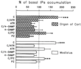
C C/ATR ATR C/4-DAMP 4-DAMP C / P Z PZ ] - - - - 4 * * * I ' I I M o d i o l u s ) )Fig. 1. Formation of IPs in the organ of Corti and in the modiolus. The data are means+S.E.M, of 6 experiments, at least, and are expressed as percentage of their respective basal levels of IPs acccu- mulation. These basal levels are 0.1255:0.014 dpm [3H]IPs/dpm [3H]inositol in the organ of Corti and 0.038 5:0.002 dpm [3H]IPs/dpm [3H]inositol in the modiolus. Carbachol was used at 1 mM while atropine, 4-DAMP and pirenzepine were applied at 1 /~M. C, carbachol; ATR, atropine; PZ, pirenzepine. * Different from control with P < 0.05; ** different from control with P < 0.01; * * * differ-
202
tubes containing 500 ~1 Krebs-Ringer buffer. The tubes were quickly transferred to a water bath thermostatically maintained at 37°C and continuously bubbled with the 95% O 2 / 5 % C O 2 mixture.
Stimulation of tritiated IP formation
LiC1 (10 mM) and the muscarinic antagonists 4 - D A M P and pirenzepine (where applicable) were added to each tube 15 min prior
to stimulation by carbachol (1 mM). Carbachol was allowed to rcacl for 20 min. T h e reaction was stopped by the application of 50 #1 ol perchloric acid (72%) per tube and by placing the tubes on ice. The cochlear tissues were homogenised by sonication and the h~- m o g e n a t e s were centrifuged at 2000 x g for 5 rain. The s u p e r n a t a n t s containing the [3H]IPs were neutralised with 1.5 M KOH/I).I)75 M HEPES. After separation from [3H]inositol and glycerophosphoinos-
Fig. 2. Loss of the o u t e r hair cells following the chronic t r e a t m e n t with amikacin. Ten-day-old rats received a daily injection of amikacin sulphate (20/*1, 479 mM) for 6 days. Following a 10-day recovery, the animals were sacrified and their cochleas processed for scanning and transmission electron microscopy, a, b and c are scanning electron micrographs of the surface of the organ of Corti. The inner hair cells (I), localised by their stereocilia bundles are present t h r o u g h o u t the organ of Corti, while the outer hair cells (O) are still present at the apex (a) but have been destroyed in the medial (b) and basal (c) turns. A semi-thin radial section of an amikacin-treated organ of Corti in the lower medial turn is shown in d. Arrows indicate the inner hair cell (I) and the Deiters' cells (D) that took the place of the destroyed o u t e r hair cells. Panel e depicts the apical pole of the Deiters'cells (D) that extended towards the surface of the organ of Corti, n u m e r o u s nerve fibers (arrows) are present in the space between the Deiters'cells and the pillar cells (P). f: high magnification of the fibers that allows the recognition of efferents (e). Bars = ll)
itols by anion-exchange chromatography, the [3H]IPs formed were measured in each cochlear sample as previously described 24.
Chronic treatment with the aminoglycoside amikacin
Ten-day-old Wistar rats from several litters received a daily subcutaneous injection of amikacin sulphate for 6 days. The injected volume was 20 /~1 of a sterile saline solution of amikacin sulphate concentrated at 479 mM. Following injections, the baby rats were returned to their mothers. This treatment represents a dosage of 300 mg of amikacin/kg body weight/day for a 12-day-old rat. At the end of the treatment, the rats were allowed to recover for 10 days. They reached then the age of 26 days, an age where morphological and physiological studies have demonstrated that the cochlea is mature. At this time, the animals were anaesthetised and killed and the cochleas were dissected, as described above, and processed for histology and measurement of the metabolism of IPs.
Section o f the crossed olicocochlear bundle
Mature Wistar rats (about 3-month-old) were deeply anaes- thetised by an intraperitoneal injection of Equithesin (300 p.l/100 g body weight) and placed in a stereotaxic instrument. The scalp was incised and the skull was opened along a 2.5-mm-wide midline, from the base of the occipital bone to the middle of the vertex in order to visualise the venous sinus. Without damaging the sinus, the brain was split by a rostro-caudal section from the vertex to the base of the occipital bone. The cut goes through the cerebellum and the brain stem to the bottom of the skull. The opening on the top of the occipital bone was closed with gel-foam and the skin was sewed. After an 11 day survival period, the animals were anaesthetised and killed, the cochleas were dissected, as described above, and pro- cessed for histology and measurement of the metabolism of IPs. The duration of the survival was determined by a preliminary time-course study (unpublished data).
Transmission electron microscopy
Following a deep anaesthesia with sodium pentobarbital and decapitation, the cochleas were dissected from the bulla and fixed in 2.5% glutaraldehyde buffered with sodium cacodylate (0.1 M, pH 7.4). The cochleas were quickly perfused with the same fixative through the windows and then kept in the fixative for 2 h. Following a buffer wash, the cochleas were further dissected and then postfixed in an aqueous solution of 1% osmium tetroxide for 1 h. Samples were dehydrated in graded series of ethanol (35-100%), embedded in Spurr resin and ultrathin sections were cut at different base-to-apex levels, in the transverse plane. The sections were observed with a Philips EM 301 electron microscope. Semithin sections were also cut for observation with Nomarski microscopy.
Scanning electron microscopy
The cochleas were processed as for the transmission electron microscopy, except that fixation time with osmium tetroxide was reduced to 10 min. During the dissection, special care was taken in the removal of the tectorial membrane. After dehydration, the speci- mens were critical point dried in liquid CO 2 and coated with gold, samples were observed with a Hitachi $4000 electron microscope examining on the outer hair cells from the base to the apex of the organ of Corti.
Data expression and statistical analysis
The data are expressed as ratios of the amount [3H]IPs (in dpm) formed to the amount of dpm of [3H]inositol taken up per cochlea (dpm [3H]IPs/dpm [3H]inositol). The total IPs are composed essen- tially of inositol trisphosphate (2%), inositol bisphosphate (23%) and inositol monophosphate (75%) 24. The amount of inositol taken up per cochlea is measured in the 'flow through' fraction of the anion- exchange chromatography used to purify the IPs. The data were not normalised to the amount of protein due to difficulty to measure very accurately the protein content of a single, dissected cochlea or cochlear region. The data are also given as percentage of the basal level of IP synthesis measured in the absence of neuroactive mole-
cules. All the data points are means_+S.E.M, of 6 experiments, at least. The statistical significance of the data was determined using one-way ANOVA.
R E S U L T S
Whatever the experimental destruction, the amounts of tritiated inositol taken up per cochlea were not different from controls.
Metabolism of IPs in the organ of Corti and the modio- lus of the mature cochlea
The basal accumulation of IPs is higher in the organ of Corti, with a level of 0.125 + 0.014 dpm [3H]IPs/ dpm [3H]inositol, than in the modiolus which the level of formation of IPs is 0.038 + 0.002 dpm [3H]IPs/dpm [3H]inositol (P < 0.001).
Following the application of carbachol (1 mM), the metabolism of IPs is stimulated in both cochlear com- partments (Fig. 1). In the organ of Corti, carbachol raises the IP response to 0.228 + 0.024 dpm [3H]IPs/ dpm [3H]inositol, corresponding to 182 + 19% of the basal level. In the modiolus, the formation of IPs is increased to 0.066 ___ 0.005 dpm [3H]IPs/dpm [3H]in- ositol (174 + 13% of control).
In the organ of Corti, the addition of the muscarinic antagonists atropine and 4-DAMP (both used at 1 ~M) completely blocks the stimulation induced by carbachol (1 mM), but they do not significantly change the basal
% of b a s a l IPs a c c u m u l a t i o n 100 2 0 0 3 0 0 4 0 0 I I I C / A T R C / 4 - D A M P c/Pz I
P
F
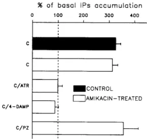
I I AMIKACIN-TREATED IFig. 3. Carbachol-induced accumulation of IPs in the amikacin- damaged cochleas. The accumulation of [3H]IPs was stimulated by 1 mM of carbachol in amikacin-damaged and control cochleas (top two bars). The data are means -I- S.E.M. of 6 experiments, at least and are expressed as percentage of their respective basal levels of IPs acccu- mulation. These basal levels are 0.020+0.003 dpm [3H]IPs/dpm [3H]inositol in control and 0.036 +0.008 dpm [3H]IPs/dpm [3H]in- ositol in amikacin-treated cochleas. The carbachol-induced synthesis of IPs was subjected to antagonism by atropine, 4-DAMP and pirenzepine (bottom three bars) that were applied at 1 /zM. C,
204
accumulation of IPs (Fig. 1). Unlike these compounds, pirenzepine, at 1 p.M, reduces the carbachol-activated synthesis of 1Ps by 63 ± 26% and the basal level by 42 _+ 1% (Fig. 1).
In the modiolus, atropine, 4-DAMP and piren- zepine, all of them applied at 1/.~M, do not affect the basal synthesis of IPs. However, the former two antag- onists completely block the stimulatory effect of carba- chol, while pirenzepine induces a 29 _+ 1% inhibition of the carbachol-enhanced formation of 1Ps (Fig. 1). Effects of the chronic treatment with amikacin
Observation of the surface of the organ of Corti using the scanning electron microscope, along the
cochlear coil, reveals that the apical surfaces of the outer hair cells have almost completely disappeared. At the apex, some outer hair cells may have survived since their apical membranes are still present (Fig. 2a). No outer hair cells are left in the medial and basal turns of the cochlea (Fig. 2b,c). Occasionally, some inner hair cells are missing in the basal turn (Fig. 2c). Based on scanning electron microscopy data, an assess- ment of the amikacin-induced outer hair cell loss indi- cates that about 90% of the outer hair cells have been destroyed. However, this assessment may under-esti- mate the extent of the hair cell loss because it has been shown that the apical surface of an outer hair cell can remain in the organ of Corti long after the degenera-
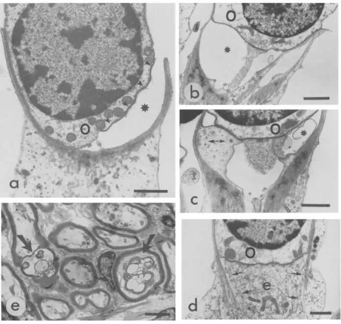
Fig. 4. Effects of crossed olivocochlear bundle section. Transmission electron micrographs a, b, c and d depict the synaptic poles of outer hair cells (O). In most of the cases the efferent terminals have d e g e n e r a t e d (a), leaving a space (stars) between the top of the Deiters' cells and the postsynaptic m e m b r a n e (arrowheads). Some relics of the terminals are also found in the spaces (b,c), together with expansions of the Deiters's cells (c, double arrow). Few large efferent terminals (e) are still connected with the outer hair cell (d) but irregularities are visible at the cytoplasmic m e m b r a n e (small arrows). Panel e shows abnormal efferent fibers (large arrows) within the intraganglionic bundle. Bars = 1 /zm
tion of the cell b o d y 16'22'35. Light microscope observa- tions of transverse sections of the organ of Corti of treated rats show that the outer hair cells are missing (Fig. 2d). The use of electron microscopy confirms this fact and also shows that Deiters' cells have expanded and taken the place of the outer hair cells (Fig. 2e). The treatment with amikacin does not affect the nerve fibers since many of them are seen in every section (Fig. 2e,f). Amongst these fibers, some large efferent terminals that contain a great amount of synaptic vesi- cles are often found (Fig. 2f).
Metabolism of IPs in the amikacin-exposed cochlea The basal synthesis of IPs is statistically similar in both control and treated cochleas, with the level o f formation of IPs being 0.020 + 0.003 dpm [3H]IPs/dpm [3H]inositol in control cochleas and 0.036 + 0.008 dpm [3H]IPs/dpm [3H]inositol in amikacin-treated cochle- as.
The activation of the IP metabolism by carbachol at 1 mM, elicits identical responses in both groups of cochleas. The carbachol-induced accumulation of IPs reaches 0.065 _+ 0.004 dpm [3H]IPs/dpm [3H]inositol (326 + 19% of basal control level) in untreated cochleas and 0.113 _+ 0.007 dpm [3H]IPs/dpm [aH]inositol (313 + 21% of basal 'treated' level) in amikacin-damaged cochleas (Fig. 3). This latter stimulation is completely abolished by the addition of 1/.LM of either atropine or 4-DAMP (Fig. 3) but it is not affected by pirenzepine (Fig. 3).
I I
I I
Effects of the section of the crossed olivo-cochlear bun- dle
Interrupting the efferent axons on the floor of the fourth ventricle results in a drastic degeneration of the efferent terminals that formed synapses with basal membranes of the outer hair cells 11 days postopera- tively (Fig. 4). Large spaces appear between the Deit- ers' cells and outer hair cells in place of the efferent terminals (Fig. 4a). Remnants of degenerated terminals are sometime found in these spaces (Fig. 4b,c), as well as expansions of Deiters' cells (Fig. 4c). Observation of cochleas from seven operated animals leads to the conclusion that about 50% of the outer hair cells have lost their synaptic contacts with the medial efferent fibers. In a quarter of cases, remains of terminals are seen associated with post-synaptic specializations of the membrane of the outer hair cells. Lastly, about 25% of the outer hair cells are still contacted by efferent synaptic boutons (Fig. 4d). At the level of the spiral ganglion, some degenerating efferent fibers are seen in the intraganglionic bundle (Fig. 4e).
Metabolism of IPs in the organ of Corti and the modio- lus following the section of the crossed olivocochlear bundle
The unstimulated, basal level of IP synthesis is sta- tistically similar in control organs and in organs of Corti from operated rats since in the latter the basal accumulation of IPs is 0.101 _+ 0.030 dpm [3H]IPs/dpm [3H]inositol (control level being 0.125_+0.014 dpm [3H]IPs/dpm [aH]inositol). In modiolar parts, the un- stimulated IP formation reach 0.029 _+ 0.003 dpm [3H]IPs/dpm [3H]inositol following the section of the crossed olivocochlear bundle. This value is not statisti- cally different from the basal production of IP in control modiolar tissue (0.038 + 0.002 dpm [3H]IPs/ dpm [3H]inositol).
The stimulation of cochlear regions of operated rats with carbachol (1 mM) enhances the synthesis of IPs to 0.149_+ 0.014 and 0.062_+ 0.008 dpm [aH]IPs/dpm [3H]inositol in the organ of Corti and the modiolus, respectively (Fig. 5). The stimulatory effects of carba-
UNSTIM UNSTIM CARB CARB C A R B / A T R IP Formotion (dpm I P / d p m inositol)xlO00 5 0 1 0 0 1 5 0 2 0 0 2 5 0 , 5 0 0 i i i i I i ~ C o n t r o l [--1Section Orgon of Corti UNSTIM UNSTIM CARB CARB CARB/ATR 2 0 4 0 6 0 8 0 1 0 0 i i i i i ~ C o n t r o l - - ~ [--lSection
P
ModiolusFig. 5. IP metabolism in the organ of Corti and in the modiolus following the section of the crossed olivocochlear bundle. Data are means + S.E.M. of 6 experiments, at least. Black bars show the basal and carbachol- (1 mM) evoked accumulations of IPs in control, unoperated cochlear regions. White bars depict the basal and carba- chol- (1 mM) evoked accumulations of IPs, as well as the block of the carbachol stimulation by atropine (1 ~zM) in operated organs of Corti (top panel) and operated modioli (bottom panel). UNSTIM, unstim-
206
chol are abolished completely by the addition of at- ropine (1 ~ M ) in both regions of the cochlea (Fig. 5). In the presence of the muscarinic antagonist, the for- mation of IPs amounts to 0.080 _+ 0.022 and 0.037 _+ 0.011 dpm [ 3 H ] I P s / d p m [3H]inositol in the organ of Corti and the modiolus, respectively.
When comparing the stimulation of the IP metabo- lism produced by carbachol in the cochlear compart- ments of control and operated rats, a difference be- comes evident in the case of the organ of Corti. A carbachol-induced rise of 47 _+ 14% above basal forma- tion of IPs is obtained following the section of the crossed efferent bundle, while the carbachol-evoked increase of the IP response represents a 82 _+ 19% rise above basal level in normal animals. The stimulated IP response is significantly smaller in organs of Corti of operated rats than in controls ( P < 0.01). No differ- ence was noticed between modiolar fractions from both groups of rats.
DISCUSSION
Stimulation of muscarinic receptors enhance the hydrolysis of phosphatidylinositol 4,5-bisphosphate in the organ of Corti and in the modiolus. In both cochlear parts, the stimulatory effect of carbachol (1 mM) is antagonised by atropine (1 /xM), showing that carba- chol elicits the synthesis of IPs via an interaction with muscarinic receptors. The application of 4-DAMP (1 txM) results in the complete blockage of the carbachol stimulation while pirenzepine (1 txM) lacks significant effects. Actually, a partial inhibition of the carbachol- induced formation of IPs is obtained with pirenzepine in the modiolus, but in fact this inhibition is due to a reduction of the basal metabolism of IPs, the reason for which remains unclear; one may speculate that some endogenous ligands for pirenzepine-sensitive sites are present in our in vitro model.
Recently, in the mice cochlea, Drescher and co- workers ~' identified mRNAs encoding three mus- carinic receptors coupled to IP metabolism, namely ml, m3 and m5 mRNAs. This result suggests the presence of the M 1, M 3 and M 5 receptors in the cochlea. While the identification of M 5 receptors is prevented by the lack of selective ligand, previous electrophysiological and biochemical observations have shown that cochlear muscarinic receptors have a very low affinity for pirenzepine (M 1 selective) and a high affinity for 4-DAMP (M 3 selective) 4'6'24'33. For in- stance, previously-reported d o s e - r e s p o n s e curves for the effects of specific antagonists on carbachol-indueed IP synthesis showed that 1 IxM of 4-DAMP abolishes completely the effect of carbachol (1 mM) while piren-
zepine at 1 /xM does not affect the carbachol-activated transduction system 4'24. These data indicate that mus- carinic receptors are mainly of the M~ type in the cochlea. In addition, binding surveys demonstrated thc existence of only one population of muscarinic sites in the cochlea 6'27'56'5~. Together with mRNA analysis ~', the whole body of data suggests that the synthesis of Mj and M 5 proteins may be regulated posttranscrip- tionally. Consequently and since, in the present study, 4-DAMP (1 /xM) is more potent than pirenzepine (I # M ) in both the organ of Corti and the modiolus, it appears that both cochlear parts contain thc same typc of functional muscarinic receptor which is probably the M 3 type.
Muscarinic receptors in the modiolus
Although binding experiments that detected mus- carinic sites in both cochlear nerves and spiral gangl- ions 27'58 suggest the transport of muscarinic receptors, the fact that carbachol activates the synthesis of IPs via an atropine-sensitive mechanism in the modiolus, demonstrates the fairly unexpected presence of func- tional muscarinic receptors in this cochlear compart- ment. It appears, therefore, that the binding sites may be related to both transported and cell surface func- tional cholinoceptors 55.
The carbachol-induced release of IPs in the modio- lus suggests that muscarinic receptors could be located at the level of the cell bodies of the spiral ganglion neurons a n d / o r nerve fibers a n d / o r Schwann cells. Although there is no previous data providing support for the presence of functional receptors on the neu- ronal cell bodies, this possibility cannot be excluded yet. The presence of functional muscarinic receptors in the fibers is questionable because the carbachol- activated IP turnover is unchanged in the modiolus despite the degeneration of some fibers, following the section of the crossed olivocochlear bundle. However. functional receptors may be located in the fibers, as this has been shown in the sciatic and vagus nerves 14'52 where phosphatidytinositol 4,5-bisphosphate is hydrol- ysed following the activation of M1 and M 3 cholinocep- tors ~4,s2, which are likely to be coupled to a calcium- sensitive phospholipases C ~7"4° via specific G proteins Gq/G71. Muscarinic receptors could be involved in the regulation of the axoplasmic transport because the stimulation of these receptors suppresses the fast ax- onal transport in cultured superior cervical neurons 54. Despite the lack of direct evidence for the expres- sion of muscarinic receptors coupled to the 1P cycle in Schwann cells, there are reports suggesting indirectly that Schwann cells may possess such receptors within their myelin sheath. Indeed, in the peripheral nervous
system, myelin sheaths are particularly rich in phos- phatidylinositol 4,5-bisphosphate 36 and contain both phospholipase C enzymes and their stimulatory G pro- teins 7'37. Moreover, the carbachol-stimulation of mus- carinic receptors enhances the IP turnover in brain myelin 28'34. Then, if cochlear Schwann cells do bear muscarinic receptors, these receptors may regulate the turnover of phospholipid within the myelin sheath.
Muscarinic receptors in the organ of Corti
The responses obtained in the rat organ of Corti show the expression of functional muscarinic receptors and support the previous demonstration of the pres- ence of muscarinic receptors linked to the activation of phospholipase C in the guinea pig organ of Corti 4~. At the cellular level, the most obvious sites of muscarinic receptors are at the cholinergic s y n a p s e s 1'13"2°'47'48. I n
order to shed more light on the synaptic position of the metabotropic cholinoceptor, we investigated, in the outer region of the organ of Corti, the effects of the loss of presynaptic and postsynaptic elements on the carbachol-evoked IP response.
Amikacin treatment leads to the degeneration of almost all outer hair cells without changing the magni- tude of the carbachol-induced formation of IPs. At first sight, this would indicate that outer hair ceils do not possess muscarinic receptors, as first suggested by Plinkert and co-workers 44. However, a reservation must be brought to this straightforward conclusion since outer hair cells represent a small proportion of the total cells in the whole cochlea, and because mus- carinic, electrophysiological responses have been recorded in isolated outer hair cells 19"29. It is possible that our assay is not sensitive enough to detect changes in the formation of IPs caused by outer hair cell loss, because the synthesis of IPs is also enhanced by carba- chol in other cochlear cells, as indicated for instance by the current results obtained in the modiolus. Thus, a more careful conclusion would be to state that the localisation of muscarinic cholinoceptors is not re- stricted to outer hair cells. The muscarinic receptors of these latter cells may be involved in changes in the cytosolic concentration of calcium ions which have been proposed to modulate the slow motility of the outer hair cells ~8.
The interruption of the crossed olivocochlear bun- dle causes a significant reduction of the level of carba- chol-enhanced release of IPs. Since the medial termi- nals which form synapses with the outer hair cells, are selectively lost following the surgery, this demonstrates that many muscarinic receptors are associated with these presynaptic terminals. Although no clue is avail- able to point to a role for these presynaptic cholino-
ceptors, a tempting hypothesis is that muscarinic recep- tors are autoreceptors which modulate the release of acetylcholine 3°. Support for this hypothesis is found in the ability of M 3 prejunctional autoreceptors to inhibit the release of acetylcholine in the guinea pig ileum 12'3~, the rat striatum ~5 and the bovine cerebral arteries 21. Besides regulating acetylcholine release, these mus- carinic receptors may control the release of A T P which increases the calcium permeability in isolated outer hair cell 2'39 as well as the IP turnover in the organ of Corti 42. Such a control of purinergic transmission has been reported in adrenal chromaffin cells 6°.
Conclusion
We demonstrate that muscarinic receptors function- ally coupled to phospholipase C are distributed in both the organ of Corti and the modiolus. T h e pharmacolog- ical features of these receptors are similar in both structures and seem indicative of M 3 muscarinic recep- tors. While the cellular localisation of the cholinocep- tors in the modiolus is still to be determined, the current results clearly show that a large number of muscarinic receptors are present on presynaptic termi- nals of the medial efferent system in the organ of Corti. Such autoreceptors may regulate the release of neurotransmitter at the synapses between the medial cholinergic axons and the outer hair cells.
Acknowledgements. We gratefully thank Dr. Marc Lenoir for his very generous help and instruction of the electron microscope tech- nics and Dr. Guy Richardson and Prof. Jochen Schacht for their useful comments. Thanks are also due to Pierre Sibleyras who skillfully printed the figures.
REFERENCES
1 Altschuler, R.A., Kachar, B., Rubio, J., Parakkal, M. and Fex, J., Immunocytochemical localization of choline acetyltransferase-like immunoreactivity in the guinea pig cochlea, Brain Res., 338 (1985) 1-11.
2 Ashmore, J.F. and Ohmori, H., Control of intracellular calcium by ATP in isolated outer hair cells of guinea pig cochlea, J. Physiol., 428 (1990) 109-131.
3 Bartolami, S., Guiramand, J., Lenoir, M., Pujol, R. and R6casens, M., Carbachol-induced inositol phosphate formation during rat cochlea development, Hear Res., 47 (1990) 229-234.
4 Bartolami, S., Lebrun, F., Mayat, E., Guiramand, J., Lenoir, M., Rebillard, G., Lippe, W.R., Pujol, R. and R6casens, M., Pharma- cological characterization of muscarinic receptors and their puta- tive involvement in the developing peripheral auditory organ. In R.J. Wegmann and M.A. Wegmann (Eds.), Recent Advances in Cellular and Molecular Biology, Vol. 3, Peeters, Leuven, 1992, pp. 251 - 260.
5 Bartolami, S., Planche, M., Ripoll, C., Pujol, R. and Lenoir, M., Effects of ototoxic drugs and selective destruction on the inositol phosphate signalling pathway coupled to cochlear muscarinic receptors, Abstr. Mol. BioL Hearing Deafness, 1 (1992) 72. 6 Bartolami, S., Planche, M. and Pujol, R., Characterization of
muscarinic binding sites in the adult and developing rat cochlea, Neurochem. Int., in press
208
T.Y., Identification of G protein subtypes in peripheral nerve and cultured Schwann cells, Z Neurochem., 59 (1992) 1729-1735. 8 Bielinsky, D.F., Morin, P.J., Dickey, B.F. and Fine, R.E., Low
molecular weight GTP-binding proteins are associated with neu- ronal organelles involved in rapid axonal transport and exocyto- sis, J. Biol. Chem., 264 (1989) 18363-18367.
9 Bobbin, R.P. and Konishi, T., Acetylcholine mimics crossed olivo- cochlear bundle stimulation, Nature, 231 (19711 222-223. 10 Bobbin, R.P. and Konishi, T., Action of cholinergic and anti-
cholinergic drugs at the crossed olivocochlear bundle-hair cell junction, Acta Otolaryngol., 77 (1974) 56-65.
11 Canlon, B., Cartaud, J. and Changeux, J.P., Localization of alpha-bungarotoxin binding sites on outer hair cells from the guinea pig cochlea, Acta Physiol. Scand., I37 (1989) 549-551/. 12 Dammann, F., Fuder, H., Giachetti, A., Giraldo, E., Kilbinger, H.
and Micheletti, R., AF-DX 116 differentiates between prejunc- tional muscarine receptors located on noradrenergic and cholin- ergic nerve, Naunyn-Schmiedeberg Arch. Pharmacol., 339 (1989) 268-271.
13 Dannhof, B., Roth, B. and Bruns, V., Anatomical mapping of choline acetyltransferase (ChAT)-like and glutamate decarboxyl- ase (GAD)dike immunoreactivity in outer hair cell efferents in adult rats, Cell Tissue Res., 266 (19911 89-95.
14 Day, N.S., Berti-Mattera, L.N. and Eichberg, J., Muscarinic cholinergic receptor-mediated phosphoinositide metabolism in peripheral nerve, J. Neurochem., 56 (1991) 1905-1913.
15 De Boer, P., Westerink, B.C.H., Rollema, H., Zaagsma, J. and Horn, A.S., An M3-1ike muscarinic receptor regulates the in vivo release of acetylcholine in rat striatum, Eur. J. Pharmacol., 179 (1990) 167-172.
16 De Groot, J.C.M.J., Huizing, E.H. and Veldman, J.E., Early ultrastructural effects of gentamicin cochleotoxicity, Acta Oto- laryngol., 111 (1991) 273-280.
17 Drescher D.G., Upadhyay S., Wilcox E. and Fex J., Analysis of muscarinic receptors subtypes in the mouse cochlea by means of the polymerase chain reaction, J. Neurochem. 59 (1992) 765-767. 18 Dulon, D. and Schacht, J., Motility of cochlear outer hair cells,
Am. J. OtolaryngoL, 13 (1992) 108-112.
19 Erostegui, C., Norris C.H. and Bobbin, R.P., Effects of acetyl- choline on isolated outer hair cells, Abstr. Assoc. Res. Otola~.n- got., 16 11993) 329.
20 Eybalin, M. and Pujol, R., Choline acetyltransferase (CHAT) immunoelectron microscopy distinguishes at least three types of efferent synapses in the organ of Corti, Exp. Brain Res., 65 (19871 261-270.
21 Ferrer, M., Galvan, R., Marin, J. and Balfagon, G., Presynaptic muscarinic recptor subtypes involved in the inhibition of acetyl- choline and noradrenaline release in bovine cerebral arteries,
Naunyn- Schmiedeberg Arch. PharmacoL , 345 11992) 619-626. 22 Forge, A., Outer hair cell loss and supporting cell expansion
following chronic gentamicin treatment, Hearing Res., 19 (1985) 171-182.
23 Galper, J.B., Dziekan, L.C., O'Hara, D.S. and Smith, T.W., The biphasic response to muscarinic cholinergic receptors in cultured heart cells to agonists, J. Biol. Chem., 257 11982) 10344-10356. 24 Guiramand, J., Mayat, E., Bartolami, S., Lenoir, M., Rumigny,
J.F., Pujol, R. and Recasens, M., A M3 muscarinic receptor coupled to inositol phosphate formation in the rat cochlea?,
Biochem. Pharmacol., 39 (19901 1913-1919.
25 Housley, G. and Ashmore, J.F., Direct measurement of the action of acetylcholine on isolated outer hair cells of the guinea pig cochlea, Proc. R. Soc. Lond. B, 244 11991) 161-167.
26 Iurato, S., Smith, C.A., Eldredge, D.H., Henderson, D., Carr, C., Ueno, Y., Cameron, S. and Richter, R., Distribution of the crossed olivocochlear bundle in the chinchilla's cochlea, J. Cornp. Neurol., 182 119781 57-76.
27 James, W.M., Cheatham, M.A. and Klein, W.L., Muscarinic acetylcholine receptor binding in the guinea pig cochlea, Hearing Res., 9 119831 113-121.
28 Kahn, D.W. and Morell, P., Phosphatidic acid and phosphoinosi- tide turnover in myelin and its stimulation by acetylcholine, J.
Neurochem., 50 11988) 1542-1550.
29 Kakehata, S., Nakagawa, T., Takasaka, T. and ,\kaike, N., (:cllu- lar mechanism of acetylcholine-induced response m dissociated- outer hair cells of guinea pig cochlea. ,L Physiol., 463 119931 227-224.
31) Kilbinger, 1-t.. Presynaptic muscarinic receptors modnlatmg acetylcholine release, Trend~ Pharmacol. Sci., March (1c)~4) 103 105.
31 Kilbinger, H., Halim, S., Lambrecht, G., Weiler, W. and Wessler, I., Comparison of affinities of muscarinic antagonists to pre- and postjunctional receptors in the guinea-pig ileum, Eur..I. Pharma-
col., 1(13 11984) 313 320.
32 Kujawa, S.G., Glattke, T.J., Fallom M. and Bobbin, R.P., lntra cochlear application of acetylcholine alters sound-induced me- chanical events within the cochlear partition, ftearing Res., 61 119921 106-116.
33 Kujawa, S.G., Fatlon, M. and Bobbin, R.P., Effect of cholinergic antagonists on contralateral suppression of DPOAEs, Abstr. A.~-
soc. Res. Otolaryngol., 16 (19931 394.
34 l_,arocca, J.N., Cervone, A. and Ledeen, R.W., Stimulation of phosphoinositide hydrolysis in myelin by muscarinic agonist and potassium, Brain Res., 436 11987) 357-362.
35 Lenoir, M. and Puel. J.L., Dose dependent changes in the rat cochlea following aminoglycoside intoxication. II. Histological study, Hearing Res., 26 (1987) 199-209.
36 Lowery, J.M., Berti-Mattera, L.N., Zhu, X., Pctcrson, R.G. and Eichberg, J., Relationship of ATP turnover, phosphoinositide metabolism, and protein phosphorylation in sciatic nerve and derived peripheral myelin subfractions from normal and strepto- zotocin diabetic rats, J. Neurochem., 52 (1989)921-932. 37 Mathew, J., Date, S. and Eichberg, J., Activity and distribution of
phosphoinositidase C in rat sciatic nerve, .L Neurosci. Res.. 33 (1992) 122-128.
38 Mountain, D.C., C'hanges in the endolymphatic potential and crossed olivocochlear bundle stimulation alter cochlear mechan- ics, Science, 210 (198/)) 71-72.
39 Nakagawa, T, Akaike, N., Kimitsuki, T., Komunc, S. and Arima, T., ATP-induced current in isolated outer hair cells of guinea pig cochlea, J. Neurophysiol., 63 (1990) 1068-11174.
40 Natarajan, V. and Schmid, H.H.O., Inositol phospholipid hydrol- ysis by rat sciatic nerve phospholipase C, J. Neurochem., 49 119871
1878-1887.
41 Niedzielski, A., Ono, T. and Schacht, J., Cholinergic regulation ot the phosphoinositide second messenger system in the guinea pig organ of Corti, Hearing Res., 59 (1992) 250-254.
42 Niedzielski, A. and Schacht, J., P2 purinoceptors stimulate inosi- tol phosphate release in the organ of Corti, NeuroReport, 3
( 19921 273-275.
43 Plinkert, P.K.. Gitter, A.tt., Zimmermann, U., Kirchner, T., Tzartos, S. and Zenner, H.P., Visualization and functional testing of acetylcholine receptor-like molecules in cochlear outer hair cells, Hearing Res., 44 (1990) 25-34.
44 Plinkert, P.K., Zenner, tt.P. and Heilbronn, E., A nicotinic acetylcholine receptor-like alpha-bungarotoxin-binding site on outer hair cells, Hearing Res., 53 (1991) 123-130.
45 Puel, J.L. and Rebillard, G.. Effect of contralateral sound stimu- lation on the distortion product 2 F ~ - F : : Evidence that the medial efferent system is involved, .l. Acoust. Soc. Am., 87 11990) 1630-1635.
46 Richardson, G.P. and Russell, l.J., Cochlear cultures as a model system for studying aminoglycoside induced ototoxicity, Hearing Res., 53 (19911 293-311.
47 Roth, B., Dannhof, B. and Bruns, V., ChAT-like immunoreactiv- ity of olivocochlear fibers on rat outer hair cells during the postnatal development, Anat EmbryoL, 183 119911 483-489. 48 Safieddine, S. and Eybalin, M., Triple immunofluorescence evi-
dence for the coexistence of acetylcholine, enkephalins and calci- tonin gene-related peptide within efferent (olivocochlear) neu- rons of rats and guinea-pigs, Eur. J. Neurosci., 4-(19921 981-992. 49 Schacht, J. and Zenner, H.-P., The phosphoinositide cascade in
isolated outer hair cells: possible role as second messenger for motile responses, Hearing Res., 22 11986) 94.
mediate motility in cochlear outer hair cells, Hearing Res., 31 (1987) 155-160.
51 Siegel, J.H. and Kim, D.O., Efferent control of cochlear mechan- ics? Olivocochlear bundle stimulation affects cochlear biome- chanical nonlinearity, Hearing Res., 6 (1982) 171-182.
52 Sierro, C.D., Vitus, J. and Dunant, Y., Effects of muscarinic agonists and depolarizing agents on inositol monophosphate ac- cumulation in rabbit vagus nerve, J. Neurochem., 59 (1992) 456- 466.
53 Spoendlin, H.Anatomy of cochlear innervation, Am. J. Otolaryn- gol., 6 (1985) 453-467.
54 Takenaka, T., Kawakami, T., Hikawa, N., Bandou, Y. and Gotoh, H., Effect of neurotransmitters on axoplasmic transport: acetyl- choline effect on superior cervical ganglion cells, Brain Res., 588 (1992) 212-216.
55 Thompson, A.K. and Fisher, S.K., Preferential coupling of cell surface muscarinic receptors to phosphoinositide hydrolysis in human neuroblastoma cells, J. BioL Chem., 266 (1991) 5004-5010.
56 van Megen, Y.J.B., Klaassen, A.B.M., Rodrigues de Miranda, J.F. and Kuijpers, W., Cholinergic muscarinic receptors in rat cochlea, Brain Res., 474 (1988) 185-188.
57 Warr, W.B., Guinan, J.J. Jr. and White, J.S., Organization of the efferent fibers: the lateral and medial olivocochlear systems. In R.A. Altschuler, D.W. Hoffman and R.P. Bobbin (Eds.), Neuro- biology of Hearing: The Cochlea, Raven, New York, 1986, pp.
159-191.
58 Whipple, M.R. and Drescher, D.G., Muscarinic receptors in the cochlear nucleus and auditory nerve of the guinea pig, J. Neu- rochem., 43 (1984) 192-198.
59 Wright, C.G. and Preston, R.E., Degeneration and distribution of efferent nerve fibers in the guinea pig organ of Corti. A light and scanning electron microscopic study, Brain Res., 58 (1973) 37-59. 60 Xu, Y., Duarte, E. and Forsberg, E.J., Calcium dependency of muscarinic and nicotinic agonist-induced ATP and catecholamine secretion from porcine adrenal chromaffin cells, J. Neurochem., 56 (1991) 1889-1896.