HAL Id: inserm-02454376
https://www.hal.inserm.fr/inserm-02454376
Submitted on 24 Jan 2020
HAL is a multi-disciplinary open access
archive for the deposit and dissemination of
sci-entific research documents, whether they are
pub-lished or not. The documents may come from
teaching and research institutions in France or
abroad, or from public or private research centers.
L’archive ouverte pluridisciplinaire HAL, est
destinée au dépôt et à la diffusion de documents
scientifiques de niveau recherche, publiés ou non,
émanant des établissements d’enseignement et de
recherche français ou étrangers, des laboratoires
publics ou privés.
an Old Field
Géraldine Gentric, Virginie Mieulet, Fatima Mechta-Grigoriou
To cite this version:
Géraldine Gentric, Virginie Mieulet, Fatima Mechta-Grigoriou. Heterogeneity in Cancer Metabolism:
New Concepts in an Old Field. Antioxidants and Redox Signaling, Mary Ann Liebert, 2017, 26 (9),
pp.462-485. �10.1089/ars.2016.6750�. �inserm-02454376�
F
ORUM
R
EVIEW
A
RTICLE
Heterogeneity in Cancer Metabolism:
New Concepts in an Old Field
Ge´raldine Gentric,1,2,* Virginie Mieulet,1,2,* and Fatima Mechta-Grigoriou1,2
Abstract
Significance: In the last years, metabolic reprogramming, fluctuations in bioenergetic fuels, and modulation of
oxidative stress became new key hallmarks of tumor development. In cancer, elevated glucose uptake and high
glycolytic rate, as a source of adenosine triphosphate, constitute a growth advantage for tumors. This represents
the universally known Warburg effect, which gave rise to one major clinical application for detecting cancer
cells using glucose analogs: the positron emission tomography scan imaging.
Recent Advances: Glucose utilization and carbon sources in tumors are much more heterogeneous than initially
thought. Indeed, new studies emerged and revealed a dual capacity of tumor cells for glycolytic and oxidative
phosphorylation (OXPHOS) metabolism. OXPHOS metabolism, which relies predominantly on mitochondrial
respiration, exhibits fine-tuned regulation of respiratory chain complexes and enhanced antioxidant response or
detoxification capacity.
Critical Issues: OXPHOS-dependent cancer cells use alternative oxidizable substrates, such as glutamine and
fatty acids. The diversity of carbon substrates fueling neoplastic cells is indicative of metabolic heterogeneity,
even within tumors sharing the same clinical diagnosis. Metabolic switch supports cancer cell stemness and
their bioenergy-consuming functions, such as proliferation, survival, migration, and invasion. Moreover,
re-active oxygen species-induced mitochondrial metabolism and nutrient availability are important for interaction
with tumor microenvironment components. Carcinoma-associated fibroblasts and immune cells participate in
the metabolic interplay with neoplastic cells. They collectively adapt in a dynamic manner to the metabolic
needs of cancer cells, thus participating in tumorigenesis and resistance to treatments.
Future Directions: Characterizing the reciprocal metabolic interplay between stromal, immune, and neoplastic
cells will provide a better understanding of treatment resistance. Antioxid. Redox Signal. 26, 462–485.
Keywords:
mitochondria, cancer, ROS, immunology, metabolism, oxidative stress
Introduction
T
he energy metabolism fieldhas emerged in the mid-19th century with the pioneering works from O. War-burg, who deciphered the nature and mode of action of en-zymes involved in respiration (Nobel Prize in Physiology, 1931) and fermentation (prevented from receiving a 2nd Nobel Prize in 1944 by the Nazi regime), A.L. Lenhinger, defining mitochondria as the main site of oxidativephos-phorylation (OXPHOS), H.A. Krebs, uncovering the tricar-boxylic acid (TCA) cycle (Nobel Prize in Physiology and Medicine, 1953), and S. Weinhouse, revisiting respiratory deficiencies in cancer cells (106, 144, 228, 229, 234, 235). Warburg’s observations revealed that adenosine triphosphate (ATP), the main source of energy, is mostly produced by aerobic glycolysis in cancer cells. Indeed, to make ATP, neoplastic cells rather produce lactate than oxidize pyruvate through the TCA cycle (212, 228, 229). This metabolic
1
Stress and Cancer Laboratory, E´ quipe Labelise´e LNCC, Institut Curie, Paris, France.
2Inserm, U830, Paris, France.
*These authors contributed equally to this work.
ª Ge´raldine Gentric, et al., 2016; Published by Mary Ann Liebert, Inc. This Open Access article is distributed under the terms of the Creative Commons Attribution Noncommercial License (http://creativecommons.org/licenses/by-nc/4.0/) which permits any noncommercial use, distribution, and reproduction in any medium, provided the original author(s) and the source are credited.
Mary Ann Liebert, Inc. DOI: 10.1089/ars.2016.6750
switch occurs under aerobic conditions, even if glycolysis is less efficient to produce ATP (2 ATP molecules instead of 36 by TCA cycle) and if it generates an acidic environment due to lactic acid production. This is the basis of the universally known Warburg effect, the gold standard process in cancer metabolism, during the last century.
Aerobic glycolysis is considered as a key feature in cancer and has recently taken place in the famous ‘‘Hallmarks of cancer’’ described by D. Hanahan and B. Weinberg (77). In many cancers, increased glucose uptake and enhanced gly-colytic rates, as a source of ATP, represent a growth advan-tage for tumor cells (63). Indeed, aerobic glycolysis represents also an effective shunting by providing carbon sources to other key metabolic pathways required for nu-cleotide, lipid, and amino acid synthesis, building blocks that are essential for highly proliferative cells (Fig. 1). For in-stance, increased levels of the glycolysis intermediate, glucose-6-phosphate (G6P), favor the pentose phosphate pathway (PPP) (Fig. 1). The PPP generates reducing equiv-alents, in the form of nicotinamide adenine dinucleotide phosphate (NADPH), used for redox homeostasis mainte-nance and fatty acid (FA) biosynthesis. Moreover, it produces ribose-5-phosphate used for nucleic acid synthesis and erythrose-4-phosphate required for aromatic amino acid production (84). Moreover, glycolysis intermediates in can-cer cells are also redirected toward the serine pathway, which starts with the conversion of 3-phosphoglycerate into phos-phohydroxypyruvate via the phosphoglycerate
dehydroge-nase (123, 162) (Fig. 1). This pathway is essential for amino acid (serine and glycine) synthesis and is also involved in the folate cycle, a major source of methyl groups for one-carbon pools and purine synthesis (122). Subsequently, this pathway provides essential precursors of proteins, nucleic acids, and glutathione-dependent antioxidant capacities. Although gly-colytic switch is now established as a key process in tumori-genesis, the cause and the mechanisms leading to this metabolic reprogramming are still under debate (24, 26, 115, 231). In brief, it was initially thought that mitochondria were bearing mutations and functionally defective, thus forcing tumor cells to adapt to this respiratory deficiency. However, mitochondria alterations are quite rare and electron micros-copy revealed that mitochondria are active. Moreover, several studies showed that cancers cells retain OXPHOS capacity and do not suffer from respiratory defects (58, 95, 170, 214, 235, 236, 239, 253). Furthermore, it has been shown that MCF7 breast cancer cells generate 80% of their ATP through mito-chondrial respiration (74). Finally, inhibiting glycolysis in neoplastic cells restores mitochondrial OXPHOS (18, 48, 135, 138), demonstrating that oxidative metabolism remains func-tional in most glycolytic cancer cells.
Physiological stresses, such as the lack of oxygen (O2), are
considered as one of the main drivers of the metabolic switch in tumor cells. As early tumors expand, they become hypoxic and require blood and nutrient supplies to keep growing. Decreased dependence on aerobic respiration becomes ad-vantageous and tumor metabolism is thus shifted toward
FIG. 1. Core metabolic pathways and enzymes in cancer cells.Here are schematically represented the main metabolic pathways altered in cancers, including the glycolysis, the PPP, the serine pathway, the fatty acid synthesis, and the TCA cycle. In cancer cells, the canonical energy metabolism pathways are often truncated (glycolysis, TCA cycle) or redirected (gluta-minolysis or serine and lipid biosynthesis). Briefly, glucose enters into cancer cells through glucose transporters and is phosphorylated to G6P by an irreversible reaction catalyzed by the hexokinase. G6P either proceeds through glycolysis to produce pyruvate or through the PPP to generate ribose-5-phosphate and NADPH. The PPP is connected at the first step of glycolysis starting with G6P dehydrogenase (G6PD) and has both an oxidative and nonoxidative arm. G6P oxidation produces the reducing equivalents, in the form of NADPH, important cellular antioxidant, and cofactor for fatty acid biosynthesis. Moreover, the PPP provides cancer cells with pentose sugars for the biosynthesis of nucleic acids. The first enzymes involved in the nonoxidative arm of the PPP are TKT and TA. Ribose-5-phosphate and xylulose-5-phosphate, generated by the oxidative PPP, can be further metabolized into F6P and G3P to reenter into glycolysis for ATP production, depending on the cell requirement. Thus, the PPP plays a key role in cancer cells to supply their anabolic demands and to counteract oxida-tive stress. The serine pathway is branched to glycolysis via 3-phosphoglycerate (3PG), which is converted by PHGDH into phosphohydroxypyruvate (P-PYR). This pathway produces serine and glycine, essential precursors for synthesis of proteins and nucleic acids through the folate cycle. Following glycolysis, pyruvate is either converted into lactate by LDHA and released through monocarboxylate transporters, MCT4 and MCT1, further causing extra cellular acidification, or converted into acetyl-CoA, through the PDH complex. Acetyl-CoA enters into TCA cycle and produces ATP, NADH, and FADH2 molecules. Reduced cofactors are then oxidized by the ETC complexes for ATP production. Glutamine and other amino acids can also replenish the TCA cycle. Indeed, the first step of glutaminolysis is the conversion of glutamine into glutamate by the GLS. Glutamate is subsequently converted into alpha-ketoglutarate (aKG) that fuels back the TCA cycle. Fatty acid degra-dation can also supply the TCA cycle through beta-oxidegra-dation, which produces acetyl-CoA. Reciprocally, citrate, a TCA cycle intermediate, can be used as a precursor for fatty acid synthesis and for NADPH production through the ACL. Citrate is subsequently converted to acetyl-CoA and OAA into the cytoplasm. Acetyl-CoA is used for fatty acid synthesis through its conversion to malonyl-CoA by ACC and to palmitic acid by the FASN. OAA is converted to malate, which is then decarboxylated into pyruvate, by the ME1 and produces NADPH. Mitochondria are represented by dotted line. ACC, acetyl-CoA carboxylase; ACL, ATP citrate lyase; aKG, alpha-ketoglutarate; ASCT2, amino acid transporter; ATP, adenosine triphosphate; DHAP, dihydroxyacetone phosphate; ETC, electron transport chain; FAD, flavin adenine dinucleotide; FASN, fatty acid synthase; F6P, fructose-6-phosphate; GADP, glyceraldehyde-3-phosphate; G3P, glyceraldehyde-3-phosphate; GLS, glutaminase; G6P, glucose-6-phosphate; GLUT, glucose transporter; LDHA, lactate dehydrogenase A; MCT, mono-carboxylate transporter; ME1, malic enzyme; NAD, nicotinamide adenine dinucleotide; NADPH, nicotinamide adenine dinucleotide phosphate; OAA, oxaloacetate; PDH, pyruvate dehydrogenase; PEP, phospho-enol-pyruvate; 6PG, 6-phospho-gluconolactone; 3PG, 3-phopho-glycerate; PGD, phosphogluconate dehydrogenase; PHGDH, phosphoglycerate dehydroge-nase; PPP, pentose phosphate pathway; P-PYR, phosphohydroxypyruvate; PSAT1, phosphoserine aminotransferase; SHMT1, serine hydroxymethyl transferase; Succ-CoA, succinyl-CoA; TCA, tricarboxylic acid; TA, transaldolase; TKT, transketolase. To see this illustration in color, the reader is referred to the web version of this article at www.liebertpub.com/ars
glycolysis, therefore balancing O2 consumption with O2
supply. Under hypoxia, the hypoxia-inducible factor-1 (HIF-1) transcription factor accumulates and provides O2 to
tu-mor cells by increasing angiogenesis, erythropoiesis, and glycolysis (21, 29, 163). HIF-1 enhances glycolysis by in-creasing expression of glucose transporters (GLUT1-3) and glycolytic enzymes, including hexokinase 2 (HK2) and lac-tate dehydrogenase A (LDHA). HIF-1 also inhibits the TCA cycle by upregulating the transcription of pyruvate dehy-drogenase kinase (PDK), which inactivates the pyruvate de-hydrogenase (PDH), preventing the conversion of pyruvate into acetyl coenzyme A (acetyl-CoA) (20, 103, 155, 163). HIF-1 is not only activated upon low O2concentration but
also under normoxic conditions by oncogenic and oxidative stress (7, 36, 65, 111). Aerobic glycolysis and concomitant
increase in glucose uptake give rise to one major clinical application for detecting cancer cells: the positron emission tomography (PET) scan, an imaging technology, which uses glucose analog tracer. In oncology, PET scan consists in measuring disease extent, lymph node localization, predic-tion of relapse, and diagnostic accuracy (4, 248). Despite the major advances provided by this technique in clinical prac-tice, some tumors do not absorb glucose and false-negative results have been diagnosed for some cancer patients. Taken together, these observations strongly suggest that glucose utilization and carbon sources in tumors could be more het-erogeneous than initially thought (33, 176).
Our review will describe tumor metabolism heterogeneity, considering both cancer cells and cells of the tumor micro-environment (TME). We will focus on recent findings
highlighting OXPHOS metabolism in neoplastic cancer cells, thereby arguing that glucose is not the only energy fuel and can be substituted, in part, by glutamine, serine/glycine, or FA for replenishing TCA cycle intermediates. Moreover, we will discuss the mechanisms regulating synthesis of mito-chondria respiratory complexes, which are absolutely re-quired to support OXPHOS metabolism. We will then describe the impact of metabolic variations on cancer cell functions, such as proliferation, survival, migration, invasion, and stemness. Finally, we will provide clues on the role of metabolism in the cross-talk between tumor cells and some key players of the TME, such as macrophages, lymphocytes, and fibroblasts, and we will show how metabolism can de-termine their differentiation.
Cancer Metabolism Heterogeneity
Over the past decade, advances in cancer research have enhanced our understanding of how metabolic reprogram-ming supports the anabolic requirements of cancer cell pro-liferation. It has become clear enough that a single metabolic program cannot be used to globally define altered tumor metabolism.
OXPHOS metabolism: modulation of reactive oxygen species levels and antioxidant response
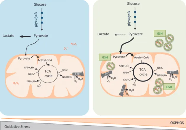
At the beginning of the 21st century, our view of cancer metabolism gained in complexity and became more attrac-tive, with the birth of new concepts (Fig. 2). The break with the Warburg dogma started when scientists studied cancer metabolism without comparing tumors with their normal counterparts. New studies emerged and proposed a dual ca-pacity for glycolytic and oxidative metabolism in tumor cells (127, 139, 147). First, gene expression profiling of 176 dif-fuse large B-cell lymphomas revealed the existence of a subset of tumors characterized by an OXPHOS gene pattern (137). In agreement with this observation, N. Danial’s labo-ratory highlighted two subgroups of lymphoma cells with either an OXPHOS or a non-OXPHOS metabolism based on the protein expression level of electron transport chain (ETC) subunits and TCA cycle enzymes (27). Moreover, metabolic functional analysis revealed that the OXPHOS lymphoma cell lines rely more on mitochondrial respiration to grow and survive than the non-OXPHOS cells (27). R. DeBerardinis confirmed that glucose could be both metabolized to lactate and oxidized by mitochondria, using an orthotopic mouse
FIG. 2. OXPHOS/glycolytic metabolism and oxidative stress heterogeneity in cancer cells. Blue left panelis a schematic representation of cancer cells relying predominantly on aerobic glycolysis. Pyruvate is preferentially oxidized into lactate (dark line). Consequently, acetyl-CoA is less incorporated into the TCA cycle (dashed line), which leads to decreased production of reducing equivalents. Some cancer cells exhibit a reciprocal phenotype, with enhancement of the OXPHOS metabolism (green right panel). Here, pyruvate is oxidized into acetyl-CoA and subsequently metabolized into the TCA cycle (dark lines), but less converted into lactate (dashed line). Mitochondrial respiration produces ATP and oxidizes electrons from reduced cofactors and reduces O2into H2O through the ETC complexes. The various single-electron intermediates can escape and react with O2
forming ROS. OXPHOS cancer cells show elevated antioxidant programs, which help them to detoxify ROS produced by the ETC and regenerate reduced GSH. GSH, glutathione; H2O2, hydrogen peroxide; H2O, water; OXPHOS, oxidative
phos-phorylation; O2, oxygen; O2(-, superoxide anion radical; ROS, reactive oxygen species. To see this illustration in color, the
model of human glioblastomas (127). Shortly after, two other studies reported that similar to large B-cell lymphoma, a subset of melanoma is also critically dependent on OXPHOS rather than on glycolysis (78, 215). Indeed, the melanocyte-specific transcription factor, MITF, upregulates the perox-isome proliferator-activated receptor-c coactivator-1a (PGC-1a), therefore resulting in increased expression of ETC proteins and enhanced OXPHOS (PGC-1a-dependent mitochondrial functions will be addressed below in the Transcriptional regulation of ETC complexes section). PGC-1a-dependent mitochondrial oxidative metabolism is essential for maintaining growth and survival of this subset of human melanomas. Loss of viability caused by suppression of PGC-1a in these melanomas is rescued by induction of glycolysis (118). These original metabolic discoveries opened up the mind of scientists in the field and pinpointed for the first time that within a group of tumors sharing the same clinical features, the metabolic program can be either glycolytic or oxidative.
Cancer cell oxidative metabolism can be associated with increased reactive oxygen species (ROS) production, thereby requiring maintenance of redox balance. By oxidizing car-bohydrates, lipids, and amino acids, mitochondria produce ATP and electrons—from NADH and succinate—and re-duce O2into water via the ETC. ETCs are major sites of
premature electron leakage, generating superoxide and po-tentially resulting in increased oxidative stress. Excessive generation of ROS or failure in antioxidant scavenging systems can disrupt cellular functions by oxidizing lipids, proteins, and DNA (141). P. Puigserver and N. Danial have both identified an enhanced antioxidant response and in-creased detoxification capacity in subgroups of melanoma and lymphoma characterized by high OXPHOS activity (27, 118, 215). In lymphoma, the authors reported higher levels of reduced glutathione in the OXPHOS subgroup compared with the non-OXPHOS subgroup (27). Similarly, in mela-noma, ROS levels are reduced in the OXPHOS subgroup due to enhanced ROS detoxification capacities mediated by PGC-1a (215). Taken as a whole, these data suggest that OXPHOS metabolism, which relies predominantly on mi-tochondrial respiration, can lead to oxidative stress resis-tance through enhanced antioxidant response and increased detoxification capacity.
Sources of carbon: beyond glucose
In addition to the importance of mitochondria for tumor cell survival and proliferation, the use of oxidizable sub-strates other than glucose, such as glutamine and FA, starts to be well appreciated (Fig. 3). The diversity of carbon sub-strates fueling cancer cells could be indicative of metabolic heterogeneity within tumors that share the same clinical features. Glucose-independent metabolism was first hypoth-esized for tumors requiring amino acids for survival.
Glutamine. Since the 1950s, glutamine is recognized as an important nutrient for tumor metabolism, in addition to its function in nitrogen storage in muscle (181). Among the other energy fuels, glutamine is the most abundant amino acid in the blood and the main donor of nitrogen. In some cell types, glutamine can be generated from intracellular gluta-mate by the glutamine synthetase (GS), the reverse reaction
being catalyzed by glutaminases. This process is important for removal of ammonia or glutamate, whose accumulation is toxic (145). Recently, there is growing evidence regarding the essential role of glutamine in cancer cells to supply cel-lular ATP by replenishing the TCA cycle (process called anaplerosis) (41). Glutamine was also considered as a building block for protein and nucleotide synthesis, as well as for its antioxidative capacity (40). Neoplastic cells undergo growth arrest and death when they are subjected to glutamine deprivation, further demonstrating their addiction to this substrate. Interestingly, such glutamine-dependent metabo-lism can be driven by increased c-MYC activity (121, 249). Moreover, stimulating the expression of amino acid trans-porters, such as SLC1A5 and SLC38A5, in glial tumor cells (241) or repressing miR-23a and miR-23b targeting mito-chondrial glutaminase in lymphoma or prostate cancer cells (61) favors glutamine-dependent metabolism. Furthermore, in tumors with PTEN mutations or expressing low levels of PTEN, the mTORC1/S6K pathway also regulates glutamine metabolism through enhanced translation of c-MYC by modulating the phosphorylation of the eukaryotic translation initiation factor, eIF4B (37). Finally, breast cancer subtypes show different exogenous glutamine addiction: basal-like breast tumors are more dependent on exogenous glutamine, compared with luminal tumors, due to reduced expression of GS (107). Another study using functional assays in 47 in-dependent breast cancer cell lines confirmed that triple-negative cancer cells consume more glutamine than luminal samples (210). These studies suggest that basal-like breast cancers might be susceptible to glutamine-targeting thera-peutics. Recently, metabolomic profiling of human breast tumors showed that the oncometabolite 2-hydroxyglutarate (2HG) accumulates in triple-negative breast cancers (209). Interestingly, this is not linked to isocitrate dehydrogenase 1 or 2 (IDH1 or IDH2) mutations, as previously shown in gli-omas and leukemia (39, 158, 230), but associated with MYC pathway activation and glutaminase overexpression (209), thereby corroborating previous data on glutamine addiction in triple-negative breast cancers. In KRAS-mutated human pancreatic ductal adenocarcinoma, glutamine supports pan-creatic cancer cell growth by a noncanonical pathway: while most cancer cells convert glutamine into alpha-ketoglutarate in the mitochondria to fuel the TCA cycle, pancreatic cancer cells rely on a distinct pathway in which glutamine-derived aspartate is transported into the cytoplasm, where it is con-verted into oxaloacetate (by the glutamic oxaloacetic trans-aminase 1, GOT1) and subsequently into malate and pyruvate (197). This increases the NADPH/NADP+ ratio and main-tains the cellular redox state. Consequently, glutamine deprivation suppresses pancreatic cancer cell growth by in-creasing ROS levels (197). Finally, glutamine metabolism is also regulated by hypoxia through HIF-1 activation. HIF-1 promotes SIAH2-mediated ubiquitination and degradation of the E1 subunit of the mitochondrial complex alpha-ketoglutarate dehydrogenase that supports glutamine-dependent lipid synthe-sis (195, 202).
Serine/glycine. Recent advances in understanding the role of metabolism in tumorigenesis have demonstrated the relevance of serine/glycine biosynthesis (2). Indeed, although glucose and glutamine are main energy sources used to maintain glycolysis and TCA cycle, the serine anabolic
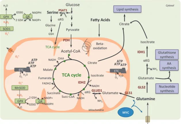
pathway represents also an important feature in glucose conversion and cancer development (97). Serine can be either imported into cells or de novo synthetized from a derived branching of glycolysis (Fig. 1). The glycolytic intermediate 3-phosphoglycerate can be converted into serine following a three-step enzymatic reaction catalyzed by phosphoglycerate dehydrogenase (PHGDH), phosphoserine aminotransferase 1 (PSAT1), and phosphoserine phosphatase (PSPH) (Fig. 1). Serine can next be converted into glycine, which is a major source of methyl groups for the one-carbon pools required for the biosynthesis of protein, purines, and DNA/histone methyl-ation, as well as glutathione. The serine/glycine pathway thus modulates cellular antioxidant capacity and is involved in redox homeostasis. Moreover, PSAT1 uses the PHGDH by-product, namely 3-phosphohydroxypyruvate, to convert glutamate into alpha-ketoglutarate, anaplerotic intermediate that fuels back the TCA cycle and sustains cancer metabolism. In cells with reduced PHGDH or PSAT1 expression, the conversion of glutamine to glutamate and to alpha-ketoglutarate is
significantly reduced, while the serine biosynthesis pathway remains the major contributor of anaplerotic supplies of TCA intermediates (123). Genetic activation of serine bio-synthesis drives cancer cell proliferation and predisposes normal mammary epithelial cells to cancer transformation (162). Moreover, cells lacking p53 fail to respond to serine starvation due to oxidative stress, which leads to reduced viability and impaired proliferation (125). Thus, p53 helps cancer cells to overcome serine starvation, preserving by this way their cellular antioxidant capacity. Finally, serine acts as an allosteric activator of PKM2, the less active iso-form of the pyruvate kinase. Upon serine deprivation, PKM2 activity is reduced, and pyruvate is diverted to a fuel-efficient mode in the mitochondria (245). Moreover, the serine/glycine pathway is also regulated through the NRF2-ATF4 signaling pathway in nonsmall cell lung carcinoma (NSCLC). Indeed, NRF2 constitutive activation through KEAP1 mutation promotes tumorigenesis via ROS detoxifi-cation and confers a poor prognosis in human NSCLC (44). FIG. 3. Glutamine, serine, and fatty acids as anaplerotic sources for TCA cycle intermediates in cancer cells.In cancer cells, glutamine can fuel the TCA cycle through aKG produced by glutaminolysis. Indeed, glutamine is converted to glutamate by the mitochondrial glutaminase (GLS1) or by the cytosolique isoform (GLS2), and glutamate can be converted to aKG in the mitochondria by the glutamine dehydrogenase 1 (GLUD1). MYC oncogene drives glutamine metabolism by promoting glutamine entry into mitochondria and its conversion into glutamate. Moreover, in cancer cells, fatty acids can be degraded through beta-oxidation, which generates acetyl-CoA subsequently fueling the TCA cycle. Indeed, glutaminolysis and fatty acid beta-oxidation provide intermediates to fuel the TCA cycle, resulting in the generation of reducing equivalents, such as NADH and FADH2. This provides electrons to the ETC and leads to ATP production. TCA cycle intermediates can also be directed into biosynthetic pathways (purple boxes) enabling production of macromolecules, such as lipids, amino acids, and nucleotides. Finally, OXPHOS metabolism through ETC not only produces high levels of ROS but generates also high levels of intermediates with antioxidant capacities, such as reduced GSH and NADPH. NADPH is used as a cofactor for antioxidant enzymes, such as glutathione reductase (GR), which reduces the oxidized glutathione (GSSG) into its reduced form (GSH). Thus, high production of reducing equivalents favors ROS scavenging and prevents deleterious accumulation of ROS in the mito-chondrial matrix and cytoplasm. GLUD1, glutamate dehydrogenase 1; GPX, glutathione peroxidase; IDH, isocitrate dehydro-genase; MnSOD, manganese superoxide dismutase; nicotinamide adenine dinucleotide; NH3, ammonia; SOD, superoxide dismutase. To see this illustration in color, the reader is referred to the web version of this article at www.liebertpub.com/ars
Fatty acids. In addition to glucose and glutamine, FAs represent a particularly relevant source of energy. They are either directly incorporated into the cells from the extracellular media or obtained from hydrolyzed triglyceride droplets. Until recently, most studies on cancer metabolism focused on gly-colysis, glutaminolysis, and FA synthesis rather than on FA beta-oxidation, while this pathway is one the most energetic and provides six times more ATP than glycogen oxidation (28). Inhibition of FA oxidation sensitizes most cancer cells to ap-optosis, supporting the key role of FAs in cancer cell survival and suggesting that FA oxidation inhibitors could provide substantial therapeutic interest (38). FA beta-oxidation takes place in the mitochondria and generates acetyl-CoA, NADH, and FADH, cofactors used by the ETC. Some cancer cells, such as the high-OXPHOS large B-cell lymphoma described above, preferentially use FA as a substrate and metabolize palmitate, rather than glucose or glutamine, for sustaining their growth (27). FA oxidation is also essential for survival of lymphoma cells by supplying ATP pools and recycling both glutathione and NADH, thereby counteracting oxidative stress (161). FA oxidation has thus a dual role: it is essential for redox balance, but, in the meantime, it can increase ROS levels in case of nonalcoholic fatty liver disease (64). Finally, P. Carmeliet’s laboratory has recently reported a new role for FA beta-oxidation in de novo nucleotide synthesis and DNA replication in endothelial cells during angiogenesis (185).
Fine-tuned regulation of mitochondrial respiratory chain complex synthesis in neoplastic cells
As previously mentioned, cancer cells with high OXPHOS activity are characterized by accumulation of ETC proteins, which are yet finely regulated. This chapter will thus describe the mechanisms involved in regulating expression of ETC-encoding genes and proteins (Fig. 4). Mitochondria biogen-esis is a complex process that requires the synthbiogen-esis, import, and assembly of proteins and lipids within the existing tochondrial reticulum. It also implies replication of the mi-tochondrial DNA (mtDNA). ETC proteins are encoded by both nuclear and mitochondrial DNA. Nucleus-encoded factors control the transcription as well as replication of mtDNA and achieve coordination of the two genomes (183). Remarkably, human mtDNA is of small size (16,569 bp), but it is present in high number of copies (around 1000). ETC complexes are synthetized by less than 100 genes, 13 of them being encoded by mtDNA. These 13 proteins participate in the composition of 4 different ETC complexes (Complex I, III, IV, V). The rest of the subunits (> 35 subunits for Complex I, 10 subunits for Complex III and IV, and 14 subunits for Complex V) and the entire complex II (4 subunits) are encoded by nuclear DNA (3, 9, 184, 191, 224). mtDNA also encodes 22 transfer RNA (tRNA) and two ribosomal RNAs (rRNA) involved in the synthesis of ETC subunits. mtDNA is not protected by histones and is thus extremely vulnerable to oxidative damages gen-erated in the matrix (1). Metabolic processes are regulated by a number of different mechanisms, including allosteric regula-tion, enzyme degradaregula-tion, and reversible post-translational modifications (PTMs). In this part, we will focus on the dif-ferent levels of regulation that could be responsible for en-hancing OXPHOS metabolism in cancer cells.
Transcriptional regulation of ETC complexes. Regulation of mitochondrial biogenesis represents a transcriptional
challenge finely orchestrated by transcription factors and coregulators (183). The nuclear respiratory factors 1 and 2 (NRF-1 and NRF-2) were the first identified nuclear tran-scription factors involved in regulating the trantran-scription of ETC-encoding genes (47, 73, 142, 182). NRF-1 and NRF-2 binding sites are evolutionary conserved in the proximal promoters of many mitochondrial genes, encoding ETC components, mitochondrial transporters, and mitochondrial ribosomal proteins (MRPs) (183). Moreover, NRF-1 acti-vates the transcription of genes encoding the mitochondrial transcription factors A (TFAM), B1 (TFB1M), and B2 (TFB2M) that mediate replication and transcription of the mitochondrial genome (102, 219). Therefore, NRF-1 coor-dinates nuclear mitochondrial gene expression with mtDNA replication (68). NRF-2 was identified as a transcriptional activator of many mitochondrial genes encoding ETC com-ponents, mitochondrial import, TFAM, TFB1M, and TFB2M (220). Estrogen-related receptors (ERRs) also regulate the expression of genes involved in OXPHOS, mitochondrial dynamics, and oxidative stress defenses (42, 186, 218). Furthermore, transcriptional coregulators ensure the coordi-nated expression of all ETC components by interacting with and coactivate several nuclear receptors through a specific LXXLL motif (130). The PGC-1 family, originally identified in the mitochondrial-rich brown adipose tissue (164), com-prises 3 members, PGC-1a, PGC-1b, and PRC (PGC-1-related coactivator), which regulate mitochondrial biogenesis in a wide variety of tissues. These 3 coactivators are able to interact with NRF-1, NRF-2, PPARs, and ERRs. While PGC-1b participates in the maintenance of basal mitochondrial function, PGC-1a promotes mitochondrial mass increase as an immediate tissue adaptation to high energetic demands (119, 182, 199). The function of PRC, the less characterized member of the family, appears to be restricted to the regu-lation of mitochondrial biogenesis in proliferating cells (216). PGC-1a expression level is either decreased in some cancer, including colon (50), breast (232), and ovary (251), or increased in other cancer types (112, 215), suggesting a dif-ferential regulation of ETC complexes and OXPHOS. In agreement with these observations, high and low OXPHOS subgroups of melanoma exhibit different PGC-1a expression rates (215). Tumor cells harboring high PGC-1a mRNA levels show elevated ETC protein levels and enhanced mi-tochondrial respiration (215). PGC-1a transcription factor is also regulated by PTMs, as discussed below (Post-translational regulation of ETC complexes section). Silencing PGC-1a is sufficient to decrease mitochondrial respiration and ETC protein expression in melanoma cells, demonstrating that PGC-1a is a key actor in OXPHOS metabolism in cancer (215). Similarly, a recent study using both mouse models and silencing strategies demonstrates that PGC-1a enhances OX-PHOS metabolism, mitochondrial biogenesis, and oxygen consumption rate in invasive breast cancers (112). In addition, increased expression of c-MYC in many aggressive tumors upregulates mitochondrial biogenesis by regulating expression of the TFAM gene (116). Thus, transcriptional regulation of mitochondrial ETC complexes varies among tumor types and plays an important role in driving OXPHOS metabolism.
Translational regulation of ETC complexes. While nu-clear DNA encodes most of the mitochondrial proteins, few of them are encoded by mtDNA and synthesized by the
mitochondrial translation system. Although the mechanisms of translation are well characterized in the cytoplasm, little is known about mitochondrial protein translation. Impaired mitochondrial translation usually results in severe respiratory dysfunction through the lack of all mtDNA-encoded proteins (193, 244). Mitochondrial mRNAs are uncapped (72), but contain a poly(A) tail (150). Unlike cytoplasmic translation, the small subunit of mitochondrial ribosomes (mitoribo-somes) binds mRNAs in a sequence-independent manner (117). The mitochondrial translation machinery consists of 2 initiation factors and 3 elongation factors. The termination process has not been entirely elucidated, although 2 release factors (mtRF1 and mtRF1a) and 1 recycling factor (mtRRF)
have been partly characterized (174). The human mitoribo-some consists of two rRNAs (12S and 16S) and about 81 MRPs (194). Levels of mitochondrial proteins are regulated by their own translational activators, which bind to 5¢-untranslated re-gions (82). Translational activator imbalance alters mitochon-drial protein levels and ultimately impairs respiration (55, 240), demonstrating the key role of mitochondrial-driven translation in OXPHOS activity.
Mutations or alterations of the mitochondrial translational process have not been deeply investigated in cancer cells (62), despite the recent identification of high OXPHOS tumors (27, 78, 215). In contrast, translational defects of nuclear-encoded mitochondrial proteins have been investigated for years as one FIG. 4. Schematic representation of the different levels of regulation of ETC complexes.The mtDNA encodes 13 ETC proteins, 22 tRNAs, as well as 12S and 16S rRNAs, whereas the nuclear DNA encodes approximately 1000 proteins belonging to ETC complexes. There are three levels of regulation: transcriptional, translational, and post-translational. (A) Transcriptional regulation: In the nucleus, PGC-1a and various transcription factors, such as NRF, ERR, and PPAR, bind to the regulatory regions of mitochondrial (mt) target genes and stimulate their expression. Inside mitochondria, TFAM, a mito-chondrial DNA transcription factor, cooperates with the mtPOLR to induce the expression of mtDNA-encoded proteins. (B) Translational regulation: Mitochondrial-addressed proteins, which are transcribed in the nucleus, are exported into the cyto-plasm, where their translation takes place via the cytoplasmic ribosomal (40S and 60S) machinery. While nuclear DNA encodes most of the mitochondrial proteins, few of them are encoded by mtDNA and synthesized by the mitochondrial translation system. mRNAs transcribed into the mitochondrial matrix are translated by the mitochondrial ribosomal 12S and 16S complexes. (C) Post-translational regulation: Upon translation, many mitochondrial proteins are synthesized as precursor proteins, with cleavable N-terminal presequences. The TOM complex allows the translocation of tagged proteins from the outer membrane barrier to the IMS. The tag signal and adjacent parts of the protein are recognized by the TOM complex, which works together with the TIM complex to translocate proteins into the mitochondria. Once imported, the N-terminal signal of the precursor protein is processed through MPP and mature isoforms are assembled into the IMS. Mitochondrial proteins, encoded by the mitochondrial or nuclear genomes, are finally assembled to form ETC complexes. ERR, estrogen-related receptor; HSP, heat shock protein; IMS, inner membrane space; MPPs, mitochondrial processing peptidases; mtDNA, mitochondrial DNA; mt, mitochondrial; NRF, nuclear respiratory factor; mtPOLR, mitochondrial RNA polymerase; PPAR, peroxisome proliferator-activated receptor; PGC-1a, peroxisome proliferator-proliferator-activated receptor-c coactivator-1a; TF, transcription factor; TIM, transporter inner membrane; TOM, translocase of the outer membrane; tRNA, transfer RNA; rRNA, ribosomal RNA. To see this illustration in color, the reader is referred to the web version of this article at www.liebertpub.com/ars
of the major pathways involved in regulating protein transla-tion, the so-called mammalian target of rapamycin (mTOR) pathway, is often deregulated in human cancers. mTOR inter-acts with several proteins to form two distinct complexes, namely mTORC1 and mTORC2, which have different up-stream regulators, downup-stream effectors, and exhibit distinct functions (110). mTORC1 senses nutrient availability and is a major regulator of protein translation. N. Sonenberg has recently reported that mTORC1 controls both mitochondrial activity and biogenesis through the eukaryotic translation initiation factor, eIF4E, in vitro and in vivo (140). mTOR inhibitors impair mi-tochondrial respiration by inhibiting translation of TFAM, subsequently reducing expression of ETC genes (140). In ad-dition to their transcriptional stimulatory activity, TFB1M and TFB2M have rRNA methyltransferase activity (129). Thus, these factors are indirectly involved in mitochondrial protein synthesis via their ability to methylate the mitochondrial 12S rRNA, which is important for mitoribosome activity.
Post-translational regulation of ETC complexes.
Mitochondrial addressing. Most mitochondrial proteins, including proteins involved in mitochondrial translation, are synthetized in the cytoplasm as nuclear DNA encodes them. Therefore, they need to be imported into mitochondria. Cy-tosolic chaperones, such as heat shock proteins, HSP70 and HSP90, escort precursor proteins toward translocation channel receptors on the mitochondrial membrane. It has been suggested that cytoplasmic and mitochondrial transla-tion machineries are in close proximity to allow efficient assembly of the ETC system (87). Nuclear DNA-encoded proteins, either tagged by an N-terminal mitochondrial tar-geting signal (N-MTS) (85) or containing an internal signal (23), are recognized by receptors of the outer mitochondrial membrane and imported into the mitochondria (9). The translocase of the outer mitochondrial membrane (TOM) complex allows the translocation of mitochondrial precursor proteins from the outer membrane barrier to the intermem-brane space (IMS) (196). The N-MTS and its adjacent parts are recognized by the TOM complex, which works together with the translocase of the inner mitochondrial membrane (TIM) complex to transfer proteins into the mitochondria (9, 168). After translocation, the N-MTS is removed by the mitochondrial processing peptidases, and ETC mature pro-teins are then released into the IMS. As shown by this short description, mitochondrial protein targeting, import, proces-sing, and assembly require a complex series of processes, which can be mutated or altered during tumorigenesis. In that sense, N-MTS mutation in one of the PDH subunits was identified as the cause of PDH import defect and PDH defi-ciency in humans (206). Similarly, genetic dimorphism in the N-MTS of the manganese superoxide dismutase (MnSOD) affects MnSOD import into mitochondria and is considered as a risk factor for severe alcoholic liver disease and hepa-tocellular carcinoma (143, 203). As translocation across the inner membrane requires an electrochemical hydrogen ion gradient (transmembrane potential DC), redox imbalance alters this process and impairs mitochondrial respiration, as observed in cancer cells (242).
Lysine acetylation. PTMs, including phosphorylation, acetylation, methylation, N-linked, and O-linked glycosylation and ubiquitination, are involved in cell signaling networks.
Lysine acetylation is conserved among species and links acetyl-CoA to cellular metabolism. Acetylation was long considered as an epigenetic modification mainly occurring on chromatin-associated proteins, but recent proteomic analyses reveal that most acetylation events occur also on non-nuclear proteins, particularly on mitochondrial proteins (35). Lysine acetylation is regulated both by lysine acetyltransferases and lysine deacetylases, which add or remove acetyl groups from proteins, respectively. Lysine acetylation is one of the most prevalent mitochondrial PTMs (233). Reciprocally, metabolic intermediates modulate lysine acetylation (81, 238). Indeed, acetyl-CoA synthesis occurs in two distinct compartments defined by the permeability of the mitochondrial membrane (238). Nuclear histone acetylation levels are directly correlated with the activity of the cytosolic ATP citrate lyase, an enzyme that converts glucose-derived citrate into acetyl-CoA in human cells and is considered as a new link between nutrient me-tabolism and histone acetylation (237).
Lysine acetylation regulates enzyme activity as lysine residues are often present in the active sites of enzymes. For instance, acetyl-CoA synthetase 2 (ACSS2) is a mitochon-drial matrix protein that produces acetyl-CoA from acetate. ACSS2 is reversibly acetylated at K642 residue (localized into its enzymatic active site) and thereby inactivated (59). In human breast cancer, the ACSS2 gene is amplified, highly expressed, and positively correlated with tumor invasiveness (187). Comparative metabolomic and lipidomic analyses demonstrated that ACSS2 promotes a switch in nutrient uti-lization from glucose to acetate to support FA and phos-pholipid biosynthesis in cancer cells. ACSS2 silencing reduces tumor growth in xenograft mouse models. Thus, acetate consumption exhibits a critical role in lipid biomass production and membrane biogenesis in cancers. This con-stitutes the rational basis for using acetate-based PET tracers in breast cancer diagnosis (187). In contrast to ACSS2, acetylation of the glycolytic enzyme, phosphoglycerate mu-tase 1 (PGAM1), enhances its activity and stimulates gly-colysis (76). Moreover, glucose deprivation triggers increased levels of sirtuin 1 (SIRT1), an evolutionary con-served NAD-dependent histone deacetylase (19, 91), leading to PGAM1 deacetylation. These results suggest that SIRT1 might have a role in the regulation of glycolysis flux by preventing depletion of glycolytic intermediates used in other biosynthetic pathways, such as the serine pathway. Ad-ditionally, SIRT1 has been previously reported to affect metabolic pathways at the transcriptional level, promoting beta-oxidation and inhibition of lipogenesis through PGC-1a regulation (75). Indeed, PGC-1a activity is regulated at post-translational levels. Mitochondrial biogenesis is induced by PGC-1a phosphorylation at threonine-177 and serine-538 residues by AMPK (89). In addition, PGC-1a is activated by deacetylation by SIRT1 (171). The cross-talk between SIRT1 and PGC-1a thus represents a key regulatory process in cancer metabolism (19, 91). Thus, examining the contribu-tion of key actors involved in acetylacontribu-tion regulacontribu-tion will likely provide novel insights into cancer metabolism.
Role of Metabolism on Cancer Cell Properties and Functions
As previously shown, metabolism in cancer is much more heterogeneous than initially thought, but the impact of these
metabolic variations (in particular OXPHOS regulation) in cancer development and progression remains unclear. In this chapter, we address this question. Indeed, oncogenic trans-formation requires metabolic adaptation to support stemness properties of cancer cells and their highly energy-consuming functions, such as proliferation, growth, migration, and in-vasion (Fig. 5).
Proliferation
Cancer metabolism is often considered as an adaptation to sustain cancer cell proliferation. Since O. Warburg’s studies, it has been assumed that cancer cell growth and proliferation require increased glycolytic and decreased oxidative metab-olism. This metabolic switch is not only required for energy production but also for the synthesis of nucleotides, proteins, and lipids to generate the building blocks supporting un-controlled tumor proliferation and growth. Indeed, there is clear evidence showing that both mitochondrial morphology and respiratory function are linked to cell cycle regulators such as cyclins. Cyclin D1, well known to promote nuclear DNA replication through phosphorylation and inactivation of the retinoblastoma protein (pRB), is involved in mitochon-drial bioenergetic coordination during G1 progression (178). The overexpression of cyclin D1 during G1 leads to a twofold decrease in mitochondrial activity mediated by CDK4 kinase activity, independently of pRB (225). Conversely, hepato-cytes lacking cyclin D1 exhibit increased mitochondrial size and activity, both associated with an increased expression of Nrf1 (225). Cyclin E controls the formation of high-energy charged mitochondria during G1/S transition (136). During
G2/M transition, the cyclin B1/CDK1 complex is a key regulator of mitochondrial fission (205), respiration, and ATP production (227). Both cyclin B1/CDK1 mitochondrial abundance and kinase activity are dramatically increased during G2/M, leading to ETC complex I phosphorylation and enhanced activity (227). Finally, cyclin-dependent regulation of mitochondrial activity is consistent with enhanced OX-PHOS activity during G2/M transition, while glycolysis ra-ther supplies the energy needs during G1 (6). Notably, in colon cancer cells, ATP production during G2/M transition is predominantly dependent on mitochondrial respiration (6).
Recently, the team of L. Fajas investigated the regulation of cell cycle by mitochondria (15, 124). They observed that mice lacking E2 transcription factor 1 (E2F1), an important regulator of S-phase entry and a downstream target of pRB, often inactivated in human cancers, consume more O2than
control mice. This phenotype is associated with increased expression of genes involved in ETC composition, mito-chondrial biogenesis, beta-oxidation, and uncoupling activity in muscle and brown adipose tissue (15). Computational analysis revealed the identification of E2F binding sites in the promoter of these OXPHOS genes, suggesting direct tran-scriptional regulation by E2F. Moreover, RB deletion affects the mitochondrial biogenesis transcription program in ery-throid progenitor cells due to a decrease in PGC-1b expres-sion (180). Exploiting a novel link between cell cycle regulators and mitochondria may help to sensitize tumors to treatment and may offer new combinatory therapeutic strat-egies. Given the recent success of CDK4/6 inhibitor (PD0332991) in clinical trials for HER2-positive advanced breast cancer patients (54, 152, 247), it would be interesting to investigate the impact of such compound on mitochondria morphology and function.
Despite an increasing number of studies showing the im-portance of mitochondrial respiration for cancer cell prolif-erative capacities, the reasons for this requirement remained unclear until recently. Similarly, it was unknown why pyru-vate supplementation allows ETC-defective cells to prolif-erate (104). Two recent studies (11, 201) have highlighted these questions. They showed that respiration-deficient cells are auxotrophic for pyruvate, which acts as an electron ac-ceptor and plays a key role in redox balance. Moreover, they demonstrated that an essential function of mitochondrial ETC complexes in cell proliferation is to enable aspartate biosyn-thesis (11, 201). Pyruvate stimulates aspartate synbiosyn-thesis in a GOT1-dependent manner, GOT1 being the cytosolic aspar-tate aminotransferase, and is sufficient to rescue proliferation in ETC-deficient cells (11, 201). Altogether, these studies provide a new perspective on the different roles of the ETC complexes in metabolism and proliferation capacity.
Migration and metastases
Whether and how metabolic reprogramming may play a role in tumor progression and metastatic process remain elusive. Recently, the identification of specific metabolites affecting cancer cell migration and invasion, as well as the characterization of bioenergetic profiling of invasive and metastatic cancer cells, brought new insights. Metabolic stress, such as hypoxia or oxidative stress, enhances stem-ness, angiogenesis, invasivestem-ness, and metastatic potential of tumor cells and is therefore of poor prognosis. Indeed, HIF-FIG. 5. OXPHOS metabolism on cancer cell functions
integrated into the 10 hallmarks of cancer from Hana-han and Weinberg, 2011. OXPHOS metabolism (purple box) supports high proliferative capabilities of cancer cells (green box). Reciprocally, cell cycle regulators are able to promote OXPHOS metabolism. Moreover, OXPHOS me-tabolism favors migration and invasion of cancer cells (or-ange box). Finally, OXPHOS metabolism plays an important role as it increases cancer cell stemness (blue box). To see this illustration in color, the reader is referred to the web version of this article at www.liebertpub.com/ars
1a activation causes E-cadherin loss and epithelium-to-mesenchymal transition (EMT), a process by which epithelial cancer cells acquire invasive properties as they loose their polarity and their cell-to-cell adhesion (36, 163). Aerobic glycolysis causes extracellular matrix (ECM) acidification, which leads to matrix metalloproteinase and cathepsin acti-vation. As a consequence, there is increased ECM degrada-tion (204), a process that takes part in most basic cell behaviors, including cell migration. Specific metabolites, such as glutamine, play a critical role in cancer cell invasion in vitro and in vivo. A recent study, using The Cancer Gen-ome Atlas database, reported that the poor survival of ovarian cancer patients is significantly correlated with elevated ex-pression of genes involved in glutaminolysis or TCA cycle and low expression of glycolytic genes (243). Highly inva-sive ovarian cancer cells preferentially use glutamine rather than glucose to replenish TCA cycle, which dramatically increases the oxygen consumption rate. Affecting oxygen consumption by using rotenone, a mitochondrial respiratory chain inhibitor, reduces both TCA cycle activity and inva-siveness of ovarian cancer cells (243). Similarly, a study aiming at identifying the bioenergetic profiles of invasive and metastatic cancer cells uncovered that human and mouse metastatic cells are characterized by increased mitochondrial biogenesis and ATP production, as opposed to primary tumor cells (112). This was associated with elevated levels of PGC-1a. Moreover, PGC-1a silencing dramatically reduced pri-mary tumor cell invasiveness and metastasis without affect-ing tumor growth. Interestaffect-ingly, patients with invasive breast cancer exhibiting high PGC-1a expression have increased metastasis and decreased survival rates. Altogether, this suggests that PGC-1a-induced OXPHOS metabolism is es-sential for the metastatic capabilities of breast cancer cells (112). Importantly, distinct metabolic signatures of breast cancer cells were found to predict the organ site of metastasis (45). While liver-metastatic breast cancer cells exhibit en-richment in glycolytic genes, bone- and lung-metastatic cells display an increased OXPHOS metabolism and glutamino-lysis associated with accumulation of TCA cycle intermedi-ates, such as citrate and succinate. Liver-metastatic breast cancer cells maintain their glycolytic functions via HIF-1a and pyruvate dehydrogenase kinase 1 (PDK1). PDK1 ex-pression is always high in liver metastases, while it is either low or high at the primary tumor site. This suggests that the metabolic switch influencing tropism of cancer cells may occur when they acquire metastatic properties (45). It would be interesting to investigate whether this results from in-trinsic heterogeneity in cancer cell metabolic program or a clonal selection, mediated by TME, such as oxygen con-centration that varies from one metastatic organ to the other.
Increased stem cell properties
Quiescent adult stem cells generally exhibit low metabo-lism associated with a slow proliferation rate and reduced risk of ROS-mediated cellular damage and long-lived renewal capacity (88, 175, 200). This low metabolism mainly depends on glycolysis (41, 154). Similar to cancer cells showing high glycolytic flux with some OXPHOS heterogeneity (as de-scribed above in the OXPHOS metabolism: modulation of reactive oxygen species levels and antioxidant response section), the new concept of bioenergetic heterogeneity of
cancer stem cells (CSCs) is now commonly recognized. While some CSCs rely on glycolytic metabolism, there is also an enrichment of CSCs exhibiting OXPHOS metabolism that would confer resistance to chemotherapy (32, 70, 156). Re-cently, the mitochondrial features and metabolic properties of CSCs and noncancer stem cells (non-CSCs) have been in-vestigated in many cancers. Lung CSCs show a lower mtDNA content, O2 and glucose consumption, ATP, and
ROS intracellular concentration compared with non-CSCs (246). Similarly, leukemia CSCs exhibit lower ROS levels and OXPHOS metabolism compared with non-CSCs (108). However, ovarian CSCs show an increased expression of genes involved in glucose uptake, OXPHOS metabolism, and FA beta-oxidation (159). Moreover, CSCs from glioma or glioblastoma also depend on OXPHOS for their energy production and survival compared with their differentiated progeny, which mainly rely on aerobic glycolysis (90, 221). However, the metabolic state (OXPHOS vs. aerobic glycol-ysis) of CSCs is less clear in breast cancer as it is still debated due to discrepant results (51, 222). Taken as a whole, these studies highlight the heterogeneity of CSC bioenergetic properties within tumors, before any treatment.
Interestingly, there is also increasing evidence that che-motherapy influences CSC metabolic status. Metastatic melanoma is a heterogeneous tumor of neuroectodermal or-igin with less than 1-year median survival. Despite the en-couraging initial tumor response to BRAF inhibitor vemurafenib, relapse occurs only after few months of treat-ment due to multiple resistance mechanisms. Among the highly proliferative melanocytes, a subpopulation of slow-cycling cells has been identified after chemotherapy. These slow proliferative cells exhibit high levels of the histone H3K4-demethylase JARID1B, high levels of mitochondrial ETC proteins, and increased OXPHOS activity (173). Similar metabolic heterogeneity and plasticity were identified in CSCs isolated from human primary pancreatic tumors and patient-derived xenografts (179). As previously reported in other cancers, pancreatic CSCs rely mainly on OXPHOS metabolism, while the non-CSCs are dependent on glycolysis (179, 217). Interestingly, the use of the OXPHOS inhibitor, metformin, to selectively kill the CSCs, leads to the positive selection of a resistant CSC subpopulation, while CSC treatment with the mitochondrial ROS inducer, menadione, does not (179). Importantly, this resistant CSC subpopulation activates a glycolytic program through an MYC/PGC-1a-dependent regulatory process (179). In conclusion, deci-phering the mechanisms involved in CSC metabolic hetero-geneity and adaptability would be crucial to identify key actors enabling CSCs to escape from chemotherapy.
Reciprocal Impact of Cancer Cell Metabolism on TME
TME plays a crucial role in tumor development, growth, and spreading. The view of cancer as a heterogeneous disease was initially restricted to genetic mutations in tumor cells. However, it is now well accepted that tumor heterogeneity also results from stromal diversity (165, 189), including tumor-associated macrophages (TAMs), tumor-infiltrating lymphocytes (TILs), and carcinoma-associated fibroblasts (CAFs). The proportion and the activation state of each cell population also increase TME heterogeneity. Some recent studies pinpoint both the importance of ROS-induced
mitochondrial metabolism and nutrient availability in TME regulation (Fig. 6).
Oxidative stress and TME activation
Tumor-associated macrophages. Macrophages are sen-tinel cells that maintain homeostasis by regulating innate immunity in response to several stimuli, such as pathogen-associated molecular patterns or cytokines found in surround-ing tissue (14). In cancer, TAMs influence several aspects of tumor progression, such as angiogenesis, metastasis, and sur-vival of cancer cells (13, 126). All these activities are delivered by different subpopulations of TAMs, TAM subset complex-ity having considerably increased in the past few years (146). To keep it simple, activated macrophages either polarize as the proinflammatory M1 phenotype or as anti-inflammatory M2 phenotype (71). The M1 phenotype is characterized by the production of inflammatory cytokines, reactive nitrogen species, and ROS (14). In contrast, the M2 phenotype is characterized by increased production of immunosuppressive cytokines and factors (148). Recently, it has been shown that ROS production is important for M2, but not for M1 differentiation (252). Indeed, inhibiting ROS production blocks the differentiation of M2 macro-phages through the ERK pathway. This is also true in vivo as antioxidant treatment blocks the occurrence of M2 macrophages in lung tumor mouse models (252). Simi-larly, in malignant melanomas, TAMs exhibiting high levels of ROS have enhanced invasive properties, medi-ated by increased TNF-a secretion (120). Recently, an HIF-1-dependent metabolic switch toward glycolysis has been detected following activation of macrophages (101). Citrate withdrawal from TCA cycle is critical for lipid
biosynthesis in TAMs, and succinate, another TCA cycle intermediate, is also crucial to promote inflammatory signaling. Because TAMs play major roles in suppressing antitumor responses, blocking M2 macrophages by im-pacting the redox balance may represent an interesting strategy for cancer treatment.
Tumor-infiltrating lymphocytes. T cells orchestrate the adaptive immune response to specific antigens. Classically, when an antigen activates a naı¨ve CD4+ T cell, cytokines determine whether it differentiates into an effector T cell (Teff) to induce an immune response or a regulatory T cell (Treg) with immunosuppressive functions (96). The main metabolic change, occurring upon T-cell activation, corre-sponds to an increased requirement for energy and for bio-synthesis of reducing equivalents. Activated T cells rely preferentially on glycolysis and lactate production, mediated by the PI3K/AKT pathway (57). However, mitochondria-dependent metabolism also plays an important role in T-cell responses, through ROS signaling rather than ATP produc-tion (94, 99, 188). During T-cell activaproduc-tion, the high glyco-lytic rate induces excessive amount of glycerol-3-phosphate that is oxidized in an FAD-dependent reaction, mediated by glycerol-3-phosphate dehydrogenase 2, which increases ROS production. Silencing this enzyme decreases ROS and IL-2 production and reduces T-cell activation (98). Similar defects in T-cell activation have been observed in mice engineered for specific inactivation in T cells of a component of the respiratory chain complex III (Uqcrfs1) as it leads to reduced ROS levels (188). Moreover, mitochondrial ROS are re-quired for antigen-specific CD4+and CD8+T-cell expansion in vivo (188). Using mice lacking the lymphocyte expansion molecule, an orphan protein that promotes CD8+ T-cell
FIG. 6. Reciprocal impact of cancer cell metabolism on TME. (A)Oxidative Stress and TME. TAM: ROS signaling promotes M2 polarization through the ERK signaling pathway; TIL: TIL activation (differentiation in CD4+, CD8+T cells) and expansion increase rates of glycolysis and generate excessive amount of glycerol-3-phosphate. G3P is oxidized by the mitochondrial G3P dehydrogenase 2, which in turn increases ROS production; CAF: ROS promote CAF activation and conversion of fibroblasts into myofibroblasts through TGF-b and CXCL12 (SDF-1)-dependent pathways. (B) Nutrient availability and TME. Both immune and stromal cells participate in a complex metabolic interplay with neoplastic cells. They can collectively adapt in a dynamic manner to the metabolic needs of cancer cells and thus participate in tumori-genesis. Metabolic competition between immune and tumor cells: Tumor cell metabolism modulates nutrient availability in TME, impacting macrophage polarization and immune response. High nutrient availability in the TME favors glycolysis through mTOR signaling and promotes M1 polarization and Teff differentiation. mTORC1 is involved in naı¨ve CD4+T-cell differentiation into T helper 1 (TH1) and T helper 17 (TH17) cells, supporting an antitumor effect. In contrast, mTORC2
promotes the differentiation of naı¨ve CD4+T cells into the protumorigenic T helper 2 (TH2) cells. Moreover, M2
mac-rophages exhibit an OXPHOS metabolism. Interestingly, blocking OXPHOS metabolism induces M1 polarization, while forcing OXPHOS metabolism in M1 macrophages potentiates M2 polarization. Glutamine deprivation promotes the dif-ferentiation of naı¨ve CD4+T cells into FOXP3+Treg cells and thus induces a shift in the immune response balance, which becomes immunosuppressive. Glutamine deprivation also impacts M2 phenotype by promoting a protumorigenic response. Moreover, glucose availability is also another layer of TIL regulation. As progressing tumors have higher glucose con-sumption than the regressing ones, TILs from progressing tumors are glucose restricted and exhibit impaired effector functions. Blocking PD-L1 in tumor cells reduces their glycolysis rates by inhibiting mTOR activity, which consequently increases extracellular glucose availability for TILs. Thus, by modulating tumor cell metabolism, one can control nutrient availability for T cells, thus promoting either their antitumor or immunosuppressive functions. Metabolic symbiosis be-tween CAFs and tumor cells: Increased ROS production by cancer cells, in particular the highly diffusible H2O2, stimulates
HIF-1 signaling in CAFs. As a consequence, CAFs switch their metabolism toward aerobic glycolysis, through an HIF-1-and oxidative stress-dependent mechanism. This highly glycolytic rate in CAFs provides nutrient HIF-1-and energetic fuels, such as lactate and ketone bodies, to cancer cells. This symbiotic relationship between CAFs and tumor cells is reversible, thus representing a metabolic optimization in cancer treatments. CAF, carcinoma-associated fibroblast; Cav 1, caveolin 1; HIF-1, hypoxia-inducible factor-1; mTOR, mammalian target of rapamycin; SMA, smooth muscle cells; TAM, tumor-associated macrophage; Teff, effector T cell; TIL, tumor-infiltrated lymphocyte; TGF-b, transforming growth factor-b; Treg, regulatory T cell. To see this illustration in color, the reader is referred to the web version of this article at www.liebertpub.com/ars
proliferation, another study confirmed that mitochondrial ROS affect respiratory chain assembly into the inner mito-chondrial membrane and CD8+ T-cell expansion, thus demonstrating that long-term protective immunity can be OXPHOS driven (151). Antineoplastic chemotherapies stimulate autophagy, which appears as an adaptive meta-bolic mechanism of resistance to treatment (113). Thus, inhibiting autophagy would be a new strategy for improving chemotherapy efficiency in some tumors, such as
triple-negative breast cancers, lacking efficient anticancer im-mune response (113). Using an immunocompetent mouse model, another study demonstrated that autophagy is re-quired for immunogenic release of ATP from dying cells to recruit immune cells following chemotherapy (134).
CAF. Although the role of fibroblasts and the concept of seed and soil have been considered in the 19th century (153), the key role of CAFs in ECM remodeling and tumor
progression has been highlighted only recently (17, 36, 56, 100, 149). While normal fibroblasts inhibit tissue progression to cancer, changes in stromal components shift the antitumor balance toward a procancerous state (12, 36, 211). Fibro-blasts are activated through specific communication with cancer cells and can promote tumor initiation, progression, and metastasis. A large proportion of activated CAFs de-tected in invasive adenocarcinomas express smooth muscle a-actin (a-SMA) and are defined as myofibroblasts (211), but other markers such as platelet-derived growth factor receptor (PDGF-R) or fibroblast activation protein (FAP) also char-acterize stromal components (22, 46). The cellular origins of myofibroblasts can be multiple. Myofibroblasts were thought to derive from EMT, bone marrow, or various mesenchymal cell types, including endothelial cells, pericytes, or pre-adipocytes (16, 66, 109, 131, 166, 177). Resident fibroblasts are also considered as one of the major source of CAFs. Indeed, ROS release from tumor cells promotes the conver-sion of fibroblasts into myofibroblasts in various types of solid tumors (8, 36, 92, 157, 198, 211). Various signaling pathways, including transforming growth factor-b (TGF-b)-and CXCL12 (SDF-1)/CXCR4-dependent pathways, are also involved in CAF activation (5, 25, 211, 226). Interestingly, targeting both CXCL12/CXCR4 axis and CAFs has a syner-gistic effect when used in combination with anti-PD-L1 im-munotherapy (49). Although stromal remodeling by caveolin 1 favors tumor invasion (69), ROS accumulation in CAFs pro-motes loss of caveolin 1 by oxidative stress-induced autophagy (128, 132, 190). Moreover, chronic oxidative stress in TME leads to HIF-1 accumulation by inhibiting prolyl hydroxylases (65). Stimulation of HIF-1 signaling is associated with in-creased glycolysis in CAFs, which in turn provide nutrients such as lactate to adjacent cancer cells (see below in the Nu-trient availability and TME differentiation section). ROS have been implicated in the metabolic reprogramming of both cancer cells and CAFs, allowing an adaptation to oxidative stress that ultimately promotes tumorigenesis and chemore-sistance. As metabolic reprogramming may impact the redox balance, it is crucial to better understand the metabolic cross-talk between neoplastic and surrounding cells.
Nutrient availability and TME differentiation
Tumor-associated macrophages. A pioneering work on metabolism in TAMs showed that activation of murine peritoneal macrophages leads to a metabolic switch as they rely on glycolysis rather than OXPHOS (79). Oxidation of glucose, glutamine, and FA are not just sources of energy but are also involved in macrophage polarization. Indeed, M1 macrophages rely preferentially on mTOR-HIF1a-mediated glycolysis, whereas M2 macrophages exhibit an OXPHOS metabolism, involving the PGC-1b-STAT6 signaling path-way (34, 172, 208, 213). Glycolysis induced in M1 macro-phages allows the synthesis of reducing equivalents and energy through the PPP (60). Decreasing aerobic glycolysis by silencing PDK1 promotes M2 polarization as it attenuates the expression of proinflammatory cytokines upon LPS or TLR2 stimulation (207). Interestingly, blocking OXPHOS metabolism induces M1 polarization, while forcing OX-PHOS metabolism in M1 macrophages potentiates M2 po-larization (172, 213). Macrophages also exhibit high rates of glutaminolysis, which promote phagocytosis, cytokine
pro-duction, and antigen presentation (223) and replenish the TCA cycle (93). Glutamine deprivation only impacts on M2 phenotype, while it has no effect on M1 polarization. Uptake and oxidation of FA, favored by PGC-1b signaling, increase OXPHOS and M2 polarization (213). Reciprocally, in-hibiting lipolysis affects M2 polarization and survival (86). Altogether, these data suggest that tumor cell metabolism should modulate nutrient availability in TME, therefore affecting macrophage polarization and immune response. Highly glycolytic tumor cells may compromise M1 polari-zation by inducing glucose deprivation, while FA abundance may influence M2 differentiation.
Tumor-infiltrating lymphocytes. Several studies based on metabolic profiling revealed that Teff lymphocytes are highly glycolytic and lipogenic (133), whereas Treg cells display increased lipolysis and lipid oxidation (10). The mTOR pathway, a major regulator of cell growth that senses nutrient availability, plays a crucial role in T-cell differentiation (43, 53, 80, 167). Indeed, mTORC1 is involved in naı¨ve CD4+ T-cell differentiation into T helper 1 (TH1) and T helper 17
(TH17) cells, supporting an antitumor effect. In contrast,
mTORC2 promotes the differentiation of naı¨ve CD4+T cells into the protumorigenic T helper 2 (TH2) cells (43). Among
the nutrients involved in T-cell differentiation, glutamine and glucose play critical functions. Glutamine deprivation pro-motes the differentiation of naı¨ve CD4+T cells into FOXP3+ Treg cells, even in the presence of cytokines that promote TH1 cell expansion (105). Adding alpha-ketoglutarate to a
glutamine-deprived medium inhibits the generation of Treg cells. Thus, glutamine deprivation in TME induces a shift in the immune response balance, which becomes immunosup-pressive (105). T-cell receptor stimulation can activate sev-eral signaling pathways that coordinate T-cell proliferation and differentiation, together with aerobic glycolysis (192). Two recent studies support the hypothesis that tumor cells can suppress antitumor T-cell responses by modulating me-tabolism through glucose deprivation (30, 83). Indeed, these studies provide some clues indicating that glycolysis is es-sential not only for biomass and ATP production but also to sustain T-cell effector functions through transcriptional and translational regulation. Using a well-established model of regressing and progressing sarcomas in immunocompe-tent mice, E.L. Pearce showed that progressing tumors have higher glucose consumption than the regressing ones (30). As a consequence, TILs from progressing tumors are glucose restricted and exhibit impaired effector functions compared with TILs in regressing tumors. Interestingly, treatments of tumor-bearing mice with immune checkpoint inhibitors (such as anti-CTLA-4, PD-1, and PD-L1 antibodies) restore glu-cose in TME, allowing T-cell glycolysis and IFN-c produc-tion. Moreover, blocking PD-L1 in tumor cells reduces their glycolysis rates by inhibiting mTOR activity, which con-sequently increases extracellular glucose availability for TILs. These observations were confirmed in a melanoma mouse model as there is an anticorrelation between glyco-lytic genes and IFNg or CD40 ligand mRNA levels. Finally, hexokinase 2 overexpression in melanoma cells confers on them high glycolytic capacities (30). In conclusion, these different studies clearly demonstrated how tumor cell metabolism deeply affects TME differentiation and func-tions. One major pathway involved in this process is the