_j
Biochemical Characterization of Murine Calprotectin and the
Host-Pathogen Competition for Manganese
by
Rose Currier Hadley B.S. Chemistry B.S. Neuroscience
University of New Hampshire, 2014
ASSACHUSETTS INSTITUTE 01- TECHNOLOGY
JUL
0
2
2019
LIBRARIES
ARCHIVES
Submitted to the Department of Chemistry in Partial Fulfillment of the Requirements of the Degree of
DOCTOR OF PHILOSOPHY IN CHEMISTRY at the
Massachusetts Institute of Technology June 2019
@Massachusetts Institute of Technology, 2019 All Rights Reserved
Signature of Author:
Signature redacted
Department f Chemistry May 10, 2019
Signature redacted
Certified by: Accepted by: Elizabeth M. Nolan Associate Professor of Chemistry Thesis Supervisor_________Signature redacted
Robert W. Field Haslam and Dewey Professor of Chemistry Chairman, Departmental Committee for Graduate Students
This doctoral thesis has been examined by a committee of the Department of Chemistry as follows:
Signature redacted
Daniel L. M. Suess Assistant Professor of Chemistry Committee Chairperson
Signature redacted
Elizabeth M. Nolan Associate Professor of Chemistry Thesis Supervisor
S ignature redacted
Mircea Dinca Associate Professor of Chemistry Committee Member
Biochemical Characterization of Murine Calprotectin and the
Host-Pathogen Competition for Manganese
by
Rose Currier Hadley
Submitted to the Department of Chemistry on 05/10/2019 in Partial Fulfillment of the
Requirements of the Degree of Doctor of Philosophy in Chemistry
ABSTRACT
Microorganisms need to acquire metal ion nutrients when they attempt to colonize the host milieu. The competition for transition metal ions between a host and an invading pathogen constitutes an important aspect of innate immunity and microbial pathogenesis. The host deploys the metal-sequestering antimicrobial protein calprotectin (CP) to sites of infection to withhold transition metal ions. The goals of this thesis are to characterize the biochemical, and Mn(Il)-binding properties of the murine orthologue of calprotectin (mCP) as well as to evaluate the molecular details of the competition for Mn(II) between calprotectin and Mn(II) transport proteins from pathogenic bacteria. In the first part of this thesis, we provide initial biochemical characterization of mCP, supporting a role of this protein in transition metal sequestration and antibacterial activity. We demonstrate that this protein is a heterodimer than can undergo Ca(Il)-induced tetramerization. We further show that mCP can bind a range of first row transition metal ions and displays antibacterial species against a panel of bacterial species. In the second part of this thesis, we characterize the Mn(Il)-binding properties of mCP, revealing Ca(Il)-dependent Mn(II) affinity at a hexahistidine site that bears a remarkable resemblance to the Mn(Il)-sequestering site in human CP (hCP). We use biochemical assays and electron paramagnetic resonance (EPR) spectroscopy to elucidate the Mn(li)-coordinating residues of mCP. Altogether, we find that mCP possesses a much lower Ca(II) sensitivity than human CP, a fact that may have consequences in vivo. In the final portion of this thesis, we use biochemical assays and EPR spectroscopy to monitor the competition for Mn(II) between hCP and the bacterial Mn(II) transport proteins MntC and PsaA. We show that in the presence of excess Ca(II), hCP rapidly outcompetes these proteins for Mn(II), revealing the notably high Mn(II) affinity of hCP and giving molecular credence to the role of CP in sequestering Mn(II) in vivo.
Abstracts
Chapter 1: Innate Immunity and Bacterial Manganese Import Systems
The innate immune system is responsible for maintaining proper homeostasis, which includes fighting off invading pathogens. Neutrophils are enlisted as the first line of defense against such insult and enact many antibacterial strategies. Calprotectin (CP) is released by neutrophils at infection sites and, in addition to other biological roles, acts to sequester transition metal ions to prevent pathogens from accessing these nutrients. The structure and metal-chelating properties of human calprotectin are well characterized. However, murine models are often used to study the host-pathogen interaction, including investigation that probe the role of CP in biology. Besides the numerous differences between human and murine innate immunity that include differences in neutrophil composition and function, rigorous biochemical characterization of the murine orthologue of CP is lacking, which represents an important gap in current knowledge. This paucity extends to the competition between murine CP and microbes for metal ions, including manganese. Microbes utilize manganese import systems to acquire manganese, and this process is often critical for virulence. The molecular details of the competition between
CP and bacterial manganese acquisition machinery is not well understood. Shedding light on the molecular details of manganese sequestration by CP and the host-pathogen competition for manganese has implications for the future development of antibacterial strategies.
Chapter 2: Initial Biochemical and Functional Evaluation of Murine
Calprotectin Reveals Ca(I)-Dependence and Its Ability to Chelate
Multiple Nutrient Transition Metal Ions
Calprotectin (CP) is an abundant host-defense protein that contributes to the metal-withholding innate immune response by sequestering nutrient metal ions from microbial pathogens in the extracellular space. Over the past decade, murine models of infectious disease have advanced understanding of the physiological functions of CP and its ability to compete with microbes for essential metal nutrients. Despite this extensive work, murine CP (mCP) has not been biochemically evaluated, and structural and biophysical understanding of CP is currently limited to the human orthologue. We present the reconstitution, purification, and characterization of mCP as well as the cysteine-null variant mCP-Ser. Apo mCP is a mS100A8/mS100A9 heterodimer and Ca(II) binding causes two heterodimers to self-associate and form a heterotetramer. Initial metal-depletion studies demonstrate that mCP depletes multiple first-row transition metal ions, including Mn, Fe, Ni, Cu and Zn, from complex microbial growth medium, indicating that
mCP binds multiple nutrient metals with high affinity. Moreover, antibacterial activity assays show that mCP inhibits the growth of a variety of bacterial species. The metal-depletion and antibacterial activity studies also provide evidence that Ca(ll) ions enhance these functional properties of mCP. This contribution provides the groundwork for understanding the similarities and differences between the human and murine orthologues of CP, and for further elucidation of its biological coordination chemistry.
Chapter 3: Murine Calprotectin Coordinates Mn(II) at a Hexahistidine
Site with Ca(II)-dependent Affinity
Manganese is an essential metal ion that bacterial pathogens need to acquire from the vertebrate host during infection. In the mammalian nutritional immunity strategy to combat bacterial infection, the host restricts access to Mn(II) by sequestering this metal nutrient using the protein calprotectin (CP). The role of murine calprotectin (mCP) in Mn(II) sequestration has been demonstrated in vivo, but the molecular basis of this function has not been evaluated. Herein, biochemical assays and electron paramagnetic resonance (EPR) spectroscopy are employed to characterize the Mn(li)-binding properties of mCP. We report that mCP has one high-affinity Mn(ll)-binding site. This site is a His6 site composed of His17 and His27 of mS100A8 and His92, His97, His105 and
His107 of mS100A9. Similar to the human orthologue, Ca(II) binding to the EF-hand domains of mCP enhances the Mn(II) affinity of the protein; however, this effect requires -10-fold more Ca(II) than what was previously observed for hCP. Mn(II) coordination to the His6 site also promotes self-association of two mCP heterodimers to form a heterotetramer. Low-temperature X-band EPR spectroscopy revealed a nearly octahedral Mn(II) coordination sphere for the Mn(lI)-His6 site characterized by zero-field splitting (ZFS) parameters D = 525 MHz and E/D = 0.3. Further electron-nuclear double resonance (ENDOR) studies with globally 1 5N-labeled mCP provided hyperfine couplings
from the coordinating c-nitrogen atoms of the His ligands (also = 4.3 MHz) as well as the distal 6-nitrogen atoms (aiso = 0.25 MHz). Mn(II)-competition assays between mCP and the bacterial Mn(il)-transport proteins staphylococcal MntC and streptococcal PsaA showed that mCP outcompetes both proteins for Mn(lI) under conditions of excess Ca(II).
striking similarities in the Mn(II) coordination sphere as well as notable differences in Ca(II) sensitivity and oligomerization behavior between hCP and mCP.
Chapter 4: Biochemical and Spectroscopic Observation of Mn(ll)
Transfer
Between
Bacterial
Mn(ll)
Transport
Machinery
and
Calprotectin
Human calprotectin (CP, S100A8/S100A9 oligomer) is a metal-sequestering host-defense protein that prevents bacterial acquisition of Mn(II). In this work, we investigate Mn(ll) competition between CP and two solute-binding proteins that Staphylococcus
aureus and Streptococcus pneumoniae, Gram-positive bacterial pathogens of significant
clinical concern, use to obtain Mn(II) when infecting a host. Biochemical and electron paramagnetic resonance (EPR) spectroscopic analyses demonstrate that CP
outcompetes staphylococcal MntC and streptococcal PsaA for Mn(II). This behavior requires the presence of excess Ca(II) ions, which enhance the Mn(II) affinity of CP. This report presents new spectroscopic evaluation of two Mn(ll) proteins important for bacterial pathogenesis, direct observation of Mn(II) transfer from bacterial Mn(ll) acquisition proteins to CP, and molecular insight into the extracellular battle for metal nutrients that occurs during infection.
Appendix A: High-field EPR Spectroscopic Characterization of Mn(ll)
Bound to the Bacterial Solute-binding Proteins MntC and PsaA
During infection, the bacterial pathogens Staphylococcus aureus and
Streptococcus pneumoniae employ ATP-binding cassette (ABC) transporters to acquire
streptococcal PsaABC attract the attention of the biophysical and bacterial pathogenesis communities because of their established importance during infection. Previous biophysical examination of Mn(Il)-MntC and Mn(Il)-PsaA using continuous-wave (=9 GHz) electron paramagnetic resonance (EPR) spectroscopy revealed broad, difficult-to-interpret spectra (Hadley et al. J. Am. Chem. Soc. 2018, 140, 110-113; Chapter 4). Herein, we employ high-frequency (>90 GHz), high-field (>3 T) EPR spectroscopy to investigate the Mn(Il)-binding sites of these proteins and determine the Spin Hamiltonian parameters. Our analyses demonstrate that the zero-field splitting (ZFS) is large for Mn(Il)-MntC and Mn(ll)-PsaA at 2.72 and 2.87 GHz, respectively. The measured 55Mn
hyperfine coupling values for Mn(Il)-MntC and Mn(Il)-PsaA of 241 and 236 MHz, respectively, demonstrate a more covalent interaction between Mn(II) and the protein compared to Mn(II) in aqueous solution (~265 MHz). These studies indicate that MntC and PsaA bind Mn(II) in a similar coordination geometry. Comparison of the ZFS values determined herein with those ascertained for other Mn(ll) proteins suggests that the
Mn(ll)-MntC and Mn(Il)-PsaA coordination spheres are not five coordinate in solution.
Appendix B: Preparation and Iron Redox Speciation Study of the
Fe(Il)-binding Antimicrobial Protein Calprotectin
Calprotectin (CP, S100A8/S100A9 heterooligomer) is an abundant metal-sequestering host-defense protein expressed by neutrophils, other white blood cells, and epithelial cells. The apo protein is a S100A8/S100A9 heterodimer that contains two sites for transition-metal binding at the S1 00A8/S1 00A9 interface: a His3Asp motif (site 1) and a His6 motif (site 2). In this chapter, we provide a step-by-step protocol for the overexpression and purification of the human and murine orthologues of CP that affords
each apo heterodimer in high yield and purity. In these procedures, the S10OA8 and
S10OA9 subunits are overexpressed in Escherichia. coli BL21(DE3), and each apo heterodimer is obtained following cell lysis, folding, column chromatography, and dialysis against Chelex resin to reduce metal contamination. Recent studies demonstrated that human CP coordinates Fe(II) and that the protein affects the redox speciation of Fe in solution. An Fe redox speciation assay that employs ferrozine is described that demonstrates the ability of both human and murine CP to shift the redox speciation of Fe from the ferric to the ferrous oxidation state over time.
Acknowledgments
I am sincerely grateful for my experience at MIT. I came to graduate school wanting to make the best future for myself that I could, both in awe of and intimidated by the outstanding prospect of enrolling in the Chemistry PhD program at MIT. I experienced a steep learning curve in my early years in the program. My subsequent commitment to improve myself marked a turning point in my life and career that would not have been possible without the generous support of my advisor Professor Elizabeth M. Nolan. Graduate school has been a very challenging and rewarding journey that has pushed me beyond what I thought possible for myself and has been central to my personal growth over the last five years. I recognize in myself greater capability, organization, scientific understanding, and communication skills than when I began and for these positive changes, among others, I am extremely grateful to Liz. I feel very lucky to have learned from such an excellent scientist.
I have also been fortunate to work with wonderful colleagues who helped me to grow as a scientist. In addition to scientific advice and suggestions, I appreciate all the help with my oral exams, writing, and presentations. Toshiki and Fabien trained me early on in protein biochemistry and peptide synthesis. Jules and Lisa taught me experimental techniques and answered many of my questions over the years. I would like to thank the rest of the lab as well for all of their help and advice and for making lab a positive place: Wilma, Tim, Claire, Phoom, Megan, Julie, Fangting, Anmol, Haritha, Emily, Chuchu, Artur, Aaron, Abraham, and Tomer. Additionally, there are two undergraduates whom I had the pleasure of working with: Yu (Vicky) Gu and Ravalika Damerla. Vicky and Rav were great people to work with and both showed impressive skill levels and commitment to research.
I am also very thankful for the advice and support of my past and present thesis committee members Professor Daniel Suess, Professor Mircea Dinc6, and Professor Stephen Lippard. I have furthermore been very fortunate to have excellent collaborators. I learned a great deal about EPR spectroscopy from Professor R. D. Britt, Dr. Derek Gagnon, and Dr. Andrew Ozarowski. I had fun visiting the Britt Lab as well as the National High Magnetic Field Lab and I am very thankful for all of their help and guidance. In addition, I would like to thank my undergraduate professors at the University of New Hampshire for their kindness and support throughout my undergraduate experience. Their excellent teaching of courses provided me with the foundation for my graduate studies. I am especially grateful for all of the guidance and support of my undergraduate advisor Professor Roy P. Planalp. He introduced me to bioinorganic chemistry and helped
me tremendously in achieving my goal of attending graduate school.
Finally, I am incredibly thankful for my family and partner who helped me to keep moving forward in graduate school and encouraged me to "just do my best." Mom and Dad: I am eternally grateful for your love, support, and encouragement that have helped me to become the person I am today and I dedicate this thesis to you. Neal: thank you for always driving me places, providing great support, and always finding a way to make me laugh. To my partner Hark: thank you for being my rock, pushing me to be better, and always putting a smile on my face. I'll never forget all the times you waited for me to get out of lab so we could go home together. I can't imagine where I would be without you.
Table of Contents
A b stra c t... . ... 3 Chapter Abstracts... 4 D e d ica tio n ... . . .... 10 Acknowledgements... 11 Table of Contents... 13 L ist o f F ig u re s ... .... 2 1 L ist of T a b le s ... . . .... 2 6 A bbrevia tio ns ... . .... 2 8 Chapter 1: Innate Immunity and Bacterial Manganese Import Systems 30 1 .1 In n a te Im m u n ity ... 3 1 1.1.1 Neutrophils in the Innate Immune Response... 311.1.2 Innate Immunity in Mice Versus Humans... 33
1.1.2.1 Antimicrobial Arsenals W ithin Neutrophils... 35
1.1.3 Nutritional Immunity... 37
1.1.3.1 Structure and Function of Human Calprotectin... 38
1.1.3.2 Manganese-binding Properties of Human Calprotectin... 40
1.1.3.3 The Function of Calprotectin: S100A9-' Mice as Infection Models...41 1.1.3.4 Competition for Manganese Between Murine Calprotectin and
42
Staphylococcus aureus....
...
.
1.1.3.5 The Gap in Knowledge Between Human and Murine
C a lp ro te ctin ... . 4 2
1.2 Manganese in Biology... 43
1.2.1 Identification of Manganese as an Essential Nutrient: an Historical P e rs p e ctiv e ... 1.2.2 Manganese Import Systems in Bacteria... 46
1.2.3 Manganese Import Systems in Virulence... 48
1.2.4 MntABC of Staphylococcus aureus... 48
1.2.4.1 MntABC is a Manganese Import System that is Important for Growth and Virulence... 49
1.2.4.2 Structure and Manganese-binding Properties of MntC... 50
1.2.4.3 MntC as a Vaccine Candidate... 52
1.2.5 PsaABC of Streptococcus pneumoniae... 53
1.2.5.1. PsaABC is a Manganese Import System that is Important for Growth and Virulence... 53
1.2.5.2. Structure and Manganese-binding Properties of PsaA... 55
1.2.5.3. PsaA as a Vaccine Candidate... 57
1.3 Summary of Thesis... 58 1.4 R efe re nce s ... ... . 6 0
Chapter 2: Initial Biochemical and Functional Evaluation of Murine Calprotectin
Reveals Ca(ll)-Dependence and Its Ability to Chelate Multiple Nutrient 84 Transition Metal Ions
2 .1 C o ntributio ns ... ... 8 5 2 .2 Intro d u ctio n ... ... . 8 5 2 .3 E xpe rim e nta l... ... . 9 0
2.3.1 General Materials and Methods... 90
2.3.2 Sub-cloning of mS100A8 and mS100A9... 91
2.3.3 Site-directed Mutagenesis... 93
2.3.4 Overexpression of mS100A8 and mS1OOA9... 93
2.3.5 Reconstitution and Purification of mCP and mCP-Ser... ... 94
2.3.6 Electrospray Ionization Mass Spectrometry (ESI-MS)... ... 96
2.3.7 Circular Dichroism Spectroscopy... 96
2.3.8 Analytical Size Exclusion Chromatography... 97
2.3.9 Metal-depletion Assay... 98
2.3.10 Inductively-coupled Plasma Mass Spectrometry (ICP-MS)... 98
2.3.11 Antimicrobial Activity Assays... 99
2 .4 R e s u lts ... 1 0 0 2.4.1 Purification of mCP and mCP-Ser... 100
2.4.2 mCP is a-Helical and Displays High Thermal Stability... ... 103
2.4.3 mCP Exhibits Ca(Il)-dependent Oligomerization... 105 2.4.4 mCP Depletes Multiple Transition Metals from Microbial Growth
2.4.5 mCP Exhibits Antimicrobial Activity... 112
2 .5 D isc u s s io n ... 1 16 2.6 Acknowledgments... 118
2 .7 R efe re n ce s ... 1 19 Chapter 3: Murine Calprotectin Coordinates Mn(lI) at a Hexahistidine Site with 128 Ca(il)-dependent Affinity 3 .1 C o ntrib utio ns ... 12 9 3 .2 In tro d u ctio n ... 12 9 3.3 Experimental Section... 135
3.3.1 General Materials and Methods... 135
3.3.2 Design of Synthetic Genes... 136
3.3.3 Site-directed Mutagenesis... 138
3.3.4 Protein Overexpression and Purification... 139
3.3.5 Analytical Size Exclusion Chromatography (SEC)... 140
3.3.6 Liquid Chromatography-Mass Spectrometry (LC-MS)... ... 141
3.3.7 Inductively-coupled Plasma Mass Spectrometry (ICP-MS)... 141
3.3.8 Circular Dichroism Spectroscopy... 142
3.3.9 Fluorescence Titrations... 142
3.3.10 EPR Spectroscopy... 143
3.3.11 EPR Data Analysis... 146
3.4.1 Preparation and Biochemical Characterization of mCP Metal-binding Site
V a ria n ts ... 1 4 6 3.4.2 Analytical Size-Exclusion Chromatography Uncovers the His6 Site and
Mn(Il)-induced Tetramerization of mCP... 156
3.4.3 Mn(ll) Competition Studies Demonstrate that Ca(II) Ions Enhance the Mn(II) Affinity of mCP... 160
3.4.4. Mn(II) Competition Studies Further Define the His6 Site... 161
3.4.5 EPR Spectroscopy of Mn(II)-mCP... 162
3.4.6 mCP Can Outcompete MntC and PsaA for Mn(II)... 170
3.4.7 Comparisons of hCP and mCP... 172
3.5 Acknowledgments... 174
3 .6 R e fe re n ce s ... 174
Chapter 4: Biochemical and Spectroscopic Observation of Mn(Il) Sequestration 185 from Bacterial Mn(II) Transport Machinery by Calprotectin 4 .1 C o n trib u tio n s ... 18 6 4 .2 In tro d u ctio n ... 18 6 4 .3 E x pe rim e nta l... 18 8 4.3.1 General Materials and Methods... 188
4.3.2 Preparation of Biotin-CP (B-CP)... 189
4.3.3 Cloning, Overexpression and Purification of MntC... 189
4.3.4 Cloning, Overexpression and Purification of PsaA... 192
4.3.6 Circular Dichroism Spectroscopy... 195
4.3.7 Liquid-Chromatography Mass-Spectrometry (LC-MS)... 195
4.3.8 Metal Analysis by Inductively-Coupled Plasma Mass Spectrometry (ICP-M S )... 1 9 6 4.3.9 Mn(II) Competition Titrations with ZP1... 197
4.3.10 Mn(II) Competition Assay Monitored by B-CP Pull-Down... 197
4.3.11 EPR Sample Preparation of Mn(Il)-MntC, Mn(II)-PsaA... 198
4.3.12 Mn(II) Transfer Assay Monitored by EPR Spectroscopy... 200
4.3.13 EPR Spectroscopy... 201
4.3.14 Antimicrobial Activity Assay... 201
4 .4 R e s u lts ... 2 0 2 4.4.1 Preparation and Characterization of MntC and PsaA... 202
4.4.2 Biotin-CP and SBP Mn Competition Assay... 207
4.4.3 Electron Paramagnetic Resonance (EPR) Spectroscopy of the Mn(ll)-S B P s ... 2 1 0 4.4.4 CP and SBP Competition for Mn Monitored by EPR Spectroscopy... 212
4.4.5 Timescale of CP and SBP Competition for Mn Monitored by EPR S pe ctro sco py ... 2 15 4 .5 C o n c lu s io n ... 2 16 4.6 Acknowledgments... 218 4 .7 R e fe re n ce s ... 2 18
Appendix A: High-field EPR Spectroscopic Characterization of Mn(ll) Bound to 225 the Bacterial Solute-binding Proteins MntC and PsaA
A .1 C o ntrib utio ns ... 2 2 6 A .2 Intro d u ctio n ... 2 2 6
A.3 Experimental and Theoretical Methods... 229
A.3.1 Sample Preparation... 229
A.3.2 EPR Measurements... 229
A.3.3 EPR Theory and Simulations... 231
A.3.4 Simulation Parameters for Field Standards... 232
A.3.5 EasySpin Phenomenological Line W idth Inputs Used in Simulations... 233
A.4 Results and Discussion... 233
A .5 C o n c lu s io n ... 2 4 1 A.6 Acknowledgments... 241
A .7 R efe re n ce s ... 2 4 1 Appendix B: Preparation and Iron Redox Speciation Study of the Fe(ll)-binding 249 Antimicrobial Protein Calprotectin B .1 . In tro d u ctio n ... 2 5 0 B .2 . M a te ria ls ... 2 5 2 B.2.1 Preparation of Expression Plasmids... 252
B.2.2 Commercial Materials and Preparation of Reagents... 253
B.2.3 Ferrozine Preparation... 254
B.2.5 Equipment for Protein Purification... 256 B.2.6 Equipment for Optical Absorption Spectroscopy... 257 B .3 . M e th o d s ... 2 5 7 B.3.1 Protein Preparation... 257 B.3.2 Biochemical Characterization of Calprotectin... 266 B.3.3 Fe Speciation Assay Using Ferrozine... 267 B .4 . N o te s ... 2 7 3 B .5 A cknow ledgm ents... 275 B .6 R efe re nce s ... 2 7 5
B iog ra p h ica l N o te ... 2 8 1 C u rricu lu m V itae ... 28 2
List of Figures
Chapter 1
Figure 1.1 Crystal structure of Mn(Il)-bound human calprotectin... 39
Figure 1.2 Depiction of the role of calprotectin in nutritional immunity...40
Figure 1.3 Crystal structure of Mn(Il)-MntC... 52
Figure 1.4 Crystal structure of Mn(ll)-PsaA... 57
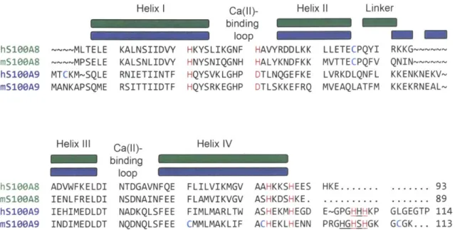
Chapter 2 Figure 2.1 Sequence alignment of human and murine S100A8 and S100A9... 90
Figure 2.2 SDS-PAGE of mCP and mCP-Ser... 102
Figure 2.3 Circular dichroism spectra of mCP and mCP-Ser... 104
Figure 2.4 Thermal denaturation plots of mCP and mCP-Ser... 105
Figure 2.5 Ca(Il)-dependent tetramerization of mCP and mCP-Ser monitored by analytical size exclusion chrom atography... 106
Figure 2.6 Metal depletion of Tris:TSB by mCP and mCP-Ser... 110
Figure 2.7 Antibacterial activity assays (8 hour timepoint) for mCP and mCP-S e r... 1 1 4 Figure 2.8 Antibacterial activity assays (20 hour timepoint) for mCP and mCP-S e r... 1 1 5 Chapter 3 Figure 3.1. Sequence alignment of human and murine S100A8 and S100A9 and predicted m anganese-binding site... 134
Figure 3.2 SDS-PAGE of mCP and variants... 153 Figure 3.3 Circular dichroism spectra of mCP variants... 154 Figure 3.4 Analytical SEC chromatograms of mCP variants ... 155 Figure 3.5 Analytical SEC chromatograms of mCP and metal-binding-site
variants with quantification of manganese and protein... 158 Figure 3.6 Analytical SEC chromatograms of mCP and mS100A9 tail variants
with quantification of manganese and protein... 159 Figure 3.7 Ca(Il)-dependent manganese affinity of mCP measured using a
Z in p y r-1 titra tio n ... 16 1 Figure 3.8 Zinpyr-1 manganese titration of mCP and variants... 162 Figure 3.9 X-band EPR spectra of Mn(Il)-bound mCP and mCP-Ser... 163 Figure 3.10 X-band EPR spectra and simulations for Mn(Il) and Ca(ll)-bound
mCP and the AHis3Asp variant... 164 Figure 3.11 Echo detected Q-band field sweep of Mn(ll)-bound mCP in the
presence of excess C a(lI)... 165 Figure 3.12 388 GHz field sweep of Mn(Il)-bound mCP in the presence of
excess Ca(ll) and corresponding simulation... 165 Figure 3.13 X-band EPR spectra of Mn(Il)- and Ca(lI)-bound mCP single-point
His-Ala variants in the mS100A9 C-terminal tail... 167 Figure 3.14. 15N-Mims ENDOR of Mn(Il)- and Ca(lI)-bound to globally labeled
15N -m C P ... 1 6 9
Figure 3.15. 15N-Mims ENDOR of globally 15N-labeled Mn(Il)- and Ca(ll)-bound
Figure 3.16 X-band EPR data of Mn(II) competition between mCP and MntC or P s a A ... 1 7 1
Chapter 4
Figure 4.1 X-band CW EPR spectra of Mn(Il)-MntC and Mn(lI)-PsaA with and
without 20% (v/v) PEG-200... 199 Figure 4.2 SDS-PAGE of purified MntC and PsaA... 204 Figure 4.3 Circular dichroism spectra of MntC and PsaA... 204 Figure 4.4 Representative Mn(II) competition titrations with ZP1 and MntC or
P s a A ... 2 0 7 Figure 4.5 Characterization of B-CP... 208 Figure 4.6 B-CP binds to streptavidin agarose resin and complexes transition
m e ta ls ... 2 0 9 Figure 4.7 SDS-PAGE analysis of B-CP pull-down assays... 209 Figure 4.8 B-CP pulldown assay to assess manganese competition with MntC
a n d P s a A ... 2 1 0 Figure 4.9 X-band CW EPR spectra of Mn(ll) bound to MntC and PsaA... 212 Figure 4.10. Comparison of X-band EPR of Mn(ll)-CP with Mn(li)-MntC and
M n (Il)-P s a A ... 2 14 Figure 4.11. X-band CW EPR spectra of 1:1:1 mixtures of Mn(Il):CP:SBP... 215 Figure 4.12. X-band CW EPR spectra showing the time-dependent loss of
Appendix A
Figure A.1. Crystal structures, zoom-in view of the metal-binding sites, and
metal-ligand distances of Mn(ll)-MntC and Mn(ll)-PsaA ... 228 Figure A.2. 2p echo detected field sweep of Mn(il)-MntC and Mn(Il)-PsaA at
1 3 0 G H z ... 2 3 4 Figure A.3. High-field/frequency CW EPR spectrum of Mn(Il)-MntC at 388 GHz
a n d 3 0 K ... 2 3 5 Figure A.4. High-field/frequency CW EPR spectrum of Mn(Il)-PsaA at 388 GHz
a n d 3 0 K ... 2 3 5 Figure A.5. High-field/frequency CW EPR spectrum of Mn(il)-MntC at 388 GHz
a n d 1 0 K ... 2 3 6 Figure A.6. High-field/frequency CW EPR spectrum of Mn(Il)-MntC at 388 GHz
a n d 3 K ... 2 3 8 Figure A.7. High-field/frequency CW EPR spectrum of Mn(ll)-PsaA at 388 GHz
a n d 5 K ... 2 3 8 Figure A.8. High-field/frequency CW EPR spectrum of Mn(Il)-MntC at 400 GHz
a n d 3 K ... 2 3 9
Appendix B
Figure B.1 Representative SDS-PAGE of whole cell lysate of E. coli BL21 (DE3)
obtained from the overexpression of S100A8 and S100A9... 260 Figure B.2 Purification of mCP and CP-Ser by anion exchange
Figure B.3 Purification of mCP and CP-Ser by size exclusion
chro m atog raphy ... 266 Figure B.4 Representative SDS-PAGE of purified hCP-Ser (hCP) and mCP... 267 Figure B.5 Absorbance profile of Fe standards in Fe speciation assay buffer
270
a nd ca lib ratio n cu rve ...
Figure B.6 Absorbance profiles of mCP and hCP-Ser samples analyzed by the
272
F e spe cia tio n a ssa y ...
Figure B.7. Bar plots showing the concentrations of Fe(II), Fe(Ill), and total Fe
List of Tables
Chapter 2
Table 2.1 Bacterial strains used... Table 2.2 Mass spectrometry analysis of mCP and mCP-Ser...
Table 2.3 Metal content of representative protein preparations... Table 2.4 Analytical size exclusion chromatography protein elution volumes... Table 2.5 Metal content of Tris:TSB media with or without protein treatment...
Chapter 3
Table 3.1 Primers and templates for site-directed mutagenesis... Table 3.2 Compositions of murine calprotectin (mCP) variants... Table 3.3 mS100A9 C-terminal tail variants... Table 3.4. Summary of results from protein mass spectrometry... Table 3.5. Metal content of representative protein preparations... Table 3.6. Metal content of representative protein preparations continued... Table 3.7. Analytical SEC elution volume and calculated molecular weights of p ro te in s ...
Chapter 4
Table 4.1 Summary of results from protein mass spectrometry... Table 4.2 Metal content of SBP samples at stages during representative p rote in purificatio ns ... 100 102 103 107 111 139 148 148 149 150 151 152 203 205
Table 4.3 Metal content of representative purified SBPs... 206
Appendix A
Table A.1. Table of Spectroscopic Parameters for Mn(lI) Bound to Various
Abbreviations
ABC ATP-binding cassette
AMA antimicrobial activity
ATCC American Type Culture Collection
BHI brain heart infusion medium
BME beta-mercaptoethanol
BPEOIA biotin polyethyleneoxide iodoacetamide
CbpA calprotectin-binding protein A
CP calprotectin
hCP human calprotectin (including hCP-Ser, the Cys+Ser variant)
mCP murine calprotectin (including mCP-Ser, the Cys-Ser variant) CD circular dichroism or cluster of differentiation receptor family
CFU colony forming units
CV column volumes
CW continuous wave
DAMPS damage-associated molecular patterns
DMSO dimethylsulfoxide
DNA deoxyribonucleic acid
DTT dithiothreitol
EDTA Ethylenediaminetetraacetic acid
ENDOR electron nuclear double resonance
EPR electron paramagnetic resonance
FPLC fast protein liquid chromatography
GE General Electric
Gu-HCI guanidinium hydrochloride
HEPES 4-(2-hydroxyethyl)-1 -piperazineethanesulfonic acid
HNP Human neutrophil peptide
ICP-MS inductive-coupled plasma mass spectrometry
IPTG isopropyl
P-D-1-thiogalactopyranoside
LB Luria-Bertani medium
LC-MS liquid chromatography mass spectrometry LD5o lethal dose that kills 50% of sample
LDPE low-density polyethylene
MES 2-(N-morpholino)ethanesulfonic acid
MRS De Man, Rogosa, and Sharpe medium
MWCO molecular weight cutoff
NARSA Network on Antimicrobial Resistance in Staphylococcus aureus
NBD nucleotide-binding domain
NCBI National Center of Biotechnology Information
NET neutrophil extracellular trap
NHMFL National High Magnetic Field Laboratory
NRAMP natural resistance-associated macrophage protein
OD600 optical density at 600 nm
PDB PMSF PQ ROS SBP SDS-PAGE SEC SOD TCEP TLR TM Tris TSB ZFS ZP1
Protein Data Bank
phenylmethylsulfonylfluoride precision quartz
reactive oxygen species solute-binding protein
sodium dodecylsulfate-polyacrylamide gel electrophoresis size exclusion chromatography
superoxide dismutase
tris(2-carboxyethyl)phosphine toll-like receptor
transmembrane
tris(hydroxymethyl)aminomethane tryptic soy broth medium
zero-field splitting Zinpyr-1
1.1 Innate Immunity
The innate immune system responds to bodily harm or danger, including alarm signals from distressed or injured cells as well as those from invading pathogenic microorganisms.' 2 Generally regarded as the first line of defense during infection, the
innate immune system detects and responds to infection before the adaptive immune response is initiated through signaling mechanisms.2 When the adaptive immune system
is activated, it orchestrates the fastidious production of novel receptors for specific antigens as well as antibodies, creating and saving unique immune system "memories" in the organism that serve its continued survival.3
1.1.1 Neutrophils in the Innate Immune Response
As the first responders in the innate immune system, neutrophils are major players in innate immunity. These white blood cells are derived from bone marrow and exist in circulating plasma at low levels.4 They are produced in quantities of =1011 per day in adult
humans.5 The lifespan of neutrophils may be between hours and days, with inflammation
extending the lifespan. 8 Upon sensing danger signals, neutrophils accumulate at the
appropriate locus to perform effector functions. Neutrophils are important for immune response mediation as well, as they are able to signal to other neutrophils, macrophages, and T-cells.5 Furthermore, these fascinating cells continue to be players as the adaptive
arm of the immune response is elaborated.9
During microbial infection, neutrophils are well known to enact degranulation, phagocytosis, and release of neutrophil extracellular traps (NETs).4 Degranulation
compartments within the cell.5 There are three main types of granules: those containing myeloperoxidase, lactoferrin, and gelatinase protein, respectively.5 Another typical process by which neutrophils combat pathogens is by phagocytosing them and treating them with antimicrobial agents within a phagocytic vacuole.5 Finally, NET-osis consists of a coordinated release of decondensed DNA, cytosolic proteins, granule contents, and histones.5 Reactive oxygen species (ROS) play a key role in the antimicrobial tactics of neutrophils as well. NADPH oxidase is present in the neutrophil membrane and generates superoxide radicals, characterizing the neutrophil oxidative burst.10 The superoxide then dismutates to hydrogen peroxide, which is utilized by myeloperoxidase to produce many reactive products, including the toxic hypochlorous and hypothiocyanous acids.10
In contrast to the prior dogma that neutrophils are a homogenous group of cells with a specific function, recent work is uncovering the bountiful phenotypic and functional diversity that these cells develop.4 0 Indeed, as a function of aging or as a response to environmental stimuli, neutrophils display divergent phenotypes that are characterized by transcriptional changes, changes in expression of surface molecules, and changes in activity.4' 1 For instance, in aberrant metabolic states such as hyperglycemia and
hypercholesterolemia, neutrophils become primed for NETosis or display increased ROS generation and myeloperoxidase release, respectively.1 2
, 13 Additionally, during methicillin-resistant Staphylococcus aureus infection, murine neutrophils develop subpopulations with differential expression of cell surface proteins, including those of the toll-like receptor (TLR) and cluster of differentiation (CD) families.1 4 These cellular
subgroups display different cytokine production and macrophage activation potentials.1 4
priming the neutrophils for greater antimicrobial activity as well as altering their lifespan.15'
16 Remarkably, neutrophil priming by peptidoglycan from the microbiota enhances the neutrophil-mediated killing of Streptococcus pneumoniae and S. aureus.16 Cumulatively,
the rich diversity of neutrophils generated in a plethora of biological contexts makes these special cells a promising subject of future discoveries.
1.1.2. Innate Immunity in Mice Versus Humans
Humans and mice diverged from a common ancestor ~65-75 million years ago.17
Though the species have much in common genetically, sharing over 90% of the same genes, their innate immune systems are notably different.18 Indeed, immune systems are
in general are especially vulnerable to evolutionary change.2 In mice, this change may
have arisen from environmental pressures such as pathogen coevolution as well as the increased evolution manifested as a result of a significantly shorter generation time.2 In a
comparative analysis of human and mouse genes, it was shown that the most divergent family of proteins are those involved in host defense.19 These proteins were -35%
divergent based on the amino acid sequences and around 3-fold more divergent compared to the average proteins in the analysis.19 It is now clear that there are numerous intricate differences in the composition and function of innate immune factors used by humans and mice that undoubtedly influence the nature of their innate immune responses.2
, 17, 20 A plethora of differences are present with respect to the secreted peptides and proteins, cytokines and chemokines, as well as toll-like receptors (TLRs), nod-like receptors, and cytokine receptors that exist in mice and humans.2 For example,
TLR1 1, TLR1 2, and TLR1 3 are present in mice but not humans; these receptors mediate sensing of various pathogenic substances.2
The degrees of similarities and differences between mice and humans, particularly in relation to innate immunity, are important to consider because mouse models are utilized to study diseases and infections, which naturally involve innate immune response activation. Mice (species mus musculus) represent the most common animal models in biomedical research.18 As model organisms that are easy to maintain and manipulate genetically while possessing great genetic similarity to humans, mice have been indispensable for decades worth of discoveries in biomedicine and immunology.18 Mouse models have been generated to study a range of innate immune functions and diseases and seen notable success; thus, there is a definite power and utility in the application of mouse model systems to understand immune functions.18 An associated immunotherapy
breakthrough, which relied on murine models, was the discovery of the role of inhibitory receptors in checkpoint blockade.2 1
However, the numerous deficiencies of murine models in imitating human biology often limit the translational ability of mouse model systems to human disease.2 2 This
includes, for example, mouse models failing to recapitulate the immune aspects of human tumor evolution in cancer.23 As is typical for mouse model systems, this disparity stems
from intrinsic biological differences between mice and humans as well as the fact that the mice studied are inbred, genetically homogenous, and sometimes raised under sterile, pathogen-free conditions that are far from capturing the natural state of humans. 23 In fact,
there is a greater disparity in immune system function between humans and inbred specific pathogen-free laboratory mice compared to wild mice.2 4 Such limitations have
spurred ongoing efforts to improve mouse model systems to increase their similarity to adult human immune systems. These efforts include consideration of bystander infections to further develop the immune systems of mice before they are used in studies as well as the development of humanized mouse strains that are engineered to have greater
immune system similarity to humans.25
1.1.2.1 Antimicrobial Arsenals within Neutrophils
There are extensive differences between neutrophils from humans and mice. Strikingly, the relative amounts of neutrophils circulating in mice and humans differs drastically; whereas white blood cells in human blood are composed of 50-70% neutrophils, in mice, neutrophils compose only 10-25% of the white blood cells.17 The
pathways by which ROS are generated in neutrophils differ between humans and mice as well.26 Among numerous other differences, mice lack the FcaRl receptor for IgA on
neutrophils, which is important for effector functions such as the oxidative burst, cytokine release, and NETosis in humans.27,28
Furthermore, the antimicrobial repertoires and regulation differ within human and murine neutrophils. For instance, a single lysozyme enzyme, an antimicrobial enzyme packaged within neutrophil granules, is present in humans, whereas two lysozyme genes exist in mice.29 Human neutrophils harbor antimicrobial defensin peptides (HNP1-4),
whereas mouse neutrophils do not.2 3 Additionally, the activities of multiple granule
enzymes, including myeloperoxidase and lysozyme, are higher in human neutrophils relative to mouse neutrophils.30 Arginase-1, an enzyme with fungicidal activity, is a
constitutive component of human azurophil granules, but is only inducibly expressed in
mouse neutrophils.3 1
There are additionally notable differences in the antimicrobial S100 proteins deployed during innate immune responses between humans and mice. Humans possess
S10OA8, S10OA9, and S10OA12 in neutrophils. S10OA8 and S10OA9, which heterooligomerize to form calprotectin, are abundant in human neutrophils and compose
~40% of the neutrophil cytoplasmic protein while S1 00A1 2 composes =5% of the cytosolic protein.32
, 33 100A8, S100A9 and S10OA12 have intracellular roles related to migration
and cytoskeletal modulation.34 These proteins are released during NETosis and are
significant components of NETs.35 Extracellularly, these S100's also act as
damage-associated molecular patterns (DAMPS; also referred to as endokines or alarmins) and
can activate immune cells and vascular endothelial cells.3 4 S100A8/S100A9 activates
TLR4 whereas S100A12 is a ligand for the receptor for advanced glycation end
products.34, 36
Mice contain S100A8 and S100A9 homologues and these proteins are abundant
in murine neutrophils; these proteins have each been detected in excess of 5 mg/mL.37
However, the amino acid identities of human and murine S100A8 and S100A9 are only
-56%. Compared to the average protein identity of -85% identified in a comparative study of over 1,100 human and murine protein sequences, the identity between the S10OA8
and S1 00A9 orthologues is relatively low.38 Indeed, the value fits within the lowest quartile
of proteins analyzed by Makalowski et al. in terms of amino acid identity.38 Consistent
with prior work by Murphy et al. identifying high divergence of host-defense proteins
antimicrobial host-defense proteins identified by Makalowski et al., including multiple interleukins and interferons.19, 38 In addition to these differences, mice completely lack a
S100A12 orthologue.39 Murine S100A8 has been shown to be chemotactic like human
S10OA12, whereas human S100A8 lacks chemotactic properties.40 Together, these differences may contribute to the differing neutrophil responses and capabilities between humans and mice, including during microbial infection.
1.1.3. Nutritional Immunity
Nutritional immunity involves the withholding of essential metal ions by host proteins in order to deprive pathogens of these nutrients.41 This process was first
recognized as a factor in the host-pathogen competition for Fe(lll).4 1 The host maintains
rigorous high-affinity chelation of Fe by proteins including ferritin, transferrin, hemoglobin, and myoglobin, which restricts available Fe to exceedingly low levels.42 In more recent
years, Zn(II) and Mn(II) sequestration became recognized as important in nutritional immunity. The host protein CP has been implicated in Mn(II) and Zn(II) sequestration in
vivo.4 3 Laser-ablation inductively-coupled plasma mass spectrometry has been used to
show that murine tissue abscesses from S. aureus infection are relatively devoid of Zn and Mn despite an adequate abundance of these metals in surrounding healthy tissues.43
Restriction of Cu in tissue abscesses has since been noted.44 Furthermore, a recent
report highlighted the heterogeneity of metal distributions between abscesses within the same tissue, providing more nuance to host-mediated nutritional immunity.44 In vitro
sequester a range of transition metal ions, including Mn(ll), Fe(ll), Ni(ll), and Zn(II), and the host protein S100A12 can sequester Zn(I1).45
, 46
Nevertheless, pathogens can often foil this host metal-withholding strategy by employing metal acquisition systems. For Fe(Ill) uptake, this includes siderophores, heme acquisition systems, and transferrin/lactoferrin receptors.4 2 Mn(II) and Zn(II) are secured
through the use of high-affinity metal uptake systems.47 One ingenious pathogen, Neisseria sp, has the ability to bind CP using the aptly named calprotectin-binding protein A (CbpA; in Neisseria meningiidis).48,49 This outer membrane protein can facilitate the
uptake of Zn(II) by Neisseria.48 Though the nature of this interaction is not yet clear, this example represents a novel adaptive tactic to combat host-mediated metal limitation.
1.1.3.1. Structure and Function of Human Calprotectin
Calprotectin is a member of the S100 family of Ca(lI)-binding proteins and a major contributor to the metal-withholding innate immune response.45
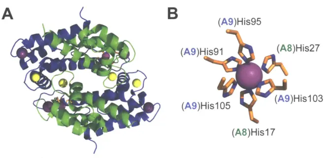
, 50 CP is a cytoplasmic protein expressed in neutrophils, macrophages, monocytes, and epithelial cells. Human calprotectin (hCP) is a heterooligomer of the S100 proteins S100A8 and S1 00A9 (Figure 1.1).51-54 S10A8 and S100A9 each harbor two EF-hand domains, with a non-canonical EF-hand in the N-terminal region of the polypeptide and a canonical EF-hand near the
C-terminus. 55 In the absence of Ca(II) and transition metal ions, hCP exists as a heterodimer.51 The protein has two transition-metal-binding sites, both present at the
S100A8/S100A9 dimer interface, and designated site 1 and site 2.54, 56, 57 Site 1 is a His3Asp motif composed of (A8)His83, (A8)His87, (A9)His2O and (A9)Asp3O.54 Site 2 is a His6 site composed of (A8)Hisl7, (A8)His27, (A9)His9l, (A9)His95, (A9)His103 and
(A9)HislO5 (Figure 1.1).58-60 The HiS6 site of hCP is of particular interest because it can
sequester a numberof divalent first-row transition metal ions including Mn(II), Fe(II), Zn(II) and Ni(Il).45
,59-64 Recent work indicated CP can bind Cu, but the details of this interaction are not yet clearly defined.65
A
B
(A9)His95
(A9)His9l
(A9)His105
(A8)His27
(A9)His103
)i
(A8)Hisl 7
Figure 1.1. Crystal structure of the Mn(Il)-, Ca(II)-, and Na(l)-bound human calprotectin heterotetramer (A) and composition of the Mn-His6 site (B). S10OA8 is shown in green and the S100A9 subunit in blue. Metal ions are shown as spheres: Mn (magenta), Ca (yellow), and Na (purple) (PDB: 4XJK). Na(l) derived from the crystallization buffer is shown occupying the non-canonical EF-hand domains.
The working model has been informed by extensive studies of this protein. It has been known for some time that Ca(II) binding by CP causes association of two heterodimers to generate a heterotetramer.52,53 Beyond promoting tetramerization, Ca(II) binding increases the transition-metal-ion affinities, antimicrobial activity, and proteolytic stability of hCP.45
, 57,66 Thus, a working model has been put forth wherein CP senses the high extracellular Ca(II) concentration (=2 mM) at an infection site and becomes a
tetramer with enhanced transition metal affinities, enabling effective metal sequestration.45
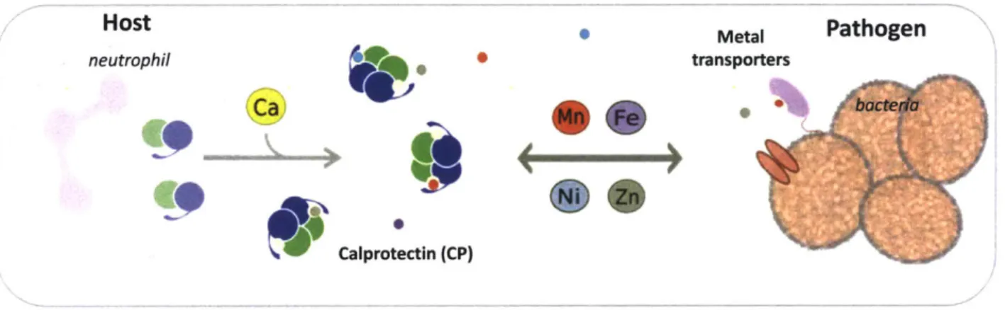
,57 It is this high-affinity metal sequestration in the extracellular space that allows CP to limit microbial growth by competing with microbes for metal nutrients (Figure 1.2)
Host Metal Pathogen
neutrophil transporters
Calprotectin (CP)
Figure 1.2. Depiction of a nutritional immunity scenario in which calprotectin (CP) competes with pathogenic bacterial metal importers for transition metal ions.
1.1.3.2. Manganese-binding Properties of Human Calprotectin
A 2008 report identified a role for CP in sequestration of Mn(II) in a murine tissue abscess model of S. aureus infection. This work represented an exciting discovery; prior to that report, only Zn(II) sequestration by CP was recognized.67 Subsequently, the molecular characteristics of Mn(II) binding to hCP were explored. In 2013, robust biochemical and structural data were reported that elucidated the Ca(ll)-modulated Mn(II) affinity and biologically unique hexahistidine Mn(II) coordination sphere of CP (Figure 1.1).58, 60, 61 Characterization by advanced EPR spectroscopy followed soon after, shedding more light on the highly symmetric Mn(Il)-binding site.59 In Chapter 4, competition between human CP and bacterial Mn transport proteins is described that reveals a sub-nanomolar Mn(II) affinity for CP.
1.1.3.3. The Function of Calprotectin: S10OA9-- Mice as Infection Models
S100A9-'- mice have been generated to study the impact of this protein, or the associated S100A8/S100A9 protein complex CP, on various immune functions.68, 69 Notably, S1 00A8-'- mice cannot be generated because the mutation is embryonic lethal.70 In fact, S100A8 plays a pivotal and non-redundant role in the absence of its S100A9 partner in the developing mouse embryo.70 Neutrophils from S1 00A9-'- mice are relatively similar to those from wildtype mice, but have some differences.71-73 While the S100A9-'-cells have normal morphology, they do display a somewhat polarized shape and show observable defects in Ca(Il)-related signaling.71-73 Despite these drawbacks,
S100A9~-mice have been useful for understanding the role of this protein in a number of inflammation-related scenarios, from arthritis to bacterial infections.69
These CP-deficient mice have been used in infection models of a number of microbial pathogens, including the bacterial pathogens S. aureus, Acinetobacter
baumannii, Streptococcus pneumoniae, Klebsiella pneumoniae, Helicobacter pylori, and Salmonella as well as the fungal pathogens Aspergillus fumigatus and Candida
albicans.35, 43, 65, 74-83 These models have provided many insights into the antimicrobial
function of CP. For example, the absence of CP leads to a greater bacterial burden in the lungs and livers of mice infected with A. baumannii.74 However, the consequence of
S100A9 deficiency is often multi-faceted and context-dependent. For instance, animal models of S. aureus infection show that CP inhibits S. aureus growth in the murine liver, does not alter bacterial growth in the kidney, and can actually promote bacterial colonization of the heart.43,
84, 85 The presence of CP can also promote bacterial growth
toxicity experienced by these species.7 8
, 80 Furthermore, CP can modulate interactions between the pathogens Pseudomonas aeruginosa and S. aureus.86 Therefore, the complex roles that CP plays in infectious disease, and the molecular mechanisms behind them, warrant continued investigation.
1.1.3.4. Competition for Manganese Between Murine Calprotectin and
Staphylococcus aureus
The competition for Mn(II) between CP and bacterial proteins has been exemplified in studies involving mice infected with S. aureus.43
, 85 S. aureus has two Mn(II) import
systems, MntABC and MntH, that contribute to infection.5 These systems are necessary for full virulence in wildtype mice but not S100A9-'- mice. In the presence of CP, a defect in Mn(II) transport translates to lower bacterial burdens in murine liver and kidney tissue.5 However, in the absence of CP, defects in Mn(II) transport do not impact the bacterial burden of organ tissues.5 The molecular details of these observations are explored in Chapters 3 and 4 of this thesis.
1.1.3.5 The Gap in Knowledge Between Human and Murine Calprotectin
Our current understanding of the function of CP is predominately informed by mouse models of infectious disease43, 74, 80, 83, 86, 87, including studies that involve the
S100A9-/- , CP-deficient, mouse strain noted above68, and molecular characterization of
the human orthologue.1,
54, 57-63, 88 A biochemical assessment of mCP is fundamentally
important to understand the function of this protein at the molecular level. Studies in this vein will serve to inform mouse models of infectious disease in addition to providing a
biochemical comparison to the human CP orthologue. Amino acid sequence alignment of the human and murine S100A8 and S100A9 polypeptides indicates, among other things, that the His3Asp and His6 sites are conserved. In an effort to supplement this gap in
knowledge, the heterologous expression and purification of mCP is described along with initial metal binding and antibacterial activity studies in Chapters 2 and 3 of this thesis.
1.2. Manganese in Biology
Manganese composes -0.085% of the earth's crust.89 The redox properties of manganese govern its solubility and therefore bioavailability; Mn(II) is soluble, whereas the Mn(lll)/Mn(IV) oxides are relatively insoluble.90 Manganese is present in seawater at
approximately low nanomolar concentrations.90, 91 Since early times, life forms have concentrated metal ions in their cells at concentrations exceeding those in natural environments, such as the sea water from which life arose.91 This observation gave an
early clue as to the biological importance of metal ions by suggesting active uptake and retention within cells. Manganese concentrations in human plasma are slightly higher than seawater, in the nanomolar range.91, 92 Bacterial species maintain varying Mn
concentrations within their cells. For instance, Escherichia coli cells contain micromolar levels of Mn, with the exact content depending on growth conditions.93, 94 In contrast, Lactobacilli sp. can accumulate millimolar intracellular Mn concentrations.95 Recent work
with Streptococcus sanguinis noted variable Mn concentrations even within different strains of the same species.96
1.2.1. Identification of Manganese as an Essential Nutrient: an Historical Perspective
Manganese was identified as a component of soil and plants as early as 1774.97
By 1892, manganese was identified in mollusk blood, with studies and hypotheses of the element's importance in mollusks extending into the 20th-century.98 In 1922, the necessity
of Mn in plant growth was established, and the role of Mn as a nutrient in mammals subsequently began to be illuminated.9 7 Studies in the 1920's and 1930's revealed that
supplementation of Mn in the rations of animals was beneficial for growth, and conversely that too much was toxic.99-102 Researchers first revealed the importance of Mn in mice
and rats, and later work reported the requirement of Mn in childhood development.100, 101, 103
The concept that Mn is essential in biology was strengthened through the 20th
century as the molecular details of Mn utilization in biology were elucidated. In the
mid-2 0th century, the importance of Mn in photosynthesis and role as a component of
photosystem 11 were becoming apparent through studies regarding the effect of Mn-deficiency on photosynthesis and oxygen evolution.10410 6 Enzymes that could be active
with added Mn, including arginase, were identified.107 In 1966, pyruvate carboxylase,
isolated from chicken liver mitochondria, was the first enzyme reported to contain bound Mn(l1).108 Multiple lines of evidence, including in vivo incorporation of 54Mn and the effect
of Mn-bound enzyme on the proton relaxation rate as measured by nuclear magnetic resonance supported the conclusion.108 A jack-bean globulin, called concanavalin A, was
found to contain bound Mn.109 An electron paramagnetic resonance spectroscopy (EPR) study was then undertaken to characterize the Mn-protein complex and assess the
observation that "immobilized" Mn bound to proteins was not detectable by EPR.11 0 The EPR characterization revealed a Mn-coordination sphere with near-cubic symmetry.110
The subsequent discovery of high-affinity Mn import systems and Mn-dependent enzymes in various microorganisms suggested an important role for this element in bacterial physiology. A high-affinity Mn uptake system in E. coli was discovered in 1969, suggesting that Mn was a crucial nutrient.1 , 112 The landmark discovery of a Mn-utilizing
superoxide dismutase (SOD) from E. coli followed in 1970.113 This work established a function for Mn in E. coli and represented the identification of a new type of SOD that bore little resemblance to the previously characterized mammalian Cu,Zn-SOD.113 Elucidation
of Mn as the required cofactor hinged on characterization of Mn(II) released from the denatured enzyme by EPR spectroscopy.'1 3
Subsequently, high-affinity, active Mn transport was verified in other species, including Bacillus subtilis, Rhodopseudomonas capsulata, and S. aureus. 1 4
-1 -16 A
Mn-requiring ribonucleotide reductase was discovered in some species of Gram-positive bacteria in 1981, adding a third type on top of the already-discovered Fe-dependent varieties.117 The Mn-requiring, non-heme "pseudocatalase" was isolated and
characterized from Lactobacillus plantarum two years later.11 8 L. plantarum is an unusual
bacterium that can accumulate intracellular concentrations of Mn in excess of 30 millimolar. In the absence of an SOD, this Mn acts to scavenge the superoxide radical anion.119 An active transport system for Mn was characterized in this organism in 1984.119 The requirement of Mn as a micronutrient in pathogens portends its relevance in virulence. Indeed, for pathogens to survive in a hostile host environment, they must successfully acquire essential elements (see section 1.1.3). The roles of the bacterial
Mn-SOD in the virulence of both S. pneumoniae and Haemophilus influenzae in murine infection models were reported by 2000.120, 121 The Mn-dependent transcriptional
repressor PerR was determined to be essential for full virulence in a murine skin abscess model of S. aureus infection.12 2 In addition, Mn-uptake systems were being discovered
and established as virulence determinants. Mn(II) acquisition systems were found to be important in the virulence of Salmonella, Yersinia pestis, Enterococcus faecalis, Streptococcus mutans, and S. pneumoniae by 2002.123-128 As elucidation of the role of
Mn in bacterial pathogenesis and virulence has unfolded in the years since, understanding and appreciation of the role that Mn import systems play in these processes has blossomed.
1.2.2. Manganese Import Systems in Bacteria
Due to the low propensity for passive diffusion of Mn across cell membranes, bacteria utilize energy-powered transport systems to acquire this ion.1 29 One type of Mn
importers in bacteria are homologs of the eukaryotic NRAMP (Natural
resistance-associated macrophage Qrotein) transporters. The bacterial proteins are referred to as MntH transporters, in reference to their proton(_H)-dependent Mn transport function.130 There are three primary groups of prokaryotic MntH transporters (A, B, and C) that diverged from each other over evolution and display relatively low amino acid sequence identity of <30%.130 Group A MntH transporters are present in Gram-positive organisms such as Bacillus subtilis and Mycobacterium tuberculosis, as well as the Gram-negative organisms E. coli, Salmonella typhimurium, Yersinia pestis, and Deinococcus radiodurans, among others.130 Of note, group A MntH transporters are the type utilized