Publisher’s version / Version de l'éditeur:
Environmental Science and Technology, 48, 22, pp. 13307-13315, 2014-10-21
READ THESE TERMS AND CONDITIONS CAREFULLY BEFORE USING THIS WEBSITE. https://nrc-publications.canada.ca/eng/copyright
Vous avez des questions? Nous pouvons vous aider. Pour communiquer directement avec un auteur, consultez la première page de la revue dans laquelle son article a été publié afin de trouver ses coordonnées. Si vous n’arrivez pas à les repérer, communiquez avec nous à PublicationsArchive-ArchivesPublications@nrc-cnrc.gc.ca.
Questions? Contact the NRC Publications Archive team at
PublicationsArchive-ArchivesPublications@nrc-cnrc.gc.ca. If you wish to email the authors directly, please see the first page of the publication for their contact information.
NRC Publications Archive
Archives des publications du CNRC
This publication could be one of several versions: author’s original, accepted manuscript or the publisher’s version. / La version de cette publication peut être l’une des suivantes : la version prépublication de l’auteur, la version acceptée du manuscrit ou la version de l’éditeur.
For the publisher’s version, please access the DOI link below./ Pour consulter la version de l’éditeur, utilisez le lien DOI ci-dessous.
https://doi.org/10.1021/es5029102
Access and use of this website and the material on it are subject to the Terms and Conditions set forth at
Sulfide oxidations for LC-MS analysis of methionine-containing
microcystins in dolichospermum flos-aquae NIVA-CYA 656
Miles, Christopher O.; Melanson, Jeremy E.; Ballot, Andreas
https://publications-cnrc.canada.ca/fra/droits
L’accès à ce site Web et l’utilisation de son contenu sont assujettis aux conditions présentées dans le site LISEZ CES CONDITIONS ATTENTIVEMENT AVANT D’UTILISER CE SITE WEB.
NRC Publications Record / Notice d'Archives des publications de CNRC:
https://nrc-publications.canada.ca/eng/view/object/?id=d0b1e963-6897-4d93-83c5-8b0808c6c840 https://publications-cnrc.canada.ca/fra/voir/objet/?id=d0b1e963-6897-4d93-83c5-8b0808c6c840Sulfide Oxidations for LC-MS Analysis of Methionine-Containing
Microcystins in Dolichospermum f los-aquae NIVA-CYA 656
Christopher O. Miles,*
,†,‡Jeremy E. Melanson,
§and Andreas Ballot
†,▽†
Norwegian Veterinary Institute, P.O. Box 750 Sentrum, N-0106 Oslo, Norway
‡
Department of Pharmaceutical Chemistry, School of Pharmacy, University of Oslo, P.O. Box 1068 Blindern, N-0316 Oslo, Norway
§
Measurement Science and Standards, National Research Council Canada, 1200 Montreal Road, Ottawa, Ontario K1A 0R6 Canada
▽
Norwegian Institute for Water Research, Gaustadalléen 21, N-0349 Oslo, Norway
*
S Supporting InformationABSTRACT: Microcystins are cyclic heptapeptides produced by a range of cyanobacteria. More than 150 microcystin analogues have been reported from cultures, algal blooms, or other contaminated samples. Relatively few analytical standards are available, making identification and quantitation of these toxins a challenge, even with LC-MS technology. We developed a two-step oxidative procedure that allows LC-MS identification of microcystins containing methionine and methionine sulfoxide, and reveals the oxidation state of the methionyl sulfur atom. The procedure was
used in parallel with mercaptoethanol derivatization and LC-MS2analysis to demonstrate the presence of [Asp3]MC-MR (12)
and MC-MR (17) in a culture of Dolichospermum flos-aquae, together with low levels of [Asp3]MC-M(O)R (5) and MC-M(O)R
(7), as well as 20 other microcystins. Fresh culture contained only traces of sulfoxides 5 and 7, but these increased during storage or sample extraction and preparation. This suggests that microcystins containing methionine sulfoxide are primarily postextraction oxidation artifacts, rather than being produced by biosynthesis in cyanobacteria. A simple, rapid extraction under inert gas followed promptly by LC-MS analysis minimized oxidation artifacts for D. flos-aquae.
■
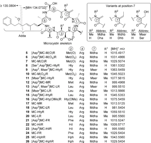
INTRODUCTIONDolichospermum flos-aquae (Brébisson ex Bornet et Flahault) P.Wacklin, L.Hoffmann & J.Komárek belongs to the cyanobacterial family Nostocaceae and is able to fix nitrogen through special cells called heterocytes.1 Formerly known as Anabaena f los-aquae ([Lyngbye] Brébisson ex Bornet et Flahault), it is a planktonic species possessing gas vesicles and was therefore assigned to the new genus Dolichospermum.2 D. flos-aquae forms blooms in lakes and reservoirs worldwide and has caused mortalities of domestic animals and wildlife.3 Several strains produce the neurotoxins anatoxin-a and anatoxin-a(s).3Production of hepatotoxic microcystins, includ-ing [Asp3]MC-HtyR (15), [Asp3]MC-LR (18), MC-HtyR
(19), and MC-LR (20) (Figure 1), has been demonstrated in strains of D. flos-aquae and unidentified Dolichospermum and Anabaena spp.4−6
Microcystins (MCs) are cyclic heptapeptides produced by a range of cyanobacteria, including Microcystis, Nostoc, and Planktothrix spp.7 Microcystins inhibit protein phosphatase-1 and -2A, and have been responsible for numerous poisonings of livestock, wild animals, and, occasionally, human beings.7 In addition to acute toxic effects, microcystins are suspected of chronic toxicity through consumption of contaminated water used for drinking and food preparation.7
The World Health Organization has issued a guideline level of 1 μg/L for MC-LR in drinking water.8,9However, more than
150 microcystin congeners have been reported in the literature,10,11making measurement of such low concentrations in water challenging. For example, a recent analysis from a Microcystis bloom in Hartbeespoort Dam, South Africa, identified 41 microcystins in the water.12 A further problem
is that analytical standards are available for only a very few MC congeners, and the quantitation of some of these commercial standards appears to be unreliable.13
LC-MS analytical methods have, to a degree, alleviated the problem of lack of standards, because tentative identification can often be obtained from characteristic ions in the MS/MS spectra14,15 although quantitation without standards remains problematic. For example, the majority of reported microcystins contain the unusual β-amino acid Adda (Figure 1), which produces a characteristic product ion during MS fragmenta-tion.14 Recently, we described a method using thiol derivatization together with LC-MS/MS analysis to identify microcystins in complex matrices, based on the reactivity of the Mdha and Dha groups (Figure 1) present in most MCs.12,16,17
This method also discriminates between microcystins contain-ing the thiol-reactive Mdha and Dha and the unreactive Mdhb
Received: June 16, 2014 Revised: October 15, 2014 Accepted: October 21, 2014 Published: October 21, 2014 Article pubs.acs.org/est copying and redist ribut ion of t he art icle or any adapt at ions for non-commercial purposes.
and Dhb (Figure 1) residues, which are otherwise difficult to differentiate by LC-MS.18
Microcystins containing methionine are relatively uncom-mon in the literature, but MC-M(O)R (7),19MC-YM(O),20,21 MC-YM19,20 and MC-LM22 ([MH]+ m/z 1029.5, 1036.5, 1020.5, and 970.5, respectively) have been reported (M, methionine; M(O), methionine sulfoxide).
While analyzing an extract from D. flos-aquae by LC-MS2, we
observed peaks at m/z 1015.5, 1029.5, 999.5, and 1013.5 whose thiol reactivities (Table 1, Supporting Information Figure S41) and MS2 spectra (Supporting Information Figures S25 and
S27−29) were consistent with Met-containing microcystins, but in the absence of high-resolution MS, these masses also corresponded to plausible non-Met-containing microcystins.
Here, we report a two-step oxidation procedure with hydrogen peroxide and potassium peroxymonosulfate that, together with unit-mass-resolution LC-MS2 and the recently
reported16,18 thiol-derivatization procedure, provides a high level of certainty in the identification of Met-containing microcystins. Subsequent application of high-resolution LC-MS before and after oxidation was used to confirm their identities and to validate the procedure.
■
MATERIALS AND METHODSChemicals and Cyanobacteria. Mercaptoethanol and potassium peroxymonosulfate (Oxone) (technical grade) were from Sigma−Aldrich, Oslo, Norway. A mixed standard (MC-RR, MC-LR (20), MC-YR, MC-LA, MC-LY, MC-LF, MC-LW; ca. 0.4−1 μg/mL in MeOH−H2O (1:1)) was prepared from
microcystins from Alexis Biochemicals (Grünberg, Germany) as
described previously,16while standards of MC-HilR (22) and MC-HtyR (19) were from AH Diagnostics (Oslo, Norway). Certified reference materials of MC-LR, [Dha7]LR,
MC-RR and NOD, and a reference material of [Asp3]MC-LR (18)
quantified by LC with a chemiluminscence nitrogen detector, were from NRC Measurement Science and Standards, Halifax, NS, Canada. Hydrogen peroxide (30%) was from Merck (Darmstadt, Germany). Solvents for LC and extraction were of gradient (Romil, Oslo, Norway) or LC/MS quality (Fisher Scientific, Fair Lawn, NJ, or Loughborough, United Kingdom). DIAION HP-20 was from Mitsubishi Chemical Corporation (Tokyo, Japan).
Cyanobacterial Culture. D. flos-aquae strain NIVA-CYA 656 (from the culture collection of the Norwegian Institute for Water Research) was originally isolated as strain AB2008/17 from Lake Scharmützelsee, Germany, in 2008.23 The culture was maintained in 50 mL flasks containing 20 mL Z8 medium24 at 22 °C and a photon flux density of 80 mol m−2s−1. Because
of the recent taxonomic revision of Anabaena, and 6 years of regular subculturing including 2 interlaboratory transfers, the species identity of this culture was confirmed by isolation of genomic DNA and a 16S rRNA gene analysis (Supporting Information). The strain was also classified based on standard morphological traits1,25 using a Leica DM2500 light micro-scope, Leica DFC450 camera and Leica Application Suite software (Leica, Oslo, Norway) (Supporting Information).
Sample Preparation. Culture 1. Aliquots of culture (1 mL) were frozen and thawed three times then ultrasonicated for 5 min. MeOH (1 mL) was then added and the samples filtered (0.2 μm, Costar Spin-X Microcentrifuge, Corning, NY).
Figure 1. Microcystins discussed in the text, with standard amino acid abbreviations, calculated m/z values, and showing the characteristic fragmentation pathway in the Adda-moiety. Note that Met(O) denotes methionine sulfoxide as a mixture of diastereoisomers, while Met(O2)
denotes methionine sulfone.
Environmental Science & Technology Article
dx.doi.org/10.1021/es5029102 | Environ. Sci. Technol. 2014, 48, 13307−13315
For thiol derivatization, sodium carbonate buffer (0.2 M, pH 9.7) was added to filtrate at 1:4 v/v, and to aliquots (200 μL) of the buffered filtrates was added 1 μL mercaptoethanol or O-(2-mercaptoethyl)-O′-methyl-hexa(ethylene glycol) (MEM-HEG)16 with vortex-mixing followed by LC-MS2 analysis
(method A) 2−3 h later. Underivatized (i.e., no thiol) buffered filtrates were used as controls. Remaining culture material was extracted with HP-20 as described previously16and analyzed by LC-MS2 (method A), then stored at −20 °C and analyzed
periodically by LC-MS2, and after 3 years storage by LC-HRMS
(methods B−D).
Culture 2.A second culture was extracted as above, except that containers used for freeze−thawing, extraction, sample storage/analysis, and filtration were, wherever possible, flushed with argon to hinder autoxidation. The methanol−water extract was used for quantitative analysis. The eluate from the HP-20 was evaporated to dryness under a stream of nitrogen and dissolved in 1:1 MeOH−H2O (10 mL). Aliquots of the
quantitative and HP-20 extracts were analyzed immediately by LC-MS2(method A) and LC-HRMS (methods B−C) and the
remainder stored under argon at −20 °C for further analysis (method D).
Oxidations.Aliquots of HP-20 extract (1 mL) were filtered (Spin-X, 0.22 μm, Costar, Corning Inc., NY). To each sample was added 50 μL of water (control), 30% hydrogen peroxide, or potassium peroxymonosulfate (5.1 mg/mL), and the progress of the reactions was followed by LC-MS methods A, B, or C starting from ∼1−2 min after addition of oxidant and at ∼15 min intervals thereafter.
LC-MS Analyses. LC-MS2 (Method A).
Liquid chromatog-raphy was performed on a Symmetry C18 column (3.5 μm, 100 × 2.1 mm; Waters, Milford, MA) as described previously,16 eluted with a linear gradient (0.3 mL/min) of acetonitrile (A) and water (B) each containing 0.1% formic acid. The gradient was from 22−75% A over 10 min, to 95% A at 11 min (1 min hold), followed be a return to 22% A with a 3 min hold to equilibrate the column. The HPLC system was coupled to a Finnigan LTQ ion trap mass spectrometer (Finnigan Thermo Electron Corp., San Jose, CA) operated as described previously.16
LC-HRMS (Method B). A Q Exactive mass spectrometer (Thermo Scientific, Bremen, Germany) was used as detector, with spray voltage 3.5 kV, capillary temperature 350 °C, probe heater 300 °C, S-lens RF level 50, with sheath and auxiliary gas 35 and 10, respectively. Chromatography was as for method A, Table 1. Microcystin Analogues Identified in Extracts from a Culture of D. f los-aquae (NIVA-CYA 656)
culture 2 Rt(min)
HP-20
extract quantitative extractc microcystin [MH]+formula measured(m/z) (ppm)Δ statusa grad.b isocrat.c % of totald (ng/ml)conc. % of totald [seco-4/5][Asp3]MC-HtyR 1 C 52H75O14N10 1063.5466 0.7 tentative 2.41 nde [seco-4/5]MC-HtyR 2 C53H77O14N10 1077.5636 1.9 tentative 2.49 nd [seco-4/5][Asp3]MC-LR 3 C 48H75O13N10 999.5517 0.8 tentative 2.60 nd [seco-4/5]MC-LR 4 C49H77O13N10 1013.5675 0.9 tentative 2.67 nd [Asp3]MC-M(O)R 5 C
47H71O13N10S 1015.4905 −1.2 tentative 3.19 2.84 0.3 trace trace
[Asp3]MC-M(O
2)R 6 C47H71O14N10S 1031.4851 −1.4 tentative nd 3.43f nd nd nd
MC-M(O)R 7 C48H73O13N10S 1029.5056 −1.7 tentative 3.47 3.69 0.3 trace trace
[Ser1,Asp3]MC-HtyRg
8 C52H73O14N10 1061.5289 −1.2 uncertain 3.37 4.05 trace nd nd
[Asp3,Mser7]MC-HtyR 9 C
52H75O14N10 1063.5432 −2.7 tentative 3.51 4.07 0.1 trace trace
MC-M(O2)R 10 C48H73O14N10S 1045.5005 −1.6 tentative nd 4.31f nd nd nd [Mser7]MC-HtyR 11 C 53H77O14N10 1077.5591 −2.3 tentative 3.60 4.56 nd nd nd [Asp3]MC-MR 12 C 47H71O12N10S 999.4963 −0.5 tentative 3.63 4.58 1.5 4.7 1.8 [Asp3, Mser7]MC-LR 13 C
48H75O13N10 999.5499 −1.0 tentative 3.63 4.64 0.1 trace trace
[Mser7]MC-LR 14 C
49H77O13N10 1013.5645 −2.1 tentative 3.66 4.71 0.1 trace trace
[Asp3]MC-HtyR 15 C 52H73O13N10 1045.5341 −1.2 tentative 3.64 4.75 14 36 14 [Asp3]MC-Hty(OMe)R 16 C 53H75O14N10 1075.5474 0.6 tentative 3.73 nd trace MC-MR 17 C48H73O12N10S 1013.5113 −1.1 tentative 3.75 5.33 2.0 6.4 2.5 [Asp3]MC-LR 18 C 48H73O12N10 981.5399 −0.5 confirmed 3.78 5.63 23 60 23 MC-HtyR 19 C53H75O13N10 1059.5507 −0.2 confirmed 3.82 5.63 19 53 20 MC-LR 20 C49H75O12N10 995.5546 −1.4 confirmed 3.88 6.00 33 87 34 [Asp3]MC-FR 21 C 51H71O12N10 1015.5233 −1.4 tentative 3.98 7.71 1.5 3.9 1.5 MC-Hilr 22 C50H77O12N10 1009.5705 −1.1 confirmed 4.06 7.78 0.8 1.4 0.5 [Asp3]MC-HilR 23 C 49H75O12N10 995.5547 −1.4 tentative 3.99 7.85 0.8 1.0 0.4 MC-FR 24 C52H73O12N10 1029.5392 −1.1 tentative 4.13 8.35 1.9 5.1 2.0 MC-HphR 25 C53H75O12N10 1043.5550 −1.0 tentative 4.28 10.50 0.5 0.8 0.3 [Asp3]MC-HphR 26 C 52H73O12N10 1029.5389 −1.4 tentative 4.19 10.79 0.3 0.5 0.2
aCompounds were considered confirmed when they had the same R
t, MS, and MS2spectra, and thiol reactivity as authentic standards. Analogues
were considered tentatively identified if the Rt, MS, and MS2spectra, and thiol reactivity were consistent with the proposed structure. All analogues
reacted rapidly with mercaptoethanol (except for Mser7-congeners 9, 11, 13, and 14), showed prominent loss of m/z 134.0732 in LC-HRMS
methods B(i) or C(i), and LC-MS2spectra are presented in the Supporting Information, together with the LC-MS AIF chromatogram showing
product ions at m/z 135.0804 (Adda fragmentation).bLC-HRMS method BcLC-HRMS method C(i)dExpressed as percent of total microcystins
5−26.eNot detected.fDetected only after treatment with K[HSO
5].gLC-MS2data is consistent with an extra oxygen atom on either amino acid 2
or, more probably, amino acid 1 (Supporting Information).
except that a Waters Acquity UPLC pump and autosampler were used. The spectrometer was operated in either: (i) full MS SIM mode scanned m/z 970−1150 with automatic gain control (AGC) target 3 × 10³, resolution 70,000, and maximum
injection time (max IT) 200 ms, or; (ii) all-ion-fragmentation (AIF) mode (full scan: scanned m/z 450−1150, AGC target 5 ×10⁶, resolution 70 000, and max IT 200 ms; AIF scanned m/z 110−1500, AGC target 3 × 10⁶, resolution 35 000, max IT 200
ms, and normalized collision energy 50).
LC-HRMS (Method C).Analysis was as for method B, except that the LC was eluted isocratically with 29% eluent A in (i) full MS SIM mode or (ii) AIF mode but with full scan m/z 840− 1070.
LC-HRMS (Method D). Chromatography was performed as for method A, except that an Agilent 1260 HPLC system (Agilent Technologies, Santa Clara, CA) and 20 μL injections were used. The mass spectrometer was an LTQ-Orbitrap with a heated electrospray (HESI-II) probe (Thermo Scientific, Bremen, Germany). Source parameters were: spray voltage 5 kV, vaporizer temperature 350 °C, capillary temperature 300 °C, capillary voltage 45 V, tube lens voltage 120 V, sheath gas 55, and auxiliary gas 15. Fragmentation in the LTQ was performed as per method A, except that a 1000 ms maximum fill time was employed. A resolution setting of 15 000 was used for detection in the Orbitrap to maximize sensitivity.
■
RESULTS AND DISCUSSIONIn a polyphasic approach using morphological and genetic criteria, cyanobacterial strain NIVA-CYA 656 was confirmed as D. f los-aquae (Supporting Information Figure S58). The filaments showed a high variability of coiling. Vegetative cells were characterized by a hemispherical to spherical form (4.8− 8.2 μm), heterocysts by an ellipsoid to spherical form (6.1−8.3 μm), and akinets by a kidney-shaped form with a cell size of up to 24 μm. The morphological traits corresponded to those described for D. flos-aquae.1The morphological determination of NIVA-CYA 656 as D. flos-aquae was supported by the 16S rRNA gene analysis. NIVA-CYA 656 clustered with other A. flos-aquae strains (Supporting Information Figure S59). The Dolichospermum/Anabaena flos-aquae cluster is supported by a bootstrap value of 99%. The sequence similarity of NIVA-CYA 656 to A. flos-aquae sequences AJ630419, AJ630422, and AJ630423 was 100%.
LC-MS2 analysis of an extract of D. flos-aquae (NIVA-CYA
656) revealed numerous putative microcystins. LC-MS2
analysis after derivatization16 with mercaptoethanol and MEMHEG identified [Asp3]MC-LR (18), MC-HtyR (19),
MC-LR (20), and MC-HilR (22) by comparison with authentic standards, and tentatively identified 20 other candidate microcystins (1−5, 7−9, 11−17, 21, and 23−26) based on their thiol reactivity and MS2spectra (Supporting Information).
Among the tentatively identified analogues were the methionine-containing [Asp3]MC-MR (12) and MC-MR
(17) and their sulfoxides (5 and 7, respectively). No standards were available for confirmation of these analogs, and only MC-M(O)R (7) has been reported in the literature.19 Several microcystins with the same nominal [MH]+m/z values as these
Met-containing analogues are known,10so it is not possible to identify these compounds solely from their nominal masses. MS2spectra can be used to identify the masses of the amino
acids at most positions in the macrocyclic ring system,15and
this approach could be used to distinguish many of the candidate microcystins (e.g., [Mser7]LR (14) from
MC-MR (17)) provided the candidate peaks do not overlap. However, not all mass spectrometers are suitable for obtaining such spectra, especially during routine analysis, and the sample must be fairly concentrated to ensure adequate signal-to-noise in the product-ion spectra. Furthermore, phenylalanine (F) is often found in microcystins and has the same nominal mass as methionine sulfoxide (M(O)). Thus, identifying the amino acid masses in the macrocycle will not necessarily afford unambiguous tentative structural assignments. The mass spectral isotopic envelope can, in principle, reveal the presence of sulfur-containing microcystins because of the increased intensities of their [MH + 2]+and [MH + 3]+ peaks resulting
from the presence of 34S.20
However, with an increase in the relative abundance of the [MH + 2]+ peak of less than 3%
compared to an MC of the same nominal mass without sulfur, as determined for 7 versus 24 by isotopic pattern simulation (Supporting Information, Figure S1), this minor difference in isotopic distribution cannot be measured reliably by conven-tional mass spectrometry. We therefore set out to develop simple approaches for identifying methionine-containing micro-cystins without high-resolution mass spectrometry (which is capable of resolving the masses of, e.g., 7 and 24). High resolution LC-MS systems subsequently became available, and were used to confirm results of the methodologies initially developed for lower resolution instruments.
Sulfides, such as Met, are easily oxidized to sulfoxides by a range of oxidants, and more powerful oxidants can oxidize sulfides and sulfoxides to their sulfones, leading to increases in mass of either 16 or 32 Da. Among the rapid and relatively selective reagents for oxidation of sulfides to sulfoxides,26 we investigated hydrogen peroxide because it is compatible with many of the amino acids commonly found in microcystins (although H2O2slowly oxidizes tryptophan-containing MCs).27
Sulfides and sulfoxides are oxidized to sulfones by excess potassium peroxymonosulfate,26and these reagents were used to develop a two-step oxidation procedure (Figure 2) for confirming the presence of M and M(O) in microcystins via LC-MS without recourse to high resolution MS.
Figure 2.Stepwise oxidation of methionine-containing microcystins for LC-MS2analysis, as exemplified by MC-MR (17). Treatment with
hydrogen peroxide efficiently and selectively oxidizes the sulfide group to the sulfoxide to form MC-M(O)R (7), which shows a characteristic loss of 64 Da (CH3S(O)H) during LC-MS2analysis. Treatment of 7
or 17 with potassium peroxymonosulfate rapidly oxidizes the methionyl residue to the corresponding sulfone, MC-M(O2)R (10).
Note that sulfone 10, as well as all the other microcystins present, also slowly degrade in the presence of the peroxymonosulfate.
Environmental Science & Technology Article
dx.doi.org/10.1021/es5029102 | Environ. Sci. Technol. 2014, 48, 13307−13315
Oxidation to Sulfoxides. Addition of hydrogen peroxide to extracts of NIVA-CYA 656 completely oxidized the putative Met-containing microcystins 12 and 17 (Figure 3) within 1−2
min. There was no overoxidation to sulfones, nor was there any oxidation of other amino acids in any of the microcystins, even after storage for several days. The only new products in the chromatograms possessed the same retention times, MS, and MS2spectra as putative sulfoxides 5 and 7 detected at low levels
prior to oxidation. Sulfoxides 5 and 7 fragmented very differently to their corresponding sulfides (12 and 17) and most other microcystins (Figure 4 and Supporting Informa-tion), in that their MS2spectra consisted almost exclusively of a
peak at [MH − 63.9983]+. The next biggest peaks in the
spectra, including the characteristic fragment at m/z 599, were less than 5% relative intensity (Figure 4B). This fragmentation is attributable to elimination of HS(O)CH3 from the M(O)
moiety to yield an olefin (Figure 2), and has been observed in other M(O)-containing peptides.28It should be noted that the [M − HS(O)CH3]+ion was not prominent in the MS2spectra
of 5 and 7 using LC-HRMS methods B or C because of the elevated energy of fragmentation in the HCD cell of the Q Exactive under the conditions used in the present study. However, LC-HRMS method D on an LTQ−Orbitrap offered MS2spectra similar to those obtained on the LTQ (method A),
but with accurate masses. The measured masses of the [M − HS(O)CH3]+ ions for 5 and 7 were m/z 951.4930 and
965.5086, respectively (Supporting Information). While use of multiple LC-HRMS platforms is generally not necessary, the Q Exactive and LTQ−Orbitrap provided complementary data in this case, as the LTQ-Orbitrap was unable to detect the diagnostic Adda-cleavage fragments of the MCs at m/z
135.0804 because of the fundamental low-mass limitation of ion traps operating in MSn-mode. By contrast, these diagnostic
ions were readily detected on the Q Exactive.
The product ion spectra of the [MH − 64]+ ions (Figure
4C) were consistent with this, being essentially identical to the MS2 spectrum of the original Met-containing congener, but
with all Met-containing fragments occurring at lower mass by m/z 48 (i.e., addition of O to Met followed by loss of HS(O)CH3). This facile elimination did not appear to
significantly affect the responses of the sulfoxide and sulfide forms in LC-HRMS method C, because the measured total contributions of 5 + 12 and 7 + 17 were essentially the same in the quantitative and partially oxidized HP-20 extracts (Table 1). Analysis by LC-HRMS was consistent with the LC-MS2
analysis. Peaks attributable to 12 and 17 disappeared completely upon treatment with H2O2, being replaced by
peaks with m/z values corresponding to sulfoxides 5 and 7. Close overlap of peaks attributable to [Asp3,Mser7]MC-LR
(13) and [Asp3]MC-MR (12) (both m/z 999) was observed in
the LC-MS chromatograms, even using isocratic elution, but with LC-HRMS the [MH]+ ions were resolved (Supporting
Information, Figure S38). Nevertheless, even with low resolution LC-MS2 method A, the presence of these two
compounds at essentially the same retention time was readily detected by their chemical reactivity; 12 reacted rapidly with thiols (at position 7) and with H2O2(at position 2), whereas
[Asp3,Mser7]MC-LR (13) was unreactive toward both reagents.
Furthermore, oxidation converted 12 to sulfoxide 5, which eluted much earlier, allowing the MS2 spectrum of 13 to be
acquired without interference from overlapping 12 (Supporting Information Figure S39). Comparison of chromatograms before and after oxidation (Figure 3) revealed diagnostic changes in peak intensities, masses, and retention times of the peaks of Met-containing microcystins which, together with thiol derivatization, clearly reveal the presence of many of the microcystins and, taken alongside the MS2 spectra, provide
strong indications as to their structural identities (Supporting Information).
Oxidation to Sulfones.Addition of potassium peroxymo-nosulfate to extracts of NIVA-CYA 656 (Figure 3) caused complete oxidation of the putative Met-containing microcystins 12and 17, as well as of sulfoxides 5 and 7 (present at low levels in HP-20 extracts and at higher levels in the stored sample) within 1−2 min. The only new products detected by LC-MS possessed masses (Table 1) and MS2 spectra (Figure 4, and
Supporting Information) consistent with sulfones 6 and 10. All microcystin congeners slowly degraded at approximately the same rate with peroxymonosulfate, so that their ratios remained similar over time after treatment, although after 2−3 days microcystins were usually no longer detectable by LC-MS. This overoxidation could probably be prevented by using lower concentrations of peroxymonosulfate and/or by quenching the reaction (e.g., by adding a small volume of DMSO to consume excess oxidant) shortly after adding the peroxymonosulfate. Nevertheless, provided the sample is analyzed within a short time, the results appear to be nearly quantitative and the relative intensities of the peaks approximately reflect their relative abundances in the sample.
Microcystin Profile. Assessment of the microcystin profile (Table 1) of the culture was greatly assisted by the stepwise oxidation (Figure 3, and Supporting Information) procedure, together with thiol-derivatization (Supporting Information).16 The latter method identifies peaks containing thiol-reactive
α,β-Figure 3. LC-HRMS chromatograms (method C(i)) of the preparative HP-20 extract from culture 2 (NIVA-CYA 656) before (A), and after treatment with hydrogen peroxide (B) or potassium peroxymonosulfate (C). The two oxidants converted [Asp3]MC-MR
(12) and MC-MR (17) to the corresponding sulfoxides (5 and 7), and sulfones (6 and 10), respectively, whereas other microcystin congeners (8, 9, 11, 13−16, and 18−26) were unaffected (except that all microcystin congeners were also slowly destroyed by peroxymono-sulfate).
unsaturated moieties such as the Mdha7- and Dha7-units
present in most microcystins, but no appreciable reaction occurs with Mdhb-7, Dhb7-, Mser7, or Ser7-containing
micro-cystins.16,18 All compounds listed in Table 1 either reacted rapidly with the thiols and showed MS2 fragment ions (e.g.,
m/z 375 and 599) consistent with Mdha7-microcystins, or else
did not react with thiols and showed MS2fragment ions (e.g.,
m/z 393 and 599) consistent with Mser7-microcystins
(Supporting Information). The microcystins were present as pairs of Masp3- and Asp3-congeners (Figure 1, R1= Me or H,
respectively) in a 3:2 ratio, with only trace amounts of the Mser7-congeners of the most abundant microcystins being
detectable. Identities of 18−20 and 22 were confirmed by comparison with authentic standards, while putative 13−15, 19, and 21−24 displayed the same retention times, MS and MS2
spectra as tentatively identified specimens in extracts of cultures and algal blooms from previous studies.12,16−18Microcystins 15
and 18−20 have been reported in Dolichospermum/Anabaena spp.,4−6,29 while the LC-MS properties of 21 would be
consistent with “toxin 3” of Harada et al.,6 and a homophenylalanine congener similar to 25 and 26 has been reported in an Anabaena culture.30Low levels (ca. 0.2% of 15) of a putative methoxyhomotyrosine congener (16) were also detected, paralleling reports of putative methoxytyrosine-containing microcystins.12,16,17
Early eluting peaks (1−4) were observed during gradient LC-MS2method A, with masses corresponding to addition of
water to the most abundant MCs (15, 18−20), and which reacted rapidly with mercaptoethanol. Their MS2 spectra
(method A) showed prominent losses of 17 and 151 Da (Supporting Information), and resembled the MS/MS spectrum of [seco-4/5]MC-LR (4).31 LC-HRMS method B
showed that 1−4 gave fragment ions at m/z 135.0804 (Adda cleavage), [MH − NH3]+, and [MH − C9H13ON]+ (Adda
cleavage with loss of NH3; Supporting Information) which,
together with the m/z of their [MH]+ ions (Table 1), was
consistent with the [seco-4/5]-congeners of 15 and 18−20 (Table 1). [Seco-4/5]MC-LR (4) was first identified in a Microcystis bloom and considered to be a biosynthetic precursor to MC-LR (20),32but is also produced by bacterial degradation of 20.311−4 eluted too early to be detected with isocratic HRMS method C(i) used for quantitation (Table 1), but LC-MS2 method A indicated levels ∼2−4% of their parent
congeners (15, 19, 18, and 20, respectively).
The Met-containing microcystins identified in NIVA-CYA 656 were relatively minor components of the microcystin profile. 12 and 17 were detected in fresh extracts of cultures 1 and 2, but HP-20 extracts contained small amounts of the corresponding sulfoxides 5 and 7. For culture 2, this was despite flushing with argon during storage and extraction. Presumably aerial oxidation occurred during filtration and
Figure 4.MSnspectra (obtained via LC-MS2method A) of selected Met-containing microcystins. (A) MS2spectrum of [Asp3]MC-MR (12) (NB
partially overlapped with [Asp3,Mser7]MC-LR (13)); (B) MS2spectrum of the corresponding, sulfoxide, [Asp3]MC-M(O)R (5); (C) MS3spectrum
of 5 using the abundant [MH − 64]+product ion from MS2; (D) MS2spectrum of the corresponding sulfone, [Asp3]MC-M(O 2)R (6).
Environmental Science & Technology Article
dx.doi.org/10.1021/es5029102 | Environ. Sci. Technol. 2014, 48, 13307−13315
elution from the HP-20. A subsample of the HP-20 extract was evaporated to dryness and stored on the bench overnight, and another subsample was similarly held in MeOH. Neither treatment increased the proportion of sulfoxide, suggesting the toxins may be more susceptible to oxidation when bound to the adsorbent. Nevertheless, during long-term storage (1:1 MeOH−H2O, −20 °C), sulfides 12 and 17 were slowly
oxidized in solution to sulfoxides 5 and 7, and within 3 years the sulfoxides were more abundant than the sulfides (Figure 5). Several Met-containing microcystins have been isolated, including MC-YM, MC-LM, as well as the Met-sulfoxide-containing MC-M(O)R and MC-YM(O), raising the possibility that there might be an effect on the susceptibility of Met to aerial oxidation arising from its location in the macrocycle (position 2 vs 4) or from the presence or absence of Arg in the structure. The present study suggests that the sulfoxides are postextraction artifacts from aerial oxidation, rather than being the products of biosynthesis or intracellular oxidation.
In summary, stepwise oxidation is effective at identifying methionine-containing microcystins and their sulfoxides by LC-MS, even without high resolution LC-MS, in the absence of
authentic standards. The procedure is simple, rapid, reveals the oxidation state of the sulfur atom, and the reagents are cheap and readily available. The characteristic fragmentation pathway for the sulfoxides provides additional confirmation, as do the fragmentation patterns for the sulfide, sulfoxide elimination product ([MH − HS(O)CH3]+), and sulfone. The oxidation
reactions are so rapid that the much slower oxidation of tryptophan-containing microcystins27should not interfere with
analysis. The analytical procedure showed that for NIVA-CYA 656, M(O)-containing microcystins were artifacts produced during extraction and storage. Although other methods are available for analyzing the proportion of methionine sulfoxides in peptide samples (e.g., LC-MS analysis after oxidation with
18O-labeled peroxide33
), the two-step oxidation procedure is suitable for routine microcystin analysis. Improvements to the oxidation procedure, to prevent overoxidation during perox-ymonosulfate-mediated oxidation to the sulfone, would increase the method’s utility further.
Figure 5.Effect of extraction and sample storage on the relative levels of [Asp3]MC-M(O)R (5), MC-M(O)R (7), [Asp3]MR (12), and
MC-MR (17) in an HP-20 extract of NIVA-CYA 656. The Figure shows LC-MS chromatograms (method B) extracted for the exact masses (±5 ppm) of the [M + H]+ions of 5, 7, 12, and 17. Chromatograms A and B are from culture 2, chromatogram C is from culture 1. Note the peaks for sulfoxides
5and 7 are broader than those for the corresponding sulfides 12 and 17 (and also for sulfones 6 and 10 produced by oxidation with K[HSO5],
Supporting Information), possibly due to their being diastereoisomers.
■
ASSOCIATED CONTENT*
S Supporting InformationLC-MS2, LC-HRMS-AIF, and LC-HRMS2chromatograms, MS
and MS2spectra, structures of [seco-4/5]microcystins (1−4),
and tabulated retention times for LC-MS methods A−D, and photomicrograph and details of morphological and phyloge-netic analysis of D. flos-aquae NIVA-CYA 656. This material is available free of charge via the Internet at http://pubs.acs.org.
■
AUTHOR INFORMATIONCorresponding Author
*Phone: +47 2321 6228. Fax: +47 2321 6201. E-mail: chris. miles@vetinst.no.
Notes
The authors declare no competing financial interest.
■
ACKNOWLEDGMENTSWe thank the Norwegian Institute for Water Research (NIVA) for D. flos-aquae culture NIVA-CYA 656, and Michael A. Quilliam and Krista M. Thomas of NRC Measurement Science and Standards, Halifax, NS, Canada for reference materials. This study was supported by grant 196085/V10 (Monitoring of Cyanotoxins in Southern Africa) from The Research Council of Norway, and by the Norwegian Institute for Water Research.
■
ABBREVIATIONSAdda 3-amino-9-methoxy-2,6,8-trimethyl-10-phenyl-4,6-decadienoic acid
AGC automatic gain control AIF all-ion fragmentation Dha dehydroalanine Dhb dehydrobutyrine ESI electrospray ionization Hph homophenylalanine Hty homotyrosine Hil homoisoleucine
Max IT maximum injection time MC microcystin
MEMHEG O-(2-mercaptoethyl)-O′-methyl-hexa(ethylene glycol)
ML maximum likelihood M(O) methionine sulfoxide M(O)2 methionine sulfone
Mdha N-methyldehydroalanine Mdhb N-methyldehydrobutyrine PCR polymerase chain reaction rRNA ribosomal ribonucleic acid SIM selected ion monitoring
■
REFERENCES(1) Komárek, J. Cyanoprokaryota Part 3: Heterocytous Genera; Springer Verlag: Berlin, 2013; Vol. 19/3, p 484 pp.
(2) Wacklin, P.; Hoffmann, L.; Komárek, J. Nomenclatural validation of the genetically revised cyanobacterial genus Dolichospermum (Ralfs ex Bornet et Flahault) comb, nova. Fottea 2009, 9, 59−64.
(3) Backer, L. C.; Landsberg, J. H.; Miller, M.; Keel, K.; Taylor, T. K. Canine cyanotoxin poisonings in the United States (1920s−2012): Review of suspected and confirmed cases from three data sources.
Toxins 2013, 5, 1597−1628.
(4) Halinen, K.; Jokela, J.; Fewer, D. P.; Wahlsten, M.; Sivonen, K. Direct evidence for production of microcystins by Anabaena strains from the Baltic Sea. Appl. Environ. Microbiol. 2007, 73, 6543−6550.
(5) Sivonen, K.; Namikoshi, M.; Evans, W. R.; Carmichael, W. W.; Sun, F.; Rouhiainen, L.; Luukkainen, R.; Rinehart, K. L. Isolation and characterization of a variety of microcystins from seven strains of the cyanobacterial genus Anabaena. Appl. Environ. Microbiol. 1992, 58, 2495−2500.
(6) Harada, K.; Ogawa, K.; Kimura, Y.; Murata, H.; Suzuki, M.; Thorn, P. M.; Evans, W. R.; Carmichael, W. W. Microcystins from
Anabaena flos-aquae NRC 525−17. Chem. Res. Toxicol. 1991, 4, 535−
540.
(7) Chorus, I.; Bartram, J. Toxic Cyanobacteria in Water. A Guide to
their Public Health Consequences, Monitoring, and Management; WHO:
London, 1999; 400 pp.
(8) WHO. Guidelines for Drinking-Water Quality; World Health Organisation: Malta, 2011; p 541.
(9) WHO. Addendum to volume 2Health criteria and other supporting information. Guidelines for Drinking-Water Quality, 2nd ed.; World Health Organization: Geneva, Switzerland, 1998; p 127.
(10) Stirling, D. J.; Miles, C. O. Marine algal toxin and cyanobacterial toxin mass lists. http://www.toxinology.no/Portals/94/media/ Services/Toxin%20mass%20list_COM_v5.xls (accessed 7 Mar 2014). (11) Neffling, M.-R. Fast LC-MS detection of cyanobacterial peptide hepatotoxinsmethod development for determination of total contamination levels in biological materials. PhD thesis, Åbo Akademi University, Turku, Finland, 2010.
(12) Ballot, A.; Sandvik, M.; Rundberget, T.; Botha, C. J.; Miles, C. O. Diversity of cyanobacteria and cyanotoxins in Hartbeespoort Dam, South Africa. Mar. Freshwater Res. 2014, 65, 175−189.
(13) Samdal, I. A.; Ballot, A.; Løvberg, K. E.; Miles, C. O. Multihapten approach leading to a sensitive ELISA with broad cross-reactivity to microcystins and nodularin. Environ. Sci. Technol. 2014,
48, 8035−8043.
(14) Diehnelt, C. W.; Dugan, N. R.; Peterman, S. M.; Budde, W. L. Identification of microcystin toxins from a strain of Microcystis
aeruginosa by liquid chromatography introduction into a hybrid linear
ion trap−Fourier transform ion cyclotron resonance mass spectrom-eter. Anal. Chem. 2006, 78, 501−512.
(15) Mayumi, T.; Kato, H.; Imanishi, S.; Kawasaki, Y.; Hasegawa, M.; Harada, K.-I. Structural characterization of microcystins by LC/MS/ MS under ion trap conditions. J. Antibiot. 2006, 59, 710−719.
(16) Miles, C. O.; Sandvik, M.; Nonga, H. E.; Rundberget, T.; Wilkins, A. L.; Rise, F.; Ballot, A. Thiol derivatization for LC-MS identification of microcystins in complex matrices. Environ. Sci.
Technol. 2012, 46, 8937−8944.
(17) Miles, C. O.; Sandvik, M.; Nonga, H. E.; Rundberget, T.; Wilkins, A. L.; Rise, F.; Ballot, A. Identification of microcystins in a Lake Victoria cyanobacterial bloom using LC-MS with thiol derivatization. Toxicon 2013, 70, 21−31.
(18) Miles, C. O.; Sandvik, M.; Haande, S.; Nonga, H.; Ballot, A. LC-MS analysis with thiol derivatization to differentiate [Dhb7]- from
[Mdha7]-microcystins: analysis of cyanobacterial blooms, Planktothrix
cultures and European crayfish from Lake Steinsfjorden, Norway.
Environ. Sci. Technol. 2013, 47, 4080−4087.
(19) Namikoshi, M.; Rinehart, K. L.; Sakai, R.; Stotts, R. R.; Dahlem, A. M.; Beasley, V. R.; Carmichael, W. W.; Evans, W. R. Identification of 12 hepatotoxins from a Homer Lake bloom of the cyanobacteria
Microcystis aeruginosa, Microcystis viridis, and Microcystis wesenbergii:
nine new microcystins. J. Org. Chem. 1992, 57, 866−872.
(20) del Campo, F. F.; Ouahid, Y. Identification of microcystins from three collection strains of Microcystis aeruginosa. Environ. Pollut. 2010,
158, 2906−2914.
(21) Botes, D. P.; Wessels, P. L.; Kruger, H.; Runnegar, M. T. C.; Santikarn, S.; Smith, R. J.; Barna, J. C. J.; Williams, D. H. Structural studies on cyanoginosins-LR, -YR, -YA, and -YM, peptide toxins from
Microcystis aeruginosa. J. Chem. Soc. Perkin Trans. 1 1985, 2747−2748.
(22) Craig, M.; McCready, T. L.; Luu, H. A.; Smillie, M. A.; Dubord, P.; Holmes, C. F. B. Identification and characterization of hydrophobic microcystins in Canadian freshwater cyanobacteria. Toxicon 1993, 31, 1541−1549.
Environmental Science & Technology Article
dx.doi.org/10.1021/es5029102 | Environ. Sci. Technol. 2014, 48, 13307−13315
(23) Ballot, A.; Fastner, J.; Wiedner, C. Paralytic shellfish poisoning toxin-producing cyanobacterium Aphanizomenon gracile in Northeast Germany. Appl. Environ. Microbiol. 2010, 76, 1173−1180.
(24) Kotai, J. Instructions for Preparation of Modified Nutrient Solution
Z8 for Algae, Publication B-11/69; Norwegian Institute for Water
Research: Oslo, Norway, 1972.
(25) Komárková-Legnerová, J.; Eloranta, P. Planktic blue−green algae (Cyanophyta) from central Finland (Jyväskylä region) with special reference to the genus. Anabaena. Archiv. Hydrobiol./Algol. Stud. 1992, 67, 103−133.
(26) Roy, K.-M. Sulfones and Sulfoxides. In Ullmann’s Encyclopedia of
Industrial Chemistry; Wiley-VCH Verlag GmbH & Co. KGaA:
Weinheim, Germany, 2000.
(27) Puddick, J.; Prinsep, M. R.; Wood, S. A.; Miles, C. O.; Rise, F.; Cary, S. C.; Hamilton, D. P.; Wilkins, A. L. Structural characterization of new microcystins containing tryptophan and oxidized tryptophan residues. Mar. Drugs 2013, 11, 3025−3045.
(28) Jiang, X.; Smith, J. B.; Abraham, E. C. Identification of a MS− MS fragment diagnostic for methionine sulfoxide. J. Mass Spectrom. 1996, 31, 1309−1310.
(29) Sivonen, K.; Skulberg, O. M.; Namikoshi, M.; Evans, W. R.; Carmichael, W. W.; Rinehart, K. L. Two methyl ester derivatives of microcystins, cyclic heptapeptide hepatotoxins, isolated from
Anabae-na flos-aquae strain CYA 83/1. Toxicon 1992, 30, 1465−1471.
(30) Namikoshi, M.; Sivonen, K.; Evans, W. R.; Carmichael, W. W.; Rouhiainen, L.; Luukkainen, R.; Rinehart, K. L. Structures of three new homotyrosine-containing microcystins and a new homophenylalanine variant from Anabaena sp. strain 66. Chem. Res. Toxicol. 1992, 5, 661− 666.
(31) Yan, H.; Wang, H.; Wang, J.; Yin, C.; Ma, S.; Liu, X.; Yin, X. Cloning and expression of the first gene for biodegrading microcystin LR by Sphingopyxis sp. USTB-05. J. Environ. Sci. (China) 2012, 24, 1816−1822.
(32) Choi, B. W.; Namikoshi, M.; Sun, F.; Rinehart, K. L.; Carmichael, W. W.; Kaup, A. M.; Evans, W. R.; Beasley, V. R. Isolation of linear peptides related to the hepatotoxins nodularin and microcystins. Tetrahedron Lett. 1993, 34, 7881−7884.
(33) Liu, H.; Ponniah, G.; Neill, A.; Patel, R.; Andrien, B. Accurate determination of protein methionine oxidation by stable isotope labeling and LC-MS analysis. Anal. Chem. 2013, 85, 11705−11709.
![Figure 5. E ff ect of extraction and sample storage on the relative levels of [Asp 3 ]MC-M(O)R (5), MC-M(O)R (7), [Asp 3 ]MC-MR (12), and MC- MC-MR (17) in an HP-20 extract of NIVA-CYA 656](https://thumb-eu.123doks.com/thumbv2/123doknet/14098032.465263/8.938.237.712.99.687/figure-extraction-sample-storage-relative-levels-extract-niva.webp)
