O R I G I N A L A R T I C L E
M. Meyer-Wittkopf Æ R. Kaulitz Æ H. Abele Æ B. Schauf M. Hofbeck Æ D. Wallwiener
Interventional fetal balloon valvuloplasty for congenital heart
disease—current shortcomings and possible perspectives
Received: 11 November 2004 / Accepted: 4 January 2005 / Published online: 13 May 2005
Ó Springer-Verlag Berlin / Heidelberg 2005
Abstract Fetal cardiac interventions are new and rela-tively unknown investigational options for modifying congenital heart disease in utero. Techniques for safer access to the fetus must be improved, and selection cri-teria for patients for whom these procedures are potentially beneficial must be developed. Currently, antenatal cardiac intervention attempts are being made to either prevent or reverse hydrops in fetuses with cardiac valve disease or outflow tract obstruction or to recruit hypoplastic ventricles. Most important are early detection and referral of these fetuses, thereby enabling timely procedures with improved outcomes. However, performing successful fetal cardiac interventions re-quires multidisciplinary collaboration between obstetri-cians, pediatric cardiologists, pediatric cardiac surgeons, and anesthesiologists, as each discipline provides specific skills for these critically ill babies.
Keywords Congenital heart disease Æ Fetus Æ Antenatal balloon valvuloplasty
Introduction
Congenital heart disease (CHD) is the most common serious congenital defect in humans, occurring as fre-quently as 1 in 100–150 live births [1]. Most forms of congenital heart defects relate to abnormal morpho-genesis and patterning during development [1–8]. During the 1980s and 1990s, remarkable advances in echocardiographic transducers, improved crystal
technology, enhanced digital processing of ultrasound signals, and expanding operator skills resulted in an increased ability to diagnose CHD long before birth [8–13].
Because of imaging difficulties encountered during the earliest weeks of gestation, fetal echocardiographic diagnosis of CHD is usually accomplished only during the 2nd and 3rd trimesters of pregnancy. However, with improved technology and techniques, most major structural heart defects can be diagnosed accurately enough from the late 1st trimester [10–12].
Several studies have proven that prenatal diagnosis of critical CHD such as hypoplastic left heart syndrome (HLHS) and transposition of great arteries not only improves the preoperative condition of neonates with these lesions but also significantly reduces intraoperative and postoperative morbidity and mortality [14, 15]. Additionally, early knowledge of these conditions prior to birth permits a better understanding of the progres-sion of human heart malformations [8–18].
Infants with HLHS or right heart syndrome or other malformations with a single ventricle physiology and associated hypoplasia of the great arteries continue to be a challenge in terms of survival. However, timely pre-natal diagnosis, peripre-natal stabilization, and improve-ments in surgical technique and perioperative care account for a substantial increase in survival after pro-cedures for complex lesions. Improved results have been reported with stage-one palliation of HLHS with the modified Norwood procedure and earlier performance of a stage-two bidirectional cavopulmonary anastomosis or a hemi-Fontan operation and have demonstrated more favorable outcomes for postnatal staged recon-structive palliation of HLHS or comparable malforma-tions [15,17–25].
However, some centers are already exploring new minimally invasive therapeutic options with a view to in-utero cardiac palliation [26–33]. The upcoming expan-sion of fetal cardiac interventions to ameliorate critically progressive fetal cardiac lesions intensifies the need to address issues about the adequacy of assessment and
M. Meyer-Wittkopf Æ R. Kaulitz Æ H. Abele Æ B. Schauf M. Hofbeck Æ D. Wallwiener
Department of Obstetrics and Gynecology,
University of Tuebingen, Calwestrasse 7, 72076 Tu¨bingen, Germany
Present address: M. Meyer-Wittkopf (&)
Ultraschall und Pra¨natalmedizin, Frauenklinik Inselspital Bern, Effingerstrasse 102, 3010 Bern, Switzerland
E-mail: m-mw@insel.ch DOI 10.1007/s10397-005-0090-z
patient selection as well as the safety of those who un-dergo these novel procedures.
Minimally invasive techniques
Despite remarkable advances, the number and type of fetal abnormalities found by ultrasound and classified as ‘‘potentially treatable’’ by in-utero interventions are still limited [33–36]. The most common noncardiac anoma-lies for which invasive fetal therapy is currently being considered are myelomeningocele, sacrococcygeal tera-toma, obstructive uropathy, and malformations of the lungs or the adjacent structures (such as diaphragmatic hernia) [34–41]. It is noteworthy that despite the ad-vances in prenatal diagnosis, studies on prenatal animal model interventions have lagged years behind and still remain limited [26–31,33–38]. Ongoing evaluation of the efficacy of these still investigational techniques is re-quired so that appropriate selection of fetuses for in-utero surgery can be made.
The timing and concepts of intrauterine intervention are now being reconsidered [27–42]. Keyhole instru-ments and catheters and three-dimensional optics and imaging modes make it possible to perform intricate intrauterine procedures with a precision that one could have achieved only through open hysterotomy in the past.
Accuracy of diagnosis and patient selection
Despite recent advances, prenatal detection and accu-racy of CHD diagnosis still remain problematic; this situation is compounded by the difficulties of assessing the adequacy of ventricular size and function with a view to future suitability for biventricular or univentricular repair in some forms of CHD [43–46]. Some cardiac lesions may evolve during pregnancy, so clarification of the implications of fetal cardiac findings may need to be delayed until later in gestation [12,25,26,32,33,43–47]. There have been many studies on the accuracy of ante-natal diagnosis of fetal cardiac malformations, but overall, most of the false negative findings occurred in hearts with multiple lesions at early gestation. These were differences that did not significantly influence the prognosis for the fetus, as the correctly identified prin-cipal cardiac defect allowed a comprehensive prediction of disease severity in most cases [8,9,24,26,33,47–49]. Reliable antenatal predictive criteria for critically progressive CHD justifying experimental fetal cardiac surgery still have to be established. A possible definition of critically progressive valvular disease could be the situation of a primarily biventricular heart progressively deteriorating into a univentricular anatomy due to increasing hypoplasia of one ventricular chamber. This would apply both to fetuses with critical pulmonary stenosis with intact ventricular septum deteriorating to pulmonary atresia as well as for fetuses with critical
aortic stenosis and progressive hypoplasia of the left ventricle (Figs. 1, 2, 3). Unfortunately, the results of attempted intrauterine transcatheter relief techniques for left ventricular outflow tract obstruction or aortic valve stenosis have been inconsistent and mostly discouraging so far [16, 17, 26, 28, 31–33, 50, 51]. However, consid-ering that the intrauterine evolution of some forms of congenital heart defects continues to have significant morbidity and mortality both in utero and in the early postnatal period, there seem to be numerous good clinical reasons to further assess the role of prenatal surgical approaches for ameliorating these conditions.
Fetal cardiac intervention
Although fetal cardiac intervention is currently not yet a conventional alternative to neonatal repair, there have been some reported successes in technique for in-utero cardiac approaches [8, 16–18,26, 28–33]. Percutaneous ultrasound-guided balloon valvuloplasty techniques have resulted in technical success in several human fe-tuses with severe aortic or pulmonary valve obstruc-tions; however, a conclusive survival figure for this investigational procedure on possibly lethal congenital heart anomalies is not appropriate in the current stage. In most cases where in-utero valvuloplasty proce-dures have been performed and have been technically successful, there has been limited or no increase in ejection fraction and only a little change in ventricular end diastolic volume, but the period over which the fe-tuses were studied prenatally was relatively short [8,26,
28, 30–33, 44,50,51, 52]. The authors have speculated that this inappropriate ventricular growth might be due to the relatively advanced gestational age of the fetuses
Fig. 1 Example of an abnormal four-chamber view of a fetal heart at 24 weeks’ gestation with severe pulmonary stenosis, a smaller right ventricle obscured by hypertrophied ventricular walls, and an intact ventricular septum
at the time of intervention and suggest an earlier yet more difficult and hazardous intrauterine approach for future cases. In view of the early mortality rate (20–30% within the first 24 h after the procedure) and the constantly improving results of the Norwood procedure at major institutions, some authors feel that prenatal procedures are less justifiable [28,33,44,50,51]. Work is in progress in animal models to improve fetoscopic approaches guided by transesophageal or intraamniotic fetal echocardiography, which improves the imaging when the balloon catheter is inside the fetal heart, but understandably, this approach has not yet been used in the human fetus [27–34]. Further modification and miniaturization of new materials such as cutting balloon catheters combined with future improvement in transcatheter techniques offer promise in reducing the risks (Fig.4) of intrauterine cardiac surgery techniques [26–33].
Neonatal balloon valvuloplasty
During the last two decades, neonatal balloon valvu-loplasty of critical aortic and pulmonary stenosis has become the treatment of choice for the majority of these patients [53–56]. In children with critical pulmonary stenosis, mortality of the neonatal intervention is well below 5% [56]. Successful balloon valvuloplasty of critical pulmonary and aortic stenosis has been per-formed in premature infants with birth weights well below 2000 g [53, 54, 56]. Therefore, if fetal echocardi-ography provides clear evidence of deteriorating hemo-dynamics, premature delivery by cesarean section can be an option in the 3rd trimester to allow neonatal inter-ventional treatment of critical stenoses with impending cardiac decompensation. This treatment should be re-stricted, however, to centers with extensive experience in the perinatal treatment of children with CHD. The possible risks and benefits for the child would have to be weighed on a case-to-case basis.
Limitations and shortcomings
In-utero balloon valvuloplasty has been performed as a means of improving the perinatal condition of affected infants [26, 28,33,44, 50, 51]. Postnatal balloon aortic valvuloplasty is a widely accepted treatment modality and represents the procedure of choice in many units for children of all ages with congenital valvular aortic ste-nosis [53,56–59]. The growing fetus who would subse-quently require a postnatal balloon aortic valvuloplasty presents a challenge to the pediatric cardiologist or cardiac surgeon to provide an early treatment that ide-ally will allow compensatory growth even before birth. The current inability to securely demonstrate a benefit of
Fig. 2 In the color-flow map imaged for the same fetus as in Fig.1, a blue jet of tricuspid regurgitation can be visualized
Fig. 3 Transuterine intraventricular needle entry in a fetus at 24 weeks’ gestation with critical aortic stenosis
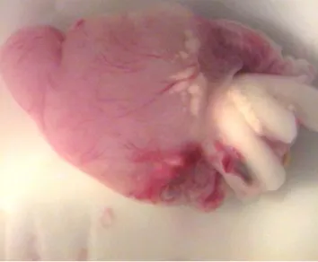
Fig. 4 Pathological specimen of a chick embryo showing some minor degree of structural damage after transamniotic intracardiac access 3 days prior to hatching
prenatal balloon valvuloplasty on the perinatal outcome of babies with severe aortic stenosis may result from at least three factors:
1. The influence of factors, such as optimized postnatal operative technique according to anatomic subtypes, on mortality and postinterventional course may outweigh lesser benefits achieved by prenatal relief of outflow obstruction. For example, postnatal studies on aortic valve repair and replacement after balloon valvuloplasty for congenital aortic stenosis in chil-dren have shown anatomic variables to be determi-nants of interventional results [53, 57–59]. Better prenatal predictive parameters will be needed to al-low prenatal interventions to be properly targeted. 2. Due to the multicenter and retrospective nature of the
analysis, the absence of uniform criteria for fetal selection, inadequate standardization of equipment for these ‘‘pioneer procedures,’’ and small patient numbers, existing studies are not yet able to assess the short- and potential long-term benefits of fetal car-diac valvuloplasty.
3. The impact of supplementary lesions (for instance, mitral stenosis in patients with aortic stenosis) or a general (genetic) programming for ventricular hypo-plasia that very well may exist in some fetuses can make it oversimplistic to view relief of valve obstruction as an adequate solution in many cases. Until the benefits of in-utero valvuloplasty attempts are determined by well-controlled clinical trials, these techniques remain investigational. Physicians and their patients who are considering these procedures must be made fully aware of the potential risks to both mother and fetus, such as preterm delivery (from membrane rupture or labor) and oligohydramnios with resulting pulmonary hypoplasia. Additionally, we are concerned that as obstetricians become more and more confident with their keyhole instrumentation for intrauterine ap-proaches, cardiac interventions might be undertaken under the guise of ‘‘last-ditch efforts’’ at various insti-tutions that have little expertise in relevant catheter techniques. Extensive informed parental counseling about alternative postnatal options for treatment re-mains critical. Few postnatal cardiac defects are cur-rently untreatable, and a balanced view of options requires counseling by a fetal/pediatric cardiologist familiar with current postnatal surgical and interven-tional options as well as outcomes. Ideally, the fetal/ pediatric cardiologist should serve as a constant member of the perinatal team planning this procedure.
Counseling
Currently, parents who receive an early prenatal diag-nosis of a cardiac lesion that may progress in severity and that might require postnatal univentricular surgical palliation often opt for termination or supportive care after counseling. This points to the extremely difficult
clinical and psychological course when surgical inter-vention is undertaken [9, 43, 45, 51]. The uncertain prognoses for these prenatally diagnosed lesions should be explained to parents as early as possible to ensure that they are in a position to make a fully informed decision about future management of the disorder. When counseling these families in early pregnancy for continuation of the pregnancy, we should always men-tion the possibility of evolumen-tion of these cardiac lesions during later pregnancy, as it is important that parents understand that there are several ‘‘hazards’’ that their fetus and later neonate might face even before reaching palliative postnatal surgery. The pediatric cardiologist is vital in the counseling process of these families no matter how early the diagnosis of fetal cardiac disease is made, as not all lesions behave similarly or have the same potential for progression. The prognostic evaluations and available prenatal and postnatal surgical options should be explained, modified, and updated accordingly as they constantly change in individual categories [11,
12, 19–23, 26, 30–33, 60, 61]. Parents who are consid-ering prenatal fetal cardiac intervention should be made aware of its investigational nature and the potential risks to both mother and fetus, and then they should be fully supported in their decision, whatever their choice is.
Conclusion
With the help of 1st-trimester fetal echocardiography, we are constantly gaining new insights into the ‘‘wors-ening’’ progressive pathophysiology of fetal cardiac le-sions during pregnancy [1–13]. Through innovations in congenital heart surgery and intrauterine interventions, as well as critical review of accepted perinatal manage-ment strategies, the outlook for fetuses with congenital heart lesions continues to improve year by year. Numerous reports have demonstrated that morbidity and mortality after staged palliation of HLHS or com-parable malformations have improved dramatically and should have a favorable impact on perinatal counseling for HLHS or its variants [14–25].
Antenatal fetal heart surgery is no more than a the-oretical alternative to postnatal repair in the human at present. Potential indications for fetal balloon valvu-loplasty include heart defects that after progression may evolve into one-ventricle (univentricular) pathology [26–
33]. Early results are still likely to be ambiguous, but in the proper hands and with long-term follow-up and further experience, the issues of timing and selection may be overcome.
Having observed the advances of the last years, we anticipate that within 10 years, advances in cardiac imaging and in-utero cardiac intervention technologies will make it possible to substantially alter the care and prognosis of the fetus with CHD. We hope that this review contributes to a more balanced understanding of what future fetal cardiac diagnosis and intervention efforts can and cannot achieve.
References
1. Epstein JA, Rader DJ, Parmacek MS (2002) Perspective: car-diovascular disease in the postgenomic era–lessons learned and challenges ahead. Endocrinology 143:2045–2050
2. Epstein JA, Buck CA (2000) Transcriptional regulation of cardiac development: implications for congenital heart disease and DiGeorge syndrome. Pediatr Res 48:717–724
3. Srivastava D (2001) Genetic assembly of the heart: implications for congenital heart disease. Annu Rev Physiol 63:451–469 4. Srivastava D, Olson EN (2000) A genetic blueprint for cardiac
development. Nature 407:221–226
5. Goldmuntz E, Bamford R, Karkera JD, dela Cruz J, Roessler E, Muenke M (2002) CFC1 mutations in patients with trans-position of the great arteries and double-outlet right ventricle. Am J Hum Genet 70:776–780
6. Hoess K, Goldmuntz E, Pyeritz RE (2002) Genetic counseling for congenital heart disease: new approaches for a new decade. Curr Cardiol Rep 4:68–75
7. Goldmuntz E, Geiger E, Benson DW (2001) NKX2.5 mutations in patients with tetralogy of Fallot. Circulation 104:2565–2568 8. Gardiner HM (2001) Fetal echocardiography: 20 years of
progress. Heart 86 :II12–22
9. Friedman AH, Kleinman CS, Copel JA (2002) Diagnosis of cardiac defects: where we’ve been, where we are and where we’re going. Prenat Diagn 22:280–284
10. Haak MC, Twisk JW, Van Vugt JM (2002) How successful is fetal echocardiographic examination in the first trimester of pregnancy? Ultrasound Obstet Gynecol 20:9–13
11. DeVore GR (2002) First-trimester fetal echocardiography: is the future now? Ultrasound Obstet Gynecol 20:6–8
12. Huhta JC (2001) The first trimester cardiologist: one standard of care for all children. Curr Opin Pediatr 13:453–455 13. Huggon IC, Ghi T, Cook AC, Zosmer N, Allan LD, Nicolaides
KH (2002) Fetal cardiac abnormalities identified prior to 14 weeks’ gestation. Ultrasound Obstet Gynecol 20:22–29 14. Bonnet D, Coltri A, Butera G, Fermont L, Le Bidois J,
Kachaner J, Sidi D (1999) Detection of transposition of the great arteries in fetuses reduces neonatal morbidity and mortality. Circulation 99:916–918
15. Tworetzky W, McElhinney DB, Reddy VM, Brook MM, Hanley FL, Silverman NH (2001) Improved surgical outcome after fetal diagnosis of hypoplastic left heart syndrome. Cir-culation 103:1269–1273
16. Saiki Y, Rebeyka IM (2001) Fetal cardiac intervention and surgery. Semin Thorac Cardiovasc Surg Pediatr Card Surg Annu 4:256–270
17. Cohen MS (2001) Fetal diagnosis and management of con-genital heart disease. Clin Perinatol 28:11–29
18. Cohen MS, Rychik J (1999) The small left ventricle: how small is too small for biventricular repair? Semin Thorac Cardiovasc Surg Pediatr Card Surg Annu 2:189–202
19. Trivedi KR, Azakie A, Benson LN (2001) Collaborative in-terventional and surgical strategies in the management of congenital heart lesions. Semin Thorac Cardiovasc Surg Pedi-atr Card Surg Annu 4:185–207
20. Mahle WT, Clancy RR, McGaurn SP, Goin JE, Clark BJ (2001) Impact of prenatal diagnosis on survival and early neurologic morbidity in neonates with the hypoplastic left heart syndrome. Pediatrics 107:1277–1282
21. Mahle WT (2001) Neurologic and cognitive outcomes in chil-dren with congenital. heart. Curr Opin Pediatr 13:482–486 22. Azakie A, McCrindle BW, Benson LN, Van Arsdell GS,
Russell JL, Coles JG, Nykanen D, Freedom RM, Williams WG (2001) Total cavopulmonary connections in children with a previous Norwood procedure. Ann Thorac Surg 71:1541–1546 23. Azakie A, Merklinger SL, McCrindle BW, Van Arsdell GS, Lee K-J, Benson LN, Coles JG, Williams WG (2001) Evolving strategies and improving outcomes of the modified Norwood procedure: a 10-year single institution experience. Ann Thorac Surg 72:1349–1353
24. Bull C (1999) Current and potential impact of fetal diagnosis on prevalence and spectrum of serous congenital heart disease at term in the UK. Lancet 354:1242–1247
25. Brackley KJ, Kilby MD, Wright JG, Brawn WJ, Sethia B, Stumper O, Holder R, Wyldes MP, Whittle MJ (2000) Out-come after prenatal diagnosis of hypoplastic left-heart syn-drome: a case series. Lancet 356:1143–1147
26. Tworetzky W, Marshall AC (2003) Balloon valvuloplasty for congenital heart disease in the fetus. Clin Perinatol 30:541–550 27. Kohl T, Strumper D, Witteler R, et al. (2000) Fetoscopic direct fetal cardiac access in sheep: an important experimental mile-stone along the route to human fetal cardiac intervention. Circulation 102:1602–1604
28. Kohl T, Sharland G, Allan LD, et al. (2000) World experience of percutaneous ultrasound-guided balloon valvuloplasty in human fetuses with severe aortic valve obstruction. Am J Cardiol 85:1230–1233
29. Kohl T, Witteler R, Stru¨mper D, et al. (2000) Operative tech-niques and strategies for minimally invasive fetoscopic fetal cardiac interventions in sheep. Surg Endosc 14:424–430 30. Kohl T (2001) Congenital heart disease. Mending the tiniest
hearts. Lancet 358:S17
31. Kohl T (2002) Fetal echocardiography: new grounds to explore during fetal cardiac intervention. Pediatr Cardiol 23:334–346 32. Cheatham JP (2001) Intervention in the critically ill neonate
and infant with hypoplastic left heart syndrome and intact at-rial septum. J Interv Cardiol 14:357–366
33. Huhta J, Quintero RA, Suh E, Bader R (2004) Advances in fetal cardiac intervention. Curr Opin Pediatr 16:487–493 34. Deprest J, Gratacos E, Nicolaides KH (2004) FETO Task
Group. Fetoscopic tracheal occlusion (FETO) for severe con-genital diaphragmatic hernia: evolution of a technique and preliminary results. Ultrasound Obstet Gynecol 24:121–126 35. Walsh DS, Adzick NS (2000) Fetal surgical intervention. Am
J Perinatol 17:277–83
36. Wilson RD (2002) Prenatal evaluation for fetal surgery. Curr Opin Obstet Gynecol 14:187–193
37. Walsh DS, Adzick NS, Sutton LN, Johnson MP (2002) The rationale for in utero repair of myelomeningocele. Fetal Diagn Ther 16:312–322
38. Mahieu-Caputo D, Senat MV, Romana S, Houfflin-Debarge V, Gosset P, Audibert F, Bessis R, Ville Y, Vekemans M, Dommergues M (2002) What’s new in fetal medicine? Arch Pediatr 9:172–186 [French]
39. Ville Y, Hecher K, Gagnon A, Sebire N, Hyett J, Nicolaides K (1998) Endoscopic laser coagulation in the management of se-vere twin-to-twin transfusion syndrome. Br J Obstet Gynaecol 105:446–453
40. Quintero RA, Morales WJ, Allen MH, Bornick PW, Johnson P (2000) Fetal hydrolaparoscopy and endoscopic cystotomy in complicated cases of lower urinary tract. Am J Obstet Gynecol 183:324–330; discussion 330–333
41. Quintero RA, Shukla AR, Homsy YL, Bukkapatnam R (2000) Successful in utero endoscopic ablation of posterior urethral valves: a new dimension in fetal urology. Urology 55:774xiii– 774xv
42. Aaronson OS, Tulipan NB, Cywes R, Sundell HW, Davis GH, Bruner JP, Richards WO (2002) Robot-assisted endoscopic intrauterine myelomeningocele repair: a feasibility study. Pe-diatr Neurosurg 36:85–89
43. Campbell S (2001) Opinion—isolated major congenital heart disease. Ultrasound Obstet Gynecol 17:370–379
44. Simpson JM, Sharland GK (1997) Natural history and out-come of aortic stenosis diagnosed prenatally. Heart 77:205–210 45. Allan LD, Apfel H, Printz B (1998) Outcome after prenatal diagnosis of the hypoplastic left heart syndrome. Heart 79:371– 373
46. Daubeney PE, Sharland GK, Cook AC, et al. (1998) Pulmo-nary atresia with intact ventricular septum: impact of fetal echocardiography on incidence at birth and postnatal outcome. UK and Eire Collaborative Study of Pulmonary Atresia with Intact Ventricular Septum. Circulation 98:562–566
47. Meyer-Wittkopf M, Cooper S, Sholler G (2001) Correlation between fetal cardiac diagnosis by obstetric and pediatric car-diologist sonographers and comparison with postnatal findings. Ultrasound Obstet Gynecol 17:392–397
48. Allan LD, Sharland G, Milburn A, Lockhart S, Groves A, Anderson R, Cook A, Fagg N (1994) Prospective diagnosis of 1,006 consecutive cases of congenital heart disease in the fetus. J Am Coll Cardiol 23:1452–1458
49. Ott WJ (1995) The accuracy of antenatal fetal echocardiogra-phy screening in high-risk and low-risk patients. Am J Obstet Gynecol 172:1741–1747; discussed 1747–1749
50. Allan LD, Maxwell DJ, Carminati M, Tynan MJ (1995) Sur-vival after fetal aortic balloon valvoplasty. Ultrasound Obstet Gynecol 5:90–91
51. Simpson JM (2000) Hypoplastic left heart syndrome. Ultra-sound Obstet Gynecol 15:271–278
52. Ohye RG, Bove EL (2001) Advances in congenital heart sur-gery. Curr Opin Pediatr 13:473–481
53. Koch A, Buheitel G, Gerling S, Klinge J, Singer H, Hofbeck M (2000) Balloon dilatation of critical left heart stenoses in low birthweight infants. Acta Paediatr 89:979–982
54. Hofbeck M, Singer H, Buheitel G, Ries M (1999) Balloon valvuloplasty of critical pulmonary valve stenosis in a pre-mature neonate. Pediatr Cardiol 20:147–149
55. Buheitel G, Hofbeck M, Singer H (1995) Ballondilatation der Pulmonalklappe innerhalb der ersten 40 Lebenstage bei
kriti-scher valvula¨rer Pulmonalstenose, Fallotkriti-scher Tetralogie und nach chirurgischer oder interventioneller Hochfrequenzero¨ff-nung einer Pulmonalatresie. Z Kardiol 84:64–71
56. Weber HS (2002) Initial and late results after catheter inter-vention for neonatal critical pulmonary valve stenosis and atresia with intact ventricular septum. Cathet Cardiovasc In-tervent 56:394–399
57. Hawkins JA, Minich LL, Shaddy RE, Tani LY, Orsmond GS, Sturtevant JE, McGough EC (1996) Aortic valve repair and replacement after balloon aortic valvuloplasty in children. Ann Thorac Surg 61:1355–1358
58. Hawkins JA, Minich LL, Tani LY, Day RW, Judd VE, Shaddy RE, McGough EC (1998) Late results and reinter-vention after aortic valvotomy for critical aortic stenosis in neonates and infants. Ann Thorac Surg 65:1758–1762; dis-cussion 1763
59. Sholler GF, Keane JF, Perry SB, Sanders SP, Lock JE (1988) Balloon dilatation of congenital aortic valve stenosis. Results and influence of technical and morphological features on out-come. Circulation 78:351–360
60. Allan LD (1995) Echocardiographic detection of congenital heart disease in the fetus: present and future. Br Heart J 74:103–106
61. Meyer-Wittkopf M (2002) Interventional fetal cardiac ther-apy—possible perspectives and current shortcomings. Ultra-sound Obstet Gynecol 20:527–531