Eur. Surg. • Vol. 3 4 . No 2 - 2 0 0 2 137
From the IDepartment of Reconstructive Surgery, the 2Department of Pathology, University Hospital, Basel, and the ~Swiss Paraplegic Centre, Nottwil, Switzerland
Optimizing the Parameters for Hydro-Jet Dissection
in
Fatty Tissue - A Morphological Ex Vivo Analysis
M . W a n n e r 1, ~, S. J a k o b 1, E S c h w a r z l 1, M. O b e r h o l z e r 2, and G. P i e r e r ]
Keywords: Hydro jet - fatty tissue - dissection parameters - vessel damage.
SchliisselwOrter: Hydro-Jet - Fettgewebe - Schneideparameter - GefdJJschaden.
S u m m a r y : Background: The advantage of water-jet dissec- tion is the preservation of vessels and nerves. Especially in liver surgery, blood loss can be significantly decreased. The use of water-jet dissectors in other fields of surgery is cur- rently under investigation. The preparation of vessels in fatty tissue is of special interest for plastic surgeons. The optimal technical parameters were investigated.
Methods: Abdominal fat tissue of fresh cadavers was cut under standardized conditions with different parameters of the water-jet dissector.
Results: One single pass at a cutting pressure between 20 and 60 Bar makes an incision of 8 mm. Deeper cuts can be achieved by repeated application on the same cut. Five passes at 40 Bar results in a depth of 1.7 cm without vessel damage. If the applied pressure is 50 or 60 Bar, up to 7 % damaged vessels can be found. The water-jet dissection leads to a water uptake of the cut tissue.
Conclusions: The optimal pressure for water-jet dissection of fatty tissue lies between 30 and 40 Bar. The effect of the mechanical irritation of the vessels has to be investigated in vivo before using the water-jet dissector for preparation of blood vessels in humans, e.g. for flap dissection.
(Eur. Surg. 2002; 34:137-141
Optimierung der Schneideparameter des Hydro-Jet-
Dissektors im Fettgewebe. Eine morphologische
Ex-vivo-Untersuchung
Zusammenfassung: Grundlagen: Der Vorteil der Hydro-Jet-Dis- sektion ist Schonung von Nerven und Geffigen. Speziell in der Leberchirurgie kann der Blutverlust signifikant gesenkt werden. Die Anwendungsmdglichkeiten des Hydro-Jet-Dissektors in an- deren chirurgischen Teilgebieten wird gegenw~irtig untersucht. Die GefS_gpr~iparation im Fettgewebe ist fiir die plastische Chir- urgie von speziellem Interesse. Die optimalen Schneideparame- ter warden untersucht.
Methodik: Bauchfettgewebe yon Frischleichen wurde unter stan- dardisierten Bedingungen mit verschiedenen Einstellungen des Hydro-Jet-Dissektors geschnitten.
Ergebnisse: Bei Drucken zwischen 20 and 60 Bar und einer Ge- webepassage werden Schnitte von 8 mm Tiefe erzielt. Tiefere In- zisionen werden durch repetitive Applikation des Wasserstrahls erreicht. Bei einem Druck von 40 Bar und 5 Passagen ist die Schnittiefe 1,7 cm ohne Geffigsch~iden. Drucke von 50 und 60 Corresponding address: M. Wanner, M.D., Department of Reconstructive Surgery, University Hospital, Spitalstrage 21, CH-4031 Basel, Switzerland.
Fax: ++41/41/9 39 58 57 E-mail: [email protected]
Bar ftihren zu Geffigsch~iden bis zu 7 %. Die Wasserstrahldissek- tion ftihrt zu einer Gewebeaufquellung.
Schlugfolgerungen: Der optimale Druck ftir die Wasserstrahldis- sektion im Fettgewebe liegt zwischen 30 and 40 Bar. Der EinfluB der mechanischen Irritation auf die geschonten Gefage mug un- tersucht werden, bevor diese Technik zur GeftiBpr@aration am Menschen, z. B. bei Lappenplastiken, eingesetzt werden daft.
Introduction
Conventional dissection of tissue with the knife cuts vessels and nerves. Bleeding vessels will thereafter be ligated or, by a faster procedure, be coagulated by mono- or bipolar electrocautery. The result is haemostasis together with a more or less extended thermal injury, depending on duration and precision of the pro- cedure. In operations with high blood loss, e.g. in liver surgery, various methods are used in order to decrease blood loss and to gain precision while coagulating vessels. Cutting with laser or electrocautery are widespread techniques, but they lead to ther- mal injury of the adjacent tissue and therefore increase the rate of necrosis (2 l).
Ultrasound dissection is frequently used in parenchymatous or- gans and fatty tissue (brain, fat, e.g. for liposuction). Small ves- sels and nerves are preserved while cutting the parenchyma, thus facilitating a precise coagulation if necessary. The advantage over the above-mentioned techniques is therefore the possibility to coagulate the spared vessels precisely with less additional in- jury to the remaining parenchyma (11). Indeed, a significant tem- perature rise can be observed, resulting in a degradation of pro- tein and nerve damage. Even blood vessels can be injured (7, 8, 12, 14, 15, 23, 24, 25).
In 1982 the use of a jet of normal saline generated by a stand- ard agricultural electric sprayer for liver resection in man was de- scribed (16). Dissection with a high pressure water jet has the ad- vantages of ultrasound dissection without the possible danger of tissue heating.
Because the devices used at this time were originally designed for industrial use, the pressure applied was very high, i.e. up to I000 Bar. In order to avoid damage caused by high pressure jets that are used to cut concrete or steel, the working distance had to be increased. This caused a reduction in the working pressure but at the same time the divergence of the jet reduced the precision and resulted in altered vision due to the dispersed water particles
(3).
Nowadays a new generation of dissectors with optimized pres- sure regulation, nozzle diameter, and jet coherence is available. In the device we use, the pressure can be varied between 0 and 150 Bar. The dissection therefore can be performed with minimal distance to the tissue or, better still, in contact mode. Moreover, the convergence is maintained by an additional helix jet around the main jet. The water jet has a diameter of 0.120 mm, thus enabling a very fine and controlled dissection without consuming too much liquid. This was demonstrated in an in vitro study using endoscopy in pig brain: pressure of 3 Bar resulted in a cutting depth of 3 to 5 mm without damaging vessels (13, 26).
Hydro-jet cutting is currently used in liver surgery, for the re- section of renal turnouts (openly and endoscopically), partially in neurosurgery and experimentally in ophthalmic surgery. First
1 3 8 Eur. S u r g . • Vol. 3 4 - N o 2 - 2 0 0 2
clinical experience was made in reconstructive surgery. It is used experimentally for the thinning of skin flaps, leaving the subder- real vasculature intact (1, 2, 4, 5, 6, 9, 10, 17, 18, 19, 20, 22, 24, 25, 27). Additionally, the preparation of flap pedicles could be of interest for plastic surgeons.
Until now the optimal pressure for dissection of fat that leaves vessels intact was not known.
The aim of this study was to identify the pressure range to be used in fat, without damaging blood vessels.
Material and m e t h o d s
Skin and fat were removed horizontally from the lower abdomen of 3 fresh cadavers. The specimen was turned 180 degrees to ex- pose the fat. Tissue blocks of roughly 6 x 5 x 2 cm were cut.
Incisions were made with the Hydro-Jet Dissector.
A jet of 0.9 % saline solution at room temperature passed over the specimen at an angle of 90 degrees with just enough distance between the nozzle and the specimen to allow translational movement. A specially constructed device allowed a constant cutting speed of 12 mm/s.
Six cuts per block were performed with the same parameters. Variables were the applied pressure and the number of passes of the jet at the same incision site. The blocks were then imme- diately fixed in formalin. After 4 days of fixation the specimens were embedded in paraffin and thin sections of 4 g m were taken and dyed with HE and vGieson. The cutting depth was analysed at 3 different distances. Eighteen measurements per parameter were theoretically possible. In the process of preparation of the histological specimen, some artefacts occurred (twisted speci- men, oblique ct~t). These thin sections were excluded. On aver- age we could measure 12 to 15 times the penetration depth and the morphology of the vessels per parameter, thus giving enough data to calculate a median penetration depth. The depth was measured with an optical scale in the microscope. In order to identify a cut, the following morphological specifications had to be present: clearly identifiable entrance point into the tissue and a cavity and/or detritus of fat or connective tissue in the direction of the applied force.
The fluid uptake of the tissue was measured by volumetric analysis of the tissue blocks before and after dissection.
The morphology of the vessels in the cavity and directly bor- dering it was analysed by a pathologist (M.O.). We included all vessels in or adjacent to the cavity formed by the dissecting water jet. The diameter was measured with an optical scale in the microscope in two directions and the area calculated from these two measurements. Vessel damage was defined if the vessels wall was disrupted.
Devices used: Water-Jet Dissector (Helix Water-Jet), Andreas Pein Medizinaltechnik, Schwerin, Germany.
Cutting device with adjustable translation speed, sterile 0.9 % NaC1 solution, connecting tubes.
Statistics: Median, average and standard deviation of the
penetration depth, volume difference, and the n u m b e r of intact and destroyed vessels are given.
Results
The penetration depth of the water jet in fat was between 7 and 9 m m for an applied pressure between 20 and 60 Bar. As ex- pected, higher pressure resulted in deeper cuts. The difference between 20 and 60 Bar, however, was only 2 ram. Table 1 shows the penetration depth depending on the applied pressure and the number of passes.
The n u m b e r of passes over the same incision had a higher in- fluence on the penetration depth than did the pressure: Applica- tion of 20 to 60 Bars five times cut between 8 and 18 ram, de- pending on the pressure.
We found a correlation between applied pressure and penetra- tion for 5 passes.
At a pressure of 20 Bar the cuts were not deeper, even after 5 passes. A pressure of 50 and 60 Bar did not cut deeper than 40 Bar.
Table 1. Median penetration depth d e p e n d i n g on the applied pres- sure (Bar) and the number o f passes (1 o r 5).
25 ... 20 . . . . ! o20Bar 15 . . . = . . . .-T.-~ ~" • .... Bar 10 ... :~:'~"~'i~- r~40 Bar
' NIIIIIIIIII N@
...
" "" N ] l J l l l l [ l [ ~ i 3 "~'~'~'~',~',~N-- , 6 0 BarMedian 1 Pass MEDIAN 5 Passes
In our model the cutting jet r e a c h e s the underlying dermal layer at pressures of 50 and above b e c a u s e the thickness of the specimens was roughly 1.7 cm. T h e r e f o r e , we could not find a correlation of pressure and p e n e t r a t i o n depth above 50 Bar. The dermis could not be penetrated b u t showed vacuoles, due to water penetration in the dermis.
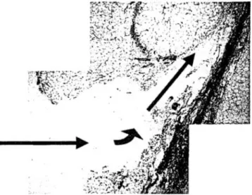
Deviation of the cutting jet by c o n n e c t i v e tissue septa occurred as shown in Fig. 1.
Thinner septa can be penetrated i f they are orientated perpen- dicularly to the jet.
In most cases the microscopically identified vessels were in- tact, even when pressure above 50 B a r was applied. T h e histo- logical section in Fig. 2 shows a typical incision with intact ves- sels in the cavity and adjacent to the cut.
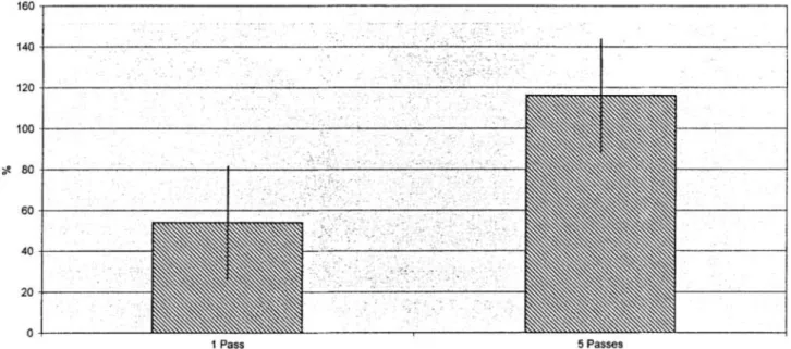
Water uptake by the tissue was significant. We found an in- crease of volume up to 215 % c o m p a r e d to the initial volume before cutting. The more cuts were performed, the more water was taken up. The applied pressure was less important, as demonstrated in Table 2.
We analysed 423 vessels morphologically in or adjacent to the cavity formed by the water jet. Table 3 shows the distribution of these vessel areas. A total of 7 d e s t r o y e d vessels were found. -) Their area ranged from 0.05 to 49 ram-. We found no vessel damage with applied pressure of 40 B a r and below (Table 4).
D i s c u s s i o n
A scalpel incision is seldom deeper t h a n 4 to 10 ram. Compara- tively, the cuts with water jet are not deeper, even after 5 passes at a pressure of 20 Bar. Pressure of 5 0 and 60 Bar did not yield a cut deeper than that of 40 Bar, but t h e risk of vessel damage is higher. If a deeper cut is desired, the pressure has to be above 20 Bar.
Eur. Surg. - Vol. 34 • N o 2 • 2 0 0 2 139
Table 2. Volume increase o f the specimens after water-jet cutting. 160 i . - 1 . L ? i . _ - I J 140 120 100 80 60 40 20 0 1 Pass 5 Passes P a s s e s
Since vessel damage could be observed only with pressures of 50 Bar and above, the optimal dissection pressure for fat lies be- tween 20 and 40 Bar.
The structures remaining will be nerves, septa, and vessels. The important structures can therefore be identified easily and be cut conventionally or coagulated if necessary.
The percentage of destroyed vessels is higher after a single pass at 60 Bar, than in the cut with 5 passes at the same pressure. We believe that this is an artefact because the remnants of the destroyed vessel wails are washed out by multiple application of the water jet. Therefore, less destruction was found in the series with 5 passes at pressures of 50 and 60 Bar. With the device used, the maximum applicable pressure can be limited, thus re-
ducing the danger of uncontrolled cuts. Septa of connective tis- sue remain undamaged if they are not perpendicular to the cut- ting jet. They could be cut at higher pressures than 60 Bar but this would lead to uncontrolled damage of vessels and should therefore be avoided. This also means that the dissection cannot be completely performed with water-jet cutting alone but needs to be completed with scissors or knife.
Water uptake by the tissue is significant. We found an increase in volume up to 215 % of the initial volume before cutting. This could lead to difficulties closing the incisions.
We presume that in clinical use the additional water in the tis- sue will be taken up as in tumescence liposuction.
Table 3.
n u m b e r
Quantity and surface o f analysed vessels in the cavity or adjacent to it.
70 60 50 40 30 20 0.0025 O. 1225 0.33 0.7 2.4 4.68 49 a r e a (ram2)
140 Eur. S u r g . . Vol. 34 • No 2 • 2002
Table 4. Percentage o f destroyed vessels depending on the different cutting parameters. 5_60 means the water jet passed the same incision
5 times with a working pressure of 60 Bar. t_40 means I pass andpressure of 40 Bar.
%
7
6
5
4
3
2-
1
0
5 60
5 50
5 40
5 30
5 20
1 60
1 50
1 4 0
1 30
1 20
Generally, our investigation showed that water-jet dissection of fat in a cadaver without damaging vessels is possible, pro- vided the cutting parameters are chosen between 30 and 40 Bar.
We conclude that it is possible to cut fat with a water jet with- out damaging vessels. The applied pressure has to range between 30 and 40 Bar. Higher pressure leads to vessel damage up to 7 %. Multiple passes of the water jet in the same incision lead to deep- er cuts, provided the applied pressure is more than 20 Bar. For vessel-sparing cutting in fat, we recommend a cutting pressure between 30 and 40 Bar.
The aim of this preliminary study was to evaluate the range of pressure that should be applied for fat dissection. In brain tissue, for example, a pressure of 3 to 5 Bar is suitable to perform cuts of 1 mm. Until now not even the pressure range for fat was known. It was therefore essential to determine the theoretical pressure range, before the cutting device could be used on living tissue.
These results were obtained in cadaverous fat, where vessels are not filled with blood. Blood-filled vessels could show a dif- ferent reaction to the cutting jet and therefore the amount of damaged vessels could differ from the above-mentioned results.
The results will have to be evaluated in living, perfused tissue and in an animaI model in order to evaluate the possibilities for vessel and nerve preparation.
Fig. 2. Typical incis'ion with intact vessels.
Acknowledgements
We thank Mr. Pein and Mr. P i t a k o of Pein, Medizintechnik, Schwerin, Germany, and Mr. D. Scherrer, Head of Microsurgical Laboratory at the University Hospital of Basel, Switzerland, for the technical support of this study. W e thank also Prof. Mihatsch, Head of the Pathology Department, for his support with the ethical committee and Mr. Schoch for his logistical support.
References
(1) Baer H, Gilg M, Maddem G J, Blumgart LH: Der Hochgeschwindigkeits-Was- serstrahI-Dissektor in der Leberchirurgie. H e i r Chir Acta 1992;59:437-442. (2) Baer HU, Maddern GJ, Blumgart LH: N e w water-jet dissector: initial experi- ence in hepatic surgery [published erratum appears in Br J Surg 1994;81(8):1103]. B r J Surg 1991; 78:502-5(13.
(3) Baer HU, Metzger A, Bah'as JR Menler D, Wheadey AM, Czemiak A: Lapa- roscoplc liver resection in the Large White pig - a comparison between waterjet dissector and ultrasound dissector. Endosc Surg Allied Technol 1994;2:189-193. (4) Baer HU, Stain SC, Guastella T, Maddern GJ. Blumgart LH: Hepatic resection using a water jet dissector. Hpb Surgery 1993"6:189-196; discussion 196-188. (5) Basting RE Corvin S, Antwerpen C, Djakovic N, Schmidt D: Use of water jet resection in renal surgery: Early clinical experiences. Eur Urol 2000;38:104-107. (6) Chan DY, Marshall FF: Partial nephrectorny for centrally located tumors. Urology 1999;54:1088-1091; discussion 1091-1082.
(7) Clarke RL, tcr Haar GR: Temperature rise recorded during lesion formation by high-intensity focused ultrasound. Ultrasound Med Biol 1997;23:299-306. (8) Fischer PD, Narayanan K, Liang MD: T h e use of high-frequency ultrasound for the dissection of small-diameter bIood vessels and nerves. Ann Plast Surg 1992;28:326-330.
(9) Guastella T, Baer HU, Maddem G J, Blumgart LH: A new fluid-jet dissector in hepatobiliary surgery. Ann Ital Chir 1992;63: t 87-191 ; discussion 19 ! - 182. (10) Hata Y, Sasaki F, Takahashi H, Ohkawa Y, Taguchi K, Une Y, Uchino J: Liver resection in children, using a water-jet. J Pediatr Surg 1994;29:648-650. (l t)Horstmann R, Kern M, Joosten U, Hohlbach G: Ultrasound dissection in laparoscopic cholecystectomy. Zentralbl Chir 1993; 118:741-745.
(12) Howard BK, Beran SJ, Kenkel JM, Krueger J, Rohrich RJ: The effects of ultrasonic energy on peripheral nerves: implications for ultrasound-assisted lipo- suction. Plast Reconstr Surg 19,99;103:984-989.
(131 Jakob S, Kehler U, Reusche E, Friedrich H, Arnold H: Endoscopic use of the water jet dissector in the cerebral ventricle system - an experimental study. Zen- train Neurochir 2000;61:14-21.
(14) Kaduk W~ Stengel B, Prhl A, Nizze H , Gundlach K: Hydro-jet cutting: a method for selective surgical dissection o nerve tissue. An experimental study on the sciatic nerve of rats. J Craniomaxillofacial Surg 2000;27:327-330.
(15) Miiller T, Pfiiller P, Vetter J, Wehner W: Basic studies of ultrasonic surgery. llI. Histomorphologic changes in various tissues following exposure to ultrasound. Z Exp Chir 1982;15:235-243.
(16) Papachristou DN, Barters R: Resection o f the liver with a water jet. B r J Surg 1982;69:93-94.
(17) Penchev RD, Losanoff JE, Kjossev KT: Reconstructive renal surgery using a water jet. J Urol 1999;162:772-774.
(18) Pick J, Wille C, Warzok R, Gaab MR: Waterjet dissection of the brain: ex- perimental and first clinical results. Technical note. J Neurosurg 1998;89:861-864. (19) Rau HG, Buttler E, Meyer G, Schardey HM, Schildberg FW: Laparoscopic liver resection compared with conventional partial hepatectomy - a prospective analysis. Hepato-Gastroenterology 1998;45:2333-2338.
(20) Ruu HG. Meyer G, Jauch K, Cohnert T, Buttler E, Schildberg F: Leberresektion mit dem Wasser-Jet: konventionell und laparoskopisch. Chirnrg 1996;67:546-55 I.
Eur, S u r g . - V o l . 3 4 - N o 2 • 2 0 0 2 141
(21) Schurr MO, Wehrmann M, Kunert W, Melzer A, Lirici MM, Trapp R, Kane- hira E, Buess G: Histologic effects of different technologies for dissection in endo- scopic surgery: Nd:YAG laser, high frequency and water-jet. Endosc Surg Allied Technot 1994;2:195-201.
(22) $hekarriz H, Pein A, Magritz R, Bgrk C, Markert U, Bruch H-P: Hydro-Jet- Dissektion in der laparoskopischen Cholezstectomie. Minimal Invasive Chirurgie 1998;7:93-97.
(23) Shimi SM: Dissection techniques in laparoseopic surgery: a review [see com- ments]. J R Coil Surg Edinb 1995;40:249-259.
(24) Siegert R, Danter J, Jurk V, Eggers R, Krtiger S: Dermal microvasculature and tissue selective thinning techniques (ultrasound and water-jet) of short-time expanded skirl in dogs. Eur Arch Otorhinolaryngol 1998;255:325-330.
(25) Siegert R, Danter J, L6ffier A, Jurk V, Eggers R, Weerda H: Tissue selective thinning of short-term expanded skin with ultrasound and a powerful water-jet. Face 1998;6:51-58.
(26) Terzis AJ, Nowak G, Rentzsch O, Arnold H, Diebold J, Barenon G: A new system for cutting brain tissue preserving vessels: water jet cutting. Br J Neuro- surg 1989;3:361-366.
(27) Une Y, Uchino J, Horie T, Sato Y, Ogasawara K, Kakita A, Sano F: Liver re- section using a water jet. Cancer Chemother Pharmacol 1989;23:$74-77.
From the Department of Plastic and Reconstructive Surgery, University of Innsbruck
Invited Commentary to:
'Optimizing the
Parameters for Hydro-Jet Dissection in Fatty
Tissue - A Morphological Ex
Vivo
Analysis'
(Eur. Surg. 2002;34:1:37-141
)
Hildegunde Piza
The main dangers associated with surgical interventions, open or endoscopic, are bleeding from cut vessels, damage to nerves or their branches innervating the area under surgery, destruction of the parenchyma and damage to the neighbouring healthy tissue. Intraoperative bleeding makes it difficult to visualize the margins of the tissue to be resected, affecting precision of surgery. In en- doscopic procedures, in particular, visual control of the blood vessels is imperative for precise cutting and reliable coagulation of the margins of the lesion. Furthermore, the need to continually clamp, ligate or coagulate bleeding vessels are time-consuming procedures that lead to considerable prolongation of the oper- ation time, cause surgeon fatigue, prolonged ischaemia time, and add unnecessary danger to the patient because of prolonged anaesthesia. All of these have negative postoperative effects on patients. Severe blood loss is also a problem that should not be underestimated. In parenchymal organs there is the additional danger of damaging the parenchyma so that partial or total, tem- porary or permanent loss of function may result, defeating one of the primary objectives of surgery.
Any innovative technique or device that successfully addresses any or all of these problems will represent a major advance in surgery. If bleeding can be avoided or reduced, damage to nerves prevented and function preserved, intraoperative and postopera- tive morbidity can be considerably reduced.
Although the use of the scalpel and scissors, the surgeon's traditional tools of trade, cannot be totally eliminated in view of the different kinds of tissues of the body that may need to be resected, e.g. glandular tissue or tough fibrous tissue, the coarseness of which presents some resistance, new techniques using laser and ul- trasound devices have facilitated surgery by reducing or avoiding intraoperative bleeding, while penetrating to the depths required. This in turn enables very precise and selective tissue cutting.
The advantages of reduced bleeding, precision in resection and shortened operation time of laser and ultrasound techniques, however, may be offset by morbidity caused by thermal/mechan- ical damage. In addition, postoperative recovery time may not necessarily be reduced by these techniques.
Corresponding address: Hildegunde Piza, M.D., Department of Plastic and Reconstructive Surgery, University of Innsbruck, Anichstra6e 35, A-6020 Innsbruck.
Fax: ++43/5 12/5 04/27 35
E-mail: hildegunde.piza@ uibk.ac.at
The relatively newly developed water-jet devices have opened whole new perspectives in surgery. The idea of using the power of water jets to cut through tissues was borrowed from industrial practices where high-pressure water jets have been in use for cut- ting through steel and concrete. Although still in a relatively early stage, this technique has found many clinical applications in a wide variety of open and endoscopic surgical interventions in soft tissues, parenchymal organs and even bony structures. However, the technique has not been uniformly successful.
In oral surgery the specific morphology of the orofacial area poses a grave danger of damage to the motor facial nerve with the devastating consequence of facial paralysis. It would be highly de- sirable if adequate penetration and dissection of the coarse parotid tissue can be achieved using jets at pressures that do not trauma- tize the branches of the facial nerve with reduction, fibrillation and severance. Experience is needed in this area. There is a report, however, that this technique proved to be fast, simple and safe in parotid gland surgery, preserving the facial nerve (5).
Experiments in pigs' eyes showed that in ophthalmology this novel technique may prove to be very promising (7, 8). In pa- tients with grey cataract, water-jet dissection might represent a less invasive procedure, helping to break up the clouded lenses and suction off the loosened particles. Subsequent cleaning of the capsule will prevent regeneration of epithelial cells and re- currence of cataract, which is one of the main unwanted conse- quences of cataract surgery.
Based on animal studies, there are unpublished reports that in the field of orthopaedics surgical interventions such as arthroscopy, synovectomy and intervertebral discectomy can be performed even faster than with laser, and with high selectivity and precision.
Water-jet dissection, however, was found to be unsuitable for intestinal dissection because of uncontrolled bloating of the rec- tal wall and tissue wall disruption. Secure haemostasis along the incision margin could not be achieved (3).
The most promising application of the water-jet technique was found to be in parenchymal organs such as the liver and the kid- ney (2). In a high blood flow organ like the liver, water-jet dis- section proved to minimize blood loss, reduce operative time, achieve excellent visibility for exposure and visualisation of in- trahepatic vessels and bile ducts, decrease the rate of complica- tion, and shorten hospital stay. Thus in every respect this tech- nique might be useful in surgery of parenchymal organs.
In reconstructive renal surgery, too (e.g. resection of turnout tissue, partial nephrectomy or nephrolithiasis), this technique fa- cilitated safe, efficient, fast and precise procedures because bleeding was reduced, vessel clamping was not required, and hy- pothermy was achieved (4). Such advantages were also found in endoscopic brain surgery where even minimal bleeding results in severe clouding of vision of the area under dissection (1). Wound debridement with the water-jet technique was experimentally tried out and has proved to be advantageous.
In plastic surgical interventions such as auricular reconstruc- tion, well-vascularized, expanded, thin skin flaps could be achieved by using a water-jet dissecting device (6). In liposuc- tion, too, this method proved to be promising. It is hoped that in other reconstructive surgical interventions such as flap pedicle preparation, this technique might prove to be useful.
Success of surgery using a water-jet dissector depends on the appropriate pressure of the water jet applied. On the one hand, the pressure applied should be adequate to penetrate the tissue to the required depth; on the other, there should be no damage to vessels, nerves or the parenchyma. At present, knowledge of the appropriate pressure to be used in the dissection of all the dif- ferent kinds of tissue is not available. In particular, there have been thus far no studies on this technique in relation to fatty tissue. The experimental investigation by Wanner et al. reported in this issue of the journal makes an important contribution to knowledge in this area.
Nearly a decade of experience with experimental and clinical use of this device has demonstrated the power of water jets to dissect selectively and safely through biological tissues while avoiding or minimizing dangers present in conventional surgical