Introduction
Central core disease (CCD) is a rare but benign con-genital myopathy, first described by Shy and Magee in 1956 [10]. Inheritance is usually autosomal dominant [10,17], but putative autosomal recessive inheritance has been described [5, 11]. While the overall incidence of CCD is rare, recent reports have documented a broader clinical spectrum of this disorder, suggesting that the actual incidence of CCD may be considerably higher than previously perceived [16, 19].
Clinical presentation of CCD includes proximal muscle weakness without or with slow progression and
hypotonia during infancy that can persist throughout adolescence/adulthood. Symptoms can include delayed motor development, reduced muscle bulk and skeletal alterations such as congenital hip dislocation, flat feet, and deformities or contractures of the joints [5,9,13–15,
17]. However, phenotypic presentation varies consider-ably from no visible disability to totally dependent ambulation [16].
In this article we describe the clinical and pathologi-cal features of two affected individuals where the diagnosis of CCD was made via a muscle biopsy taken during scoliosis surgery. Both patients presented with severe progressive scoliosis and unexplained muscle Kirsten D. Mertz
Bernhard Jost Markus Glatzel Kan Min
Progressive scoliosis in central core disease
Received: 30 September 2004 Revised: 5 March 2005 Accepted: 12 March 2005 Published online: 31 May 2005
Ó Springer-Verlag 2005
Abstract Central core disease (CCD) is a rare congenital myopa-thy with autosomal dominant inheritance. Here, we report on two cases of progressive scoliosis in CCD, pointing out the value of a muscle biopsy to establish the cor-rect diagnosis. The first case involves a 13-year-old boy with severe pro-gressive scoliosis and joint contrac-tures. The patient was initially diagnosed with arthropgryposis multiplex congenita. The second case involves a 45-year-old man with severe scoliosis that had slowly pro-gressed over the years. Both patients suffered from unexplained muscle weakness and severe restriction of pulmonary function. The correct diagnoses were established through muscle biopsies taken from the par-avertebral musculature during scoli-osis surgery. Correction of the spinal
deformities was achieved through posterior instrumentation in both patients, with prior anterior release in one patient. Although scoliosis is a common feature in CCD, the cor-rect diagnosis can be missed in sco-liosis patients. Therefore, we recommend a muscle biopsy in pa-tients with scoliosis, unexplained muscle weakness and multiple joint problems.
Keywords Scoliosis Æ Central core disease Æ Arthrogryposis Æ Muscle biopsy
K. D. Mertz Æ M. Glatzel Institute of Neuropathology, University Hospital Zurich, Schmelzbergstrasse 12, CH, 8091 Zurich, Switzerland B. Jost Æ K. Min (&)
Balgrist Clinic, Department of Orthopedics, University of Zurich, Forchstrasse 340, CH, 8008 Zurich, Switzerland
E-mail: [email protected] Tel.: +0041-1-3861111 Fax: +0041-1-3861609 Present address: K. D. Mertz Brigham and Women’s Hospital Department of Pathology,
Eugene Braunwald Research Center 414, 221 Longwood Avenue,
weakness. In the literature, only a few patients have been reported who present scoliosis as the predominant skeletal feature of CCD [13, 15]. The purpose of this report is to highlight potential difficulties in establishing the correct diagnosis of CCD in scoliosis patients.
Case reports
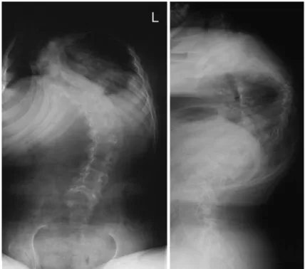
Patient 1, a 13-year-old white male, was referred to our hospital with severe kyphoscoliosis. He had hypotonic muscles, normal mental development, but delayed mo-tor development. He was able to walk only a few steps with orthopedic walking aids. Joint contractures of hips and knees developed very early so that the initial diagnosis of arthrogryposis multiplex congenita had been made. Scoliosis developed at the age of 4 years. At age ten, he had a right thoracic curve T2–T12 of 74 degrees with pulmonary vital capacity of 65% of nor-mal. At the time of presentation, the deformity had progressed to 80 degrees of scoliosis and 100 degrees of kyphosis (Fig.1). The vital capacity of the lung had decreased to 26%. Deep tendon reflexes in the upper extremities were hypoactive and missing in the lower extremities. All extremities exhibited reduced muscle tone with muscle weakness. Serum creatine kinase level was within normal limits: 47 IU/l (normal<110 IU/l). When the family history was researched, the father and paternal grandmother of this patient were known to have slight unexplained muscle weakness and joint
problems. The father had undergone muscle biopsy in 1985, having suffered from multiple musculoskeletal problems since childhood. He was able to walk nor-mally, but complained of muscle weakness and increasing problems with shortened tendons and hy-permobile joints, especially in the lower extremities. The grandmother of this patient had presented with various musculoskeletal problems at a young age and was therefore diagnosed with poliomyelitis. The pedi-gree of the family is depicted in Fig.2.
The kyphoscoliosis of Patient 1 was treated with posterior instrumentation and fusion from T1 to L5. During surgery, a muscle biopsy was taken from the erector spinae muscle at the convex side of the curve. We reexamined the muscle biopsy of this patient’s father, which had been judged inconclusive in 1985. The father’s muscle biopsy evidenced the typical pattern of CCD. The features were similar to those observed in the biopsy of Patient 1: structural abnormalities of the muscle fibers with accumulation of basophilic material in the center of the majority of muscle fibers were observed in the hematoxylin and eosin (HE) stain (Fig.3).
Patient 2, a 45-year-old white male, presented with severe scoliosis, which was evident since childhood. The scoliosis deformity had progressed slowly over the years. His motor skills have always been poor; he had never been as physically active as his peers. Many examinations had been made in several European countries without conclusive results. Because of his tall, thin stature, he underwent examinations to exclude
Fig. 1 Pre-operative ap and lateral radiographs of Patient 1. Note the severe thoraco-lumbar kyphoscoliosis
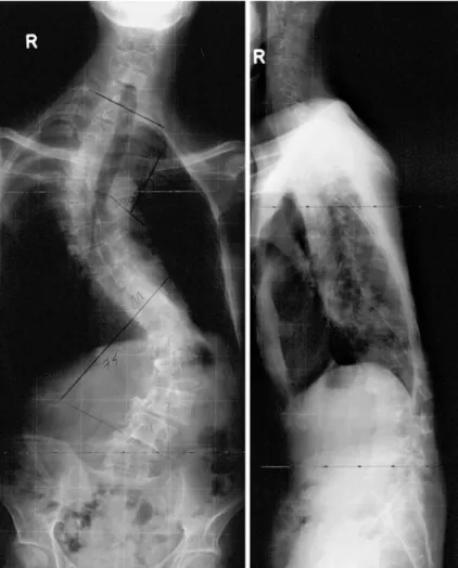
Marfan Syndrome. He was presented to our hospital with the diagnosis of severe idiopathic scoliosis. At the time of scoliosis surgery, he had a right thoracic curve T1–T10 of 75 degrees and a left lumbar curve T11–L4 of 80 degrees (Fig.4). He suffered from restricted pul-monary function with a vital capacity of 53%. Previous lung function tests were not available, but Patient 2 complained of progressive shortness of breath in the last 3–4 years. Physical examination revealed that he had slight weakness of proximal muscle groups in the upper limbs as well as atrophy of shoulder and
Fig. 3 (a) Immunohistochemical stain for desmin of the biopsy taken from patient 1. There is a disturbance of the normal architecture of the muscle fibers with single and central cores (asterix). (b) Electron microscopic examination of the biopsy taken from patient 1. Note the accumulation of electron dense material within the central region of the muscle fibers. The insert shows a magnification of the border zone between intact (triangle) and disturbed myofibrils (arrow). (c) Nicotinic acid-dehydrogenase (NADH) stain of the biopsy taken from patient 1. In the area of the central cores (asterix) there is no enzymatic activity detectable, while the rest of the muscle fibers show normal enzyme activity. (d) Periodic Acidic Schiff (PAS) stain of the biopsy taken from the Fig. 2 Pedigree of Family 1.
Three of the family members, the paternal grandmother, the father and the son, show typical clinical features of central core disease. In case of the father (2) and the son (1) this was con-firmed by muscle biopsy (black). In case of the paternal grandmother (3), who is de-ceased, similar musculoskeletal problems were reported by the family members. The diagnosis of central core disease is prob-able but not confirmed by muscle biopsy (grey)
parascapular musculature. Deep tendon reflexes were absent in the upper limbs, but normal in the lower limbs. Serum creatine kinase level was normal: 59 IU/l (normal<110 IU/l). He had no family history of neu-romuscular disease or scoliosis. A staged ventral release from T7 to L2 and dorsal instrumentation with fusion from T4 to L4 were performed to adjust the scoliosis deformity. A muscle biopsy was taken from the lateral vastus during this operation. The muscle biopsy of Patient 2 evidenced features similar to Patient 1 and his father (Fig.3).
Scoliosis surgery did not cause any complications in either patient; there were no signs of malignant hyperthermia during general anesthesia, nor any neu-rological complications. Intraoperative neuromonitor-ing with sensory and motor evoked potentials revealed no pathological activity. Scoliosis was adjusted to 50 degrees in Patient 1, and to 35 degrees thoracic and 35 degrees lumbar in Patient 2. Three years after our initial examination, pulmonary vital capacity of Pa-tient 1 was 30% of normal, and 41% of normal in Patient 2.
Discussion
Although scoliosis is a common musculoskeletal feature in patients with neuromuscular diseases, usually appar-ent in advanced stages of the disease, the correct diag-nosis of CCD is often missed in soliosis patients [13,15]. Both patients in our report suffered from severe pro-gressive scoliosis and showed further clinical signs of CCD [16], namely the symmetrical muscle weakness with varying pattern and extent, involving the trunk and the proximal muscles of the limbs more than the distal groups, and decreased or absent deep tendon reflexes, but normal sensation. In addition, Patient 1 had multi-ple joint contractures, another common musculoskeletal problem in CCD [5, 17]. Patient 2 had only slight weakness of proximal muscles of the upper limbs, mus-cular atrophy in his shoulders, and absent deep tendon reflexes in the upper limbs.
These case reports illustrate the wide spectrum of clinical appearance of CCD, even within the same family, ranging from apparently clinically normal to
Fig. 4 Pre-operative ap and lateral radiographs of Patient 2 showing severe thoraco-lumbar scoliosis
severely disabled individuals. To our knowledge, respi-ratory insufficiency has not been reported as a feature of CCD. Therefore, the findings of both patients might be considered unusual. In conjunction with their muscle biopsy findings, the symptoms of the patients presented could have led to the incorrect diagnosis of mini-core myopathy, a condition with autosomal recessive inheri-tance, having no linkage with malignant hyperthermia, but association with nocturnal hypoventilation. Exami-nation of the father of Patient 1 suggested an autosomal dominant disorder in this family. The muscle biopsy of Patient 1 confirmed the diagnosis of CCD. An interest-ing findinterest-ing in CCD is that children are usually more severely affected than their parents [16].
The level of serum creatine phospokinase was normal in both patients, a finding consistent with documented reports [14]. Cases with elevated levels of serum creatine phospokinase have also been described [9,15] which can be associated with episodes of malignant hyperthermia [2, 4]. Inherited as a dominant trait, malignant hyper-thermia is now considered to be a genetically heteroge-neous skeletal muscle disorder characterized by a hypermetabolic response to common inhalational anes-thetics and depolarizing muscle relaxants [1]. Reliable diagnosis of the malignant hyperthermia phenotype is made using an in vitro contracture test (IVCT), where contractile responses to caffeine and halothane are measured in muscle biopsies obtained from individuals suspected to be susceptible to malignant hyperthermia [3, 8]. Genetic analysis has indicated that mutations in the ryanodine receptor gene of the skeletal muscle (RYR1), encoding the calcium release channel in the
sarcoplasmic reticulum, may be responsible for both malignant hyperthermia and CCD [6, 20, 21]. To date, over 35 mutations/deletions in the RYR1 gene have been identified as co-segregating with the CCD trait [12,18]. Out of these, twelve have been definitively shown as resulting in susceptibility to malignant hyperthermia. Thus, it seems likely that some mutations in RYR1 may lead to CCD in the absence of malignant hyperthermia. Nevertheless, CCD has been shown to be closely linked with malignant hyperthermia and CCD patients should not be anesthetized with any malignant hyperthermia-triggering agents [2,4].
Conclusions
The patients described in this report highlight the importance of examining children and adults with pro-gressive scoliosis of unknown etiology and a family history of musculoskeletal problems such as muscle weakness and joint disorders. In such cases, we recom-mend a muscle biopsy which, in case of CCD, allows an easy and correct diagnosis due to the typical histological pattern. It is important for patients and treating physi-cians to know that CCD does not necessarily indicate a shortened life expectancy, but rather the possibility of a slow progression of the disease [7]. Furthermore, the autosomal dominant mode of inheritance and the in-creased susceptibility to malignant hyperthermia should be considered when advising individuals affected by this disease.
References
1. Denborough M (1998) Malignant hyperthermia. Lancet 352:1131–1136 2. Denborough MA, Dennett X,
Ander-son RM (1973) Central-core disease and malignant hyperpyrexia. Br Med J 1:272–273
3. Ellis FR, Harriman DG (1973) A new screening test for susceptibility to malignant hyperpyrexia. Br J Anaesth 45:638
4. Frank JP, Harati Y, Butler IJ et al (1980) Central core disease and malig-nant hyperthermia syndrome. Ann Neurol 7:11–17
5. Gamble JG, Rinsky LA, Lee JH (1988) Orthopaedic aspects of central core disease. J Bone Joint Surg Am 70:1061– 1066
6. Gillard EF, Otsu K, Fujii J et al (1991) A substitution of cysteine for arginine 614 in the ryanodine receptor is poten-tially causative of human malignant hyperthermia. Genomics 11:751–755 7. Lamont PJ, Dubowitz V, Landon DN
et al (1998) Fifty year follow-up of a patient with central core disease shows slow but definite progression. Neuro-muscular Disord 8:385–391
8. Larach MG (1989) Standardization of the caffeine halothane muscle contrac-ture test. North American Malignant Hyperthermia Group. Anesth Analg 69:511–515
9. Ma JS, Mak SC, Liu AM et al (1997) Central core disease associated with scoliosis: report of one case. Zhonghua Min Guo Xiao Er Ke Yi Xue Hui Za Zhi 38:297–299
10. Magee KR, Shy GM (1956) A new congenital non-progressive myopathy. Brain 79:610–621
11. Manzur AY, Sewry CA, Ziprin J et al (1998) A severe clinical and pathologi-cal variant of central core disease with possible autosomal recessive inheri-tance. Neuromuscular Disord 8:467– 473
12. McCarthy TV, Quane KA, Lynch PJ (2000) Ryanodine receptor mutations in malignant hyperthermia and central core disease. Hum Mutat 15:410–417 13. Merlini L, Mattutini P, Bonfiglioli S et al (1987) Non-progressive central core disease with severe congenital sco-liosis: a case report. Dev Med Child Neurol 29:106–109
14. Nagai T, Tsuchiya Y, Maruyama A et al (1994) Scoliosis associated with central core disease. Brain Dev 16:150– 152
15. Pozio G, De Giorgi G, Trizio W et al (1985) Central core disease and con-genital scoliosis. Study of one case. Acta Neurol (Napoli) 7:425–431
16. Quinlivan RM, Muller CR, Davis M et al (2003) Central core disease: clini-cal, pathologiclini-cal, and genetic features. Arch Dis Child 88:1051–1055 17. Ramsey PL, Hensinger RN (1975)
Congenital dislocation of the hip asso-ciated with central core disease. J Bone Joint Surg Am 57:648–651
18. Scacheri PC, Hoffman EP, Fratkin JD et al (2000) A novel ryanodine receptor gene mutation causing both cores and rods in congenital myopathy. Neurol-ogy 55:1689–1696
19. Sewry CA, Muller C, Davis M et al (2002) The spectrum of pathology in central core disease. Neuromuscular Disord 12:930–938
20. Urwyler A, Deufel T, McCarthy T et al (2001) Guidelines for molecular genetic detection of susceptibility to malignant hyperthermia. Br J Anaesth 86:283–287 21. Zhang Y, Chen HS, Khanna VK et al
(1993) A mutation in the human ry-anodine receptor gene associated with central core disease. Nat Genet 5:46–50