CHROMOSOMAL PROTEINS IN ACTIVATION OF TRANSCRIPTION
by
Benjamin Michael Shykind B.A., Biochemistry
Columbia College, New York, NY
Submitted To The Department Of Biology In Partial Fulfillment Of The Requirements
For The Degree Of
DOCTOR OF PHILOSOPHY at the
MASSACHUSETTS INSTITUTE OF TECHNOLOGY CAMBRIDGE, MASSACHUSETTS
February 1996
© Benjamin Michael Shykind, 1996 All rights reserved.
The author hereby grants MIT permission to reproduce and to distribute publicly copies of this thesis document in whole or in part.
Signature of Author Certified by Accepted by ... ,SACH-USETTS INS'f [ OF TECHNOLOGY Department of Biology ,/Dr. Phillip K Sharp Thesis Supervisor D•GPrakt-Selomon JTE Graduate Committee
FEB 131996
LIBRARIESAbstract
CHROMOSOMAL PROTEINS IN ACTIVATION OF TRANSCRIPTION by
Benjamin Michael Shykind
Submitted to the department of Biology in partial fulfillment for the degree Doctor of Philosophy
Transcription by RNA polymerase II requires multiple auxiliary factors for accurate initiation. The positive modulation of this reaction requires a further set of factors, collectively called coactivators, that are not required for the unregulated or basal initiation of transcription. Coactivators can be divided into two groups: TATA binding protein (TBP) associated, and soluble. Both groups appear to be required for activated levels of transcription. The exact mechanism by which activators, in collaboration with coactivators, enhance the basal reaction remains unknown. This thesis presents experiments designed to advance the understanding of this fundamentally important biological process.
Using an in vitro transcription system unresponsive to activators as a functional assay, an activity was identified that potentiated activation. This coactivator was purified to homogeneity by standard biochemical means and identified by amino acid sequencing as the non-histone chromosomal protein 2. Subsequently, the highly related HMG-1 protein, and the 80 amino acid HMG box B were demonstrated to by coactivators. These proteins have DNA binding and bending activity and have been included in a group of protein known as "architectural factors" for their ability to impart structure to DNA. Coactivation with HMG-proteins was shown to require the factor TFIIA and the TFIID-TFIIA stage of initiation was the first step in transcription altered in the activation process. The inability to detect a quantitative change at this critical step led to the proposal that a qualitative change takes place during this stage of activation that facilitates transcription.
Another chromosomal protein that coactivates stimulation, DNA topoisomerase I, was examined in vitro. This coactivator also functioned at the TFIID-TFIIA stage of activation as determined by order-of-addition transcription. The activation event was associated by a dramatic enhancement in the formation of the TFIID-TFIIA complex as demonstrated by electrophoretic mobility shift assay (EMSA). This enhancement of complex formation required the concomitant participation of TFIID, TFIIA, activator and topoisomerase I, exactly analogous to the conditions required for maximal activation. DNase I footprinting confirmed the EMSA analysis. The effect of enhanced complex formation on the activation process is likely both quantitative and qualitative.
Thesis Supervisor: Phillip A. Sharp
Table of contents
Title page.... ... ... ... 1...
Abstract... ... ... 2
T able of C ontents... ... 3
A cknow ledgm ents... ... 4
Chapter One: Introduction... ... 5
O verview ... ... . . . . 6
B asal Transcription ... 8
Overview of Activation... ... ... 17
Coactivators...18
M echanism s of A ctivation...24
Conclusion and Introduction to Thesis...33
R eferen ces...3 5 Chapter Two: HM G Proteins in Activation...55
A b strac t... ... 5 7 Introduction ... 58
R esults... ... 60
D iscu ssio n ... 6 8 Figures... 73
Materials and Methods... ... 90
R efe ren ces...9 6 A cknow ledgm ents...104
Chapter Three: DNA topoisomerase I in Activation...105
A b stract...10 6 In tro du ctio n ... 10 7 R esults ... 111
D iscu ssio n ... 12 2 Figures... 126
Materials and Methods... ... 148
R eferences ... 150
Acknowledgm ents... ... 159
Chapter Four: Conclusions and Perspectives... 160
Sum m ary of D ata ... 161
A rchitectural Factor Coactivators... 162
The TFIID-TFIIA Complex... ... 164
T he R ole of T F IIA ... 165
C onclusion...166
R eferen ce s...16 8 Appendix A: Mechanism of Basal Repression by DNA Topoisomerase I...171
Appendix B: DNA Topoisomerase I Effect on Linear Template...189
Appendix C: Coactivator-coactivator Interaction...207
Acknowledgments
I foremost thank my thesis advisor Phil Sharp, for atfording me the chance to work in the stimulating and exciting environment of his lab. I further am indebted to him for his enthusiasm, honesty, and professionalism. Finally, I am very grateful to him for the opportunity to have pursued science in a climate supportive of independent research. I salute the memory of Richard Wistar in whose lab at the National Naval Medical Center I first learned the work and joy of biochemistry many years ago.
I thank Keiko Ozato at the N.I.H. for the chance to have learned molecular immunology in her lab, and for her support and friendship over the years.
I heartily thank my once and future mentor Richard Axel for an influential exposure to molecular cell biology in his unique lab, and for his support, friendship, and interest. The following friends contributed subtly and overtly to my efforts over the years: Kris Niyogi, Kevin Lee, Rachel Meyers, Jae Kim, and Andrew MacMillan. Their generosity and camaraderie has sustained me over the years.
Many Sharp lab associates over the years deserve thanks for having made life in the lab richer and more enjoyable including: Sean Harper, Robbyn Issner, Marc Timmers, Jeff Parvin, Joel Pomerantz, Patrick McCaw, Lee Lim, Charles Query, Ben Blencowe, Dan Chasman, Jorgen Kjems, Marcia Morton, and Margarita Sciafaca.
I thank Frank Solomon, Arthur Lander and Ann Calof for their advice, generosity, and sense of humor.
I give thanks and credit to my editors Milos Konopasek, Lyuba Konopasek, and Andrew MacMillan for their thoroughness and persuasiveness.
I thank my thesis committee members Michael Green, David Balti~ore,Tyler Jacks, and David Housman for their participation, support, and incisiveness during a provocative defense.
I gratefully thank my family Edwin and Arlene Shykind and new additions Milos and Ludmilla Konopasek for years of support and patience for my endeavor.
Finally, I thank .my wife Lyuba Konopasek for her support, love, and boundless good humor.
Chapter I
Introduction Overview
The regulation of the production of mRNA in the cell mirrors the complexity of all of development, as the determination of cell fate depends primarily on the correct expression of the genes transcribed by RNA polymerase II. Therefore, the elucidation of the process that delivers polymerase II to correct loci is of great importance to the understanding of biological processes. Since the first demonstration of accurate initiation of RNA polymerase II (pol II) transcription in a cell extract seventeen years ago (Weil et al., 1979), the understanding of the initiation of transcription in vitro has advanced to the point where the earliest steps in the process have now been described by x-ray crystallography to roughly 2.5 A resolution (Kim et al., 1993a; Kim et al., 1993b; Nikolov et al., 1995). Despite this progress, the critical aspects of regulation of the pol 11 machinery, are still being uncovered.
This thesis describes experiments to advance the understanding of the mechanism of activation of pol II transcription. The determination of the factors involved, and their function in the process, has been undertaken. In the first part of this introductory chapter, the components of the process that effects accurate initiation of pol II transcription will be described. These factors, many of which have now had their cDNAs isolated, fulfill specific duties in the transcription cycle. Each step in this cycle is carried out by protein-DNA and protein-protein interactions with enough specificity to fix the site of the start of transcription with high fidelity. The cell exerts a great deal of control on this process and there is evidence of positive and negative modulation of each step in the reaction.
The DNA segment that directs the accurate initiation of transcription by pol II is the promoter, a region of DNA immediately upstream of the gene, encompassing
approximately fifty nucleotides from around +10 to -40. This region typically contains a TATA element at -30, and often an initiator (Inr) element centered on +1 (Smale and Baltimore, 1989). While this region is capable of directing accurate initiation, more
distal cis-acting elements, typically upstream, are required for the dramatic induction of gene expression observed in vivo (Banerji et al., 1981). These distal elements function as binding sites for sequence specific factors (activators) that mediate the activation or enhancement of the process. Transcriptional activation will be reviewed in the second part of this chapter.
The enzyme that polymerizes ribonucleotides into mRNA using DNA as a template, the RNA polymerase II, cannot accurately initiate transcription from the promoter on its own. Additional protein factors are required. These proteins, termed the general transcription factors (GTFs), recognize the promoter, recruit and help position the polymerase, enable it to enter the coding region where mRNA is transcribed, and finally, promote the successful full length elongation of nascent RNA chain. Although able to carry out accurate initiation of transcription (referred to as the "basal" reaction), this ensemble of almost fifty polypeptides cannot on its own be positively modulated by upstream elements. Additional factors are required. These factors have variously been termed coactivators, adaptors, or mediators. This thesis is primarily concerned with these factors and their mechanisms of action. The second part of the introduction will describe current understanding about them.
Knowledge about the regulation of transcription has moved forward by the dissection of the basal reaction. We now turn to the description of the factors that effect basal, unregulated transcription and their roles in forming the structure that positions and launches RNA polymerase II.
The Basal Reaction
The recruitment of RNA polymerase II to the promoter is accomplished through the step-wise assembly of a protein complex. This assembly can be broken down into functional stages as described in Figure 1. The factors responsible for many of these
steps have been identified and their roles are now being clarified. As derepression can be considered a positive modulation of the reaction, the first step we will consider is the
initial and stable recognition of the promoter by protein factors or "nucleation."
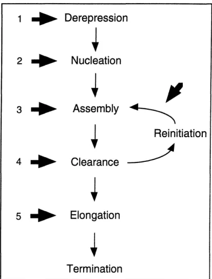
Figure 1. Schematic view of the pol II transcription cycle
Nucleation
Upon successful early fractionation of the mammalian cell extracts that supported accurate initiation (Matsui et al., 1980; Samuels et al., 1982), those crude fractions that reconstituted transcription were examined by DNA interference and template
Derepression
Nucleation
Assembly
4Reinitiation
Clearance
Elongation
Termination
_ ~ __ ~I ~ I_ ~ _ Icommitment assays to determine roughly their stage of participation in the reaction. Two second column fractions required for transcription, DB and AB (TFIID and TFIIA respectively), were found to form a complex on the adenovirus major late promoter which was resistant to inhibition by addition of excess DNA (Fire et al., 1984). Similar results were obtained on the conalbumin promoter (Davison et al., 1983). This stable complex was proposed to be the first step in promoter recognition and to give long term activity to the promoter, although there was no direct evidence of promoter binding. This evidence came from DNase I footprinting of the Drosophila and mammalian factors (Parker and Topol, 1984; Sawadogo and Roeder, 1985) The requirement for TFIIA in the formation of the committed complex on the promoter was subsequently brought into question by studies that demonstrated the ability of the TFIID fraction alone to stably commit (Sawadogo and Roeder, 1985; Van Dyke et al., 1989). However, the purity of TFIID remained a critical issue in the interpretation of these results. The complexes formed on the major late promoter during initiation were convincingly demonstrated by gel shift assay using the yeast TFIID. In this study, TFIIA was shown to alter the TFIID footprint, extending it upstream (Buratowski et al., 1989).
The mammalian TFIID activity proved extremely difficult to purify. However, in S. cerevisiae an activity that complemented an in vitro transcription reaction lacking TFIID was identified (Buratowski et al., 1988). Yeast TFIID supported accurate initiation in a mammalian in vitro system and footprinted over the TATA box. This activity was estimated to be 23-27kD in mass, much smaller than the estimate of almost 1000kD for mammalian TFIID. Additionally, it differed from its mammalian counterpart by the production of a smaller footprint over the TATA region. Yeast TFIID purification was amenable to conventional chromatography which led to the isolation of its cDNA clone (Cavallini et al., 1989; Eisenmann et al., 1989; Hahn et al., 1989; Horikoshi et al., 1989; Schmidt et al., 1989). The corresponding Drosophila and human counterparts to the yeast TFIID were cloned using P.C.R. with degenerate primers based on the yeast
sequence (Hoey et al., 1990; Peterson et al., 1990). Again, the cloned TFIIDs did not behave like the purified TFIIDs: the recombinant protein was unable to support regulated transcription by upstream activators, was smaller (43kD for the human) than the endogenous activity, and had a smaller footprint. The physical and functional differences between cloned and recombinantly expressed, and endogenous TFIIDs led to the proposal that the endogenous TFIID was a multisubunit complex (Hoey et al., 1990; Pugh and Tjian, 1990). Definitive proof for this proposal came when the endogenous Drosophila TFIID was immunoprecipitated and found to be complexed with several associated factors. The fly TFIID activity was composed of the 45kD TATA binding protein (TBP) and TBP associated factors (TAFs) of 150, 110, 80, 60, 40, and 32kD, which were also shown to potentiate the response to activation (Dynlacht et al., 1991). Subsequently, a TAF-TBP complex from human TFIID was also identified (Tanese et al., 1991; Zhou et al., 1992). The properties of endogenous TFIID were recapitulated by combinations of TBP and TAFs. The extended footprint seen with endogenous TFIID was found in Drosophila to be mediated by TAF 150 (Verrijzer et al., 1994). Recently, TAFs have been demonstrated to promote TFIID binding to different core promoter elements (Verrijzer et al., 1995). The coactivating properties of TAFs will be discussed in the second part of this introduction. The discovery of TAFs in higher metazoans prompted a reexamination of yeast TBP. This led to the demonstration that yeast TBP could be immunoprecipitated in the company of a number of other polypeptides (Poon and Weil, 1993; Poon et al., 1995). This yeast TBP complex has been shown to be required for stimulated transcription in vitro (Reese et al., 1994).
The role of TFIIA in nucleation, or formation of the first stable promoter-protein complex, has been confirmed by recent experiments. Mammalian TFIIA was purified to homogeneity and found to be made up of polypeptides of 35, 19, and 12kD; the larger two subunits were found to be derived from the same gene (DeJong and Roeder, 1993; Ma et al., 1993). The cDNA of the smaller subunit was also cloned, making it possible to
polymerase. TFIIB is believed to determine this start-site position; yeast TFIIB was first isolated as a start-site alteration suppressor (Pinto et al., 1992). The role of TFIIB in start-site selection was convincingly demonstrated in experiments where an S. pombe start-site was utilized in an S. cerevisiae in vitro reaction only when both S. pombe polymerase and TFIIB were substituted into the system (Li et al., 1994). Recently the x-ray structure of the TBP-TFIIB-DNA complex was reported. This structure shows TFIIB binding underneath TBP and making contacts with the DNA backbone and the c-terminal stirrup of TBP (Nikolov et al., 1995).
RNA polymerase II is composed of ten subunits and has a molecular weight of about 500kD. A prominent feature of the largest subunit is the repetitive C-terminal domain (CTD) which is a substrate for phosphorylation. The phosphorylation state of the CTD is thought to play a role in transcription regulation. The Ilo form of pol II is heavily phosphorylated while the IIa form in unphosphorylated. Pol IIA is found in the assembled initiation complex while Pol IIO is the form found in elongating complexes (Payne et al., 1989). However, the CTD is not required for transcription from the adeno major late promoter. Interestingly, there may be core promoter specificities for the requirement of the CTD (Akoulitchev et al., 1995)
Clearance
At the end of assembly, the promoter is bound by TFIID, TFIIA, TFIIB, TFIIF, and Pol II, with the polymerase correctly positioned. With the addition of NTPs, this
complex can send off an elongation complex, depending on the superhelical state and
type of the promoter (Parvin and Sharp, 1993). In the absence of negative superhelical tension, the assembled complex requires TFIIE, TFIIH, and ATP hydrolysis for the elongation complex to clear the immediate promoter region (Goodrich and Tjian, 1994). TFIIE is a heterodimer with subunits of 34 and 56kD (Ohkuma et al., 1990; Inostroza et
al., 1991). TFIIH is composed of at least eight polypeptides and is associated with two helicase activities, an ATPase, and a kinase activity that has been shown to phosphorylate the CTD of polymerase. TFIIH also shares subunits with the nucleotide excision repair mechanism (Drapkin et al., 1994). The exact mechanism of promoter clearance is not yet understood. However, it is fair to say that TFIIE-TFIIH-ATP driven clearance is obviated by the energy stored in negative superhelical tension, suggesting that the helicase activity in TFIIH may function in moving the elongation complex away from the immediate promoter region. Additionally, the phosphorylation state of the polymerase may play a part in the conversion of the initiating polymerase to the elongating form (reviewed by (Buratowski, 1994).
Reinitiation
The study of multiple-round transcription has yet to come under the intense scrutiny given initiation. It could be proposed that the study of reinitiation, rather than is initiation, is more germane to activation. This is due to the fact that increased initiations per time, or activation, must by definition involve mechanisms of reinitiation more often than strategies of initiation. Control of initiation may be more related to regulation by derepression. However, several interesting features that distinguish initiation from reinitiation have come to light in the past few years. The fate of the general factors has been traced by Zawel and Reinberg following the departure of the polymerase from the promoter (Zawel et al., 1995). As was expected, TFIID remained bound to its site. Upon the formation of the first phosphodiester bonds, TFIIB was released from the complex. TFIIF and TFIIE were released somewhere within the first ten nucleotides. Interestingly, TFIIH was the last GTF released; this occurred somewhere between +30 and +68. Important to the process of reinitiation, TFIIB once released from the initiation complex, promptly reassociated with the promoter bound TFIID. Thus presumably signaling the start of the next assembly process. A factor that boosted reinitiation was identified by
polymerase collision assay that was subsequently identified as the elongation factor TFIIS (Szentirmay and Sawadogo, 1991). Interestingly, first round transcription was found to be less dependent on TFIIS than were reinitiates, highlighting a difference in factor requirement between the two processes (Szentirmay and Sawadogo, 1993). It is possible that multiple round transcription will have an increased need for topoisomerase I (see below) over single round transcription as the torsional tension generated by elongation should increase with each polymerase loaded (Wang, 1992).
Elongation
Polymerase II is not only unable to accurately initiate transcription on its own, but likely is unable to complete elongation or full-length transcription without help. Several factors have been shown to have elongation or anti-pausing activity including TFIIF (Flores et al., 1989; Price et al., 1989), TFIIS (Reinberg and Roeder, 1987; Bengal et al., 1991) and the elongins (Aso et al., 1995). Additionally, topoisomerase I has been shown to promote elongation (Egyhazi and Durban, 1987). The elongating polymerase may traverse the gene on its own, and recruit elongation factors when it gets stuck; TFIIF has been shown to be associated with stalled polymerases (Zawel et al., 1995). As an example of the pitfalls of biochemical identification of factors, the reported elongation factor TFIIJ (Cortes et al., 1992) turned out to be an RNAse inhibitor (D. Reinberg, personal communication). This spurious identification occurred when rRNAsin was omitted from in vitro transcription reactions.
Derepression
The dissection of the transcription reaction has primarily been carried out on DNA templates devoid of chromosomal proteins. However, the physiological state of the gene is a nucleoprotein complex called chromatin. The packaging of DNA into nucleosomes can have repressive effects on transcription (reviewed in Paranjape et al., 1994). This
repression can be circumvented by prebinding of initiation factors, such as TFIID, to the template, prior to chromatinization (Workman and Roeder, 1987). Alleviation of chromatin repression is also believed to occur through the active reconfiguration of nucleosome structure. This active derepression has been reported in yeast to be mediated by a multicomponent complex of SWI and SNF gene products (Hirschhorn et al., 1992, and references therein). Such nucleosome remodeling activities have recently been found in human and Drosophila extracts (Kwon et al., 1995; Tsukiyama et al., 1995). This active reconfiguration is believed to make promoters accessible to transcription factor binding. Thus the first step in transcription initiation may well be the active derepression of the chromatinized promoter region.
Derepression may take place at other points in the pathway as well. Partially formed complexes can become blocked by specific inhibitors, preventing subsequent assembly. A mammalian factor called Drl has been shown to bind to TBP and prevent assembly of the TBP-TFIIA complex by formation of a competitive TBP-Drl complex on the promoter. The TBP-TFIIB complex is similarly unable to form in the presence of Dri unless TFIIA is present in sufficient quantity. Another factor, ADI, which was isolated from yeast prevents the binding of TBP to the promoter (Auble and Hahn, 1993). ADI requires ATP for its inhibitory function and like Drl is blocked by preassociation of TBP with TFIIA. The abundant chromosomal enzyme topoisomerase I has been shown to inhibit transcription in a manner similar to Drl (Merino et al., 1993).
Holocomplexes
The primacy of the stepwise model of assembly of the initiation complex has recently been challenged by the finding that polymerase can be isolated in a large complex containing many of the GTFs (Koleske et al., 1992; Thompson et al., 1993;
Koleske and Young, 1994; Ossipow et al., 1995). Thus some of the GTFs may arrive at the promoter preassembled, bypassing some of the steps of assembly.
We can now draw a schematic summary of the transcription cycle and map it onto an idealized control region (Figure 2). The locations of factor function on the promoter are correlated with their stage of action in the transcription cycle.
Figure 2. Transcription cycle mapped onto an idealized gene
Activators, which bind to specific sites, typically, but not necessarily, upstream of the basal machine, positively modulate this process. The remainder of this introduction will focus on the factor requirements for activation and the differences between the basal and activated initiation complexes.
Activation Overview
There are now a large number of well characterized sequence-specific DNA binding proteins that stimulate transcription from binding sites distant from the region of initiation. The mechanism by which these activator proteins increase the rate of initiation
rInranmcnQin W/N
nucleation
assem bly
r -
I
I
[1l
r activatorFactivator
binding sites
I
GTFs
RNA pol 11
\AVIVrl VVV1VI t ~17 or Ir401.0..r Iis the subject of intense research. As summarized in the first part of this introduction, the factors that bring the polymerase to the start site have been defined. However, the basal reaction, the target of activation, has been found to be deficient in its ability to be positively modulated by activators in vitro. The missing activities that bridge the functional and perhaps literal gap between the activator and the basal machinery are known as coactivators. These activities can be subdivided into soluble or non-TBP associated, and TBP-bound or TAFs. Many of the defined soluble coactivators have been shown to act generally, by mediating activation equally well from all activators. There are also reports of activator specific coactivators. The TAFs, as an ensemble, are general coactivators but individually have activator specificities. Activator proteins are bipartite, containing separable DNA binding and activation domains (Hope and Struhl, 1986). Activation domains fall into classes based on the amino acid moieties they contain. Many groups have demonstrated interactions between members of the GTFs and activation domains. Such interactions have been proposed to mediate some aspect of activation.
Using functional assays a number of groups have attempted to define the step of initiation potentiated by activators. Despite various lines of evidence implicating different stages of initiation as being altered by activators, no conclusive picture has emerged of the mechanism of activation. It may be that there is no single mechanism and that activator alters many stages of initiation. The assorted interactions between activation domains and GTFs may all define relevant intermediates of the process. It could be that the templates used by many labs to study the reaction may be too far removed from the actual, physiologically significant, chromatinized promoter. Thus a definitive activating event may be detected by standard biochemical means, once the process is studied in nucleosome packaged templates.
Coactivators
TBP associated factors
As soon as yeast TFIID was isolated, and found to be unable to support activation in an in vitro reaction, it became clear that either mammalian TFIID, which supported activation, was a quite different protein, or was associated with enabling factors. A number of unsuccessful attempts were made to force the putative enabling factors to function with yeast TBP. When human TBP was cloned and also found to fail to support stimulation in vitro, the case for the existence of associated factors (as opposed to post-translational modifications) that enabled activation was strengthened (Pugh and Tjian, 1990). Subsequently immunoprecipitation of Drosophila TFIID revealed that TBP was adorned with several stably bound proteins. Crude isolation of these associated proteins revealed them to possess coactivating activity (Dynlacht et al., 1991). Human TFIID was found to be complexed with TAF coactivators as well (Tanese et al., 1991). The large number of TAFs were proposed to potentially accommodate the diversity of activation domains. Several TAF-activator interactions have been demonstrated including: TAF 110 with Spl, TAFs 40 and 60 with VP 16, TAFs 40 and 60 with p53, and TAF 40 with EBNA-2 (Goodrich et al., 1993; Hoey et al., 1993; Tjian and Maniatis, 1994; Thut et al., 1995). The functional significance of many of these activator-TAF interactions have been confirmed by mutant studies where loss of activation and loss of binding were correlated. The TAFs isolated from yeast have recently been shown to possess coactivator function in vitro (Reese et al., 1994).
Soluble coactivators USA
A set of GTFs of sufficient purity, and a third column TFIID fraction (containing TAFs) that responded very weakly to various activators, led to the discovery of a family of coactivators not tightly associated with TBP (Meisterernst et al., 1991b). This activity, termed USA for "upstream factor stimulatory activity," was further separated into
positive and negative cofactors (PCs and NCs). Of the PCs 1-4, the best characterized are PC-4 and PC-3. PC-4 was purified to homogeneity and its cDNA isolated (Ge and Roeder, 1994; Kretzschmar et al., 1994). PC-4 was shown to bind to activation domains and to the TBP-TFIIA complex, thus having the potential to bridge the activator and the basal complex. PC4 had non-specific DNA binding activity and its coactivating activity was negatively modulated by phosphorylation (Kretzschmar et al., 1994).
DNA Topoisomerase I/PC-3
In the course of the in vitro analyses of factors required for activation, Merino et al. purified an activity from HeLa cells that potentiated stimulation by Gal4-AH called Dr2. This activity additionally repressed the basal reaction in the absence of TFIIA. Dr2 was purified to homogeneity and identified as topoisomerase I (Merino et al., 1993) . Subsequently, Roeder's group also identified topoisomerase I as the coactivator PC3 (Kretzschmar et al., 1993). Topoisomerase I was shown to coimmunoprecipitate with the TFIID complex and to interact with TBP as shown by Far Western. Interestingly, Merino et al. demonstrated a promoter specificity for repression which suggested the requirement for a TATA element for repression by topoisomerase I. The transcriptional activity of topoisomerase I in these in vitro reactions was convincingly demonstrated to be independent of its relaxing activity, as a point mutant of topoisomerase I, unable to relax supercoils, retained its ability to affect transcription. Further, wild type topoisomerase I from yeast, vaccinia virus, and E. coli had no transcriptional properties in this mammalian transcription system.
The helical structure of DNA had suggested to some, a priori, that a "swivel" would be required in transcription such that polymerases could avoid rotating about the axis of the coding region DNA during elongation (Wang, 1985, and references therein). Thus topoisomerase I has been implicated in transcription elongation, a post-initiation step. Substantiating this proposal, a number of studies have localized topoisomerase I to
actively transcribed regions of the genome. Nucleosomes from actively transcribed genes were found to contain topoisomerase I (Weisbrod, 1982). Additionally, heat shock loci on Drosophila polytene chromosomes stained with anti-topoisomerase I antibodies after induction, but not before (Fleischmann et al., 1984). Further, UV crosslinking trapped topoisomerase I on active genes in Drosophila (Gilmour et al., 1986). Finally, topoisomerase I-coding region DNA covalent intermediates were demonstrated by the inhibitor camptothecin, to be tightly associated with induction of the tyrosine aminotransferase gene (Stewart and Schutz, 1987), and the c-fos gene (Stewart et al., 1990). In addition to the localization of topoisomerase I to the coding regions of genes, there is evidence of a topoisomerase I site in a promoter region of a gene (Stewart and Schutz, 1987). Thus convincing evidence exists demonstrating the ability of topoisomerase I to coactivate transcription in vitro. Meanwhile, in vivo efforts have focused on demonstrating a role for the enzyme in elongation. It is possible that topoisomerase I joins the initiation complex during activation and is then loaded into the elongation complex, but no data exists for this presumption.
HMG proteins
Those cellular proteins that eluted from isolated nuclei at 350mM NaC1, were soluble in 2% trichloroacetic acid, and migrated rapidly in electrophoresis were given the operational name high mobility group or HMG proteins (reviewed in Bustin et al., 1990; Grosschedl et al., 1994). There are three types of HMG proteins, unrelated by sequence homology. They do share some interesting properties such as the ability to bind to DNA and nucleosomes, and the possession of highly charged regions. All three groups, HMG-1/2, HMG-14/17 and HMG-I(Y), have now been identified as transcriptional coactivators.
The HMG-1/2 proteins have been described as modulators of pol II transcription in a variety of crude systems (Tremethick and Molloy, 1988; Watt and Molloy, 1988; Singh and Dixon, 1990). Recently they have also been demonstrated to coactivate stimulation by a variety of activators in a defined reaction (Shykind et al., 1995).
HMG14 and 17
This HMG family has been implicated in transcription by its association with active chromatin (Weisbrod, 1982). HMG-17 was shown to be a chromatin specific coactivator with Gal4-VP16 (Paranjape et al., 1995). Its coactivating activity functioned on nucleosomal but not naked DNA. The highly related HMG-14 has been reported to promote elongation on transcription from SV40 mini-chromosomes (Ding et al., 1994).
HMG I(Y)
Induction of the human interferon- 3 gene by virus was demonstrated to require both the activator NF-icB and I(Y) in vivo (Thanos and Maniatis, 1992). HMG-I(Y) was shown to bind to the promoter along with the activator such that mutations which abolished HMG-I(Y) binding dramatically reduced in vivo activation levels. HMG-I(Y) was further shown to enhance the in vitro binding of NF-KCB homo and heterodimers possibly by altering the structure of the promoter region DNA.
ADAs
In vitro transcriptions with the potent activator Gal4-VPl6 in yeast extracts led to the observation that over-addition of activator diminished activated levels of transcription before basal levels were altered (Berger et al., 1990). Further, over-expression of Gal4-VPl6 was found to be toxic in yeast, dependent on a functional activation domain. This led to a genetic screen that identified suppressors of the activator toxicity including
ADA2, ADA3, ADA5 and GCN5 (Berger et al., 1992, and reviewed in Guarente, 1995). The products of ADA genes have properties of coactivators.
Holopolymerase/Mediator
The heptapeptide repeats of the CTD of RNA polymerase II play an important regulatory role in yeast. Truncations of these repeats leads to various conditional growth phenotypes. Suppressors were isolated that reverted the growth phenotypes of CTD truncations (Nonet and Young, 1989). These suppressors, called SRBs "for suppressors of RNA polymerase B," were shown to form a massive complex with the polymerase containing in addition TFIIB, TFIIH, and TFIIF which was called the holopolymerase. Activation in vitro with this holoenzyme required the addition of only TBP, TFIIE and the activator (Koleske and Young, 1994). A similar polymerase complex termed the mediator was isolated from yeast and reported to contain SRBs, TFIIF, SUG1, and GALl1 (Kim et al., 1994). A mutation in the GALl 1 component that generated an interaction domain with the GAL4 dimerization domain was shown to be sufficient for activation, dependent on GAL4 (Barberis et al., 1995). SUG1 had been identified in a screen for suppressors of a GAL4 activation domain mutation (Swaffield et al., 1995).
Activator-specific coactivators
A number of soluble factors have been shown to potentiate stimulation specifically from a single activator, in contrast to the general coactivators described above. These specific coactivators include CBP, which binds to the cAMP-regulated enhancer binding protein (CREB) when CREB is activated by protein kinase A phosphorylation (Kwok et al., 1994). Interestingly, CBP can bind to the GTF TFIIB. Further, CBP shares homology with the yeast coactivator ADA-2. The adenovirus E1A protein, which mediates both repression and activation of gene expression, binds to a cellular factor called p300. This large protein has been implicated as a coactivator by the
ability of a mutant form to restore activity to an SV40 enhancer silenced by E1A (Eckner et al., 1994). Recently, p300 was shown to be a functional homologue of CBP in vivo (Lundblad et al., 1995). Another candidate for an activator restricted coactivator is the factor Bob-1, which was isolated by virtue of its ability to interact with the lymphoid-restricted activator Oct-2 (Gstaiger et al., 1995). Bob-1 boosts activation by Oct-2 and to a lesser extent Oct-1, in vivo. The potent Herpes virus activator VPl6 functions through an octamer-like motif via association with Oct-1 mediated by a factor called C 1 (Kristie and Sharp, 1990). The assembly of this large complex likely controls the activity of this element.
Thus the participation of sequence-specific activating proteins in the stimulation of transcription requires yet another level of protein-protein interaction. This level is the domain of coactivators, which may be working upon the basal factors as intermediates of the activator, or enabling direct and functional interaction between activator and the basal machine. Coactivators may be classified as activator-specific or activator-general. The TAFs as an ensemble can be considered activator-general although certain TAFs may mediate activation from specific classes of activators. It is likely that the TAFs and the soluble coactivators must both be present to support stimulation although it is possible that certain promoter contexts or activators may break this rule. The next section of this introduction will examine mechanism of activation, using the paradigm of assembly of the initiation complex described earlier in this introduction.
Mechanisms of Activation
The schematic view of assembly on the promoter can now be modified to highlight the stages of initiation proposed to be altered by activators (Figure 3).
1. (Workman et al., 1991; Croston et al., 1992)
2. (Sawadogo and Roeder, 1985; Abmayr et al., 1988; Horikoshi et al., 1988;
Stringer et al., 1990; Wang et al., 1992; White et al., 1992; Lieberman and Berk, 1994; Kaiser et al., 1995; Sauer et al., 1995; Shykind et al., 1995)
3. (Lin and Green, 1991; Choy and Green, 1993; Kim and Roeder, 1994; Chi et al., 1995) 4. (Xiao et al., 1994)
5. (Yankulov et al., 1994)
Figure 3. Points of activation described in the literature.
From the number of references cited for nucleation (step 2), it is clear that much attention has been focused on the stage where TFIID and TFIIA form a stable complex. In addition, compelling data exists for the derepression of transcription by activators, and elegant and convincing experiments have demonstrated the significance of the TFIIB binding step as well. In this last section I will review the reported stages and possible
1
Derepression
*
2
m4
Nucleation
3
o.Assembly
4
Reinitiation
4 w-
Clearance
4
5 m.
Elongation
Termination
Termination
mechanisms of activator function, basing the analysis on the steps of transcription schematically described in Figures 2 and 3.
Activator derepression
A paradigm for gene regulation has been proposed wherein the gene can exist in three states: an inactive or ground state, a derepressed state, and an activated state (Grunstein et al., 1992; Paranjape et al., 1994). The inactive state is mediated by nucleosome position, the condensation of chromatin, or by non-histone chromosomal protein repressors. Derepression brings the gene from the ground state to a basal state, and true activation occurs on the derepressed gene. The role of activators then is to first derepress the promoter, allowing free access for the GTFs, and second to aid in the assembly of those GTFs such that initiations per time are increased. In vitro experiments with synthetic Gal4 based activators have shown that the acidic activation domain can function more efficiently in assembled chromatin by dramatically increasing the induced to uninduced ratio, as the basal level of transcription drops away due to chromatin mediated repression (Workman et al., 1991). This effect, which depended on the presence of the activation domain, not simply the DNA binding domain, led the authors to conclude that the activator was functioning by helping the initiation complex compete for binding with nucleosomes. An investigation using histone H1 and H1 containing chromatin showed similarly that activator could prevent repression of transcription (Croston et al., 1992). These authors showed that local nucleosome structure in the promoter was disrupted by the activator DNA binding domain alone, but that this was not sufficient for derepression.
Nucleation
The stage of initiation complex assembly where GTF (TFIID +/- TFIIA) first recognizes the cis-regulatory elements of the promoter has received a great deal of
scrutiny in studies of activation. Studying the effect of the upstream regulatory factor USF (MLTF) on adeno major late promoter transcription, a link was made between activator and TFIID binding (Sawadogo and Roeder, 1985). In this study, a crude TFIID fraction was shown to stabilize USF binding and thus an interaction between the two factors was proposed. DNase I footprinting showed that the TFIID fraction made contact with the TATA box and also with almost forty base pairs of downstream DNA. This extended footprint was not dependent on the activator, but subtle changes in the footprint upon binding of both factors led the authors to propose that the interaction between USF and TFIID could result in a conformational change in TFIID that may be accompanied by extensive wrapping of the DNA around the complex and facilitation of the subsequent steps in initiation. The TFIID binding step was also implicated in the mechanism of pseudorabies immediate early protein activation (Abmayr et al., 1988). In this study, prebinding of a crude (second column) TFIID fraction to the promoter obviated the need for activator to reach stimulated levels of transcription. This suggested to the authors that facilitated binding of TFIID was the mechanism of activation. Using the adenovirus E4 promoter, a synthetic Gal4 binding domain based activator was demonstrated to have dramatic effects on the downstream footprint of a crude TFIID fraction (Horikoshi et al., 1988). In this study, the extended TFIID footprint observed upon TFIID binding to the major late promoter alone, was induced on the E4 promoter by functional activator protein. Thus a direct interaction between the activator and the TFIID was proposed by the authors. The mammalian activator ATF gave a similar alteration of TFIID footprint on the E4 promoter (Horikoshi et al., 1988b). Using oligonucleotide competition to remove activator from the promoter, this study concluded that once established, the altered TFIID footprint was maintained in the absence of activator. In an accompanying report, ATF function was examined in transcription assays with oligonucleotide competition (Hai et al., 1988). ATF binding site oligo inhibited transcription if added at time zero with all the partially purified GTFs and ATF. However, the reaction was
refractory to oligo addition if the template was preincubated with TFIID, TFIIB, and pol II along with ATF. The ATF-TFIID complex, which showed an altered footprint, was not an oligo-refractory complex, indicating that the ATF altered TFIID complex was not the activated complex. However, this study did emphasize the early action of ATF in activation. These early forays into the mechanism of activation used only partially purified factors at a time when the entire complement of GTFs had not yet been elucidated. Further, the existence of coactivators was then unknown. Thus factor-factor cross contamination diminishes the resolution of some of these results but not the inference of activator function early in the process. Further suggestion of an early stage of activator function resulted from the demonstration that the potent activation domain of VP16, when immobilized, could retain TFIID activity from nuclear HeLa extract (Stringer et al., 1990). Further, this domain could bind to recombinant yeast TBP. A control for the biological significance of this result was the correlation of poor TFIID binding and low activity of a transcriptionally compromised point mutant VP 16 (Ingles et
al., 1991).
Using potassium permanganate footprinting to assess start-site melting (open complex formation) TFIID-TFIIA complex formation was identified as the rate-limiting step facilitated by activator (Wang et al., 1992). Interestingly, this activator increase in start-site melting would not occur if TFIID alone, or TFIID plus TFIIA were preincubated with the template, even if activator was added subsequently. This work was carried out with partially purified factors. The TFIID used here likely contained topoisomerase I (see chapter III). Sarkosyl (a detergent which prevents initiation complex formation but does not inhibit elongation) and anti-Gal4-VP16 antibodies were used to probe activator function. This produced a result suggesting (through extremely circuitous logic) that the activator acted just after template commitment by TFIID and TFIIA (White et al., 1992).
The use of reagents purified to apparent homogeneity, and a direct physical assay (gel-shift), suggested that the role of the activator was the recruitment of TFIID,
dependent on TFIIA and the TAFs (Lieberman and Berk, 1994). The TFIID used in this study was immunoaffinity purified an estimated 15,000 fold (holoTFIID) and could be detected binding to radiolabeled probe in agarose gel EMSA (Zhou et al., 1992). In this study, the viral activator Zta was shown to increase the rate and extent of formation of the TFIID-TFIIA complex, and to stabilize the complex. DNase I footprinting revealed alterations in the TFIID footprint in the presence of Zta similar to those seen with ATF and cruder TFIID. This activator dependent loading was not completely correlated with activation, as stimulation in an in vitro transcription assay further required the coactivator fraction USA. However, an oligonucleotide challenge transcription assay demonstrated that the Zta-TFIID-TFIIA-promoter complex was a rapidly forming, oligonucleotide resistant entity. The authors concluded that Zta increased the formation of an intermediate in the initiation pathway and induced a conformational change in the TFIID-TFIIA complex that may alter the binding of subsequent GTFs. The TFIID-TFIID-TFIIA stage was also identified as a first step in activation in a highly purified transcription system using the coactivator PC4 (Kaiser et al., 1995). PC4 and the activator Gal4-AH were found to stimulate formation of the TFIID-TFIIA complex at limiting levels of TFIID, and to have modest effects on later GTF assembly as well. This work used a restriction enzyme cleavage of the activator site to temporally dissect activation. This showed the requirement for the continued presence of activation, after TFIID-TFIIA formation, for full activation in the system.
Recently recombinant Drosophila TFIID subcomplexes were assembled which showed the TAF requirements of activational synergy brought about by the two activators Bicoid and Hunchback (Sauer et al., 1995). In this study TAFs 60, 110, and 250 were required to be present in the TBP complex in order to support synergy. DNase I footprinting demonstrated that the activators recruited the TBP-TAF complex to the promoter, suggesting that a critical feature of activation is TFIID recruitment via TAF-activator interaction. It is interesting to note that TFIIA was not required for this
recruitment. It is unclear whether the Drosophila in vitro transcription system used here was free of soluble coactivators.
The recognition of the promoter by TFIID and TFIIA generates a stable complex. Extensive contacts are made with the DNA by a combination of TBP and TAF interactions which may be facilitated, and are certainly altered, by TFIIA. The TBP component of TFIID was the first GTF shown to bind to an activator and the first GTF implicated in the activation process. The main conflict in the data implicating nucleation as the stage of initiation altered in activation, is whether activation is equal to the facilitation of TFIID binding, or whether qualitative changes in the TFIID complex, induced by the activator and coactivator, result in the facilitation of subsequent steps in initiation. Both quantitative and qualitative changes may be occurring during the process. The variable requirement for TFIIA in the process may reflect template differences whereby different core promoter elements necessitate different TFIID conformations for activation that are stabilized by TFIIA. Additionally, there may be differential activator requirements for TFIIA. A combination of the two scenarios is likely as well. Suggestively, studies in Drosophila cells in culture have shown TFIID occupation of promoters can be rate limiting on TATA containing but not TATA-less promoters (Colgan and Manley, 1992). Recently, in vivo experiments in yeast showed that a TBP mutant that failed to support activation and also failed to bind TFIIA. Activation was restored by the fusion of the small subunit of yeast TFIIA to the mutant TBP (Stargell and Struhl, 1995). Further verification in vivo came from the fusion of the yeast TBP to an exogenous and potent DNA binding domain which generated a hybrid that obviated the need for activators to achieve stimulated levels of expression (Chatterjee and Struhl, 1995). An analogous in vivo manipulation yielded similar results (Klages and Strubin, 1995). Thus, significant in vitro and in vivo evidence exists supporting the model that activation occurs at the nucleation step.
Activated assembly of the initiation complex
The effects of activator have been shown to occur post-nucleation (Lin and Green, 1991). In this investigation, an immobilized template transcription system demonstrated that the assembly of initiation complexes in nuclear extract stalled after the binding of TFIID, at the TFIIB binding stage. Further, using moderately defined GTFs, activator was shown to recruit TFIIB to the TFIID complex. Additionally, TFIIB was specifically retained by immobilized activator but not by point mutant activator (Cress and Triezenberg, 1990), compromised for activation. Subsequently, the recruitment of TFIIB by activator was shown to occur in the absence of TAFs where activation would not be supported (Choy and Green, 1993). Thus activation was not equivalent to TFIIB recruitment. However, the authors in this study further showed that activator increased the incorporation of pol II, TFIIF, and TFIIE into the initiation complex dependent on TAFs. This led to a two step model of activation wherein activator induces a conformational change in TFIIB that facilitates recruitment of subsequent GTFs in the pathway. The importance of TFIIB in this process was reinforced by the analysis of mutants that did not bind activator (Roberts et al., 1993). Here, mutant TFIIBs, compromised in their ability to bind to Gal4-VP16 and Gal4-AH, supported basal levels of transcription as wild type, but failed to support activation. Finally, the hypothesized conformational change in TFIIB was verified by in vitro studies (Roberts and Green, 1994). Recently, experimental findings using holoTFIID gel shift and DNase I footprinting assays were reported showing that TFIIB binds more stability to the activated TFIID-TFIIA complex, and further, that TFIIB adds stability to this complex (Chi et al., 1995). The authors postulated that this dual enhancement of initiation complex assembly, by multiple activator contacts, may account for transcriptional synergy.
The data describing the significance of TFIIB interaction with the early initiation complex in activation are compelling. Interestingly, the conflict over its role in the
process is again a question of quantitative versus qualitative changes in its participation. Multiple protein-protein interactions between TFIIB and factors critical to activation including activation domains and TBP, have been elucidated. Further, TAF 40 has been shown to bind to TFIIB (Goodrich et al., 1993); and maps by protease footprinting, to the region required for VP16 interaction (Hori et al., 1995). TFIIB may be the pivotal factor in stimulation, transducing the activating signal from the TFIID-TFIIA complex to foster the enhanced assembly of the initiation complex.
Clearance
The GTFs that facilitate polymerase clearance, TFIIE and TFIIH, join the initiation complex at the last stage of assembly. Thus it is formally possible that clearance is a target of activation, but little evidence has come to light to support this possibility. Recently the activators VPl6 and p53 were shown to bind to the 62kD subunit of TFIIH (Xiao et al., 1994). In this report, mutations that decreased activator potency also decreased retention of TFIIH. The previously described experiments of Choy and Green (1993) indicated an increased recruitment of TFIIE during activation. Hence increased promoter clearance could participate in activation.
Elongation
Recently polymerase processivity has been reported to be increased by activators
in vivo (Yankulov et al., 1994). Measuring elongation efficiency through natural pause sites in from c-myc and HIV-2 TAR, activators were found to boost full-length elongation. The mechanism for this action and the generality of this phenomenon are presently unknown. The possibility of activator-influenced elongation also arises from a study showing the interaction of the TFIIF 74kD subunit with the activator SRF (Joliot et
al., 1995). This interaction may aid in the assembly of the initiation complex or could increase the elongation effects reported for TFIIF (Flores et al., 1989).
Reinitiation
On the single gene, activation, or increased initiation per time, must alter reinitiation frequency. The assembly of the initiation complex and the generation of the first elongation complex differ from the process of reinitiation, which is the reloading of the TFIID-TFIIA complex that remains promoter bound after the first initiation (Szentirmay and Sawadogo, 1993; Zawel et al., 1995). Reinitiation does not have to overcome the proposed slow step of TFIID binding. Nor is it likely to have to rescue the promoter from nucleosomal repression (Workman and Roeder, 1987). Thus reinitiation leaves the activator with less work to do to reassemble the complex. Along these lines, the rerecruitment of TFIIB by activator, after its rapid release from the promoter after initiation, has been proposed to be a possible activation mechanism (Zawel et al., 1995). It is possible that different mechanisms are utilized to produce activated initiation complexes, than to maintain activated reinitiation; there are clearly different slow steps to each process. The mechanism by which genes are maintained in their activated state is not yet understood.
Holoenzyme complexes
The recent discovery of partially assembled complexes of GTFs has introduced the compelling new possibility of single step activation (reviewed in Carey, 1995). The discovery of coactivating activities in these ensembles has strengthen the proposal that recruitment of a holocomplex could account for activation. Indeed, single protein-protein interactions between a holopolymerase complex and a promoter bound factor have been reported to be sufficient for activation (Barberis et al., 1995). The involvement of such holopolymerase complexes still allows for activation to occur at the nucleation stage: the
entire complex could then be recruited in one step by an activated TFIID-promoter structure. The recently described holocomplex in the mammalian system contains TFIID and was demonstrated to support basal transcription (Ossipow et al., 1995). However, this complex has not been reported to support activation.
Summary and Introduction to the Thesis
The factors that comprise the basal reaction form a machine that recruits, accurately positions, and launches RNA polymerase II into transcription. The extent of factors required for this reaction has been circumscribed, but has some variability depending on the promoter and the topological conditions. The ability of upstream regulatory elements to potentiate this reaction depends on additional factors, called coactivators, whose extent and function are not yet known.
This thesis has sought, through biochemical analysis, to define coactivators and characterize their function in the reaction; thus asking the fundamental question "what is the mechanism of activation?" In this thesis, a highly purified in vitro transcription system was used. This system does not support positive modulation by activators to a significant degree. In chapter two, the application of this system as a sensitive functional assay led to the identification of the non-histone chromosomal protein HMG-2 as a coactivator. The stage of initiation first altered during activation potentiated by HMG-2 was found to be the TFIID-TFIIA binding step. This activating step was not correlated with an increase in the amount of TFIID-TFIIA complex formed as judged by two physical methods. Thus the activating event was proposed to involve a qualitative change in the TFIID-TFIIA complex with activator and HMG-2.
In chapter three data is presented demonstrating that coactivating topoisomerase I in conjunction with activator enhanced the rate and extent of formation of the TFIID-TFIIA complex. This is the first physical demonstration of a soluble coactivator physically altering a stage in initiation of activated transcription. The functional staging
of the transcription reaction exactly correlated with the physical examination of the event with respect to factor and temporal requirements.
Three sections are appended to the end of this thesis that elaborate on points brought out in the two main chapters. In the first appendix, the mechanism of repression of the basal reaction by topoisomerase I in the absence of TFIIA is elucidated. This repression and its mechanism may be important for a full description of activation in vivo. In the second appendix, the function of topoisomerase I in activation, based on the topology of the promoter, is examined. In the final appendix, the ability of three of the best defined mammalian coactivators to function in the same reaction is determined.
The fourth and final chapter summarizes and examines the conclusions of the work, focusing on the TFIID-TFIIA complex. The formation of activated initiation complexes with architectural- factor coactivators is discussed.
References
Abmayr, S. M., J. L. Workman, and R. G. Roeder. 1988. The pseudorabies immediate early protein stimulates in vitro transcription by facilitating TFIID: promoter interactions. Genes Dev 2: 542-53.
Akoulitchev, S., T. P. Makela, R. A. Weinberg, and D. Reinberg. 1995. Requirement for TFIIH kinase activity in transcription by RNA polymerase II. Nature 377: 557-560.
Aso, T., W. S. Lane, J. W. Conaway, and R. C. Conaway. 1995. Elongin (SIII): a multisubunit regulator of elongation by RNA polymerase II. Science 269: 1439-1443.
Auble, D. T., and S. Hahn. 1993. An ATP-dependent inhibitor of TBP binding to DNA. Genes Dev 7: 844-56.
Banerji, J., S. Rusconi, and W. Schaffner. 1981. Expression of a beta-globin gene is enhanced by remote SV40 DNA sequences. Cell 27: 299-308.
Barberis, A., J. Pearlberg, N. Simkovich, S. Farrell, P. Reinagel, C. Bamdad, G. Sigal, and M. Ptashne. 1995. Contact with a component of the polymerase II holoenzyme suffices for gene activation. Cell 81: 359-368.
Bengal, E., O. Flores, A. Krauskopf, D. Reinberg, and Y. Aloni. 1991. Role of the mammalian transcription factors IIF, IIS, and IIX during elongation by RNA polymerase II. Mol Cell Biol 11: 1195-206.
Berger, S. L., W. D. Cress, A. Cress, S. J. Triezenberg, and L. Guarente. 1990. Selective inhibition of activated but not basal transcription by the acidic activation domain of VP 16: evidence for transcriptional adaptors. Cell 61: 1199-208.
Berger, S. L., B. Pina, N. Silverman, G. A. Marcus, J. Agapite, J. L. Regier, S. J. Triezenberg, and L. Guarente. 1992. Genetic isolation of ADA2: a potential transcriptional adaptor required for function of certain acidic activation domains. Cell 70: 251-65.
Buratowski, S. 1994. The basics of basal transcription by RNA polymerase II. Cell 77: 1-3.
Buratowski, S., S. Hahn, L. Guarente, and P. A. Sharp. 1989. Five intermediate complexes in transcription initiation by RNA polymerase II. Cell 56: 549-61.
Buratowski, S., S. Hahn, P. A. Sharp, and L. Guarente. 1988. Function of a yeast TATA element-binding protein in a mammalian transcription system. Nature 334: 37-42.
Burton, Z. F., M. Killeen, M. Sopta, L. G. Ortolan, and J. Greenblatt. 1988. RAP30/74: a general initiation factor that binds to RNA polymerase II. Mol Cell Biol 8: 1602-13.
Bustin, M., D. A. Lehn, and D. Landsman. 1990. Structural features of the HMG chromosomal proteins and their genes. Biochim Biophys Acta 1049: 231-43.
Cavallini, B., I. Faus, H. Matthes, J. M. Chipoulet, B. Winsor, J. M. Egly, and P. Chambon. 1989. Cloning of the gene encoding the yeast protein BTF1Y, which can substitute for the human TATA box-binding factor. Proc Natl Acad Sci U S A 86: 9803-7.
Chatterjee, S., and K. Struhl. 1995. Connecting a promoter-bound protein to TBP bypasses the need for a transcriptional activation domain. Nature 374: 820-822.
Chi, T., P. Lieberman, K. Ellwood, and M. Carey. 1995. A general mechanism for transcriptional synergy by eukaryotic activators. Nature 377: 254-257.
Choy, B., and M. R. Green. 1993. Eukaryotic activators function during multiple steps of preinitiation complex assembly. Nature 366: 531-6.
Colgan, J., and J. L. Manley. 1992. TFIID can be rate limiting in vivo for TATA-containing, but not TATA-lacking, RNA polymerase II promoters. Genes & Dev. 6: 304-315.
Cortes, P., O. Flores, and D. Reinberg. 1992. Factors involved in specific transcription by mammalian RNA polymerase II: purification and analysis of transcription factor IIA and identification of transcription factor IIJ. Mol Cell Biol 12: 413-21.
Cress, W. D., and S. J. Triezenberg. 1990. Critical structural elements of the VP16 transcriptional activation domain. Science 251: 87-90.
Croston, G. E., P. J. Laybourn, S. M. Paranjape, and J. T. Kadonaga. 1992. Mechanism of transcriptional antirepression by GAL4-VP 16. Genes Dev 6: 2270-81.
Davison, B. L., J. M. Egly, E. R. Mulvihill, and P. Chambon. 1983. Formation of stable preinitiation complexes between eukaryotic class B transcription factors and promoter
sequences. Nature 301: 680-6.
DeJong, J., and R. G. Roeder. 1993. A single cDNA, hTFIIA/alpha, encodes both the p35 and p19 subunits of human TFIIA. Genes Dev 7: 2220-34.
Ding, H.-F., S. Rimsky, S. C. Batson, M. Bustin, and U. Hansen. 1994. Stimulation of RNA polymerase II elongation by chromosomal protein HMG-14. Science 265: 796-799.
Drapkin, R., A. Sancar, and D. Reinberg. 1994. Where transcription meets repair. Cell 77: 9-12.
Dynlacht, B. D., T. Hoey, and R. Tjian. 1991. Isolation of coactivators associated with the TATA-binding protein that mediate transcriptional activation. Cell 66: 563-76.
Eckner, R., M. E. Ewen, D. Newsome, M. Gerdes, J. A. DeCaprio, J. D. M. Livingston. 1994. Molecular cloning and functional analysis E1A-associated 300kD protein (p300) reveals a protein with transcriptional adaptor. Genes & Dev 8: 869-884.
B. Lawrence, and of the adenovirus properties of a
Egyhazi, E., and E. Durban. 1987. Microinjection of anti-topoisomerase I immunoglobulin G into nuclei of Chironomus tentans salivary gland cells leads to blockage of transcription elongation. Mol Cell Biol 7: 4308-16.