HAL Id: hal-02165026
https://hal.archives-ouvertes.fr/hal-02165026
Submitted on 3 Jul 2019HAL is a multi-disciplinary open access
archive for the deposit and dissemination of sci-entific research documents, whether they are pub-lished or not. The documents may come from teaching and research institutions in France or abroad, or from public or private research centers.
L’archive ouverte pluridisciplinaire HAL, est destinée au dépôt et à la diffusion de documents scientifiques de niveau recherche, publiés ou non, émanant des établissements d’enseignement et de recherche français ou étrangers, des laboratoires publics ou privés.
Structure and dynamics of receptor-bound
neuropeptides
Guillaume Ferré, Olivier Saurel, Georges Czaplicki, Pascal Demange, Jackie
Marie, Jean-Alain Fehrentz, Jean-Louis Banères, Raymond C Stevens, Vadim
Cherezov, Alain Milon
To cite this version:
Guillaume Ferré, Olivier Saurel, Georges Czaplicki, Pascal Demange, Jackie Marie, et al.. Structure and dynamics of receptor-bound neuropeptides. Biomembranes 2018, Gordeliy, V., Oct 2018, Moscou, Russia. pp.489-490, �10.1007/s10863-018-9775-7�. �hal-02165026�
STRUCTURE AND DYNAMICS OF RECEPTOR-BOUND NEUROPEPTIDES
Guillaume Ferré1, Olivier Saurel1, Georges Czaplicki1, Pascal Demange1, Jacky Marie2, Jean-Alain Fehrentz2, Jean-Louis Banères2, R.C. Stevens3, V. Cherezov3, Alain Milon1*
1 Institut de pharmacologie et de biologie structurale, Université de Toulouse, CNRS, UPS, 205
route de Narbonne, 31077 Toulouse, France
2 Institut des Biomolécules Max Mousseron, Université de Montpellier, CNRS, ENSCM, 15
Av. Charles Flahault BP 14 491, 34093 Montpellier Cedex 5, France
3 Department of Chemistry and Department of Biological Science, Bridge Institute, University
of Southern California, Los Angeles, CA 90089, USA
* Speaker and corresponding author, Email: [email protected]
G-protein coupled receptors (GPCR) play an essential role in human physiology. They are prominent pharmacological targets and the understanding of molecular mechanisms underlying their activity is fundamental for the design of new drugs. Since a decade, a number of 3D structures have been elucidated, but sparse in complex with peptide agonists. We will present two examples of the characterization by liquid state NMR of the structure and dynamics of neuropeptides bound to their receptor, dynorphin and ghrelin.
Dynorphin:
The opioid receptor family contains four subtypes MOP, DOP, KOP and NOP, each activated by specific neuropeptides. The kappa-opioid receptor (KOP) mediates the actions of opioids with hallucinogenic, dysphoric, and analgesic activities. Its endogenous agonist is dynorphin A, a 17 amino acid long peptide, of sequence YGGFLRRIRPKLKWDNQ. The KOP 3D structure was first solved in 2012, KOP being in complex with an antagonist, JDTic (1). More recently, a new KOP structure was solved, in the presence of a nanobody mimicking G-proteins and an agonist (2). These two structures reveal the major structural changes associated with KOP activation by an agonist and provide a wealth of detailed information on the molecular
interactions driving these conformational changes. However, they do not fully explain the activation mechanism by dynorphin itself.
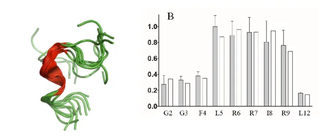
Using NMR, we characterized the structure and dynamics of dynorphin 1-13 (a shorter, fully active analogue of Dynorphin A) bound to KOP, and in the absence of G-protein (3). Using transferred NOE experiments we observed the formation of a helical conformation extending from residue L5 to R9 (Fig. 1A). 15N relaxation rate measurements provided a profile of N-H vector order parameters along the peptide sequence (Fig 1B). This is a direct measure of the conformational flexibility of dynorphin bound to KOP. It confirmed the flexibility of the last four residues, which was expected in the context of the “message-address” concept (4). We also found that the first 4 residues retained a high degree of internal motion when bound to KOP. This was much less expected since it was known that the receptor - peptide recognition is extremely dependent on the exact sequence in the “message” part of opioid peptides, YGGFL. It provided the first accurate quantification of internal dynamics of a neuropeptide in its receptor-bound state. Receptor and peptide dynamics are known to be essential parameters of GPCRs activation (5) and one should consider this residual dynamics as an essential feature of the receptor activation mechanism.
A
Figure 1: A) receptor-bound conformation of dynorphin 1-13: a helical conformation is formed between F4 and R9; B) Order parameter profile of dynorphin N-H bonds in the receptor-bound state. Grey: experimental data; White: calculated S2 profiles from molecular dynamics simulations of dynorphin – receptor complexes. Note that both the N- and C-termini remain flexible in the receptor-bound state.
Dynorphin is known to bind to KOP in its G-protein decoupled state with a 10 fold reduced affinity compared to its G-protein coupled, fully activated state. It is thus expected that both the structure and dynamics of dynorphin should be different in the activated state. Based on the recent publication of the 3D structure of KOP bound to an agonist and a nanobody (2), we could extend our NMR observations to the ternary complex dynorphin – KOP – nanobody, and we will present the results during the Biomembranes’18 conference.
Ghrelin:
Ghrelin is a lipopeptide hormone involved in phenomena such as appetite, growth hormone secretion and reward-seeking behaviors. It is the natural ligand of the growth hormone secretagogue receptor (GHSR). A strategy similar to the one presented above was applied to the Ghrelin - GHSR pair. The perdeuterated receptor reconstituted into lipid nanodiscs after expression in E. coli (6) allowed studying Ghrelin in its bound state by liquid state NMR. We performed transferred 1H NOE experiments to determine the hormone’s conformation and we measured 15N transverse relaxation to decipher the conformational flexibility along its sequence. Consistent with pharmacological data, Ghrelin amino-terminal part folds with its acyl chain to form a hydrophobic core essential for GHSR binding and activation. In contrast, the carboxy-terminal part remains flexible and may participate in the binding with the receptor through electrostatic interactions. Furthermore, we combined NMR data with molecular dynamics simulations to build an ensemble of Ghrelin-GHSR complexes and thus reproduce the order parameter profile determined experimentally.
A B
Figure 2: A) peptide sequence of the Ghrelin analogue used in the present study. The differences with the natural sequence are highlighted in red. B) Ghrelin receptor-bound conformation determined from trNOE data. C) It is characterized by a well-defined hydrophobic core formed by the acyl chain (which is essential for ghrelin’s activity) and the Phe4 and Leu5 side chains.
This approach provides the first active structure of Ghrelin and a novel insight into the molecular mechanisms responsible for its activity with respect to the activation of GHSR. Here again, the results were obtained in the absence of G-proteins, and extension of this work will involve using a ternary complex peptide – receptor – G protein (or a mimic of G proteins). Considering the pharmacological relevance of Ghrelin and its receptor, our results may lead to the design of new drugs with applications in the treatment of obesity, diabetes and addiction.
References:
1. Wu, H., D. Wacker, M. Mileni, V. Katritch, G. W. Han, E. Vardy, W. Liu, A. A. Thompson, X.-P. Huang, F. I. Carroll, S. W. Mascarella, R. B. Westkaemper, P. D. Mosier, B. L. Roth, V. Cherezov, and R. C. Stevens. 2012. Structure of the human κ-opioid receptor in complex with JDTic. Nature 485:327.
2. Che, T., S. Majumdar, S. A. Zaidi, P. Ondachi, J. D. McCorvy, S. Wang, P. D. Mosier, R. Uprety, E. Vardy, B. E. Krumm, G. W. Han, M. Y. Lee, E. Pardon, J. Steyaert, X. P. Huang, R. T. Strachan, A. R. Tribo, G. W. Pasternak, F. I. Carroll, R. C. Stevens, V. Cherezov, V. Katritch, D. Wacker, and B. L. Roth. 2018. Structure of the Nanobody-Stabilized Active State of the Kappa Opioid Receptor. Cell 172:55-+.
3. O'Connor, C., K. L. White, N. Doncescu, T. Didenko, B. L. Roth, G. Czaplicki, R. C. Stevens, K. Wuthrich, and A. Milon. 2015. NMR structure and dynamics of the agonist dynorphin peptide bound to the human kappa opioid receptor. Proc Natl Acad Sci U S A 112:11852-11857.
4. Schwyzer, R. 1986. Estimated conformation, orientation, and accumulation of dynorphin A-(1-13)-tridecapeptide on the surface of neutral lipid membranes. Biochemistry 25:4281-4286.
5. Hilger, D., M. Masureel, and B. K. Kobilka. 2018. Structure and dynamics of GPCR signaling complexes. Nat Struct Mol Biol 25:4-12.
6. Damian, M., S. Mary, M. Maingot, C. M'Kadmi, D. Gagne, J. P. Leyris, S. Denoyelle, G. Gaibelet, L. Gavara, M. Garcia de Souza Costa, D. Perahia, E. Trinquet, B. Mouillac, S. Galandrin, C. Gales, J. A. Fehrentz, N. Floquet, J. Martinez, J. Marie, and J. L. Baneres. 2015. Ghrelin receptor conformational dynamics regulate the transition from a preassembled to an active receptor:Gq complex. Proc Natl Acad Sci U S A 112:1601-1606.