HAL Id: hal-02300965
https://hal-univ-pau.archives-ouvertes.fr/hal-02300965
Submitted on 9 Dec 2020HAL is a multi-disciplinary open access archive for the deposit and dissemination of sci-entific research documents, whether they are pub-lished or not. The documents may come from teaching and research institutions in France or abroad, or from public or private research centers.
L’archive ouverte pluridisciplinaire HAL, est destinée au dépôt et à la diffusion de documents scientifiques de niveau recherche, publiés ou non, émanant des établissements d’enseignement et de recherche français ou étrangers, des laboratoires publics ou privés.
Inhibitory effect of metals on animal and plant
glutathione transferases
Dirk Dobritzsch, Konstantin Grancharov, Corinna Hermsen, Gerd-Joachim
Krauss, Dirk Schaumlöffel
To cite this version:
Dirk Dobritzsch, Konstantin Grancharov, Corinna Hermsen, Gerd-Joachim Krauss, Dirk Schaumlöffel. Inhibitory effect of metals on animal and plant glutathione transferases. Journal of Trace Elements in Medicine and Biology, Elsevier, 2020, 57, pp.48-56. �10.1016/j.jtemb.2019.09.007�. �hal-02300965�
1
Inhibitory effect of metals on animal and plant glutathione
transferases
#Dirk Dobritzsch¹,4*, Konstantin Grancharov², Corinna Hermsen¹, Gerd-Joachim Krauss¹, Dirk Schaumlöffel3
¹Martin-Luther-Universität Halle-Wittenberg, Institut für Biochemie und Biotechnologie, Abteilung 5
Ökologische und Pflanzen-Biochemie, Kurt-Mothes-Str. 3, 06120 Halle (Saale), Germany
²Institute of Molecular Biology, Dept. Biochemical Pharmacology and Drug Design, Bulgarian Academy of Sciences, 1113 Sofia, Bulgaria
3CNRS / Université de Pau et des Pays de l'Adour / E2S UPPA, Institut des Sciences Analytiques et de
Physico-Chimie pour l'Environnement et les Matériaux, UMR 5254, 64000 Pau, France 10
4Present address: Martin-Luther-Universität Halle-Wittenberg, Core Facility Proteomic Mass Spectrometry,
Proteinzentrum Charles Tanford, Kurt-Mothes-Str. 3a, 06120 Halle (Saale), Germany *corresponding author: dirk.dobritzsch@biochemtech.uni-halle.de
#Accepted manuscript :
Dobritzsch, D., Grancharov, K., Hermsen, C., Krauß, G.-J., Schaumlöffel, D. (2020) 15
Inhibitory effect of metals on animal and plant glutathione transferases.
J. Trace Elem. Med. Biol., 57, 48-56
https://doi.org/10.1016/j.jtemb.2019.09.007
Abstract
Glutathione transferases (GSTs) represent a widespread enzyme superfamily in eukaryotes and prokaryotes 20
catalyzing different reactions with endogenous and xenobiotic substrates such as organic pollutants. The latter are often found together with metal contamination in the environment. Besides performing of essential functions, GSTs protect cells by conjugation of glutathione with various reactive electrophiles. The interference of toxic metals with this functionality of GSTs may have unpredictable toxicological consequences for the organisms. In this review results from the recent literature are summarized and 25
discussed describing the ability of metals to inhibit intracellular detoxification processes in animals and plants.
Keywords
Glutathione transferase, metal, man, animal, plant
Introduction
30
Metal pollutants are one of the greatest challenges to the environment and human health. Metals enter the biosphere by natural processes (e.g. volcanism, weathering) and human activities such as mining, fuel combustion and many other industrial processes [1]. The toxicological relevance of metals depends on their chemical species, i.e. the chemical compound and the oxidation state of the metal, as well as the bioavailability of these metal species for organisms. In the environment biogeochemical transformations of 35
metal species have to be taken into account. For example in aquatic systems the inorganic mercury species Hg2+ can be transformed by microorganisms into highly toxic methylmercury (MeHg) that represents the
most environmental concern regarding this element [2]. Another example are acid mine drainage waters which are acidic effluents from abandoned mines which can contain very high amounts of arsenic. Biogeochemical processes involving microorganisms oxidize arsenite (AsO33-) to arsenate (AsO43-) together
2
with the oxidation of iron (Fe2+ to Fe3+) resulting in As-Fe coprecipitation and thus the formation of the As(V)–
Fe(III) hydroxysulfate sediments which reduce drastically the bioavailability of As [3].
Beside other toxic effects, metals can for example interfere with intracellular detoxification mechanisms of organic pollutants and thus amplify the toxicity of these compounds. Glutathione transferases (GSTs) play a key role in the detoxification of organic xenobiotics as well as endobiotic metabolites. When metals enter an 45
organism, protective mechanism against oxidative stress and other detrimental effects involve also the increase of GST activity (for example in plants see [4]). However, there is increasing evidence that metals at toxic concentrations affect GSTs either by direct inhibition of enzyme activity or indirectly by decreasing the concentration of the GST substrate reduced glutathione (GSH, Figure 1). The latter is involved in the cellular defense against metals by high chemical affinity to bind to soft metal ions, like Hg2+ and Cd2+ and/or
metal-50
induced oxidative stress by changing the GSH/GSSG equilibrium [5, 6]. As consequence, metal-induced inhibition of GSTs interferes with intracellular detoxification of organic xenobiotics or endobiotic metabolites which may lead to unpredictable toxicological consequences for organisms. This fact is of particular importance as under ecological conditions organic contaminants are often found together with metal contaminants [7, 8].
55
Our review gives a brief overview on the role of GST in organisms and discusses recent data for both in vivo and in vitro inhibitory effects on animal and plant GSTs by toxic metals.
Characteristics of glutathione transferase and its role in biological
systems
The glutathione transferase (formerly reported as glutathione S-transferases, EC 2.5.1.18) supergene family 60
is composed of several isoenzymes which are involved in phase II detoxification of electrophilic xenobiotics and endogenous compounds in procaryotes [9, 10] and eucaryotes including non-mammalian vertebrates [11], mammals and humans [12, 13] and plants [14-16]. GSTs catalyze the conjugation of organic compounds to the nucleophilic sulfur of the major intracellular thiol compound, glutathione (GSH) rendering them more hydrophilic and less toxic.
65
Recently, a graphical overview of the different classes of glutathione transferase was proposed by Kumar and Trivedi [16] (Figure 2). At least 16 genes encode GSTs in the mammalian cytosol, whereas for plants a number of 40-60 is proposed [17]. They are mostly dimeric enzymes of 45-55 kDa and include eleven classes of mammalian cytosolic GSTs: A (alpha), M (mu), P (pie), C (chie), D (delta), B( beta), E (epsilon), O (omega), S (sigma), T (theta), and Z (zeta), both latter classes are found in plants, too . To date six classes of plant 70
cytosolic GSTs have been identified Z (zeta), T (theta), F (phi), U (tau), L (lambda), and DHAR (dehydroascorbate reductase)) [17, 18]. Except for L (lambda) and DHAR (dehydroascorbate reductase), which are monomers, plant GSTs also occur as dimers. In plants phi and tau class GSTs catalyze peroxidase reactions, while zeta class GSTs catalyze isomerase reactions [19]. In each class, multiple isoenzymes were found whereas tandem duplication can be regarded as the major expansion mechanism [20].
75
In mammals, cell specific distribution of GST isoforms has been reported, as GSTP in the biliary tract and GSTA in hepatocytes [21]. A high expression of cytosolic GST P, M, A, and T classes in various tumor cells has been observed [22, 23]. Typical GST-catalyzed reactions include: i) nucleophilic aromatic substitution, ii) Michael-type addition, iii) nucleophilic addition to epoxides, iv) hydroperoxide reduction, and v) double bond isomerization[24].
80
Besides detoxification, GSTs perform other essential functions in animal and plant cells [25, 26]. GSTs have regulatory roles in cellular signaling (e.g. by mediating S-glutathionylation of specific clusters of target proteins and in reactions that define a negative regulatory role in some kinase pathways through ligand or protein:protein interactions, example for the role of GSTs´ in cellular signaling [27] and inactivate
3
endogenous products of oxidative stress (such as 13-oxooctadecadienoic acid and 4-hydroxynonenal). These 85
compounds are known to elicit cellular responses, such as apoptosis and cell proliferation. Conjugation with GSH eliminates these effects [28, 29].
GSTs also have non-catalytic functions. Originally, GSTs were identified as “ligandins” due to their ability to bind and transport across the plasma membrane organic molecules that are not enzyme substrates [30]; e.g. steroids, thyroid hormones, bile acids, bilirubin and heme [31]. This however might just be some sort of cross 90
reaction due to the chemical abilities rather than a specific, regulated function of GSTs.
Plant GSTs are active as hormone and flavonoid ligandins [26, 32]. Tau-type GSTs are able to bind GSH conjugates of fatty acids [33].
Probably, plant GSTs are involved in in vivo synthesis of sulfur-containing secondary metabolites such as volatiles and glucosinolates as well as in conjugation, transport and storage of reactive oxylipins, phenolics 95
and flavonoids [14, 15, 34]. During plant developmental processes and stress response GSTs play also an important role in the regulation of cellular detoxification processes under different light conditions [35]. Gullner et al. [36] summarize diverse roles of plant GST isoenzymes in the interaction of plants with pathogenic bacteria and fungi.
In mammalian systems, GSTs participate in cell signaling through protein-protein interactions with many 100
regulatory signaling proteins, such as c-Jun N-terminal kinase (JNK) and apoptosis signal regulating kinase ASK1 [37]. Activation of these kinases leads either to apoptosis or cellular proliferation [38]. GSTs can inhibit JNK and ASK1 activity by forming complexes [23, 39]. Thus, GSTs play a decisive cytoprotective role against carcinogenic electrophiles and products of oxidative stress. Perturbation of their function may have unpredictable toxicological consequences. Previously the concept of GST inhibition in ameliorating 105
anticancer drug resistance was reviewed [40, 41]. In recent years novel functions of GSTs have been suggested such as regulation of cell signaling pathways and ion channels [25].
Inhibition of GST by toxic metals in man, animals and plants
A principal function of the GST enzymes is the detoxification of xenobiotic substances by catalysing their conjugation with glutathione. This GST activity can be interfered by metals which may lead to detrimental 110
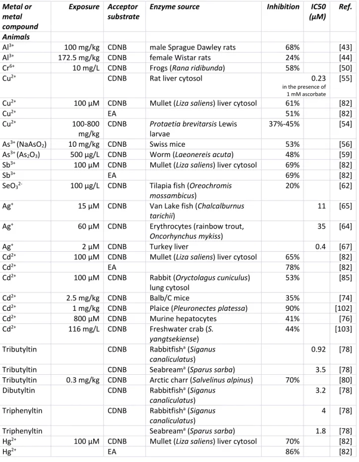
toxic effects for the organism (Figure 1). Data on in vivo and in vitro inhibitory effects of metal on GSTs are summarized in Table 1. In most of the studies the influence of toxic metals or surplus essential biometals to total GST activity was determined by measuring the absorbance (340 nm) of the conjugation of the model xenobiotic 1-chloro-2,4-dinitrobenzene (CDNB) with reduced GSH (Figure 3), usually according to a method published by Habig et al.[42]. The increase of absorbance is directly related to the GST activity. The method 115
shows a good reproducibility, when summarizing the different studies reviewed here. The overall standard deviation for GST activity determination was around 3% in most of the studies. In the following sections the metals are listed according to Periodic Table of the Elements with increasing average atomic mass from light to heavy.
GST inhibition in animals
including men
120Although most studies have been conducted with laboratory animals or fish, here we report published effects that will obviously occur in all types of animals, including humans. Although the qualitative and quantitative expression of the effects may vary greatly according to the type of organism and its physiological state, as well as the type of metals and mode of application.
125
Aluminium
Aluminium can be found ubiquitously in the environment either naturally or introduced due to human activity. This metal has no known essential biological function. However, it has the potential to cause neurotoxic effects in man and animals. For example, Al has been connected to neurodegenerative diseases
4
such as Alzheimer’s and Parkinson’s disease, as well as amyotrophic lateral sclerosis. Therefore, the few 130
studies describing Al influence on GST activity have been mainly done in brain tissue. A recent study of Singla and Dhawan [43] detected a highly significant decrease of GST activity by 68% in cerebellum and to 41% in cerebrum of male Sprague Dawley rats after oral administration of AlCl3 at a daily dose of 100 mg kg-1 for a
total duration of 8 weeks. Unfortunately, the used GST conjugate was not mentioned which limits the comparability with other studies. However, these results confirmed an earlier study of Sharma et al.[44] who 135
measured a decrease of GST activity (CDNB conjugate) by 24% in brains of female Wistar rats after oral administration of AlCl3 (172.5 mg kg-1 day-1). Even at lower doses of AlCl3 (orally 34 mg kg-1 day-1) a decrease
of GST activity (p-nitrobenzylchloride conjugate) to 19-24% was detected in plasma, liver, kidney, testes but not brain of male Sprague Dawley rats [45].
Chromium
140
CrIII was considered to be required as a trace element in metabolism [46] but results of new studies indicate
that chromium currently can only be considered pharmacologically active with beneficial effects have not been definitively shown to occur in animals [47]. Hexavalent chromium easily enters the cell where it is reduced to trivalent chromium compounds by thiols and cytochrome P450-dependent monooxygenases (P450-MO) [48]. CrVI is very toxic and mutagenic [49]. The coordinated regulation and expression of both
145
enzyme systems GST and P450-MO could result in diminished detoxification capacity of the organism, and severe toxicity of CrVI compounds. Exposure of the frog Rana ridibunda to chromium (10 ppm K
2Cr2O7, 14
days) caused decreased liver (58%) and kidney (56%) GST activities [50]. Similar decline has been also reported for P450-MO.
Copper
150
In low concentrations copper is an essential micronutrient. Copper-containing proteins play a role in energy production, signalling and proliferation. Copper deficiency or excess can lead to acute or chronic toxicity triggered by redox-dependent chelation and regulatory mechanisms. For humans, the toxicity limit is more than 10 μM, [51, 52]. Excess bioavailable copper thus acts as a genotoxic pollutant and leads to oxidative stress, DNA and protein damage. CuSO4 significantly inhibited GST activity in the gill homogenate of
155
Oncorhynchus mykiss (rainbow trout, [53] and as recently shown in the larvae of the insect Protaetia brevitarsis which play an important role for composting and rotting of fungal and plant residues [54]. In the
latter case, firstly an increase of GST activity was observed after 7 days of Cu exposure followed by a significant decrease after 28 d. Letelier et al. [55] reported that Cu2+ either alone or in the presence of
ascorbate (ROS generating system) produced dose- and time-dependent inactivation of GST in rat liver 160
cytosol. Copper significantly modified GST affinity to GSH, (Vmax ↓, Km ↑) while substrate affinity for
xenobiotics remained unchanged. The major mechanism of Cu2+-induced GST inhibition is evidenced to
oxidation of its thiol groups and not direct binding to thiol groups.
Arsenic
Arsenic is one of the most harmful metalloids in the environment. Chronic exposure to AsO33-is associated
165
with oxidative stress and increased risk of cancer (skin, bladder, and lung). Swiss mice exposed to arsenic (NaAsO2, 10 mg × kg-¹, 2 days, orally) showed accumulation in cardiac and renal tissues, accompanied with
significant reduction of antioxidant enzyme activities [56, 57]. Also, cardiac (53%) and renal (63%) GST activities were reduced. Chronic exposure of C57B16 mice to low As levels (50 ppb, NaAsO2, 4 weeks) fully
blocked GST-ω expression in the lung-lining fluid [58]. Arsenic at low concentrations (500 µg × L-¹ of As 2O3, 7
170
days) interfered with antioxidant system of the polychaete Laeonereis acuta. Thus, 48% lower conjugation capacity of GST compared to controls was observed [59].
Antimony
In the environment natural concentration of the toxic metalloid antimony is low. However, Sb may occur on higher levels at industrial sites such as mining and processing which raises concerns about human and 175
5
erythrocytes, trivalent antimony (Sb³+, antimony potassium tartrate) was found to be the most efficient
inhibitor (IC50: 50 µM), followed by Hg²+, Cu²+, Cd²+, Cr³+ and Fe²+ [61]. While Sb³+ was a competitive inhibitor
towards CDNB (Ki: 18 µM), evidenced by interaction of Sb³+ with GSH, Sb5+ failed to exert any effect on GST.
Selenium
180
Selenium is an essential trace element but at high concentrations it becomes toxic. For example, Se toxicity to aquatic organisms has been studied with respect to the GST activity by Gobi et al. [62] in gill and liver cytosols of the fish Oreochromis mossambicus native to southern Africa. In liver GST activity decreased about 20% when exposed to 100 µg/L Se in comparison to water only (control) for periods of 96 h. Interestingly, at the same Se concentration the GST activity in gill increased by approximately 30%. One conceivable 185
hypothesis is that Se supplementation leading to an increase of GPXs activities which may enable a decrease of GST activity / GSH synthesis to minimize efforts for antioxidant capacity.
Tellurium
The influence of tellurium at toxic concentrations on GST activity in animal tissue has been rarely investigated. Tellurium, a rare element of raising importance, occurs in dusts from blasts furnace refining of 190
lead. Given to mice (orally, 15 days, 2 mg × kg-¹, sodium salt), it caused dose-dependent decrease of activity
of GSH and antioxidant enzymes as well as reduced GST activity in cerebrum (37%), cerebellum (26%) and brain stem (29%) [63].
Silver
Silver levels are high in mineral rich areas and polluted soils. The monovalent silver ions reduce cell viability. 195
In all inhibitory studies referred here, Ag+ shows the highest inhibitory effect to GSTs. Using purified rainbow
trout erythrocytes the in vitro effects of various metal ions on GST activities were measured [64]. Inhibition constant (Ki values) and IC50 values (concentration of inhibitor necessary to halve the reaction rate of an enzyme-catalyzed reaction) demonstrate that Ag+ show the highest inhibitory effect. But, Cd2+ and Cr2+
inhibited the GST strongly, too (Tab. 1). GST in muscle tissue from Van Lake fish (Chalcalburnus tarichii) was 200
inhibited mostly strong in a non-competitive manner by Ag+, but also by other metals (Tab.1). Cd2+, Co2+ and
Fe2+ caused no inhibitory effect [65]. Similar results are shown for Van Lake fish gills [66]. Highly purified GST
from turkey liver is inhibited in the following order: Ag+ > Cu2+ > Hg2+ [67].
Within the last decades the use of nanomaterials in various products has increased. Among different nanomaterials, silver nanoparticles (AgNPs) are extensively used in many commercial products [68]. Thus, 205
AgNPs will reach the environment and cause effects at various levels in aquatic organisms [69] or soil organisms [70]. The induction of GSTs mRNA triggered by Cd and AgNPs seems to show that GST are important for defense in aquatic organisms [71]. However, change of GST activity was not significant with increasing concentration of AgNPs in Folsomia candida (Collembola) but effects of AgNPs is similar to the one from AgNO3 [70].
210
Cadmium
Cadmium, one of the most hazardous pollutants, can be accumulated in aquatic organisms. The gills of fishes are major targets for acute metal toxicity in these organisms due to their permanent contact with water contaminants. Long term (2 weeks) low-level (2 µg × L-¹) exposure to Cd²+ led to a distinct decrease of GST
activity in gills (> 80%) , but also in digestive glands (> 60%) of the freshwater crayfish Astacus astacus [72]. 215
In Cd2+ exposed freshwater crab (Sinopotamon yangtsekiense) GSH and antioxidant enzymes activities were
gradually reduced in gills with increasing concentrations (7-116 mg × L-¹) and duration [73]. GST activity was
decreased by 44% after exposure to 116 mg× L-¹ Cd2+ for 96 h.
In mice liver GST showed a cadmium (up to 2.5 mg × L-¹ CdCl
2) induced, duration (up to 7 alternate days)
-dependent depression of conjugating capacity (35%) [74]. This was correlated with elevated hepatic lipid 220
peroxidation (102%) and chromosomal aberrations (30%). CdCl2 (4 mg × kg-¹) also caused organ specific
6
accumulation of Cd in these tissues increased lipid peroxidation products and protein carbonyl contents, as well as decreased activities of other antioxidant enzymes. Murine hepatocytes, treated with 800 µM CdCl2
for 3 h showed reduced GST activity associated with significant reduction of cell viability, increased levels of 225
lipid peroxidation products and oxidized glutathione [76].
Tin
Organotin compounds such as tributyltin (TBT), triphenyltin (TPT) and dibutyltin (DBT), are extensively used as plastic stabilizers, wood preservatives and antifouling agents in paints. Due to their widespread use, high amounts of butyltins have polluted the environment causing harmful toxic effects. Fishes are suitable 230
organisms to evaluate the effect of such contaminants in aquatic ecosystems [77]. Organotins strongly inhibit GST activities in marine fish liver and kidney cytosol showing a 10 (liver) to 100 (kidney) times higher inhibition compared to inorganic tin [78]. This highlights the fact that inhibition depends on the chemical species. IC50 values for hepatic GST ranged between 7.4-18 µM for Siganus canaliculatus (Rabbitfish), and
between 17-45 µM for Sparus sarba (Arabian seabream). The order of inhibitory potency was TBT > TPT > 235
DBT; reversible inhibition was observed in all cases. Several times lower IC50 values have been recorded for
kidney GST (0.92-4 µM) indicating significantly higher sensitivity of renal isoenzymes to organotins. The results were consistent with previous in vitro competitive and reversible inhibition of rat liver GSTs by TBT and TPT [79]. Organotin compounds are a special case when studying GST inhibition as they represent metals and organic xenobiotics in the same time. All studies proposed an inhibitory effect via a direct binding of the 240
organotins to the active site of GST. Recent in vivo studies on TBT towards hepatic GST in Arctic charr (Salvelinus alpinus, 0.3 mg × kg-¹, at long-term exposure) confirmed the potent inhibitory effect but discussed
also the possibility that organotins may compete with the detoxification of other organic xenobiotics for binding to GST and GSH [80].
Mercury
245
Mercury is present in the environment as several different chemical species showing a range of toxicological properties. These chemical forms can be classified as elemental (Hg0), inorganic (e.g. Hg2+), and organic
mercury, mainly alkyl mercury such as methyl mercury (MeHg). Goodrich and Basu [81] proved the influence of mercury among many other metals to genetically engineered GST allozymes. All allozymes were inhibited strongly by Hg2+ (but also MeHg and selenium) (Tab.1). In a comprehensive in vitro study Sen and Semiz [82]
250
investigated the effects of mercury and other toxic metals on cytosolic liver GST activities in the fish leaping mullet (Liza saliens). Both CDNB- and EA-GST were mainly inhibited by Hg²+ (CDNB-GST 70% and EA-GST 86%,
resp.), but also by Sb²+ (69% and 78%), and Cd²+ (65% and 69%). Other tested metals Cu²+, Zn²+, Co²+, Fe3+,
and Fe2+ exhibited more moderate effects (41-61%). The fail of fully reactivation by GSH pointed to inhibition
mechanisms other than simple binding to SH-groups by toxic metals. In a recent study of Gunderson et al. 255
[83] in the signal crayfish Pacifastacus leniusculus a similar decrease of the GST activity in hepatopancreas cytosol was observed under Hg2+ exposition at concentrations comparable to previous studies.
Lead
A continuous, low-level exposure of the freshwater crayfish Astacus astacus to lead (20 µg × L-¹) resulted in
strong decrease of GST activity in gills and digestive gland [72]. Accordingly, increased metal accumulation 260
and severe structural impairment in these organs were observed. Thus, a chronic low-level metal stress may have a biological impact as great as short-term acute dose insults in aquatic ecosystems. Long term low-level lead exposure (0.1% in drinking water) of rats has been reported to profoundly impair phase I and phase II metabolising enzyme activity in the lenticular system of the animals. A very pronounced decrease (up to 55% inhibition) was noticed in GST activity [84].
265
Cd2+ and Pb2+ have been shown to be potent inhibitors (by more than 50% at 100 µM) of pulmonary GST in
rabbit lung cytosol [85]. Since GST and other detoxifying enzymes were found to be extremely poor in lung,
7
GST inhibition in plants
Trace elements such as Cu, Zn, Mn, Fe, Mo and Ni are essential in small amounts for plant metabolism while 270
higher concentrations can cause severe toxic effects [86]. Only few studies investigated the inhibition of GST activity in plants by metals such as Cu, Zn, Cd, As, Pb, and Ni. Recently, Kumar and Trivedi [16] gave an overview regarding the role of GST for arsenic detoxification in plants.
Copper
Due to its redox activity (Cu+, Cu²+) copper is of high significance for plants (respiratory chain: cytochrome
275
oxidase, photosynthesis: plastocyanine). On the other hand, copper induce reactive oxygen species (ROS) by autoxidation and Fenton [87]. Ahsam et al. [88] did a general study of Cu²+ influence in rice (Oryza sativa)
germination. During proteomic research in the first 4 days of germination, GST (identified by peptide-mass fingerprinting) showed a slightly decrease upon CuSO4 exposure (1.5 mM, 4 d). Unfortunately the authors
did not assay enzyme activities. However, in a recent study Li et al. [89] measured the GST activity in roots of 280
two varieties of Oryza sativa seedlings. In the Cu sensitive variety GST activity decreased by about 52% after 3 days Cu exposure (8 µM) while the Cu tolerant variety showed only a GST activity decrease of 30%. In duckweed (Lemna minor) GST activity was inhibited time- and concentration-dependently by CuSO4 [90]. For
example, low Cu exposure (0.25 μM to 0.5 μM) lad to a stimulation (114.4% to 125%) of GST as a consequence of CuSO4 addition. While higher Cu content (2 μM) reduced GST activity (60% of the control level).
285
Zinc
Zinc is typically the second (after iron) most abundant transition metal in plants [91]. In poplar lines different effects of ZnSO4 for GST activity were measured [92]. Leaf disks of 2 hybrids (Populus nigra (N-SL clone), P. canescens (P. termula P. alba)) and 2 transgenic (P. canescens) clones, overexpressing E. coli γ-ECS (11ggs:
cytosol, 6LgI: chloroplast)) were exposed to Zn (10-5-10-1 M, 21 days). In clone 11ggs GST activity was
290
consistently lowered by 10-5-10-1 M ZnSO
4. In both hybrids GSTs were inhibited at high (cytotoxic) Zn2+
concentrations (10-2 and 10-1 M).
Cadmium, arsenic, lead and nickel
Cadmium which chemically is similar to zinc, generally causes toxic effects in plant species. Interest in Cd tolerance mechanisms [93, 94] is often driven by potential applications in phytoremediation [95, 96]. While 295
GST activities in 25 day old hydroponic cultures of Indian mustard (Brassica juncea L. cv. Vitasso) after Cd2+
(5 days, 10 µM) which is environmentally relevant, were not influenced, higher metal concentrations decreased GST activity significantly to 61.3% (30 µM), 82.4% (50 µM) and 66.4% (100 µM) [97]. Interestingly no continuous decrease was observed. A 10 days exposure of Cd (40 mg L-1) to rice (Oryza sativa) showed a significant inhibition of GST activity by 63% in rice roots in contrast to a minimal increase in shoots [98]. 300
Typha latifolia is some of the potent wetland plants for phytoremediation. It is used in constructed wetlands,
an alternative to traditional industrial wastewater treatment [99]. After 72h exposures of Typha latifolia either to Pb, arsenite, or Cd showed remarkable GST inhibitions in leaves [95].
Arsenic and lead are taken up by plants from the soil, water or air. Spruce cell culture (Picea abies L.Karst.) were exposed for 16 h to 50-500 µM CdSO4, 1.5-80 µM Na2HAsO4 and 50-400 µM PbCl2 [100]. To distinguish
305
between different GST isoforms 4 different standard substrates for activity assay were used. Only assays with 1,2-epoxy-3-(p-nitrophenoxy)-propane (EPNP) as substrate showed remarkable inhibition effects (60%) caused by cadmium. Under lead exposure the same effect was observed with a slightly higher residual enzyme activity (56%). Arsenic inhibited EPNP conjugation continuous with higher concentration. GST was assayed in horseradish (Armoracia rusticana L.) hairy root culture, incubated for 6 h with 0.1 and 0.5 mM 310
Cd2+, Ni2+ and Pb2+ using 1-chloro-2,4-dinitrobenzene (CDNB) and 1,2-dichloro-4-nitrobenzene (DCNB) in
order to detect different GST isoforms [101]. While GST activity with CDNB was not influenced, assays with DCNB showed decreasing activity levels in a metal dependent manner in the range of 35% (for Ni2+) to 63%
8
Conclusions
315
Metals represent a major ecological concern because they can accumulate in organisms in the food web and finally, they can be a threat to human health. Impact of metals to aquatic and terrestrial ecosystems at sub-lethal contaminant concentrations can cause biological responses of the glutathione antioxidative network including glutathione transferases in organisms living in polluted habitats. On cellular level, metals are able to inhibit different intracellular processes such as enzyme activities. Inhibition of GSTs in animals and plants 320
represents an early warning biomarker indicating a sensitive physiological response of organism to metals. In this context the measurement of GST activity is an important tool. In most studies, metals decreased GST activity supporting the fact that GST is part of the cellular antioxidant defense system where it is serving as bifunctional enzyme: GST and as glutathione peroxidase, too. Furthermore, animal and plant glutathione transferases are involved in detoxification of xenobiotics and endobiotic metabolites. Therefore, 325
interferences with the functionality of GSTs caused by toxic metals or imbalanced essential biometals may have unpredictable toxicological consequences for the organisms. However, it is difficult to make a general assessment of the level at which natural exposure, dietary supplementation, over-nutritional dietary supplementation or poisoning will show effects.
When reviewing the literature on animal studies it can be concluded that the majority of articles are dealing 330
with organisms living in aquatic ecosystems. Here, fishes are of particular interest because in contaminated aquatic environments they are in direct contact with toxic substances in contrast to terrestrial animals which have a rather selective contact with toxic substances in a contaminated site. Fish gills are organs and tissue which are primarily affected because they provide a large surface area in contact with contaminants dissolved in water. Gill cells respond rapidly to metals and thus gills are major targets for measurement of acute toxicity 335
via GST activity. Moreover, some fish species such as signal crayfish are living in relatively small habitats; therefore they can serve as good bioindicators for specific exposed areas.
Rather few studies have investigated the impact of metals to GST activity in plants although plants as sessile organisms represent suitable bioindicators for specific contaminated sites as well as for long-term observation of pollution. With respect to metal polluted aquatic ecosystems only one plant study has been 340
published so far. It investigated the aquatic macrophyte Typha latifolia which represents a common wetland plant used in some cases for phytoremediation in constructed wetlands for sustainable waste water treatment. This plant can accumulate metals, however, after long-term exposure a decrease of GST activity was observed. This particular case demonstrates that there is a lack of studies in this field. In future, more investigations on GST activity in plants exposed to toxic metal in aquatic as well as in terrestrial ecosystems 345
would be desirable in order to fill this gap of knowledge.
Acknowledgements
This work was supported by the bilateral (German-French) DAAD - Campus France program PROCOPE funded by the German ‘Bundesministerium für Bildung und Forschung (BMBF)’, the French ‘Ministère des Affaires étrangères et du développement International (MAEDI)’ and ‘Ministère de l’Education nationale, de 350
l’Enseignement supérieur et de la Recherche (MENESR)’.
Conflict of interest
9
Figure legends
Figure 1: General scheme of the glutathione transferase (GST) catalysed detoxification of organic xenobiotics 355
and endobiotic metabolites and its impairment by metals at toxic concentration, either by direct inhibition of enzyme activity* or indirectly by decreasing the concentration of the GST substrate glutathione (GSH) via changing the GSH/GSSG equilibrium**.
Figure 2: A graphical overview of the different classes of glutathione transferase (modified from Kumar and 360
Trivedi [16], Copyright © 2018 Kumar and Trivedi, doi: 10.3389/fpls.2018.00751).
Figure 3: Measurement of the GST activity and its inhibition by metals by using the model xenobiotic CDNB (1-chloro-2,4-dinitrobenzene). GST catalyses the reaction of reduced GSH with CDNB via a nucleophilic aromatic substitution, the absorbance of the resulting GS-conjugate measured at 340 nm indicates the GST 365
10
Figures
370
11 Figure 2 375
12 Figure 3
13
Table 1. In vitro and in vivo inhibitory effects of metals on cytosolic GST* activity 380 Metal or metal compound Exposure Acceptor substrate
Enzyme source Inhibition IC50
(μM)
Ref.
Animals
Al3+ 100 mg/kg CDNB male Sprague Dawley rats 68% [43]
Al3+ 172.5 mg/kg CDNB female Wistar rats 24% [44]
Cr6+ 10 mg/L CDNB Frogs (Rana ridibunda) 58% [50]
Cu2+ CDNB Rat liver cytosol 0.23
in the presence of 1 mM ascorbate
[55] Cu2+ 100 µM CDNB Mullet (Liza saliens) liver cytosol 61% [82]
Cu2+ EA 51% [82]
Cu2+ 100-800
mg/kg
CDNB Protaetia brevitarsis Lewis
larvae
37%-45% [54]
As3+ (NaAsO
2) 10 mg/kg CDNB Swiss mice 53% [56]
As3+ (As
2O3) 500 μg/L CDNB Worm (Laeonereis acuta) 48% [59]
Sb3+ 100 µM CDNB Mullet (Liza saliens) liver cytosol 69% [82]
Sb3+ EA 69% [82]
SeO32- 100 μg/L CDNB Tilapia fish (Oreochromis mossambicus)
20% [62]
Ag+ 15 µM CDNB Van Lake fish (Chalcalburnus tarichii)
11 [65] Ag+ 60 µM CDNB Erythrocytes (rainbow trout,
Oncorhynchus mykiss)
35 [64]
Ag+ 2 µM CDNB Turkey liver 0.4 [67]
Cd2+ 100 µM CDNB Mullet (Liza saliens) liver cytosol 65% [82]
Cd2+ EA 78% [82]
Cd2+ 100 µM CDNB Rabbit (Oryctolagus cuniculus)
lung cytosol
53% [85]
Cd2+ 2.5 mg/kg CDNB Balb/C mice 35% [74]
Cd2+ 1 mg/kg CDNB Plaice (Pleuronectes platessa) 90% [102]
Cd2+ 800 µM CDNB Murine hepatocytes 41% [76]
Cd2+ 116 mg/L CDNB Freshwater crab (S. yangtsekiense)
44% [103] Tributyltin CDNB Rabbitfisha (Siganus
canaliculatus)
0.92 [78]
Tributyltin CDNB Seabreama (Sparus sarba) 3.5 [78]
Tributyltin 0.3 mg/kg CDNB Arctic charr (Salvelinus alpinus) 70% [80] Dibutyltin CDNB Rabbitfisha (Siganus
canaliculatus)
3.2 [78] Triphenyltin CDNB Rabbitfisha (Siganus
canaliculatus)
4 [78]
Triphenyltin Seabreama (Sparus sarba) 1.8 [78]
Hg2+ 100 µM CDNB Mullet (Liza saliens) liver cytosol 70% [82]
14 Metal or metal compound Exposure Acceptor substrate
Enzyme source Inhibition IC50
(μM)
Ref.
Hg2+ 0.9 µg/kg CDNB Signal crayfish (Pacifastacus leniusculus) hepatopancreas
cytosol
70% [83]
Pb2+ 20 µg/L CDNB Freshwater crayfish (Astacus astacus) 77% in gills 44% in digestive gland [72]
Pb2+ 100 µM CDNB Rabbit (Japanese White) lung
cytosol
74% [85]
Pb2+ 0.1%b CDNB Sprague-Dawley rats 55% [84]
Plants
Cu2+ 1.5 mM Rice (Oryza sativa) seedlings [88]
Cu2+ 8 µM CDNB Rice (Oryza sativa) seedlings,
roots, Cu sensitive variety
~52% [89]
Cu2+ 5 µM CDNB Duckweed (Lemna minor) ~40% [90]
Zn2+ 100 mM CDNB Poplar (Populus) [92]
Cd2+ 30 µM CDNB Indian mustard (Brassica juncea)
40% [97]
Cd2+ 50 µM EPNP Spruce (Picea) 60% [100]
Cd2+ 100 µM DCNB Horseradish (Armoracia rusticana)
55% [101]
Cd2+ 50 µM CDNB Typha latifolia 50% [95]
AsO42- 80 µM EPNP Spruce (Picea) 67% [100]
AsO42- 50 µM CDNB Typha latifolia 80% [95]
Pb2+ 100 µM DCNB Horseradish (Armoracia rusticana)
42% [101]
Pb2+ 50 µM EPNP Spruce (Picea) 45% [100]
Pb2+ 50 µM CDNB Typha latifolia 80% [95]
Ni2+ 500 µM DCNB Horseradish (Armoracia rusticana)
15
*Membrane-associated glutathione S-transferases not involved in intracellular detoxification processes were
not considered here. These GSTs are more likely to be involved in special developmental processes (e.g. heat shock in stationary phase and ability to grow at 39 °C, [104]. Membrane bound microsomal GSTs are involved in the endogenous metabolism of leukotrienes and prostaglandins [105].
385 aPMS of kidney homogenate EPNP = 1,2-epox-y-3-(p-nitrophenoxy)-propane DCNB = 1,2-dichloro-4-nitrobenzene CDNB = 1-chloro-2,4-dinitrobenzene EA = ethacrynic acid 390
16
References
[1]
R.G. Garrett, Natural sources of metals to the environment, Hum. Ecol. Risk Assess. 6 (2000)
945-963.
[2]
J.M. Benoit, C.C. Gilmour, A. Heyes, R.P. Mason, C.L. Miller, Geochemical and biological
395controls over methylmercury production and degradation in aquatic ecosystems, in
Biogeochemistry of Environmentally Important Trace Elements, ed. by Y. Cai and O.C.
Braids, Washington: Amer Chemical Soc, (2003), pp. 262-297.
[3]
O. Bruneel, A. Volant, S. Gallien, B. Chaumande, C. Casiot, C. Carapito, A. Bardil, G. Morin,
G.E. Brown, C.J. Personne, D. Le Paslier, C. Schaeffer, A. Van Dorsselaer, P.N. Bertin, F.
400Elbaz-Poulichet, F. Arsene-Ploetze, Characterization of the Active Bacterial Community
Involved in Natural Attenuation Processes in Arsenic-Rich Creek Sediments, Microb. Ecol.
61 (2011) 793-810.
[4]
N.A. Anjum, I. Ahmad, I. Mohmood, M. Pacheco, A.C. Duarte, E. Pereira, S. Umar, A.
Ahmad, N.A. Khan, M. Iqbal, M.N.V. Prasad, Modulation of glutathione and its related
405enzymes in plants' responses to toxic metals and metalloids-A review, Environ. Exp. Bot. 75
(2012) 307-324.
[5]
E.V. Kalinina, N.N. Chernov, M.D. Novichkova, Role of Glutathione, Glutathione
Transferase, and Glutaredoxin in Regulation of Redox-Dependent Processes, Biochemistry
(Moscow) 79 (2014) 1562-1583.
410
[6]
F.M. Rubino, Toxicity of Glutathione-Binding Metals: A Review of Targets and Mechanisms,
Toxics 3 (2015) 20-62.
[7]
G.J. Krauss, D. Schlosser, Sensing of Pollutant Effects and Bioremediation, in Ecological
Biochemistry: Environmental and Interspecies Interactions, ed. by G.J. Krauss and D.H.
Niess, Weinheim: Wiley-VCH Verlag GmbH & Co. KGaA, (2015), pp. 333-340.
415
[8]
S. Kurwadkar, Occurrence and distribution of organic and inorganic pollutants in
groundwater, Water Environment Research. doi:10.1002/wer.1166 (2019).
[9]
N. Allocati, L. Federici, M. Masulli, C. Di Ilio, Glutathione transferases in bacteria, Febs J.
276 (2009) 58-75.
[10]
N. Allocati, L. Federici, M. Masulli, C. Di Ilio, Distribution of glutathione transferases in
420Gram-positive bacteria and Archaea, Biochimie 94 (2012) 588-596.
[11]
D. Sheehan, G. Meade, V.M. Foley, C.A. Dowd, Structure, function and evolution of
glutathione transferases: implications for classification of non-mammalian members of an
ancient enzyme superfamily, Biochem. J. 360 (2001) 1-16.
[12]
D. Dourado, P.A. Fernandes, M.J. Ramos, Mammalian cytosolic glutathione transferases,
425Curr. Protein Pept. Sci. 9 (2008) 325-337.
[13]
P. Jancova, P. Anzenbacher, E. Anzenbacherova, Phase II drug metabolizing enzymes,
Biomed. Pap-Olomouc 154 (2010) 103-116.
[14]
D.P. Dixon, M. Skipsey, R. Edwards, Roles for glutathione transferases in plant secondary
metabolism, Phytochemistry 71 (2010) 338-350.
430
[15]
I. Cummins, D.P. Dixon, S. Freitag-Pohl, M. Skipsey, R. Edwards, Multiple roles for plant
glutathione transferases in xenobiotic detoxification, Drug Metab. Rev. 43 (2011) 266-280.
[16]
S. Kumar, P.K. Trivedi, Glutathione S-Transferases: Role in Combating Abiotic Stresses
Including Arsenic Detoxification in Plants, Front. Plant Sci. 9 (2018).
[17]
C. Frova, Glutathione transferases in the genomics era: New insights and perspectives,
435Biomol. Eng. 23 (2006) 149-169.
[18]
G.E. Edwards, V.R. Franceschi, M.S.B. Ku, E.V. Voznesenskaya, V.I. Pyankov, C.S. Andreo,
Compartmentation of photosynthesis in cells and tissues of C-4 plants, J. Exp. Bot. 52 (2001)
577-590.
[19]
D.P. Dixon, A. Lapthorn, R. Edwards, Plant glutathione transferases, Genome Biol. 3 (2002)
44017
[20]
Y.H. Chi, Y. Cheng, J. Vanitha, N. Kumar, R. Ramamoorthy, S. Ramachandran, S.Y. Jiang,
Expansion Mechanisms and Functional Divergence of the Glutathione S-Transferase Family
in Sorghum and Other Higher Plants, DNA Res. 18 (2011) 1-16.
[21]
J.D. Hayes, L.I. McLellan, P.K. Stockman, J. Chalmers, G.J. Beckett, Glutathione
S-445transferases in man: the relationship between rat and human enzymes, Biochem. Soc. Trans.
15 (1987) 721-725.
[22]
K.D. Tew, Glutathione-associated Enzymes in Anticancer Drug Resistance, Cancer Res. 54
(1994) 4313-4320.
[23]
D.M. Townsend, K.D. Tew, The role of glutathione-S-transferase in anti-cancer drug
450resistance, Oncogene 22 (2003) 7369-7375.
[24]
A.S. Johansson, B. Mannervik, Human glutathione transferase A3-3, a highly efficient
catalyst of double-bond isomerization in the biosynthetic pathway of steroid hormones, J.
Biol. Chem. 276 (2001) 33061-33065.
[25]
P.G. Board, D. Menon, Glutathione transferases, regulators of cellular metabolism and
455physiology, Biochim. Biophys. Acta-Gen. Subj. 1830 (2013) 3267-3288.
[26]
I. Nianiou-Obeidat, P. Madesis, C. Kissoudis, G. Voulgari, E. Chronopoulou, A. Tsaftaris,
N.E. Labrou, Plant glutathione transferase-mediated stress tolerance: functions and
biotechnological applications, Plant Cell Reports 36 (2017) 791-805.
[27]
K.D. Tew, Y. Manevich, C. Grek, Y. Xiong, J. Uys, D.M. Townsend, The role of glutathione
460S-transferase P in signaling pathways and S-glutathionylation in cancer, Free Radic. Biol.
Med. 51 (2011) 299-313.
[28]
I. Hubatsch, M. Ridderstrom, B. Mannervik, Human glutathione transferase A4-4: an Alpha
class enzyme with high catalytic efficiency in the conjugation of 4-hydroxynonenal and other
genotoxic products of lipid peroxidation, Biochem. J. 330 (1998) 175-179.
465
[29]
R. Sharma, D. Brown, S. Awasthi, Y.S. Yang, A. Sharma, B. Patrick, M.K. Saini, S.P. Singh,
P. Zimniak, S.V. Singh, Y.C. Awasthi, Transfection with 4-hydroxynonenal-metabolizing
glutathione S-transferase isozymes leads to phenotypic transformation and immortalization of
adherent cells, Eur. J. Biochem. 271 (2004) 1690-1701.
[30]
E. Tipping, B. Ketterer, P. Koskelo, Binding of porphyrins by ligandin, Biochem. J. 169
470(1978) 509-516.
[31]
U.H. Danielson, B. Mannervik, Kinetic independence of the subunits of cytosolic glutathione
transferase from the rat, Biochem. J. 231 (1985) 263-267.
[32]
L.A. Mueller, C.D. Goodman, R.A. Silady, V. Walbot, AN9, a petunia glutathione
S-transferase required for anthocyanin sequestration, is a flavonoid-binding protein, Plant
475Physiol. 123 (2000) 1561-1570.
[33]
D.P. Dixon, R. Edwards, Selective Binding of Glutathione Conjugates of Fatty Acid
Derivatives by Plant Glutathione Transferases, J. Biol. Chem. 284 (2009) 21249-21256.
[34]
M. Hasanuzzaman, K. Nahar, T.I. Anee, M. Fujita, Glutathione in plants: biosynthesis and
physiological role in environmental stress tolerance, Physiol. Mol. Biol. Plants 23 (2017)
249-480268.
[35]
A. Galle, Z. Czekus, K. Bela, E. Horvath, A. Ordog, J. Csiszar, P. Poor, Plant Glutathione
Transferases and Light, Front. Plant Sci. 9 (2019).
[36]
G. Gullner, T. Komives, L. Kiraly, P. Schroder, Glutathione S-Transferase Enzymes in
Plant-Pathogen Interactions, Front. Plant Sci. 9 (2018).
485
[37]
M. Edalat, M.A.A. Persson, B. Mannervik, Selective recognition of peptide sequences by
glutathione transferases: A possible mechanism for modulation of cellular stress-induced
signaling pathways, Biol. Chem. 384 (2003) 645-651.
[38]
Y.M. Liu, W. Min, Thioredoxin promotes ASK1 ubiquitination and degradation to inhibit
ASK1-mediated apoptosis in a redox activity-independent manner, Circ.Res. 90 (2002)
1259-49018
[39]
R. Monaco, F.K. Friedman, M.J. Hyde, J.M. Chen, S. Manolatus, V. Adler, Z. Ronai, W.
Koslosky, M.R. Pincus, Identification of a glutathione-S-transferase effector domain for
inhibition of jun kinase, by molecular dynamics, J. Protein Chem. 18 (1999) 859-866.
[40]
P. Ruzza, A. Rosato, C.R. Rossi, M. Floreani, L. Quintieri, Glutathione Transferases as
495Targets for Cancer Therapy, Anti-Cancer Agents Med. Chem. 9 (2009) 763-777.
[41]
A. Sau, F.P. Tregno, F. Valentino, G. Federici, A.M. Caccuri, Glutathione transferases and
development of new principles to overcome drug resistance, Arch. Biochem. Biophys. 500
(2010) 116-122.
[42]
W.H. Habig, M.J. Pabst, W.B. Jakoby, Glutathione S-Transferases - First Enzymatic Step in
500Mercapturic Acid Formation, J. Biol. Chem. 249 (1974) 7130-7139.
[43]
N. Singla, D.K. Dhawan, Zinc modulates aluminium-induced oxidative stress and cellular
injury in rat brain, Metallomics 6 (2014) 1941-1950.
[44]
P. Sharma, Z.A. Shah, A. Kumar, F. Islam, K.P. Mishra, Role of combined administration of
Tiron and glutathione against aluminum-induced oxidative stress in rat brain, J. Trace Elem.
505Med. Biol. 21 (2007) 63-70.
[45]
F.M. El-Demerdash, Antioxidant effect of vitamin E and selenium on lipid peroxidation,
enzyme activities and biochemical parameters in rats exposed to aluminium, J. Trace Elem.
Med. Biol. 18 (2004) 113-121.
[46]
W. Mertz, Chromium in human nutrition - a review, J. Nutr. 123 (1993) 626-633.
510[47]
J.B. Vincent, New Evidence against Chromium as an Essential Trace Element, The Journal
of Nutrition 147 (2017) 2212-2219.
[48]
L. Gerhardsson, S. Skerfving, Concepts on biological markers and biomonitoring for metal
toxicity, in Toxicology of Metals, ed. by L. Chang, New York: Lewis Publishers, (1966),
pp. 81-107.
515
[49]
Toxicological Profile: Chromium, The Agency for Toxic Substances and Disease Registry
(ATSDR), (2012).
[50]
I. Kostaropoulos, D. Kalmanti, B. Theodoropoulou, N. Loumbourdis, Effects of exposure to
a mixture of cadmium and chromium on detoxification enzyme (GST, P450-MO) activities in
the frog Rana ridibunda, Ecotoxicology 14 (2005) 439-447.
520
[51]
P.G. Georgopoulos, A. Roy, M.J. Yonone-Lioy, R.E. Opiekun, P.J. Lioy, Environmental
copper: Its dynamics and human exposure issues, J. Toxicol. Env. Health, Part B. 4 (2001)
341-394.
[52]
National_Research_Council, Copper in Drinking Water (2000), The National Academies
Press, Washington, DC, https://doi.org/10.17226/19782.
525
[53]
M. Arabi, M.A. Alaeddini, Metal-ion-mediated oxidative stress in the gill homogenate of
rainbow trout (Oncorhynchus mykiss) - Antioxidant potential of manganese, selenium, and
albumin, Biol. Trace Elem. Res. 108 (2005) 155-168.
[54]
S.Z. Yin, G.D. Li, M.M. Liu, C.L. Wen, Y.Y. Zhao, Biochemical responses of the Protaetia
brevitarsis Lewis larvae to subchronic copper exposure, Environ. Sci. Pollut. Res. 25 (2018)
53018570-18578.
[55]
M.E. Letelier, M. Martinez, V. Gonzalez-Lira, M. Faundez, P. Aracena-Parks, Inhibition of
cytosolic glutathione S-transferase activity from rat liver by copper, Chem.-Biol. Interact. 164
(2006) 39-48.
[56]
P. Manna, M. Sinha, P.C. Sil, Arsenic-induced oxidative myocardial injury: protective role of
535arjunolic acid, Arch. Toxicol. 82 (2008) 137-149.
[57]
M. Sinha, P. Manna, P.C. Sil, Arjunolic acid attenuates arsenic-induced nephrotoxicity,
Pathophysiology 15 (2008) 147-156.
[58]
R.C. Lantz, B.J. Lynch, S. Boitano, G.S. Poplin, S. Littau, G. Tsaprailis, J.L. Burgess,
Pulmonary biomarkers based on alterations in protein expression after exposure to arsenic,
54019
[59]
J. Ventura-Lima, J.Z. Sandrini, M.F. Cravo, F.R. Piedras, T.B. Moraes, D. Fattorini, A. Notti,
F. Regoli, L.A. Geracitano, L.F.F. Marins, J.M. Monserrat, Toxicological responses in
Laeonereis acuta (annelida, polychaeta) after arsenic exposure, Environ. Int. 33 (2007)
559-564.
545
[60]
M.J. Tamas, Cellular and molecular mechanisms of antimony transport, toxicity and
resistance, Environ. Chem. 13 (2016) 955-962.
[61]
R. Poon, I. Chu, Effects of trivalent antimony on human erythrocyte
glutathione-S-transferases, J. Biochem. Mol. Toxicol. 14 (2000) 169-176.
[62]
N. Gobi, B. Vaseeharan, R. Rekha, S. Vijayakumar, C. Faggio, Bioaccumulation, cytotoxicity
550and oxidative stress of the acute exposure selenium in Oreochromis mossambicus, Ecotox.
Environ. Safe. 162 (2018) 147-159.
[63]
P. Kaur, S. Yousuf, M.A. Ansari, A. Siddiqui, A.S. Ahmad, F. Islam, Tellurium-induced
dose-dependent impairment of antioxidant status - Differential effects in cerebrum, cerebellum, and
brainstem of mice, Biol. Trace Elem. Res. 94 (2003) 247-258.
555
[64]
V. Comakli, M. Ciftci, O.I. Kufrevioglu, Effects of some metal ions on rainbow trout
erythrocytes glutathione S-transferase enzyme: an in vitro study, J. Enzym. Inhib. Med. Chem.
28 (2013) 1261-1266.
[65]
M. Aksoy, M.S. Ozaslan, O.I. Kufrevioglu, Purification of glutathione S-transferase from Van
Lake fish (Chalcalburnus tarichii Pallas) muscle and investigation of some metal ions effect
560on enzyme activity, J. Enzym. Inhib. Med. Chem. 31 (2016) 546-550.
[66]
M.S. Ozaslan, Y. Demir, O.I. Kufrevioglu, M. Ciftci, Some metals inhibit the glutathione
S-transferase from Van Lake fish gills, J. Biochem. Mol. Toxicol. 31 (2017).
[67]
E. Akkemik, P. Taser, A. Bayindir, H. Budak, M. Ciftci, Purification and characterization of
glutathione S-transferase from turkey liver and inhibition effects of some metal ions on
565enzyme activity, Environ. Toxicol. Pharmacol. 34 (2012) 888-894.
[68]
A.D. Maynard, R.J. Aitken, T. Butz, V. Colvin, K. Donaldson, G. Oberdörster, M.A. Philbert,
J. Ryan, A. Seaton, V. Stone, S.S. Tinkle, L. Tran, N.J. Walker, D.B. Warheit, Safe handling
of nanotechnology, Nature 444 (2006) 267-269.
[69]
P.V. Asharani, Y.L. Wu, Z.Y. Gong, S. Valiyaveettil, Toxicity of silver nanoparticles in
570zebrafish models, Nanotechnology 19 (2008) 255102.
[70]
P. Sillapawattana, M.C.H. Gruhlke, A. Schäffer, Effect of silver nanoparticles on the standard
soil arthropod Folsomia candida (Collembola) and the eukaryote model organism
Saccharomyces cerevisiae, Environmental Sciences Europe 28 (2016) 27.
[71]
P.M.G. Nair, J. Choi, Identification, characterization and expression profiles of Chironomus
575riparius glutathione S-transferase (GST) genes in response to cadmium and silver
nanoparticles exposure, Aquat. Toxicol. 101 (2011) 550-560.
[72]
W. Meyer, M. Kretschmer, A. Hoffmann, G. Harisch, Biochemical and histochemical
observations on effects of low-level heavy metal load (lead, cadmium) in different organ
systems of the freshwater crayfish, Astacus astacus L. (Crustacea: Decapoda), Ecotox.
580Environ. Safe. 21 (1991) 137-156.
[73]
L. Wang, B. Yan, N. Liu, Y.Q. Li, Q. Wang, Effects of cadmium on glutathione synthesis in
hepatopancreas of freshwater crab, Sinopotamon yangtsekiense, Chemosphere 74 (2008)
51-56.
[74]
R. Karmakar, S. Banik, S. Bandyopadhyay, M. Chatterjee, Cadmium-induced alterations of
585hepatic lipid peroxidation, glutathione S-transferase activity and reduced glutathione level and
their possible correlation with chromosomal aberration in mice: a time course study, Mutat.
Res.-Fundam. Mol. Mech. Mutagen. 397 (1998) 183-190.
[75]
P. Manna, M. Sinha, P.C. Sil, Amelioration of cadmium-induced cardiac impairment by
taurine, Chem.-Biol. Interact. 174 (2008) 88-97.
20
[76]
M. Sinha, P. Manna, P.C. Sil, Attenuation of cadmium chloride induced cytotoxicity in murine
hepatocytes by a protein isolated from the leaves of the herb Cajanus indicus L, Arch. Toxicol.
81 (2007) 397-406.
[77]
L.P. Fang, C.H. Xu, J. Li, O.K. Borggaard, D.S. Wang, The importance of environmental
factors and matrices in the adsorption, desorption, and toxicity of butyltins: a review, Environ.
595Sci. Pollut. Res. 24 (2017) 9159-9173.
[78]
S.M. Al-Ghais, B. Ali, Inhibition of glutathione-S-Transferase catalyzed xenobiotic
detoxification by organotin compounds intropical marine fish tissues, Bulletin Environmental
Contamination Toxicology 62 (1999) 207-213.
[79]
R.A. Henry, K.H. Byington, Inhibition of glutathione-S-aryltransferase from rat liver by
600organogermanium, lead and tin compounds, Biochem. Pharmacol. 25 (1976) 2291-2295.
[80]
J. Padros, E. Pelletier, C.O. Ribeiro, Metabolic interactions between low doses of benzo a
pyrene and tributyltin in arctic charr (Salvelinus alpinus): a long-term in vivo study, Toxicol.
Appl. Pharmacol. 192 (2003) 45-55.
[81]
J.M. Goodrich, N. Basu, Variants of glutathione s-transferase pi 1 exhibit differential
605enzymatic activity and inhibition by heavy metals, Toxicol. Vitro 26 (2012) 630-635.
[82]
A. Sen, A. Semiz, Effects of metals and detergents on biotransformation and detoxification
enzymes of leaping mullet (Liza saliens), Ecotox. Environ. Safe. 68 (2007) 405-411.
[83]
M.P. Gunderson, B.T. Nguyen, J.C.C. Reyes, L.L. Holden, J.M.T. French, B.D. Smith, C.
Lineberger, Response of phase I and II detoxification enzymes, glutathione, metallothionein
610and acetylcholine esterase to mercury and dimethoate in signal crayfish (Pacifastacus
leniusculus), Chemosphere 208 (2018) 749-756.
[84]
R.S. Dwivedi, Lead exposure alters the drug metabolic activity and the homeostasis of
essential metal ions in the lenticular system of the rat, Environ. Pollut. 94 (1996) 61-66.
[85]
T. Nakahama, Y. Inouye, M. Fukuhara, Comparative study on in vitro inhibitory effects of
615heavy metals on rabbit drug-metabolizing enzymes, J. Health Sci. 47 (2001) 14-20.
[86]
R. Hansch, R.R. Mendel, Physiological functions of mineral micronutrients (Cu, Zn, Mn, Fe,
Ni, Mo, B, Cl), Curr. Opin. Plant Biol. 12 (2009) 259-266.
[87]
C. Jonak, H. Nakagami, H. Hirt, Heavy metal stress. Activation of distinct mitogen-activated
protein kinase pathways by copper and cadmium, Plant Physiol. 136 (2004) 3276-3283.
620[88]
N. Ahsan, D.G. Lee, S.H. Lee, K.Y. Kang, J.J. Lee, P.J. Kim, H.S. Yoon, J.S. Kim, B.H. Lee,
Excess copper induced physiological and proteomic changes in germinating rice seeds,
Chemosphere 67 (2007) 1182-1193.
[89]
L. Li, M.J. Hou, L. Cao, Y. Xia, Z.G. Shen, Z.B. Hu, Glutathione S-transferases modulate Cu
tolerance in Oryza sativa, Environ. Exp. Bot. 155 (2018) 313-320.
625
[90]
H. Teisseire, V. Guy, Copper-induced changes in antioxidant enzymes activities in fronds of
duckweed (Lemna minor), Plant Sci. 153 (2000) 65-72.
[91]
M.R. Broadley, P.J. White, J.P. Hammond, I. Zelko, A. Lux, Zinc in plants, New Phytol. 173
(2007) 677-702.
[92]
A. Bittsanszky, T. Komives, G. Gullner, G. Gyulai, J. Kiss, L. Heszky, L. Radimszky, H.
630Rennenberg, Ability of transgenic poplars with elevated glutathione content to tolerate
zinc(2+) stress, Environ. Int. 31 (2005) 251-254.
[93]
W.H.O. Ernst, G.J. Krauss, J.A.C. Verkleij, D. Wesenberg, Interaction of heavy metals with
the sulphur metabolism in angiosperms from an ecological point of view, Plant Cell Environ.
31 (2008) 123-143.
635
[94]
M. Rother, G.J. Krauss, G. Grass, D. Wesenberg, Sulphate assimilation under Cd2+ stress in
Physcomitrella patens - combined transcript, enzyme and metabolite profiling, Plant Cell
Environ. 29 (2006) 1801-1811.
[95]
L. Lyubenova, P. Schroder, Plants for waste water treatment - Effects of heavy metals on the
detoxification system of Typha latifolia, Bioresour. Technol. 102 (2011) 996-1004.
21
[96]
A.R. Memon, P. Schroder, Implications of metal accumulation mechanisms to
phytoremediation, Environ. Sci. Pollut. Res. 16 (2009) 162-175.
[97]
Y.K. Markovska, N.I. Gorinova, M.P. Nedkovska, K.M. Miteva, Cadmium-induced oxidative
damage and antioxidant responses in Brassica juncea plants, Biol. Plant. 53 (2009) 151-154.
[98]
C.-h. Zhang, Y. Ge, Response of Glutathione and Glutathione S-transferase in Rice Seedlings
645Exposed to Cadmium Stress, Rice Science 15 (2008) 73-76.
[99]
A. Guittonny-Philippe, V. Masotti, P. Höhener, J.-L. Boudenne, J. Viglione, I.
Laffont-Schwob, Constructed wetlands to reduce metal pollution from industrial catchments in aquatic
Mediterranean ecosystems: A review to overcome obstacles and suggest potential solutions,
Environ. Int. 64 (2014) 1-16.
650
[100] P. Schröder, C. Fischer, R. Debus, A. Wenzel, Reaction of detoxification mechanisms in
suspension cultured spruce cells (Picea abies L. Karst.) to heavy metals in pure mixture and
in soil eluates, Environ. Sci. Pollut. Res. 10 (2003) 225-234.
[101] A. Nepovím, R. Podlipná, P. Soudek, P. Schröder, T. Vanek, Effects of heavy metals and
nitroaromatic compounds on horseradish glutathione S-transferase and peroxidase,
655Chemosphere 57 (2004) 1007-1015.
[102] S.G. George, P. Young, The time course of effects of cadmium and 3-methylcholanthrene on
activities of enzymes of xenobiotic metabolism and metallothionein levels in the plaice,
Pleuronectes platessa, Comp. Biochem. Physiol. C-Toxicol. Pharmacol. 83 (1986) 37-44.
[103] C.S. Liu, Y.B. Du, B.S. Zhou, Evaluation of estrogenic activities and mechanism of action of
660perfluorinated chemicals determined by vitellogenin induction in primary cultured tilapia
hepatocytes, Aquat. Toxicol. 85 (2007) 267-277.
[104] J.H. Choi, W. Lou, A. Vancura, A novel membrane-bound glutathione S-transferase functions
in the stationary phase of the yeast Saccharomyces cerevisiae, J. Biol. Chem. 273 (1998)
29915-29922.
665